Embed Size (px)
Citation preview

viruses
Article
Global Prevalence of Adaptive and Prolonged Infections’Mutations in the Receptor-Binding Domain of the SARS-CoV-2Spike Protein
Johan Lennerstrand 1 and Navaneethan Palanisamy 2,*
�����������������
Citation: Lennerstrand, J.;
Palanisamy, N. Global Prevalence of
Adaptive and Prolonged Infections’
Mutations in the Receptor-Binding
Domain of the SARS-CoV-2 Spike
Protein. Viruses 2021, 13, 1974.
https://doi.org/10.3390/v13101974
Academic Editors: Kenneth
Lundstrom and Alaa A. A. Aljabali
Received: 6 August 2021
Accepted: 27 September 2021
Published: 30 September 2021
Publisher’s Note: MDPI stays neutral
with regard to jurisdictional claims in
published maps and institutional affil-
iations.
Copyright: © 2021 by the authors.
Licensee MDPI, Basel, Switzerland.
This article is an open access article
distributed under the terms and
conditions of the Creative Commons
Attribution (CC BY) license (https://
creativecommons.org/licenses/by/
4.0/).
1 Department of Medical Sciences, Section of Clinical Microbiology, Uppsala University,751 85 Uppsala, Sweden; [email protected]
2 Chester Medical School, University of Chester, Chester CH2 1BR, UK* Correspondence: [email protected]
Abstract: Several vaccines with varying efficacies have been developed and are currently administeredglobally to minimize the spread of severe acute respiratory syndrome coronavirus 2 (SARS-CoV-2).Despite having an RNA-dependent RNA polymerase with a proofreading activity, new variants ofSARS-CoV-2 are on the rise periodically. Some of the mutations in these variants, especially mutationson the spike protein, aid the virus in transmission, infectivity and host immune evasion. Further, thesemutations also reduce the effectiveness of some of the current vaccines and monoclonal antibodies(mAbs). In the present study, using the available 984,769 SARS-CoV-2 nucleotide sequences on theNCBI database from the end of 2019 till 28 July 2021, we have estimated the global prevalence ofso-called ‘adaptive mutations’ and ‘mutations identified in the prolonged infections’, in the receptor-binding domain (RBD) of the spike (S) protein. Irrespective of the geographical region, in the case ofthe adaptive mutations, N501Y (48.38%) was found to be the dominant mutation followed by L452R(17.52%), T478K (14.31%), E484K (4.69%), S477N (3.29%), K417T (1.64%), N439K (0.7%) and S494P(0.7%). Other mutations were found to be less prevalent (less than 0.7%). Since the last two months,there has been a massive increase of L452R and T478K mutations (delta variant) in certain areas. Inthe case of prolonged infections’ mutations (long-term SARS-CoV-2 infections), V483A (0.009%) wasfound to be dominant followed by Q493R (0.009%), while other mutations were found in less than0.007% of the studied sequences. The data obtained in this study will aid in the development ofbetter infection control policies, thereby curbing the spread of this virus.
Keywords: SARS-CoV-2; COVID-19; coronavirus; spike; prevalence
1. Introduction
Since its discovery in Wuhan, China, at the end of 2019, severe acute respiratorysyndrome coronavirus 2 (SARS-CoV-2: COVID-19) has been wreaking havoc globally,both in terms of human lives and the economy. SARS-CoV-2 belongs to the Coronaviridaefamily, and the members of this family are enveloped, spherical and carry a single-strandedpositive-sense RNA (+ssRNA) genome of 27–32 kb [1]. The RNA is capped at the 5′ end, andthe 3′ end carries a polyA tail [1]. The genomic RNA codes for two polyproteins, namelyORF1a and ORF1b (via ribosomal frameshifting), are later cleaved by viral proteasesinto individual functional non-structural proteins [1,2]. These proteins play a role inviral RNA replication and processing [1,2]. The sub-genomic RNA codes for four majorstructural proteins: spike (S), nucleocapsid (N), membrane (M) and envelope (E) [1].At the time of writing this manuscript, globally, 199,374,999 SARS-CoV-2 cases havebeen documented with 4,245,791 deaths (https://www.worldometers.info/coronavirus/,accessed on 3 August 2021).
Coronaviruses are classified into α-CoVs, β-CoVs and δ-CoVs [3,4]. SARS-CoV-2 iscategorized under β-CoV. Coronaviruses infect both animals and humans. In humans, they
Viruses 2021, 13, 1974. https://doi.org/10.3390/v13101974 https://www.mdpi.com/journal/viruses
Viruses 2021, 13, 1974 2 of 15
can cause both upper and lower respiratory tract infections, and the symptoms can rangefrom a mild cold to bronchitis, pneumonia and SARS [5]. Some coronaviruses that initiallyexisted in the animal populations (enzootic) can jump into humans and can successfullyestablish diseases in humans [4]. The SARS epidemic between 2002 and 2004 caused bysevere acute respiratory syndrome coronavirus 1 (SARS-CoV or SARS-CoV-1), and theperiodic Middle East respiratory syndrome (MERS) outbreaks caused by the Middle Eastrespiratory syndrome-related coronavirus (MERS-CoV) since its discovery in 2012, are thebest examples of coronaviruses that have crossed the animal–human barrier [6,7]. It isstill not clear how these viruses came into the human population in the first place. Somepieces of evidence indicate that these viruses might have jumped from bats to humansvia an intermediary host [8,9]. On the genome level, SARS-CoV-2 is 86.85% identical toSARS-CoV-1 and 81.25% identical to MERS-CoV [10].
SARS-CoV-2 spreads mainly via droplets/aerosols from an infected person to a healthyperson and via fomites. Further, debates are still ongoing regarding the airborne trans-mission of SARS-CoV-2 [11–13]. After inhalation, in the human body, the virus attachesto membrane-bound angiotensin-converting enzyme 2 (ACE2) using the S protein, andthe host serine protease TMPRSS2 primes the S protein for membrane fusion [14]. Onceinside the cell, like any other virus, SARS-CoV-2 uses the host’s resources for replication.The pathology is mainly due to the destruction of lung tissues. Currently, there is noeffective drug against SARS-CoV-2, although remdesivir and other immunosuppressantsare prescribed to reduce the severity of the disease [15,16].
The S protein of coronaviruses elicits a strong T-cell immune response, and since thisprotein is on the viral surface, especially the S1 subunit, it is a major inducer of virus-neutralizing antibodies [1]. Unlike SARS-CoV-1 and MERS-CoV, approved vaccines havebeen developed against SARS-CoV-2 and are currently administered to people to reducethe spread of this virus. Many of these vaccines use the S protein as a target [17]. Due to theinfidelity of the viral RNA polymerase, unique random template switching during RNAreplication and genome plasticity, new variants of SARS-CoV-2 are on the rise [4]. Many ofthese new variants accumulate mutations on the S protein and several of these mutationsaid the virus in transmission, infectivity and host immune evasion. Further, preliminaryresults suggest that some of these mutations might reduce the effectiveness of some of thecurrent vaccines that target the S protein [18,19].
In addition to vaccines, monoclonal antibody (mAb) therapy for treating SARS-CoV-2infections has shown promise recently [20]. In addition to use as a treatment, mAbs can alsobe used as prophylaxis [20,21]. Recently, several neutralizing mAbs have been developedagainst SARS-CoV-2 and are in different stages of clinical trials. In 2020, the US Foodand Drug Administration (FDA) approved the emergency use of neutralizing mAbs forless severe SARS-CoV-2 cases in the USA [20]. Bamlanivimab has been approved to beused either alone or together with etesevimab, and casirivimab with imdevimab [20]. Theemergence of new SARS-CoV-2 variants acts as a hurdle to mAb therapy as well as vaccineprotection. Similar to the vaccines that target the S protein, some SARS-CoV-2 mutationscan also resist neutralization by mAbs currently in clinical trials, to convalescent plasma andplasma from vaccinated individuals [22–39]. Table 1 shows mAbs that are in different stagesof clinical trials and their effectiveness against different mutations. Therefore, to effectivelytreat the patients with mAbs and vaccinate high-risk populations, it becomes necessary toknow the type of mutation(s)/variant(s) that these patients carry and that are in circulation,respectively. Mutations that aid SARS-CoV-2 in adaptation to the host are called adaptivemutations. Escape mutations, a subgroup of adaptive mutations, help the virus to slip pastthe host’s immune defenses. Some mutations only emerge/dominate during prolongedinfections in immunocompromised patients (prolonged infections’ mutations: long-termCOVID-19 infections).

Viruses 2021, 13, 1974 3 of 15
Table 1. List of adaptive and prolonged infections’ mutations in the RBD of the SARS-CoV-2 S protein studied and their
resistance to mAbs that are in emergency use and clinical trials
Viruses 2021, 13, 1974 3 of 15
Table 1. List of adaptive and prolonged infections’ mutations in the RBD of the SARS-CoV-2 S protein studied and their resistance to mAbs that are in emergency use and clinical trials ⁑.
Type Mutation Resistance to mAbs # (Fold Change Compared to SARS-CoV-2 Wuhan-Hu-1 Isolate)
Adaptive
R346K C135 (≥100-fold) PVH, PVV [37] K417N casirivimab (43-fold) PVM [40], (5.7-fold) PVH [36], (7-fold) PVV [32] K417T data not available N439K C135 (≥100-fold) PVH [37], (33.1-fold) PVV [37]; imdevimab (5-fold) PVV [41], (>100-fold) PVV [32] L452Q data not available L452R bamlanivimab (≥100-fold) PVV [32]; bamlanivimab/etesevimab (7.4-fold) PVV [32] Y453F casirivimab (≥100-fold) PVV [29,32], (>25.6-fold) PVV [41]; casirivimab/imdevimab (3.5-fold) PVV [29] S477N no resistance to current mAbs T478K no resistance to current mAbs T478R data not available
E484K
bamlanivimab (≥100-fold) PVV [31,32]; bamlanivimab/etesevimab (17-fold) PVV [32]; C135 (3.5-fold) PVH [37]; C144 (≥100-fold) PVH, PVM [37,40]; casirivimab (20.2-fold) PVV [29], (55-fold) PVM [40], (4.4-fold) PVV [41], (25-fold) PVV [32]; casirivimab/imdevimab (4-fold) PVM [40]; etesevimab (4.4-
fold) PVV [31]
E484Q bamlanivimab (≥100-fold) PVV [31,32]; bamlanivimab/etesevimab (22-fold) PVV [32]; C144 (≥100-fold) PVH, PVV [37,42]; casirivimab (19-fold) PVV [32], (63.3-fold) PVH [43]; casirivimab/imdevimab
(4.5-fold) PVH [43] F490S bamlanivimab (≥100-fold) PVV [31,32]; C144 (4.9-fold) PVH [37]
S494P bamlanivimab (>100-fold) PVV [31], (>71-fold) PVV [32]; C144 (>100-fold) PVH [42], (60.8-fold) PVH [37], (17-fold) PVV [37]; casirivimab (5-fold) PVV [32]
N501Y etesevimab (20-fold) PVM [44], (5-fold) PVV [32]; imdevimab (15.6-fold) PVM [44]
Pro-longed infec-tions
T415A data not available T470N data not available V483A bamlanivimab (48-fold) PVV [32] E484A C144 (≥100-fold) PVH, PVV [37,42] F486I data not available
Q493K C144 (≥100-fold) PVH, PVV [37]; casirivimab (≥100-fold) PVV [29,32], (>25.6-fold) PVV [41]
Q493R bamlanivimab (≥100-fold) PVV [32]; bamlanivimab/etesevimab (≥100-fold) PVV [32]; casirivimab (70-fold) PVV [32]; C144 (≥100-fold) PVH, PVV [37,42]; imdevimab (5-fold) PVV [32]
⁑ Residue positions and resistance data were taken from Stanford University’s coronavirus antiviral and resistance data-base (CoV-RDB) using the following link: https://covdb.stanford.edu/ (accessed on 29 May 2021) [45]. # Resistance ≤3.4-fold is not shown here. PVH = pseudovirus (HIV), PVM = pseudovirus (MLV), PVV = pseudovirus (VSV).
In the present study, using the available nucleotide sequence data on the NCBI COVID-19 database, we estimated the global prevalence of adaptive and prolonged infec-tions’ mutations, in the receptor-binding domain (RBD) of the S protein, i.e., amino acid positions between 333 and 527. While similar prior studies exist [22,46], this study offers the latest update (along with CovMT) and uniquely focuses on the adaptive and pro-longed infections’ mutations. Further, in this study, we have shown a simple way of han-dling a large dataset (roughly 25 GB of sequence data) and acquiring the necessary data within a very short time without the need for massive computing power and advanced programming knowledge. During the last 2 months, a massive increase in the delta vari-ant was reported in the human population (especially in the UK). Thus, we also compared the dynamics of both the adaptive and prolonged infections’ mutations in the global pop-ulation by initially splitting the whole dataset into two timelines: end of 2019 till 29 May 2021 and 30 May 2021 till 28 July 2021. Further, end of 2019 till 29 May 2021 dataset was split into six timelines and time course of the appearance of the mutations until 29 May
.
Type Mutation Resistance to mAbs # (Fold Change Compared to SARS-CoV-2 Wuhan-Hu-1 Isolate)
Adaptive
R346K C135 (≥100-fold) PVH, PVV [37]
K417N casirivimab (43-fold) PVM [40], (5.7-fold) PVH [36], (7-fold) PVV [32]
K417T data not available
N439K C135 (≥100-fold) PVH [37], (33.1-fold) PVV [37]; imdevimab (5-fold) PVV [41], (>100-fold) PVV [32]
L452Q data not available
L452R bamlanivimab (≥100-fold) PVV [32]; bamlanivimab/etesevimab (7.4-fold) PVV [32]
Y453F casirivimab (≥100-fold) PVV [29,32], (>25.6-fold) PVV [41]; casirivimab/imdevimab (3.5-fold) PVV [29]
S477N no resistance to current mAbs
T478K no resistance to current mAbs
T478R data not available
E484K
bamlanivimab (≥100-fold) PVV [31,32]; bamlanivimab/etesevimab (17-fold) PVV [32];C135 (3.5-fold) PVH [37]; C144 (≥100-fold) PVH, PVM [37,40]; casirivimab (20.2-fold) PVV [29],
(55-fold) PVM [40], (4.4-fold) PVV [41], (25-fold) PVV [32]; casirivimab/imdevimab (4-fold) PVM [40];etesevimab (4.4-fold) PVV [31]
E484Qbamlanivimab (≥100-fold) PVV [31,32]; bamlanivimab/etesevimab (22-fold) PVV [32];C144 (≥100-fold) PVH, PVV [37,42]; casirivimab (19-fold) PVV [32], (63.3-fold) PVH [43];
casirivimab/imdevimab (4.5-fold) PVH [43]
F490S bamlanivimab (≥100-fold) PVV [31,32]; C144 (4.9-fold) PVH [37]
S494P bamlanivimab (>100-fold) PVV [31], (>71-fold) PVV [32]; C144 (>100-fold) PVH [42],(60.8-fold) PVH [37], (17-fold) PVV [37]; casirivimab (5-fold) PVV [32]
N501Y etesevimab (20-fold) PVM [44], (5-fold) PVV [32]; imdevimab (15.6-fold) PVM [44]
Prolongedinfections
T415A data not available
T470N data not available
V483A bamlanivimab (48-fold) PVV [32]
E484A C144 (≥100-fold) PVH, PVV [37,42]
F486I data not available
Q493K C144 (≥100-fold) PVH, PVV [37]; casirivimab (≥100-fold) PVV [29,32], (>25.6-fold) PVV [41]
Q493R bamlanivimab (≥100-fold) PVV [32]; bamlanivimab/etesevimab (≥100-fold) PVV [32];casirivimab (70-fold) PVV [32]; C144 (≥100-fold) PVH, PVV [37,42]; imdevimab (5-fold) PVV [32]
Viruses 2021, 13, 1974 3 of 15
Table 1. List of adaptive and prolonged infections’ mutations in the RBD of the SARS-CoV-2 S protein studied and their resistance to mAbs that are in emergency use and clinical trials ⁑.
Type Mutation Resistance to mAbs # (Fold Change Compared to SARS-CoV-2 Wuhan-Hu-1 Isolate)
Adaptive
R346K C135 (≥100-fold) PVH, PVV [37] K417N casirivimab (43-fold) PVM [40], (5.7-fold) PVH [36], (7-fold) PVV [32] K417T data not available N439K C135 (≥100-fold) PVH [37], (33.1-fold) PVV [37]; imdevimab (5-fold) PVV [41], (>100-fold) PVV [32] L452Q data not available L452R bamlanivimab (≥100-fold) PVV [32]; bamlanivimab/etesevimab (7.4-fold) PVV [32] Y453F casirivimab (≥100-fold) PVV [29,32], (>25.6-fold) PVV [41]; casirivimab/imdevimab (3.5-fold) PVV [29] S477N no resistance to current mAbs T478K no resistance to current mAbs T478R data not available
E484K
bamlanivimab (≥100-fold) PVV [31,32]; bamlanivimab/etesevimab (17-fold) PVV [32]; C135 (3.5-fold) PVH [37]; C144 (≥100-fold) PVH, PVM [37,40]; casirivimab (20.2-fold) PVV [29], (55-fold) PVM [40], (4.4-fold) PVV [41], (25-fold) PVV [32]; casirivimab/imdevimab (4-fold) PVM [40]; etesevimab (4.4-
fold) PVV [31]
E484Q bamlanivimab (≥100-fold) PVV [31,32]; bamlanivimab/etesevimab (22-fold) PVV [32]; C144 (≥100-fold) PVH, PVV [37,42]; casirivimab (19-fold) PVV [32], (63.3-fold) PVH [43]; casirivimab/imdevimab
(4.5-fold) PVH [43] F490S bamlanivimab (≥100-fold) PVV [31,32]; C144 (4.9-fold) PVH [37]
S494P bamlanivimab (>100-fold) PVV [31], (>71-fold) PVV [32]; C144 (>100-fold) PVH [42], (60.8-fold) PVH [37], (17-fold) PVV [37]; casirivimab (5-fold) PVV [32]
N501Y etesevimab (20-fold) PVM [44], (5-fold) PVV [32]; imdevimab (15.6-fold) PVM [44]
Pro-longed infec-tions
T415A data not available T470N data not available V483A bamlanivimab (48-fold) PVV [32] E484A C144 (≥100-fold) PVH, PVV [37,42] F486I data not available
Q493K C144 (≥100-fold) PVH, PVV [37]; casirivimab (≥100-fold) PVV [29,32], (>25.6-fold) PVV [41]
Q493R bamlanivimab (≥100-fold) PVV [32]; bamlanivimab/etesevimab (≥100-fold) PVV [32]; casirivimab (70-fold) PVV [32]; C144 (≥100-fold) PVH, PVV [37,42]; imdevimab (5-fold) PVV [32]
⁑ Residue positions and resistance data were taken from Stanford University’s coronavirus antiviral and resistance data-base (CoV-RDB) using the following link: https://covdb.stanford.edu/ (accessed on 29 May 2021) [45]. # Resistance ≤3.4-fold is not shown here. PVH = pseudovirus (HIV), PVM = pseudovirus (MLV), PVV = pseudovirus (VSV).
In the present study, using the available nucleotide sequence data on the NCBI COVID-19 database, we estimated the global prevalence of adaptive and prolonged infec-tions’ mutations, in the receptor-binding domain (RBD) of the S protein, i.e., amino acid positions between 333 and 527. While similar prior studies exist [22,46], this study offers the latest update (along with CovMT) and uniquely focuses on the adaptive and pro-longed infections’ mutations. Further, in this study, we have shown a simple way of han-dling a large dataset (roughly 25 GB of sequence data) and acquiring the necessary data within a very short time without the need for massive computing power and advanced programming knowledge. During the last 2 months, a massive increase in the delta vari-ant was reported in the human population (especially in the UK). Thus, we also compared the dynamics of both the adaptive and prolonged infections’ mutations in the global pop-ulation by initially splitting the whole dataset into two timelines: end of 2019 till 29 May 2021 and 30 May 2021 till 28 July 2021. Further, end of 2019 till 29 May 2021 dataset was split into six timelines and time course of the appearance of the mutations until 29 May
Residue positions and resistance data were taken from Stanford University’s coronavirus antiviral and resistance database (CoV-RDB) using the following link: https://covdb.stanford.edu/ (accessed on 29 May 2021) [45]. # Resistance ≤3.4-fold is not shown here.PVH = pseudovirus (HIV), PVM = pseudovirus (MLV), PVV = pseudovirus (VSV).
In the present study, using the available nucleotide sequence data on the NCBI COVID-19 database, we estimated the global prevalence of adaptive and prolonged infections’mutations, in the receptor-binding domain (RBD) of the S protein, i.e., amino acid positionsbetween 333 and 527. While similar prior studies exist [22,46], this study offers the latestupdate (along with CovMT) and uniquely focuses on the adaptive and prolonged infections’mutations. Further, in this study, we have shown a simple way of handling a large dataset(roughly 25 GB of sequence data) and acquiring the necessary data within a very short timewithout the need for massive computing power and advanced programming knowledge.During the last 2 months, a massive increase in the delta variant was reported in thehuman population (especially in the UK). Thus, we also compared the dynamics of boththe adaptive and prolonged infections’ mutations in the global population by initiallysplitting the whole dataset into two timelines: end of 2019 till 29 May 2021 and 30 May 2021till 28 July 2021. Further, end of 2019 till 29 May 2021 dataset was split into six timelines and

Viruses 2021, 13, 1974 4 of 15
time course of the appearance of the mutations until 29 May 2021 was studied. The datafrom this study can be used in conjuncture with the coronavirus antiviral and resistancedatabase (CoV-RDB) of Stanford University [45]. We found the prevalence of adaptivemutations in the global population to be quite significant, especially the N501Y, L452R,T478K, E484K and S477N mutations.
2. Materials and Methods
A total of 984,769 SARS-CoV-2 nucleotide sequences was retrieved in FASTA for-mat from the NCBI COVID-19 database (https://www.ncbi.nlm.nih.gov/sars-cov-2/)(accessed on 28 July 2021). Based on the release date, the dataset was split into two time-lines: end of 2019 till 29 May 2021, and 30 May 2021 till 28 July 2021. For convenience indata handling, the large FASTA files were further split into smaller files using the FASTASplitter Perl script developed by Kirill Kryukov (http://kirill-kryukov.com/study/tools/fasta-splitter/) (accessed on 28 July 2021). Nucleotide sequences were aligned using theMAFFT v7 alignment program [47]. To do the alignment, a free web server providedby the Osaka University, Japan, was used (https://mafft.cbrc.jp/alignment/server/add_fragments.html?frommanualnov6) (accessed on 28 July 2021). Alignment was performedwith default parameters, except for ambiguous sequences, which were not removed duringthis alignment. SARS-CoV-2 isolate Wuhan-Hu-1 (NCBI accession number: NC_045512.2)was used as the reference for the alignment. The aligned sequences were then processedusing AliView (https://ormbunkar.se/aliview/) (accessed on 28 July 2021) [48]. Dur-ing this stage, sequences that did not contain the region of interest were removed. Thenucleotide sequences were then translated into protein sequences using BioEdit v7.2.5,and the protein sequences were exported as an XML file [49]. Table 1 shows the spe-cific mutations studied. These mutations were chosen based on information provided onStanford University’s CoV-RDB (https://covdb.stanford.edu/page/mutation-viewer/)(accessed on 29 May 2021). Some adaptive mutations, namely Y453F, S477N, T478K, E484K,S494P and N501Y, were also found in long-term COVID-19 infections. Unless stateddifferently, we considered these mutations as only adaptive. Mutations were counted semi-manually. In the case of countries having more than 50 sequences, Search+ written by AmarGhosh (https://github.com/amarghosh/searchplus) (accessed on 28 July 2021), a pluginof Notepad++, was used. Compiled Search+ plugin (ready-to-use) for use with Notepad++32-bit is provided in the Supplementary Material. Specific tetrapeptide sequences wereprovided as the input for the search.
3. Results and Discussion
SARS-CoV-2 is rapidly evolving, and countries around the world are struggling tocope with new variants. On 31 May 2021, the WHO named these variants from alpha tokappa (https://www.who.int/en/activities/tracking-SARS-CoV-2-variants/) (accessedon 31 May 2021). These variants have been shown to exhibit reduced susceptibility tosome of the mAbs that are under clinical trials, to convalescent plasma and plasma fromvaccinated people [22–39]. Table 1 shows mutations and their level of resistance to mAbsthat are in different stages of clinical trials currently. Further, pieces of evidence suggestthat several of these mutations might reduce the effectiveness of some of the vaccines thatare given to the public [19,50]. Knowing the prevalence of these mutations will aid inimplementing better vaccination strategies and better treatment for patients. Further, as theinfection is spread via infected humans to a healthy human, it will aid in postulating bettercontrol strategies. Understanding the importance of these mutations, several countriesaround the globe regularly sequenced the virus collected from their patients and depositedsequence information in some of the public repositories. To this end, we have retrieved984,769 SARS-CoV-2 nucleotide sequences from the NCBI database for this study. Afteralignment, nearly 1400 sequences were removed from further analysis, as these sequenceslacked the region of interest. Figure 1A shows the residue positions (region of interest)and their corresponding mutation(s) that we studied. To reduce complexity and to make

Viruses 2021, 13, 1974 5 of 15
sense of the available mutation data, we considered only specific mutation(s) at specificresidue positions, and even in this case, we did not study the prevalence of the entirespectrum of amino acid substitutions at the studied residue positions. Further, we didnot study the prevalence of mutations in combinations. The main reason for this is thatvariants are classified as alpha, beta, etc., based on the presence/absence of signaturemutation(s), and not all members within a classification carry the same set of additionalmutations. Moreover, as new mutations arise in the human population at regular intervals,the way we infer resistance might change. Thus, we studied stand-alone mutations thathave a significant effect on treatment with mAbs. Figure 1B shows the continent/country-wise distribution of the studied SARS-CoV-2 sequences (timeline: end of 2019 till 29 May2021). From the figure, it can be seen that a major proportion of these sequences comefrom Switzerland (3.5%), the UK (34.2%) and the USA (57.4%). This is not surprising, ascountries with good economies understood the severity of the threat this virus holds andhave invested significant resources in sequencing patient samples and became frontrunnersin tracking these variants. A report from the Wall Street Journal on 30 Jan 2021 stated thatthe UK became the world leader in sequencing the coronavirus genome as they alertedthe world about the identification of the alpha variant, while other countries are lagging(report by Joanna Sugden). Because of this, the prevalence data shown here might alsobe biased. This could be avoided in the future if more countries around the world comeforward in sequencing their patient samples.
Viruses 2021, 13, 1974 5 of 15
specific residue positions, and even in this case, we did not study the prevalence of the entire spectrum of amino acid substitutions at the studied residue positions. Further, we did not study the prevalence of mutations in combinations. The main reason for this is that variants are classified as alpha, beta, etc., based on the presence/absence of signature mutation(s), and not all members within a classification carry the same set of additional mutations. Moreover, as new mutations arise in the human population at regular inter-vals, the way we infer resistance might change. Thus, we studied stand-alone mutations that have a significant effect on treatment with mAbs. Figure 1B shows the conti-nent/country-wise distribution of the studied SARS-CoV-2 sequences (timeline: end of 2019 till 29 May 2021). From the figure, it can be seen that a major proportion of these sequences come from Switzerland (3.5%), the UK (34.2%) and the USA (57.4%). This is not surprising, as countries with good economies understood the severity of the threat this virus holds and have invested significant resources in sequencing patient samples and became frontrunners in tracking these variants. A report from the Wall Street Journal on 30 Jan 2021 stated that the UK became the world leader in sequencing the coronavirus ge-nome as they alerted the world about the identification of the alpha variant, while other countries are lagging (report by Joanna Sugden). Because of this, the prevalence data shown here might also be biased. This could be avoided in the future if more countries around the world come forward in sequencing their patient samples.
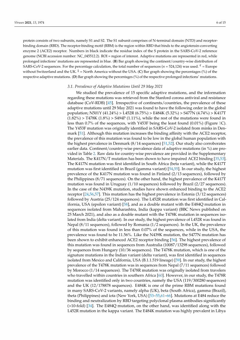
Figure 1. Global prevalence of adaptive and prolonged infections’ mutations in the receptor-binding domain (RBD) of the SARS-CoV-2 spike (S) protein until 29 May 2021. (A) SARS-CoV-2 virion (left) and schematics of the S protein (right). The
Figure 1. Global prevalence of adaptive and prolonged infections’ mutations in the receptor-binding domain (RBD) of theSARS-CoV-2 spike (S) protein until 29 May 2021. (A) SARS-CoV-2 virion (left) and schematics of the S protein (right). The S
Viruses 2021, 13, 1974 6 of 15
protein consists of two subunits, namely S1 and S2. The S1 subunit comprises of N-terminal domain (NTD) and receptor-binding domain (RBD). The receptor-binding motif (RBM) is the region within RBD that binds to the angiotensin-convertingenzyme 2 (ACE2) receptor. Numbers in black indicate the residue index of the S protein in the SARS-CoV-2 referencegenome (NCBI accession number: NC_045512.2). ROI = region of interest. Adaptive mutations are represented in red, whileprolonged infections’ mutations are represented in blue. (B) Bar graph showing the continent/country-wise distribution ofSARS-CoV-2 sequences. For the percentage calculation, the total number of sequences (n = 524,124) was used. € = Europewithout Switzerland and the UK. ‡ = North America without the USA. (C) Bar graph showing the percentages (%) of therespective adaptive mutations. (D) Bar graph showing the percentages (%) of the respective prolonged infections’ mutations.
3.1. Prevalence of Adaptive Mutations Until 29 May 2021
We studied the prevalence of 15 specific adaptive mutations, and the informationregarding these mutations was retrieved from the Stanford corona antiviral and resistancedatabase (CoV-RDB) [45]. Irrespective of continents/countries, the prevalence of theseadaptive mutations until 29 May 2021 was found to have the following order in the globalpopulation; N501Y (41.24%) > L452R (6.75%) > E484K (5.32%) > S477N (4.74%) > K417T(1.82%) > T478K (1.8%) > S494P (1.11%), while the rest of the mutations were found inless than 0.7% of the sequences, with Y453F being the least found (0.01%) (Figure 1C).The Y453F mutation was originally identified in SARS-CoV-2 isolated from minks in Den-mark [51]. Although this mutation increases the binding affinity with the ACE2 receptor,the prevalence of this mutation was found to be low in the global human population withthe highest prevalence in Denmark (8/14 sequences) [51,52]. Our study also corroboratesearlier data. Continent/country-wise prevalence data of adaptive mutations (in %) are pro-vided in Table 2. Raw data for country-wise prevalence are provided in the SupplementaryMaterials. The K417N/T mutation has been shown to have impaired ACE2 binding [35,53].The K417N mutation was first identified in South Africa (beta variant), while the K417Tmutation was first identified in Brazil (gamma variant) [54,55]. In our study, the highestprevalence of the K417N mutation was found in Finland (2/13 sequences), followed bythe Philippines (8/71 sequences). On the other hand, the highest prevalence of the K417Tmutation was found in Uruguay (1/10 sequences) followed by Brazil (2/27 sequences).In the case of the N439K mutation, studies have shown enhanced binding to the ACE2receptor [24,56,57]. This mutation has the highest prevalence in Estonia (3/12 sequences)followed by Austria (25/124 sequences). The L452R mutation was first identified in Cal-ifornia, USA (epsilon variant) [58], and as a double mutant with the E484Q mutation insequences isolated from Maharashtra, India (kappa variant) (BBC News published on25 March 2021), and also as a double mutant with the T478K mutation in sequences iso-lated from India (delta variant). In our study, the highest prevalence of L452R was found inNepal (8/11 sequences), followed by Romania (1/2 sequences). In the UK, the prevalenceof this mutation was found in less than 0.07% of the sequences, while in the USA, theprevalence was found to be 11.56%. Like the N439K mutation, the S477N mutation hasbeen shown to exhibit enhanced ACE2 receptor binding [56]. The highest prevalence ofthis mutation was found in sequences from Australia (10087/13298 sequences), followedby sequences from Hungary (10/36 sequences). The T478K mutation, which is one of thesignature mutations in the Indian variant (delta variant), was first identified in sequencesisolated from Mexico and California, USA (B.1.1.519 lineage) [59]. In our study, the highestprevalence of the T478K mutation was in sequences from Nepal (7/11 sequences) followedby Morocco (1/14 sequences). The T478R mutation was originally isolated from travelerswho travelled within countries in southern Africa [60]. However, in our study, the T478Rmutation was identified only in two countries, namely the USA (119/300280 sequences)and the UK (12/178878 sequences). E484K is one of the prime RBM mutations foundin many SARS-CoV-2 variants, namely alpha (UK), beta (South Africa), gamma (Brazil),theta (Philippines) and iota (New York, USA) [53–55,61–66]. Mutations at E484 reduce thebinding and neutralization by RBD targeting polyclonal plasma antibodies significantly(>10-fold) [34]. The E484Q mutation, on the other hand, was identified along with theL452R mutation in the kappa variant. The E484K mutation was highly prevalent in Libya

Viruses 2021, 13, 1974 7 of 15
(28/34 sequences), followed by South Africa (2/3 sequences), while the E484Q mutationwas highly prevalent in Nepal (1/11 sequences) followed by Bahrain (12/269 sequences).The F490S mutation was earlier identified in a few sequences isolated in Peru and Chile [67].This mutation was found to be highly prevalent in sequences isolated from Peru (26/127sequences) followed by sequences isolated from the West Bank (6/62 sequences). The S494Pmutation was earlier identified in sequences isolated from Santa Cruz County, USA [68].This mutation has been predicted to have enhanced binding with the ACE2 receptor [69]and was highly prevalent in sequences isolated from Russia (31/304 sequences), followedby sequences isolated from the USA (5167/300280 sequences). Finally, N501Y is anotherprime RBM mutation found in many SARS-CoV-2 variants, namely alpha, beta, gammaand theta [54,55,65,70,71]. This mutation has been shown to increase the ACE2 binding andenhances the viral replication in the upper respiratory tract [56,72–75]. More than half ofthe sequences isolated in the UK until 29 May 2021 were found to carry this mutation, whilearound 40% of the sequences isolated in the USA during the same period were found tocarry this mutation. This mutation was found in greater proportions in sequences isolatedfrom nearly 41 countries. Some of the adaptive mutations that we have studied here havebeen shown by others to have a moderate to a very high level of resistance (≥100-fold)towards some of the mAbs that are currently either in emergency use in patients or differentstages of clinical trials, to convalescent plasma and plasma from vaccinated individuals(Table 1) [22–39].
Table 2. Continent/country-wise prevalence of adaptive mutations in the receptor-binding domain of the SARS-CoV-2spike protein until 29 May 2021.
Continent/Country No. of StudiedSequences
Mutation (%)
R346K K417N K417T N439K L452Q L452R Y453F S477N T478K T478R E484K E484Q F490S S494P N501Y
Africa 1728 0.347 0.289 5.845 0.058 1.678 0.058 4.456 0.174 0.058 9.028
Asia 3703 0.135 1.242 0.351 0.270 0.027 1.836 0.513 1.053 1.782 0.702 0.189 7.399
Europe € 6092 0.033 0.509 0.312 1.346 0.575 0.230 5.368 0.066 1.625 0.033 0.657 26.133
Switzerland 18,234 0.362 0.516 0.126 4.442 0.005 1.475 0.016 21.213 0.801 0.856 0.038 0.033 0.104 17.906
UK 178,878 0.003 0.084 0.004 1.348 0.002 0.070 0.004 1.094 0.010 0.007 0.214 0.013 0.051 0.323 51.055
North America ‡ 99 2.020 9.091 1.010
USA 300,280 0.041 0.424 3.147 0.034 0.119 11.556 0.010 2.836 3.067 0.040 9.005 0.089 0.162 1.721 39.631
Oceania 13,299 75.848 0.008 0.278
South America 603 0.663 4.312 1.990 4.312 0.995
€ = Europe excluding Switzerland and the UK; ‡ = North America excluding the USA; empty cell = 4 or more decimal places or 0%.
3.2. Prevalence of Prolonged Infections’ Mutations Until 29 May 2021
We studied the prevalence of seven specific prolonged infections’ mutations (Table 3).Globally, these mutations were less prevalent in the studied sequences with the V483Amutation having the highest prevalence (0.014%) (Figure 1D). Table 3 shows the conti-nent/ country-wise prevalence of prolonged infections’ mutations (in %) in the receptor-binding domain of the SARS-CoV-2 spike protein. Country-wise, V483A mutation wasfound only in the USA (63/300280 sequences) and the UK (11/178878 sequences). World-wide, except for the V483A mutation, other mutations were found in <0.008% of thesequences: Q493R (0.007%) > Q493K (0.006%) > T415A (0.005%) > E484A (0.002%) >T470N (0.001%) (Figure 1D). The T415A mutation was found only in sequences fromthree countries, namely Italy (2/258 sequences), the USA (21/300280 sequences) and theUK (2/178878 sequences). Similar to the V483A mutation, the T470N mutation was alsofound only in sequences from the USA (4/300280 sequences) and the UK (2/178878 se-quences). The E484A mutation was identified only in sequences from Hong Kong (1/339sequences), the UK (3/178878 sequences) and the USA (4/300280 sequences). The F486Imutation was not found in any of the studied sequences. The Q493K mutation was iden-tified in sequences from four countries, with the highest prevalence in Spain (1/1173sequences) followed by the UK (13/178878 sequences), Switzerland (1/18234 sequences)and the USA (14/300280 sequences), while the Q493R mutation was identified in sequences

Viruses 2021, 13, 1974 8 of 15
from three countries with the highest prevalence in Italy (1/258 sequences) followed by theUSA (25/300280 sequences) and the UK (11/178878 sequences). Some of these mutationswere either predicted and/or tested to have resistance towards bamlanivimab and etese-vimab [76]. The V483A mutation was shown to resist bamlanivimab roughly 48-fold [32],while the E484A mutation was shown to resist C144 mAb greater than 100-fold [37].Q493K/R mutations, although rare, were found to confer a very high resistance towardsbamlanivimab [32], etesevimab [32], casirivimab [23,29,33], C121 [37] and C144 [37].
Table 3. Continent/country-wise prevalence of prolonged infections’ mutations in the receptor-binding domain of theSARS-CoV-2 spike protein until 29 May 2021.
Continent/Country No. of Studied SequencesMutation (%)
T415A T470N V483A E484A F486I Q493K Q493R
Africa 1728
Asia 3703 0.027
Europe € 6092 0.033 0.016 0.016
Switzerland 18,234 0.005
UK 178,878 0.001 0.001 0.006 0.002 0.007 0.006
North America ‡ 99
USA 300,280 0.007 0.001 0.021 0.001 0.005 0.008
Oceania 13,299
South America 603€ = Europe excluding Switzerland and the UK; ‡ = North America excluding the USA; empty cell = 4 or more decimal places or 0%.
3.3. Prevalence of Adaptive Mutations from 30 May 2021 Till 28 July 2021
From 30 May 2021 till 28 July 2021, 460,645 SARS-CoV-2 nucleotide sequences werereleased in the NCBI database. Of these sequences, 177 sequences were removed fromfurther analysis due to the lack of the region of interest/poor sequence quality. In thisdataset, sequences from Germany, Switzerland, the UK and USA constituted about 98%of the total sequences (Figure 2A). Irrespective of continents/countries, the prevalence ofthese adaptive mutations between 30 May 2021 and 28 July 2021 were found to have thefollowing order in the global population: N501Y (56.61%) > L452R (29.8%) > T478K (28.54%)> E484K (3.97%) > S477N (1.65%) > K417T (1.45%). The rest of the mutations were foundin less than 0.8% of the sequences with Y453F being the least found (0.004%) (Figure 2B).During this period, N501Y, L452R and T478K mutations increased by 15.39, 23.06 and26.75% points, respectively, while E484K, S477N, K417T and S494P mutations decreased by1.35, 3.09, 0.37 and 0.88% points, respectively. As mentioned earlier, L452R and T478K aresignature mutations in the delta variant. Our data confirm that the prevalence of the deltavariant has increased considerably in the last two months in the global population. Onthe continent/country-level, the highest prevalence of the L452R mutation was observedin Asia (57.90%), followed by the UK (49.1%) and the USA (23.34%) (Table 4), and thehighest prevalence of the T478K mutation was again observed in Asia (50.59%) followedby the UK (48.63%) and the USA (18.64%) (Table 4). Comparing the two timelines, theprevalence of L452R increased by roughly 56.07, 49.01 and 11.78% points in Asia, the UKand USA, respectively. Similarly, the prevalence of T478K increased roughly by 49.54,48.62 and 15.57% points in Asia, the UK and USA, respectively. These data indirectlyshow that the prevalence of the delta variant in the UK has increased tremendously inthe last two months. Within Asia, the highest prevalence of the L452R mutation wasobserved in Uzbekistan (92%) followed by India (75.65%), Bahrain (73.03%) and Myanmar(71.43%) (Supplementary Material). Similarly, the highest prevalence of the T478K mutationwas observed in Uzbekistan (76%), followed by Bahrain (73.03%), India (61.36%) andBangladesh (44.33%) (Supplementary Material). As mentioned before, E484K is one of the

Viruses 2021, 13, 1974 9 of 15
prime RBM mutations found in several SARS-CoV-2 variants, and mutations at E484 reducethe binding and neutralization by RBD targeting polyclonal plasma antibodies significantly(>10-fold) [34]. Though a reduction in the prevalence of E484K was observed globally, theprevalence of this mutation has increased by 2.09, 1.06, 0.45 and 5.13% points in Germany,Switzerland, the UK and USA, respectively, in the last 2 months (Figures 1 and 2).
Viruses 2021, 13, 1974 9 of 15
observed in Uzbekistan (76%), followed by Bahrain (73.03%), India (61.36%) and Bangla-desh (44.33%) (Supplementary Material). As mentioned before, E484K is one of the prime RBM mutations found in several SARS-CoV-2 variants, and mutations at E484 reduce the binding and neutralization by RBD targeting polyclonal plasma antibodies significantly (>10-fold) [34]. Though a reduction in the prevalence of E484K was observed globally, the prevalence of this mutation has increased by 2.09, 1.06, 0.45 and 5.13% points in Germany, Switzerland, the UK and USA, respectively, in the last 2 months (Figures 1 and 2).
Figure 2. Global prevalence of adaptive and prolonged infections’ mutations in the receptor-binding domain (RBD) of the SARS-CoV-2 spike (S) protein from 30 May 2021 till 28 July 2021. (A) Bar graph showing the continent/country-wise dis-tribution of SARS-CoV-2 sequences. For the percentage calculation, the total number of sequences (n = 460,645) was used. § = Europe without Germany, Switzerland and the UK. ‡ = North America without the USA. (B) Bar graph showing the percentages (%) of the respective adaptive mutations. (C) Bar graph showing the percentages (%) of the respective pro-longed infections’ mutations.
Table 4. Continent/country-wise prevalence of adaptive mutations in the receptor-binding domain of the SARS-CoV-2 Scheme 30 May 2021 till 28 July 2021.
Continent/ Country
No. of Stud-ied Se-
quences
Mutation (%)
R346K K417N K417T N439K L452Q L452R Y453F S477N T478K T478R E484K E484Q F490S S494P N501Y
Africa 271 11.808 11.439 1.107 20.664 0.369 0.738 3.321 49.815 Asia 1354 7.090 0.148 0.074 57.903 0.148 50.591 0.074 6.499 6.721 0.074 23.560
Europe § 7400 0.014 0.311 0.041 3.608 0.027 0.122 1.041 0.027 0.554 0.014 0.041 0.027 17.270 Germany 120,429 0.033 1.476 0.453 2.148 0.121 3.310 0.002 2.352 2.819 0.011 3.582 0.134 0.542 0.174 79.022
Switzerland 17,578 0.233 0.688 0.484 2.463 0.017 2.890 9.148 2.020 0.006 1.911 0.091 0.307 0.165 58.095 UK 229,321 0.007 0.256 0.065 0.090 0.003 49.075 0.025 48.633 0.666 0.171 0.112 0.054 48.698
North Amer-ica ‡
66 3.030 1.515 6.061 1.515
USA 83,122 0.066 0.929 6.950 0.011 0.217 23.336 0.018 3.621 18.640 0.017 14.132 0.109 0.393 0.829 50.363 Oceania 634 0.946
Figure 2. Global prevalence of adaptive and prolonged infections’ mutations in the receptor-binding domain (RBD) of theSARS-CoV-2 spike (S) protein from 30 May 2021 till 28 July 2021. (A) Bar graph showing the continent/country-wise distributionof SARS-CoV-2 sequences. For the percentage calculation, the total number of sequences (n = 460,645) was used. § = Europewithout Germany, Switzerland and the UK. ‡ = North America without the USA. (B) Bar graph showing the percentages (%) ofthe respective adaptive mutations. (C) Bar graph showing the percentages (%) of the respective prolonged infections’ mutations.
Table 4. Continent/country-wise prevalence of adaptive mutations in the receptor-binding domain of the SARS-CoV-2Scheme 30 May 2021 till 28 July 2021.
Continent/Country No. of StudiedSequences
Mutation (%)
R346K K417N K417T N439K L452Q L452R Y453F S477N T478K T478R E484K E484Q F490S S494P N501Y
Africa 271 11.808 11.439 1.107 20.664 0.369 0.738 3.321 49.815
Asia 1354 7.090 0.148 0.074 57.903 0.148 50.591 0.074 6.499 6.721 0.074 23.560
Europe § 7400 0.014 0.311 0.041 3.608 0.027 0.122 1.041 0.027 0.554 0.014 0.041 0.027 17.270
Germany 120,429 0.033 1.476 0.453 2.148 0.121 3.310 0.002 2.352 2.819 0.011 3.582 0.134 0.542 0.174 79.022
Switzerland 17,578 0.233 0.688 0.484 2.463 0.017 2.890 9.148 2.020 0.006 1.911 0.091 0.307 0.165 58.095
UK 229,321 0.007 0.256 0.065 0.090 0.003 49.075 0.025 48.633 0.666 0.171 0.112 0.054 48.698
North America ‡ 66 3.030 1.515 6.061 1.515
USA 83,122 0.066 0.929 6.950 0.011 0.217 23.336 0.018 3.621 18.640 0.017 14.132 0.109 0.393 0.829 50.363
Oceania 634 0.946
South America 293 37.201 0.341 1.024 62.457 1.024 0.341 39.249
§ = Europe excluding Germany, Switzerland and the UK; ‡ = North America excluding the USA; empty cell = 4 or more decimal places or0%. Prevalence of prolonged infections’ mutations from 30 May 2021 till 28 July 2021.

Viruses 2021, 13, 1974 10 of 15
3.4. Prevalence of Prolonged Infections’ Mutations from 30 May 2021 Till 28 July 2021
Similar to the previous timeline, globally, these mutations were less prevalent. How-ever, unlike the previous timeline, during this period, Q493R (0.011%) was found to bethe dominant mutation followed by Q493K (0.007%), E484A (0.005%), V483A (0.004%),T470N (0.002%), T415A and F486I (Figure 2C). During this period, the prevalence ofQ493R, Q493K, E484A and T470N increased by 0.004, 0.001, 0.003 and 0.001% points,respectively, while the prevalence of V483A and T415A decreased by 0.01 and 0.005%points, respectively. As mentioned before, the Q493R mutation alone confers signif-icant resistance towards bamlanivimab, etesevimab, casirivimab and C144 [32,37,42].On the continent/country level, this mutation was found in only sequences from threecountries: the USA (24/83122 sequences), the UK (23/229321 sequences) and Germany(4/120429 sequences) (Table 5). Q493K and E484A mutations were found in only sequencesfrom Switzerland, the UK, USA and Germany. Of these four countries, both these mutationswere found to be highly prevalent in sequences isolated from Switzerland (Table 5). Unlikethe previous timeline, V483A was additionally identified in sequences from Egypt andGermany. It is worth noting that 3 out of 65 sequences from Egypt carried this mutation(Supplementary Material). Until 29 May 2021, the T470N mutation was identified onlyin sequences from the UK and USA. During the last two months, this mutation was con-fined within Europe, especially in Estonia, Germany, Switzerland and the UK. In Estonia,5 out of 1635 sequences carried this mutation (Supplementary Material). During the sameperiod, on the other hand, the T415A mutation was confined within the USA (Table 5), andthe prevalence almost doubled. Finally, until 29 May 2021, the F486I mutation was notidentified in any of the sequences. Between 30 May 2021 and 28 July 2021, this mutationwas identified in one of the 83,122 studied sequences from the USA.
Table 5. Continent/country-wise prevalence of prolonged infections’ mutations in the receptor-binding domain of theSARS-CoV-2 spike protein from 30 May 2021 till 28 July 2021.
Continent/Country No. of Studied SequencesMutation (%)
T415A T470N V483A E484A F486I Q493K Q493R
Africa 271 1.107
Asia 1354
Europe § 7400 0.068
Germany 120,429 0.001 0.004 0.002 0.002 0.003
Switzerland 17,578 0.006 0.057 0.011
UK 229,321 0.002 0.001 0.003 0.010 0.010
North America ‡ 66
USA 83,122 0.002 0.010 0.006 0.001 0.006 0.029
Oceania 634
South America 293§ = Europe excluding Germany, Switzerland and the UK; ‡ = North America excluding the USA; empty cell = 4 or more decimal places or 0%.
3.5. Time Course of the Appearance of the Mutations Until 29 May 2021
To study the time course of the appearance of the mutations, we split the data intosix timelines, with each timeline covering about 3 months except the last timeline, whichcovered approximately 4 months: Timeline 1 (1 November 2019 till 31 January 2020: T1),Timeline 2 (1 February 2020 till 30 April 2020: T2), Timeline 3 (1 May 2020 till 31 July 2020:T3), Timeline 4 (1 August 2020 till 31 October 2020: T4), Timeline 5 (1 November 2020 till31 January 2021: T5) and Timeline 6 (1 February 2021 till 29 May 2021: T6). T1, T2, T3,T4, T5 and T6 had the following respective sequence distributions: 0.006%, 0.32%, 2.07%,4.68%, 3.81% and 90.13%. This shows that a similar number of SARS-CoV-2 sequences

Viruses 2021, 13, 1974 11 of 15
were released by the NCBI from 1 February 2021 to 29 May 2021 and from 30 May 2021 to28 July 2021. In T1, none of the sequences carried any adaptive and prolonged infections’mutations. In T2, nearly 0.76% of sequences from the USA carried the V483A mutation.Country-wise raw data on the prevalence of SARS-CoV-2 receptor-binding domain (RBD)mutations concerning different timelines is provided in the Supplementary Material. Noother mutation was observed in T2. In T3, mutations N439K, Y453F, S477N, V483A, E484K,E484Q, S494P and N501Y were observed. While N439K, Y453F, V483A, E484K, S494P andN501Y mutations were only observed in sequences from the USA (0.015%), Netherlands(33.33%), USA (0.21%), USA (0.01%), USA (0.04%) and Australia (0.99%), respectively,S477N and E484Q were identified in more than one country with the highest prevalence inLebanon (25%) and India (0.44%), respectively. In T4, apart from the mutations identifiedin T3, mutations R346K, L452Q, L452R and T478K were observed. While R346K and L452Rmutations were observed only in sequences from Iraq with a prevalence of 7.41% and 1.85%,respectively, the other two mutations were observed only in sequences from the USA, witheach having a prevalence of approximately 0.009%. In T5, except for the L452Q mutation,all other mutations discussed above were observed. Additionally, mutations K417N, K417T,T470N, T478R, E484A, F490S and Q493K were observed. All these mutations were observedonly in sequences from the USA except for the K417T mutation, which was also found in asequence from Italy. The prevalences of these mutations were quite low (≤0.04%). Finally,mutations T415A and Q493R were observed in T6 apart from the other mutations discussedabove. Both these mutations were identified only in sequences from the USA, UK and Italy.It is worth noting that both these mutations were found to be highly prevalent in sequencesfrom Italy compared to those from the USA and UK.
In conclusion, globally, we found a high prevalence of N501Y mutation in the RBDof the SARS-CoV-2 S protein using the available sequence data from the NCBI databaseup to 28 July 2021. Further, we observed a considerable prevalence of other adaptivemutations in the RBD, namely L452R, T478K, E484K, S477N, K417T, N439K and S494P.Prolonged infections’ mutations were observed only in a few sequences from a few coun-tries. A few drawbacks in our study, like any other epidemiological study, are the availablenumber of sequences from each country, the context to which the sequencing was donein the first place, etc. Because of these reasons, the data presented here might be skewedand should be used with caution. Further, we did not use the sequences from the GI-SAID (https://www.gisaid.org/) (accessed on 3 August 2021) database, which contained2,568,717 sequences at the time of writing this manuscript, as one needs prior permissionto use the data. Thus, we used only sequences from the NCBI database as they are readilyaccessible by anyone without any registration and/or signing any agreement. Moreover,the GISAID database has inbuilt visualization software using which variants (mutations)can be analyzed. In addition, many online visualization tools (e.g., CovMT) use data fromthe GISAID database. To our knowledge, no such tools use sequence data deposited in theNCBI database. To analyze large sequence data, in many cases, specialized computationalskills/hardware requirements are needed. This motivated us to use the data from theNCBI database and develop a methodology. It should be noted that not all SARS-CoV-2sequences are deposited in the NCBI and GISAID databases. Anyone interested in studyingthe prevalence of mutations (variants of concern, variants of interest and variants undermonitoring) from sequences isolated locally or regionally can follow our methodology, andour methodology is not limited only to the SARS-CoV-2 sequences. Like the UK, USA,Germany and Switzerland, many countries should actively sequence as many SARS-CoV-2sequences as possible from patients and deposit them in the public repository. This willaid in a better understanding of the viral evolution in the human population. As moresequence information gets deposited in the public database regularly, we might/will ob-serve a change in the prevalence landscape shortly, as we have observed for the deltavariant. Thus, it is necessary to do this type of study at regular intervals to keep ourselvesupdated about the latest dominant mutation(s), both globally and regionally (or locally),

Viruses 2021, 13, 1974 12 of 15
and thereby we can evolve our control strategies and put a full stop to the spread of thisdevastating viral infection.
Supplementary Materials: The following are available online at https://www.mdpi.com/article/10.3390/v13101974/s1, Country-wise raw data on the prevalence of SARS-CoV-2 receptor-bindingdomain (RBD) mutations: 2019 till 29 May 2021, 30 May 2021 till 28 July 2021 and different time lines(T1–T6) are provided as Excel files. Compiled Search+ plugin (ready-to-use) for use with Notepad++32-bit is also provided.
Author Contributions: N.P. conceived the study and performed all the sequence analyses. N.P. andJ.L. wrote the manuscript together. Both authors have read and agreed to the published version ofthe manuscript.
Funding: J.L. received financial support from Scandinavian Society for Antimicrobial Chemotherapy(SLS-961049) and from the Erik, Karin and Gösta Selander Foundation.
Institutional Review Board Statement: Not applicable.
Informed Consent Statement: Not applicable.
Acknowledgments: N.P. would like to thank Chester Medical School, University of Chester, forproviding IT support for this study. N.P. would also like to thank Kate Harrison for her criticalreading of this manuscript. N.P. and J.L. would like to thank Robert W. Shafer, Stanford University,for the helpful discussion.
Conflicts of Interest: The authors declare no conflict of interest.
References1. King, A.M.Q.; Adams, M.J.; Carstens, E.B.; Lefkowitz, E.J. (Eds.) Order—Nidovirales. In Virus Taxonomy; Elsevier: San Diego, CA,
USA, 2012; pp. 784–794, ISBN 978-0-12-384684-6.2. Snijder, E.J.; Decroly, E.; Ziebuhr, J. The Nonstructural Proteins Directing Coronavirus RNA Synthesis and Processing. Adv. Virus
Res. 2016, 96, 59–126. [CrossRef]3. Jimenez-Guardeño, J.M.; Nieto-Torres, J.L.; DeDiego, M.L.; Regla-Nava, J.A.; Fernandez-Delgado, R.; Castaño-Rodriguez, C.;
Enjuanes, L. The PDZ-Binding Motif of Severe Acute Respiratory Syndrome Coronavirus Envelope Protein Is a Determinant ofViral Pathogenesis. PLoS Pathog. 2014, 10, e1004320. [CrossRef] [PubMed]
4. Woo, P.C.Y.; Lau, S.K.P.; Huang, Y.; Yuen, K.-Y. Coronavirus Diversity, Phylogeny and Interspecies Jumping. Exp. Biol. Med. 2009,234, 1117–1127. [CrossRef]
5. Van der Hoek, L. Human Coronaviruses: What Do They Cause? Antivir. Ther. 2007, 12, 651–658. [PubMed]6. A Chronicle on the SARS Epidemic. Chin. Law Gov. 2003, 36, 12–15. [CrossRef]7. Zumla, A.; Hui, D.S.; Perlman, S. Middle East Respiratory Syndrome. Lancet Lond. Engl. 2015, 386, 995–1007. [CrossRef]8. Andersen, K.G.; Rambaut, A.; Lipkin, W.I.; Holmes, E.C.; Garry, R.F. The Proximal Origin of SARS-CoV-2. Nat. Med. 2020, 26,
1–3. [CrossRef]9. Zhou, P.; Yang, X.-L.; Wang, X.-G.; Hu, B.; Zhang, L.; Zhang, W.; Si, H.-R.; Zhu, Y.; Li, B.; Huang, C.-L.; et al. A Pneumonia
Outbreak Associated with a New Coronavirus of Probable Bat Origin. Nature 2020, 579, 270–273. [CrossRef]10. Ellis, P.; Somogyvári, F.; Virok, D.P.; Noseda, M.; McLean, G.R. Decoding Covid-19 with the SARS-CoV-2 Genome. Curr. Genet.
Med. Rep. 2021, 9, 1–12. [CrossRef]11. Baraniuk, C. Covid-19: What Do We Know about Airborne Transmission of SARS-CoV-2? BMJ 2021, 373, n1030. [CrossRef]12. Tang, J.W.; Bahnfleth, W.P.; Bluyssen, P.M.; Buonanno, G.; Jimenez, J.L.; Kurnitski, J.; Li, Y.; Miller, S.; Sekhar, C.; Morawska,
L.; et al. Dismantling Myths on the Airborne Transmission of Severe Acute Respiratory Syndrome Coronavirus-2 (SARS-CoV-2).J. Hosp. Infect. 2021, 110, 89–96. [CrossRef]
13. Greenhalgh, T.; Jimenez, J.L.; Prather, K.A.; Tufekci, Z.; Fisman, D.; Schooley, R. Ten Scientific Reasons in Support of AirborneTransmission of SARS-CoV-2. Lancet 2021, 397, 1603–1605. [CrossRef]
14. Hoffmann, M.; Kleine-Weber, H.; Schroeder, S.; Krüger, N.; Herrler, T.; Erichsen, S.; Schiergens, T.S.; Herrler, G.; Wu, N.-H.;Nitsche, A.; et al. SARS-CoV-2 Cell Entry Depends on ACE2 and TMPRSS2 and Is Blocked by a Clinically Proven ProteaseInhibitor. Cell 2020, 181, 271–280.e8. [CrossRef] [PubMed]
15. Beigel, J.H.; Tomashek, K.M.; Dodd, L.E.; Mehta, A.K.; Zingman, B.S.; Kalil, A.C.; Hohmann, E.; Chu, H.Y.; Luetkemeyer, A.; Kline,S.; et al. Remdesivir for the Treatment of Covid-19—Final Report. N. Engl. J. Med. 2020, 383, 1813–1826. [CrossRef] [PubMed]
16. Schoot, T.S.; Kerckhoffs, A.P.M.; Hilbrands, L.B.; van Marum, R.J. Immunosuppressive Drugs and COVID-19: A Review. Front.Pharmacol. 2020, 11, 1333. [CrossRef]
17. Dai, L.; Gao, G.F. Viral Targets for Vaccines against COVID-19. Nat. Rev. Immunol. 2021, 21, 73–82. [CrossRef]18. Williams, T.C.; Burgers, W.A. SARS-CoV-2 Evolution and Vaccines: Cause for Concern? Lancet Respir. Med. 2021, 9, 333–335. [CrossRef]

Viruses 2021, 13, 1974 13 of 15
19. Wise, J. Covid-19: The E484K Mutation and the Risks It Poses. BMJ 2021, 372, n359. [CrossRef]20. Taylor, P.C.; Adams, A.C.; Hufford, M.M.; de la Torre, I.; Winthrop, K.; Gottlieb, R.L. Neutralizing Monoclonal Antibodies for
Treatment of COVID-19. Nat. Rev. Immunol. 2021, 21, 382–393. [CrossRef]21. Marovich, M.; Mascola, J.R.; Cohen, M.S. Monoclonal Antibodies for Prevention and Treatment of COVID-19. JAMA 2020,
324, 131. [CrossRef]22. Liu, Z.; VanBlargan, L.A.; Bloyet, L.-M.; Rothlauf, P.W.; Chen, R.E.; Stumpf, S.; Zhao, H.; Errico, J.M.; Theel, E.S.; Liebeskind,
M.J.; et al. Identification of SARS-CoV-2 Spike Mutations That Attenuate Monoclonal and Serum Antibody Neutralization. CellHost Microbe 2021, 29, 477–488.e4. [CrossRef] [PubMed]
23. Starr, T.N.; Greaney, A.J.; Addetia, A.; Hannon, W.W.; Choudhary, M.C.; Dingens, A.S.; Li, J.Z.; Bloom, J.D. Prospective Mappingof Viral Mutations That Escape Antibodies Used to Treat COVID-19. Science 2021, 371, 850–854. [CrossRef] [PubMed]
24. Chen, R.E.; Zhang, X.; Case, J.B.; Winkler, E.S.; Liu, Y.; VanBlargan, L.A.; Liu, J.; Errico, J.M.; Xie, X.; Suryadevara, N.; et al.Resistance of SARS-CoV-2 Variants to Neutralization by Monoclonal and Serum-Derived Polyclonal Antibodies. Nat. Med. 2021,27, 717–726. [CrossRef] [PubMed]
25. Wang, P.; Nair, M.S.; Liu, L.; Iketani, S.; Luo, Y.; Guo, Y.; Wang, M.; Yu, J.; Zhang, B.; Kwong, P.D.; et al. Antibody Resistance ofSARS-CoV-2 Variants, B.1.351 and B.1.1.7. Nature 2021, 593, 130–135. [CrossRef] [PubMed]
26. Wang, L.; Zhou, T.; Zhang, Y.; Yang, E.S.; Schramm, C.A.; Shi, W.; Pegu, A.; Oloninyi, O.K.; Ransier, A.; Darko, S.; et al. Antibodieswith Potent and Broad Neutralizing Activity against Antigenically Diverse and Highly Transmissible SARS-CoV-2 Variants.bioRxiv 2021. [CrossRef]
27. Copin, R.; Baum, A.; Wloga, E.; Pascal, K.E.; Giordano, S.; Fulton, B.O.; Zhou, A.; Negron, N.; Lanza, K.; Chan, N.; et al.REGEN-COV Protects against Viral Escape in Preclinical and Human Studies. bioRxiv 2021. [CrossRef]
28. Li, Q.; Wu, J.; Nie, J.; Zhang, L.; Hao, H.; Liu, S.; Zhao, C.; Zhang, Q.; Liu, H.; Nie, L.; et al. The Impact of Mutations inSARS-CoV-2 Spike on Viral Infectivity and Antigenicity. Cell 2020, 182, 1284–1294.e9. [CrossRef]
29. Baum, A.; Fulton, B.O.; Wloga, E.; Copin, R.; Pascal, K.E.; Russo, V.; Giordano, S.; Lanza, K.; Negron, N.; Ni, M.; et al. AntibodyCocktail to SARS-CoV-2 Spike Protein Prevents Rapid Mutational Escape Seen with Individual Antibodies. Science 2020, 369,1014–1018. [CrossRef]
30. Wang, R.; Zhang, Q.; Ge, J.; Ren, W.; Zhang, R.; Lan, J.; Ju, B.; Su, B.; Yu, F.; Chen, P.; et al. SARS-CoV-2 Variants Resist AntibodyNeutralization and Broaden Host ACE2 Usage. bioRxiv 2021. [CrossRef]
31. Gottlieb, R.L.; Nirula, A.; Chen, P.; Boscia, J.; Heller, B.; Morris, J.; Huhn, G.; Cardona, J.; Mocherla, B.; Stosor, V.; et al. Effect ofBamlanivimab as Monotherapy or in Combination With Etesevimab on Viral Load in Patients With Mild to Moderate COVID-19:A Randomized Clinical Trial. JAMA 2021, 325, 632–644. [CrossRef]
32. FDA Authorizes Revisions to Fact Sheets to Address SARS-CoV-2 Variants for Monoclonal Antibody Products under EmergencyUse Authorization. Available online: https://www.fda.gov/drugs/drug-safety-and-availability/fda-authorizes-revisions-fact-sheets-address-sars-cov-2-variants-monoclonal-antibody-products-under (accessed on 28 July 2021).
33. Starr, T.N.; Greaney, A.J.; Dingens, A.S.; Bloom, J.D. Complete Map of SARS-CoV-2 RBD Mutations That Escape the MonoclonalAntibody LY-CoV555 and Its Cocktail with LY-CoV016. Cell Rep. Med. 2021, 2, 100255. [CrossRef] [PubMed]
34. Greaney, A.J.; Loes, A.N.; Crawford, K.H.D.; Starr, T.N.; Malone, K.D.; Chu, H.Y.; Bloom, J.D. Comprehensive Mapping ofMutations in the SARS-CoV-2 Receptor-Binding Domain That Affect Recognition by Polyclonal Human Plasma Antibodies. CellHost Microbe 2021, 29, 463–476.e6. [CrossRef] [PubMed]
35. Greaney, A.J.; Starr, T.N.; Gilchuk, P.; Zost, S.J.; Binshtein, E.; Loes, A.N.; Hilton, S.K.; Huddleston, J.; Eguia, R.; Crawford,K.H.D.; et al. Complete Mapping of Mutations to the SARS-CoV-2 Spike Receptor-Binding Domain That Escape AntibodyRecognition. Cell Host Microbe 2021, 29, 44–57.e9. [CrossRef] [PubMed]
36. Wang, Z.; Schmidt, F.; Weisblum, Y.; Muecksch, F.; Barnes, C.O.; Finkin, S.; Schaefer-Babajew, D.; Cipolla, M.; Gaebler, C.; Lieber-man, J.A.; et al. MRNA Vaccine-Elicited Antibodies to SARS-CoV-2 and Circulating Variants. Nature 2021, 592, 616–622. [CrossRef]
37. Weisblum, Y.; Schmidt, F.; Zhang, F.; DaSilva, J.; Poston, D.; Lorenzi, J.C.; Muecksch, F.; Rutkowska, M.; Hoffmann, H.-H.;Michailidis, E.; et al. Escape from Neutralizing Antibodies by SARS-CoV-2 Spike Protein Variants. eLife 2020, 9, e61312. [CrossRef]
38. Jangra, S.; Ye, C.; Rathnasinghe, R.; Stadlbauer, D.; Krammer, F.; Simon, V.; Martinez-Sobrido, L.; Garcia-Sastre, A.; Schotsaert,M. The E484K Mutation in the SARS-CoV-2 Spike Protein Reduces but Does Not Abolish Neutralizing Activity of HumanConvalescent and Post-Vaccination Sera. medRxiv 2021. [CrossRef]
39. Tada, T.; Dcosta, B.M.; Samanovic-Golden, M.; Herati, R.S.; Cornelius, A.; Mulligan, M.J.; Landau, N.R. Neutralization of Viruseswith European, South African, and United States SARS-CoV-2 Variant Spike Proteins by Convalescent Sera and BNT162b2 MRNAVaccine-Elicited Antibodies. bioRxiv 2021. [CrossRef]
40. Yuan, M.; Huang, D.; Lee, C.-C.D.; Wu, N.C.; Jackson, A.M.; Zhu, X.; Liu, H.; Peng, L.; van Gils, M.J.; Sanders, R.W.; et al.Structural and Functional Ramifications of Antigenic Drift in Recent SARS-CoV-2 Variants. Science 2021, 373, 818–823. [CrossRef]
41. Rothenberger, S.; Walser, M.; Malvezzi, F.; Mayor, J.; Ryter, S.; Moreno, H.; Liechti, N.; Hälg, S.; Bosshart, A.; Iss, C.; et al. Multi-specific DARPin® Therapeutics Demonstrate Very High Potency against SARS-CoV-2 Variants in Vitro. bioRxiv 2021. [CrossRef]
42. Muecksch, F.; Weisblum, Y.; Barnes, C.O.; Schmidt, F.; Schaefer-Babajew, D.; Lorenzi, J.C.C.; Flyak, A.I.; DeLaitsch, A.T.;Huey-Tubman, K.E.; Hou, S.; et al. Development of Potency, Breadth and Resilience to Viral Escape Mutations in SARS-CoV-2Neutralizing Antibodies. bioRxiv 2021. [CrossRef]
Viruses 2021, 13, 1974 14 of 15
43. Tada, T.; Zhou, H.; Dcosta, B.M.; Samanovic, M.I.; Mulligan, M.J.; Landau, N.R. The Spike Proteins of SARS-CoV-2 B.1.617and B.1.618 Variants Identified in India Provide Partial Resistance to Vaccine-Elicited and Therapeutic Monoclonal Antibodies.bioRxiv 2021. [CrossRef]
44. Yao, W.; Wang, Y.; Ma, D.; Tang, X.; Wang, H.; Li, C.; Lin, H.; Li, Y.; Zhong, G. Circulating SARS-CoV-2 Variants B.1.1.7, 501Y.V2,and P.1 Have Gained Ability to Utilize Rat and Mouse Ace2 and Altered in Vitro Sensitivity to Neutralizing Antibodies andACE2-Ig. bioRxiv 2021. [CrossRef]
45. Tzou, P.L.; Tao, K.; Nouhin, J.; Rhee, S.-Y.; Hu, B.D.; Pai, S.; Parkin, N.; Shafer, R.W. Coronavirus Antiviral Research Database(CoV-RDB): An Online Database Designed to Facilitate Comparisons between Candidate Anti-Coronavirus Compounds. Viruses2020, 12, 1006. [CrossRef]
46. Alam, I.; Radovanovic, A.; Incitti, R.; Kamau, A.A.; Alarawi, M.; Azhar, E.I.; Gojobori, T. CovMT: An Interactive SARS-CoV-2Mutation Tracker, with a Focus on Critical Variants. Lancet Infect. Dis. 2021, 21, 602. [CrossRef]
47. Katoh, K.; Misawa, K.; Kuma, K.; Miyata, T. MAFFT: A Novel Method for Rapid Multiple Sequence Alignment Based on FastFourier Transform. Nucleic Acids Res. 2002, 30, 3059–3066. [CrossRef] [PubMed]
48. Larsson, A. AliView: A Fast and Lightweight Alignment Viewer and Editor for Large Datasets. Bioinformatics 2014, 30, 3276–3278.[CrossRef] [PubMed]
49. Hall, T.A. BioEdit: A User-Friendly Biological Sequence Alignment Editor and Analysis Program for Windows 95/98/NT. NucleicAcids Symp. Ser. 1999, 41, 95–98.
50. Noh, J.Y.; Jeong, H.W.; Shin, E.-C. SARS-CoV-2 Mutations, Vaccines, and Immunity: Implication of Variants of Concern. SignalTransduct. Target. Ther. 2021, 6, 1–2. [CrossRef]
51. Bayarri-Olmos, R.; Rosbjerg, A.; Johnsen, L.B.; Helgstrand, C.; Bak-Thomsen, T.; Garred, P.; Skjoedt, M.-O. The SARS-CoV-2Y453F Mink Variant Displays a Pronounced Increase in ACE-2 Affinity but Does Not Challenge Antibody Neutralization. J. Biol.Chem. 2021, 296, 100536. [CrossRef] [PubMed]
52. Gobeil, S.M.-C.; Janowska, K.; McDowell, S.; Mansouri, K.; Parks, R.; Stalls, V.; Kopp, M.F.; Manne, K.; Saunders, K.; Edwards,R.J.; et al. Effect of Natural Mutations of SARS-CoV-2 on Spike Structure, Conformation and Antigenicity. bioRxiv 2021. [CrossRef]
53. Collier, D.A.; De Marco, A.; Ferreira, I.A.T.M.; Meng, B.; Datir, R.P.; Walls, A.C.; Kemp, S.A.; Bassi, J.; Pinto, D.; Silacci-Fregni,C.; et al. Sensitivity of SARS-CoV-2 B.1.1.7 to MRNA Vaccine-Elicited Antibodies. Nature 2021, 593, 136–141. [CrossRef]
54. Tegally, H.; Wilkinson, E.; Giovanetti, M.; Iranzadeh, A.; Fonseca, V.; Giandhari, J.; Doolabh, D.; Pillay, S.; San, E.J.; Msomi,N.; et al. Detection of a SARS-CoV-2 Variant of Concern in South Africa. Nature 2021, 592, 438–443. [CrossRef]
55. Faria, N.R.; Mellan, T.A.; Whittaker, C.; Claro, I.M.; Candido, D.d.S.; Mishra, S.; Crispim, M.A.E.; Sales, F.C.; Hawryluk, I.;McCrone, J.T.; et al. Genomics and Epidemiology of a Novel SARS-CoV-2 Lineage in Manaus, Brazil. medRxiv 2021. [CrossRef]
56. Starr, T.N.; Greaney, A.J.; Hilton, S.K.; Ellis, D.; Crawford, K.H.D.; Dingens, A.S.; Navarro, M.J.; Bowen, J.E.; Tortorici, M.A.;Walls, A.C.; et al. Deep Mutational Scanning of SARS-CoV-2 Receptor Binding Domain Reveals Constraints on Folding and ACE2Binding. Cell 2020, 182, 1295–1310.e20. [CrossRef]
57. Thomson, E.C.; Rosen, L.E.; Shepherd, J.G.; Spreafico, R.; da Silva Filipe, A.; Wojcechowskyj, J.A.; Davis, C.; Piccoli, L.; Pascall, D.J.;Dillen, J.; et al. Circulating SARS-CoV-2 Spike N439K Variants Maintain Fitness While Evading Antibody-Mediated Immunity.Cell 2021, 184, 1171–1187.e20. [CrossRef]
58. Deng, X.; Garcia-Knight, M.A.; Khalid, M.M.; Servellita, V.; Wang, C.; Morris, M.K.; Sotomayor-González, A.; Glasner, D.R.; Reyes,K.R.; Gliwa, A.S.; et al. Transmission, Infectivity, and Antibody Neutralization of an Emerging SARS-CoV-2 Variant in CaliforniaCarrying a L452R Spike Protein Mutation. medRxiv 2021. [CrossRef]
59. Giacomo, S.D.; Mercatelli, D.; Rakhimov, A.; Giorgi, F.M. Preliminary Report on Severe Acute Respiratory Syndrome Coronavirus2 (SARS-CoV-2) Spike Mutation T478K. J. Med. Virol. 2021, 93, 5638–5643. [CrossRef]
60. De Oliveira, T.; Lutucuta, S.; Nkengasong, J.; Morais, J.; Paixão, J.P.; Neto, Z.; Afonso, P.; Miranda, J.; David, K.; Inglês, L.; et al.A Novel Variant of Interest of SARS-CoV-2 with Multiple Spike Mutations Detected through Travel Surveillance in Africa.medRxiv 2021. [CrossRef]
61. Annavajhala, M.K.; Mohri, H.; Zucker, J.E.; Sheng, Z.; Wang, P.; Gomez-Simmonds, A.; Ho, D.D.; Uhlemann, A.-C. A NovelSARS-CoV-2 Variant of Concern, B.1.526, Identified in New York. medRxiv 2021. [CrossRef]
62. West, A.P.; Wertheim, J.O.; Wang, J.C.; Vasylyeva, T.I.; Havens, J.L.; Chowdhury, M.A.; Gonzalez, E.; Fang, C.E.; Di Lonardo, S.S.;Hughes, S.; et al. Detection and Characterization of the SARS-CoV-2 Lineage B.1.526 in New York. bioRxiv 2021. [CrossRef]
63. Bascos, N.A.D.; Mirano-Bascos, D.; Saloma, C.P. Structural Analysis of Spike Protein Mutations in the SARS-CoV-2 P.3 Variant.bioRxiv 2021. [CrossRef]
64. Ferrareze, P.A.G.; Franceschi, V.B.; Mayer, A.d.M.; Caldana, G.D.; Zimerman, R.A.; Thompson, C.E. E484K as an InnovativePhylogenetic Event for Viral Evolution: Genomic Analysis of the E484K Spike Mutation in SARS-CoV-2 Lineages from Brazil.bioRxiv 2021. [CrossRef]
65. Tablizo, F.A.; Kim, K.M.; Lapid, C.M.; Castro, M.J.R.; Yangzon, M.S.L.; Maralit, B.A.; Ayes, M.E.C.; la Paz, E.M.C.; Guzman,A.R.D.; Yap, J.M.C.; et al. Genome Sequencing and Analysis of an Emergent SARS-CoV-2 Variant Characterized by MultipleSpike Protein Mutations Detected from the Central Visayas Region of the Philippines. medRxiv 2021. [CrossRef]
66. Voloch, C.M.; da Silva Francisco, R.; de Almeida, L.G.P.; Cardoso, C.C.; Brustolini, O.J.; Gerber, A.L.; Guimarães, A.P.d.C.; Mariani,D.; da Costa, R.M.; Ferreira, O.C.; et al. Genomic Characterization of a Novel SARS-CoV-2 Lineage from Rio de Janeiro, Brazil.J. Virol. 2021, 95, e00119-21. [CrossRef] [PubMed]

Viruses 2021, 13, 1974 15 of 15
67. Grabowski, F.; Kochanczyk, M.; Lipniacki, T. L18F Substrain of SARS-CoV-2 VOC-202012/01 Is Rapidly Spreading in England.medRxiv 2021. [CrossRef]
68. Thornlow, B.; Hinrichs, A.S.; Jain, M.; Dhillon, N.; La, S.; Kapp, J.D.; Anigbogu, I.; Cassatt-Johnstone, M.; McBroome, J.; Haeussler,M.; et al. A New SARS-CoV-2 Lineage That Shares Mutations with Known Variants of Concern Is Rejected by AutomatedSequence Repository Quality Control. bioRxiv 2021. [CrossRef]
69. Chakraborty, S. Evolutionary and Structural Analysis Elucidates Mutations on SARS-CoV2 Spike Protein with Altered HumanACE2 Binding Affinity. Biochem. Biophys. Res. Commun. 2021, 534, 374–380. [CrossRef] [PubMed]
70. Preliminary Genomic Characterisation of an Emergent SARS-CoV-2 Lineage in the UK Defined by a Novel Set of Spike Mutations.Available online: https://virological.org/t/preliminary-genomic-characterisation-of-an-emergent-sars-cov-2-lineage-in-the-uk-defined-by-a-novel-set-of-spike-mutations/563 (accessed on 17 June 2021).
71. Phylogenetic Relationship of SARS-CoV-2 Sequences from Amazonas with Emerging Brazilian Variants Harboring MutationsE484K and N501Y in the Spike Protein. Available online: https://virological.org/t/phylogenetic-relationship-of-sars-cov-2-sequences-from-amazonas-with-emerging-brazilian-variants-harboring-mutations-e484k-and-n501y-in-the-spike-protein/585 (accessed on 17 June 2021).
72. Cheng, M.H.; Krieger, J.M.; Kaynak, B.; Arditi, M.; Bahar, I. Impact of South African 501.V2 Variant on SARS-CoV-2 SpikeInfectivity and Neutralization: A Structure-Based Computational Assessment. bioRxiv 2021. [CrossRef]
73. Liu, Y.; Liu, J.; Plante, K.S.; Plante, J.A.; Xie, X.; Zhang, X.; Ku, Z.; An, Z.; Scharton, D.; Schindewolf, C.; et al. The N501Y SpikeSubstitution Enhances SARS-CoV-2 Transmission. bioRxiv 2021. [CrossRef]
74. Supasa, P.; Zhou, D.; Dejnirattisai, W.; Liu, C.; Mentzer, A.J.; Ginn, H.M.; Zhao, Y.; Duyvesteyn, H.M.E.; Nutalai, R.;Tuekprakhon, A.; et al. Reduced Neutralization of SARS-CoV-2 B.1.1.7 Variant by Convalescent and Vaccine Sera. Cell 2021, 184,2201–2211.e7. [CrossRef]
75. Zhu, X.; Mannar, D.; Srivastava, S.S.; Berezuk, A.M.; Demers, J.-P.; Saville, J.W.; Leopold, K.; Li, W.; Dimitrov, D.S.; Tuttle,K.S.; et al. Cryo-EM Structures of the N501Y SARS-CoV-2 Spike Protein in Complex with ACE2 and Two Potent NeutralizingAntibodies. bioRxiv 2021. [CrossRef]
76. Laurini, E.; Marson, D.; Aulic, S.; Fermeglia, A.; Pricl, S. In Silico Molecular-Based Rationale for SARS-CoV-2 Spike CirculatingMutations Able to Escape Bamlanivimab and Etesevimab Monoclonal Antibodies. bioRxiv 2021. [CrossRef]


![Hepatitis screening Chinese [Kompatibilitetstilstand] 2014/Oral presentations...collaboration with the Chinese community High prevalence of hepatitis B infections (6%) Low prevalence](https://img.pdfslide.net/doc/110x75/5e175694a1c2c52b8c4f477f/hepatitis-screening-chinese-kompatibilitetstilstand-2014oral-presentationscollaboration.jpg)