Embed Size (px)
Citation preview

Glutamine-dependent Asparagine Synthetase
from Leukemia Cells
(‘HI,OItII1E DEI’I~JSII)ESCE, -\II5CHAKISM 01; ACTION, ANI> InTHIBITIOK*
(Ileceived for p~~blicntion, J11nc 5, IWL)
SUMMARY
Asparagine synthetase, purified from an asparaginase-re- sistant mouse leukemia (RADAI), catalyzes the formation of L-asparagine, AMP, PPi, and L-glutamate from L-aspartate, L-glutamine, and ATP. L-Glutamine may be replaced by NH4+ as the amide nitrogen donor. The postulated inter- mediate, P-L-aspartyl adenylate, was prepared by organic synthesis. The enzyme catalyzes the synthesis of ATP from PPi and /3-L-aspartyl adenylate. Asparagine synthe- tase was found to exhibit glutaminase activity both in the presence and absence of the other substrates needed for asparagine synthesis. Both the glutaminase and glutamine- dependent (but not the NH,+-dependent) synthetase ac- tivities require chloride ions; Cl- can be replaced by Br- or I-, but with much less activity. The enzyme is inhibited by L-Z-amino-4-oxo-5-chloropentanoic acid (which binds, proba- bly covalently , to the enzyme), aminomalonic acid, N- methyl-DL-aspartic acid, and several other compounds. These compounds or similar ones may be useful in augment- ing asparaginase therapy of tumors.
T11r biosy1lthesis of asparagi11e has l)een studied in l)lants (l--4), bacteria (5-7)) and mammalian cells (8-12). There is rvidenre that the synthesis of asparagi1le in I;actobacillus arabi- noses (5)) Streptococcus bovis (6), :uld l~scherichia coli (7) takes place by conversion of aspartate and ammonia to asparagine in :I reaction in which I\TP is cleaved to lZhll’ and inorganic pyro- phosphate; glutarnine callnot replace anm~onia in this reaction. That the amide nitrogen atom of gluixmine is directly utilized for aslxnxgine biosynthesis in HeLa rells was discovered bJ Levint,ow (13), who found that whe11 these cells were grown on media containing [amide-‘jN]glutan1irlr, substantial amounts of liK were incorporated into the I”oteill-:as~ara~ine; similar studies with [‘“S]amrnonia failed to reveal al)l)reciable incorporation of label into asparagine. Subsequent studies in which cell-free prel)arations of asparagine synthetasr were used suggested that
* This work was supported in part by Public Health Service l:c~st::trc~h Contract NIH W-60 from the National Cancer Institute.
1 l’rfadoctoral Research Fellow of the I’llblic Health Service, Piation:ll Institutes of Health.
1nammalian asparagine synthetase catalyzes the following re- actiorl (where %I++ = Mg++ or Mn++).
iu++ 1,.Aspartntr + 1,.glutamine + ATI’ + ITLO A
L-asparagine + 1,.glrltamate+ AhIP + I’P,
The reaction proceeds with either magnesium or manganese ions; glutamine may be replaced by ammo1lium ion. :I partiall? purified preparation of asparagille synthetase from Sovikoff hepatorna (9) was found to catalyze stoichiometric formation of L-asl)aragine , AMP, ard inorganic pgrophosphate: no data mere reported for glutamine or glutamate.
The present work on tumor asparagi11e synthetase developed from earlier studies in this laboratory WI the asparagine syn- the&e activities of a series of mouse leukemia cells and normal 1nouse tissues (14). It was found that a variety of normal mouse tissues exhibited asparagine synt’hetase activity, and that leuke- mic cells known to be sensitive to treatment with asparaginase exhibited either 110 asparagine synthetase or remarkably low levels of this e1lzyme. In contrast, a 11umber of 1nouse leuke- 1nias k1lown to be resistant to theral)\- with asparaginase ex- hibited 1noderate to high asparagine synthetase activities. A number of variant tumor lines were obtained by transplantation of asl)arapi1iase-sensitive tumors to ailiinals that were given theral)eutically suboptirnal doses of asl)araginase; these tumors, which acquired resistalice to asl)aragillase, were found to have substantial levels of asl)aragine s\-ntlietase. There is thus a strikillg correlatio1l between resistance to asparaginase and possession of asparagine synthetase activity on the one hand, and sensitivity to asparagillase and absence of asparagine synthetase on the other. It appenrs that tumors tllat lack asparagine syn- the&e activity must obtain asparagine from the extracellular fluid; when this process is interrupted by treatment with aspa- raginase, the tumor cells cannot survive. These findings (14) and observations made in other laboratories (I 5-17)) therefore indicated a clear-cut biochemical difference between certain tumors and normal tissues which (aan be exploited therapeuti- cally. The accumulated data, which indicate that asparagine synthetase is of central significance in relation to the effectiveness of asl)aragiriase theral)y of certain tumors, suggested the im- portance of further investigation of asparagine synthetase.
1~1 this communication, we describe the purification and char- acterization of asparagine synthetase from RXD:il cells, an nsparagi1lase.resistant mouse leukemia. The findings have
6708
by guest on April 3, 2019
http://ww
w.jbc.org/
Dow
nloaded from

6709
relevance to the mechanism of action of this enzyme; thus, direct evidence for the intermediate participation of P-aspartyl adenyl- ate was obtained. A study of the stoichiometry of the reaction revealed that the asparagine synthetase preparation exhibits substantial glutaminase activity; both this activity and the glutarnine-dependent asparagine synthetase activity of the enzyme require chloride. ?\lany compounds were examined for ability to inhibit asparagine synthetase and several effective inhibitors of the enzyme were found.
EXl'ERIMEiYTAL PROCEDURE
Xaterials
L-[4-‘4C]Asl,artate and dithiothreitol were obtained from Calbiochem. VPi and Omnifiuor were obtained from New England Nuclear. iL’-Benzyloxycarbonyl-or-benzyl-B-L-aspartic acid was purchased from Cycle Chemical Corp. Unlabeled and L-2-amino-4-oxo-5-chloro[5-1%]pentanoic acid were prepared in this laboratory by Mr. Lawrence Pinkus by a modification of the procedure of Khedouri el al. (18). [2-14C]Aminomalonate was prepared in this laboratory by Dr. Anil G. Palekar by the method of Matthew and Neuberger (19). The preparation of aminomalonamic acid previously described (20) was used. Pal- ladium black was purchased frorn Fisher Scientific Co. Dicyclo- hexylcarbodiimide was purchased from Calbiochern and Eastman Kodak Co. Sephadex gel chromatography materials were pur- chased from Pharmacia Fine Chemicals, Inc. OLIN-Polygram sil G/UV previously coated plastic sheets were purchased from Rrinkmann Instruments, Inc. Glutamate decarboxylase (am- monium-free), pyruvate kinase, lactate dehydrogenase, mgoki- nase, and inorganic pyrophosphatase were purchased from Sigma Chenlical (10. Aspartate pmdecarboxylase was isolated from Alcaligenes j’aecnlis by !VIiss Noel Relyea in this laboratory by the procedure of Tate and Meister (21). Diaflow XM 50 membranes were purchased from .Smicon Corp. Aspartyl- tRN*4 and aspartyl-tRNA synthetase were prepared by the rnethod of Moldave (22). The ability of aspartgl-tRNg ayn- thetase to charge the isolated tRNAAS” was confirmed in studies with L-[VC]aspartate. Several strain A4 mice injected in- traperitoneally with R-4DhI leukemia cells (23) were given to us by Doctors L. J. Old and E. ,4. Royse. Strain X/JAX mice w-ere obtained from Jackson Laboratories and from the National Institutes of Health. Earle’s balanced salt solution was ob- tained from Grand Island Biological Co. L-2-hmino-4-oxo-5- hydroxypentanoic acid was a gift from Dr. A. Miyake of Takeda Chemical Industries, Osaka, Japan. S-illethyl-L-cysteiiie- (RS)-sulfosirnille was prepared by Dr. T. Ubuka in this labo- ratory. L- and 1,.Asparagine, (R)-@-hydroxg-nL-aspartic acid, and n;-methyl-uL-asp:Lrtic acid were obtained from Calbiochem. L-;\lbizziin alld the dicwrboxyljyridine derivatives were obtained from Aldrich Chemical Co. 1V-uenzSloxvcarbotl~l-L-asl)aragine, cY-methyl-l)L-asl):lrtic acid, and (RS) -P-methyl-uL-aspartic acid were obt,ained frorn Sigma Chemical Co. 2.Carboxypyridine and 3.csrbox~pSridit]e were obtained from Eastman Kodak Co. 4-Carbosypyridine, nicotinamide, and phthalic acid were ob- tained from Haker Chemical Co. The a,/%methglene and p, y-methylene analogs of ATP were obtained from >Iiles Labo- tories. The other materials were obtained commercially and were of the higllest grade available.
Protein
Xethods
Protein collcelrtration was determined by the method of Lowry et al. (24) with bovine serum albumin as the standard.
Determination of Bsparagine Synthetase Activity
Asparagine synthetase was measured by following the toll- version of labeled L-aspartate to L-asparagine. The enzyme preparation was added to a mixture (final volume, 0.5 ml; final pH, 7.@) containing Tris-HCl (50 pmoles; pH 7.6), sodiun- ATP (1 pmole), MgC12 (2.5 pmoles), L-glutamine (10 pmoles), and L&‘%]aspartate (0.5 pmole; 1.67 Ci per mole); where indicated, NH&l (50 pmoles) was used in place of L-glutamine. Sfter incubation for 30 min at 37”, one of the following two procedures was used to measure the formation of ~-[4-‘~C]as~ paragine.
Procedure A-The reaction was stopped by the addition of 0.05 ml of 15% trichloroacetic acid and an aliquot (50 ~1) was streaked on a strip (60 x 2.5 cm) of Whatman No. 3MM paper and sub- jected to electrophoresis iu 0.04 M sodium acetate buffer (pH 5.5) for 45 min at 50 volts per cm. Under these condit,ions, as- paragine and aspartate are effectively separated; their respertive mobilities were -2 and +15 cm. The radioactivity in the asparagine area was quantitat,ed with a Nuclear Chicago ITuilux
II scintillation counter. The paper strips were cut into I-cm sections and each was placed in a scintillatiorl vial containing toluene (10 ml) and Omnifluor (40 mg).
Procedure B-This method can be carried out more rtrpidly than Procedure ,4 and was principally used for monitoring columns
effluents. The reaction was stopped by adding 0.05 ml of 1 31 sodium acetate buffer (pH 4.7); then 5 ~1 of 0.01 M sodium cr- ketoglutarate and 5 ~1 of an aspartate P-decarboxylase solut’ion (1 mg per ml) were added in sequence. This mixture was ill- cubated at 37” for 10 min. iiri aliquot (0.1 ml) of the mixture was the11 removed and added to a scintillation vial containing 0.1 ml of 0.5 N HCl. After 10 min, 5 ml of Bray’s solution (25) were added and the radioactivity was determined with IL scin- tillation counter.
That the asparagine formed was of the L configuration was shown by studies in which guinea pig serum asparaginase was added to the reaction mixtures; no radioactivity appeared in the asparagine area in sucli experiments. Similar controls were carried out previously in studies on tumor asparagine synthetase
(14). One unit of erizyme is defined as the amount that catalyzes
the formation of 1 pmole of L-asparagine per hour under the above conditions.
The ot,her products of the enzgmat)ic reaction were determined by published methods. L-Glutamate was determined by the method of Rernt and Bergmeyer (26) in which L-glutamate is converted to cr.ketoglut:rrat8e in the presence of glutamxte delry- drogenase, DPN+, alld hgdrazine, and the amount of DPNH formed is determilled hpectropllotometrically. hhlI’ was deter- mined as described by Adam (27) wit’h myokinase, pyruvate killase, and lactate dehydrogenase. Pyrophosphate was deter- mined with yeast inorganic pyrophosphatase; the orthophosphat,e produced was est#imated by the method of Dryer et al. (28). [V]ATP was measured after separation by paper electrophoresis at pH 3.8 and 40 volts per cm as described by Sate et al. (29). The amount of [V]ATI’ formed was determined with a Nuclear Chicago Actigraph III Strip Scanner.
Determination of Glutanzinase Activity
Glutaminase was determined by measuring the formation of L-glutamate or NH4+. When glutamate was determined, the enzyme was added t,o :I mixture (final volume, 0.5 ml) contaiilillg
1 The pH of all sollltions was adjusted at 22-24”.
by guest on April 3, 2019
http://ww
w.jbc.org/
Dow
nloaded from

(ii10
Tris-IICI (50 pmoles; 111-1 7.6) and r,-glutamille (5 to 10 pmoles) ; the misture was incubated at 37” for 30 min. Tkle reaction was stol)l)ed 1)~. adding 0.1 ml of 1 N HCl; the mixture was neutralized k)y adding 0.1 ml of 1 M Tris, and the L-glutamate 1 )resellt was measured by the method of Berllt arid l<ergmeyer (26). When SK+ was determined, the enzyme was added to a misture (final volume, 0.5 ml) containing sodium or potassium phosphate (20 I.tmole-;; pH 7.2), sodium or potassium chloride (10 Fmoles), :u~d L-glutamine (5 pmoles); the mixture was incubated at 37” for 30 mill. The reaction was stopped by adding 3.75 ml of 0.14 M phenol, and NH4+ was deterrnined kly the method of Chsykin (30). Phosphate m-as used in place of Tris, which reacts with the colored conil)les formed in this procedure.
Growth and Collection of RADAl Cell.9
‘l’hc turnor was transplanted weekly by injecting strain A mice (age 12 to 20 weeks) intraperitoneally witkl 3 to 6 x lo6 RADAl cells suspended in 0.5 1111 of Earle’s salt solution (22-24’). After 1 week, the ascitic fluid was collected; the peritoneal space was leashed with 2 to 3 ml of Earle’s balanced salt solution containing 20 pg per ml of hepsrin. The cell suspension was centrifuged at 200 x g for 10 min, and the pellet was resuspended in an equal volume of Earle’s solution. The erythrocytes were lysed by adding an equal volume of distilled water at 22-24”, and the suspension was imrnediately centrifuged at 1000 x g for 3 min. The pellet of leukemia cells was then washed twice more with Earle’s solution and finally suspended in an equal volume of 0.06 M Tris-HCl buffer (pH 7.6) containing 1 mM DTT2 and 0.5 m&f EDTA. Approximately 6 X lo7 leukemia cells were ob- tained per mouse.
l’uri.fication of RI1DAl Asparagine Synthetase3
Step 1: Ceell Disruption-The suspended RADAl cells were homogenized through 16 cycles with a Potter-Elvehjem type llomogellizer fitted with a Teflon-coated pestle, and the homoge- Ilate was centrifuged at 27,000 x g for 10 min. The supernatant solution was collected and the pellet was resuspended in an equal volume of the same buffer and subjected to eight cycles of ho- mogenization. ;Uter centrifugation, the two supernatant solu- tions were combined.
Step 2: Centrijugation-The supenlatant solution obtained in Step 1 was centrifuged at 105,000 X g for I hour and tkle clear supernatant solution was collected. The enzyme activity of this solution and of the supernatant solution obtained in Step 1 was stable for at least 6 rnontkls when stored at -20”.
Steps 3 and 4: Selectizle Heat Denaturation and (NII&S04 Practionation-Nine volumes of the supernatant solution ob- tained in Step 2 were mixed with 1 volume of a solution con- taitlillg 0.1 M sodium ATP and 0.1 M MgClz (pH 7.6), and then sufficient solid L-asparagine was dissolved to achieve a final con- celltration of 10 rnnr; additional solid DTT was added to increase its concentration from 1 to 2 mti1. The solution was brought to 54” in a water bath (SO’) and maintained at 54” for 13 min. The solution was then cooled rapidly in ice, and the precipitated prot.ein was removed by centrifugwtion at 27,000 x g for 10 min. (Determinations of asparagine synthetase activity of the super- natwnt solutioli are unreliable at this stage because the synthetase activity is markedly inhibited by L-asparagine; see below.)
The clear centrifuged solution was treated with saturated (NH&SO4 (adjusted aft,er saturation to pH 7.6 by addition of
NH~OH) to achieve 3i.5’i;, of (SIIJ,SO., saturation and it was then allowed to stand for 30 min. The 1)recipitated protein was removed by centril’ugation at 27,000 x g for 10 mill. The super- natant solution was brought to 500/, of (NHJ2S04 saturation by adding additional saturated, (NH,)2S04, and, after 30 rnin, the suspension was cetltrifuged. The pellet was dissolved in a srnall (8 to 12 ml) volume of 0.06 M Tris-HCl buffer (111-1 7.6) containing 0.5 mRI EDT,\ and 1 InsI DTT. The enzyme solution could be stored frozen at -20” at this stage for at least 1 month without appreciable loss of activity.
Step 5: Gel P’iltration--ll portion (5 to 7 ml) of the enzyme solution obtained in Step 4 n-as layered 011 a column of Sephadex G-100 (81 X 2.5 cm) which had heen equilibrated with 0.06 M Tris-HCl (1~1-1 7.6) containing 105; (v/v) gl>-cerol, 2 mM DTT, and 0.5 nlM EDTA; elut,ion was carried out with the same buffer. Fractions (3.5 ml) were collected at a flow rate of 10 to 15 ml per hour. The A280 and synthetase activity of tkle individual frac- t.ions were determined. The enzyme emerged from the column after the major protein component. The eluted enzyme was concentrated by ultrafiltration with a IMlow XM50 membrane. The concentrated enzyme solution was stored frozen at -20”: under these conditions, only 2On of its initial activity was lost after 5 months.
Step 6: Second (K/1,),80, Fraction&ion-The lyophilized enzyrne was dissolved in 0.06 M Tris-I-ICl (pH 7.6) containing 1 rnM DTT and 0.5 mM EDTA and fractionated with a saturated solution of (NH&SO4 as described above. hsparagine syn- thetase precipitated between 44 and 49% saturation; it was dissolved in 0.66 M Tris-HCl (pH 7.6) containing 1 rnM DTT and 0.5 rnM EDTA. Enzyme carried to this stage was used im- mediately after preparation; no data are available on the stability of this enzyme solutioll.
Commsnts on Purijication Procedure--.-I tgpical purification of the enzyme from the axcitic fluid obtained from 600 mice is sumrnarized in Table I. A considerable 1)urification of the enzyme was achieved (about 170.fold), but the enzyme prepara- tion was not homogeneous. Acrylamide gel electrophoresis (31) of the material obtained at Step 5 of the purification procedure revealed about 12 to 14 bands; at Step 6 of the purification procedure about six to eight bands of protein were found. It is estimated that tkle most purified preparation of the enzyme is about 30 % pure. The molecular weight of asparagine syntkletase was estimated by Sephades G-100 colurnrl chromatography (32) ; in these studies, the elution of the enzyme was determined by measurement of asparagine synthetase activity. The data ob- tained, which are given in Fig. 1, indicate an apparent molecular weight of 105,000 for asparagine synthet,ase.
It is notable that about 50y0 of tkle enzymatic activity de- tected in the homogenate sedimented OJI centrifugation at 27,000 x g and was therefore discarded in tkle present work. A similar
situation has been reported for anthranilate synthetase (33). Attempts to solubilize the enzyme in such a sedimented pellet by prolonged homogenization or sonification, or by treatment with deoxycholate or Triton X-100 failed. In a11 attempt to deter- mine to which cellular component (or components) the enzyme was bound, homogenates mere differentially centrifuged by the method of Hogeboom (34). All of the sedimenting enzyme was found in the “nuclear fraction” (700 x g); however, when the nuclear fraction was dispersed in 0.06 M Tris-HCl (pH 7.6) con- taining 1 mM DTT and 0.5 mM EDTA and tklen stored overnight at 4”, 70 % of the synthetase activity remained in the supernatant
2 The abbreviation used is: I>TT, dit hiothreitol. solution after the suspension was centrifuged at 1100 x g for 20 8 ~Jnless otherwise stated, all solutions nerc at O-4”. min. Of this newly solubilized activity, 40 % now sedimented at
by guest on April 3, 2019
http://ww
w.jbc.org/
Dow
nloaded from

6711
I’uri$cation of asparagine synthelu.se Jwm RAIjAf cell.7
I:Al)Al cells \\-ere harvested from the ascitic fluid obtained from BOO mice; the enzyme was pluified as described in the text.
FitelI
1. TTomogenization.. 2. Ccntrifugation.
3, 4. Selective heat denatruation and
(NH~),804 fractionation 5. Gel filtration 6. (NHd),SOa fractionation..
Volume
ml nzg mg/ml
480 15,700 32.8 290 4,350 15.0
12.5 200 lG.O 57.0 28.5 0.50
2.0 8.2 4.1
Protein
Total
w ASPARAGINE SYNTHETASE
z- \ BO”,NE SERUM ALBUMIN DIMER -
I I I I IIIII I I
10,000 30,000 50,000 70,000 100,000 200,000
MOLECULAR WEIGHT
FIG. 1. Estimation of molecular weight by gel filtration (32). A Sephadex G-100 column (80 X 2.5 cm) was equilibrated and eluted with 0.06 M Tris-HCl buffer (pH 7.6) containing 1 mM DTT, 0.5 mu lSI)TA, and 0.15 M NaCl. The column was calibrat,ed with proteins of known molecular weight. Asparagine synthe- tase activity was determined by the aspartate p-decarboxylase procedure.
5900 x g; the activity remaining in the supernatant solution was subjected to centrifugation at 54,000 x g and no activity sedi- mented under these conditions. Since the enzyme which origi- nally sedimented at 700 x g might be contained within the nucleus, the nuclei from RADAl cells were isolated by the method described by Busch (35) and tested for asparagine syn- thetase activity; no activity was detected. While extensive studies are needed to clarify the relationship between the soluble and the sedimentable synthetase, the available evidence suggests that the sedimentable synthetase is not associated with a specific cell organelle but rather may be nonspecifically bound to mem- branous structures. It is unlikely that the sedimentable syn- thetase activity is obligatorily associated with the presence of unbroken cells, since intact cell suspensions were found to exhibit very little synthetase activity.
RESULTS
Catalytic Properties of Asparagine Xynthetase--The purified
asparagine synthetase preparation catalyzed the conversion of L-[4-14C]aspartate to L-[4-14C]asparagine in a reaction mixture containing ATI’, magnesium chloride, and either L-glutamine or NI-Id+. No asparagine was formed when ATP, magnesium ions, or glutamine (or ammonia) was separately omitted. The for- mation of asparagine was linear with time for 30 min under the
conditions employed. When the concentration of L-aspartate,
Concentration
Activity
Total ~ Specific activity
310 1.55 25.0 224
1
7.85 127
83.0 10.7 173
Purification Recovery
[l.Ol 1.i
32 23
9.0
6.5 7.0 7.5 8.0 8.5 9.0
PH
FIG. 2. Effect of pH on xsparagine synthetase and glutaminase activities. Tris-HCl (50 pmoles) of the indicated pH value was added to the otherwise complete asparagine synthetase and glu- taminase assay mixtures containing 0.35 unit (Curve 1) and 0.054 unit (Curves 2 and 3) of enzyme. Czrlve 1, glntaminase; Curve 2, glutamine-dependent synthetase; Curve 3, ammonia-dependent synthetase. The ordinates give enzymatic activity expressed as nanomoles of product formed per 30 min.
L-glutamine, or ATP was separately varied hyperbolic curves of activity against substrate concentration were obtained. The K, values for L-aspartate, ATl’, L-glutamine, and ammonium chloride were 0.9, 0.2, 1, and 9 mM, respectively. The effect of varying pH on the reactioll is described in Fig. 2. Glutamine- dependent asparagine synthetase activity varied relatively little over the pl-I range 6.7 to 8.8; optimal activity was observed in the range 7.6 to 8.2. Ammonia-dependent asparagine syn- thetase activity exhibited a much greater increase as the pH was raised from 6.7 to 7.8; the optimal pH range was about the same as that observed for the glutamine-dependent activity.
No [YJasparagine was formed when L-glutamine was replaced by either u-glutamine or L-asparagine. Replacement of gluta- mine by hydroxylamine (0.1 M) led to the formation of P-aspartyl hydroxamate (as determined by paper electrophoresis and appli- cation of the ferric chloride reaction). The formation of fi- aspartyl hydrosamate by tumor (9) and bacterial (5) asparagine synthetase has been previously reported. Addition of D-aspar- tate (20 mM) did not inhibit the synthesis of asparagine (from 1 InM L-aspartate) significantly. Substantially lower asparagine synthetase activity was observed when manganese ions were
substituted for magnesium ions; thus, the maximal activity found with manganese chloride (at concentrations between 1 and
by guest on April 3, 2019
http://ww
w.jbc.org/
Dow
nloaded from

6712
TABLE II
Stoichiometry of product formation
The reaction mixtures contained L-[4-14Claspartate (1.5 pmoles; 1.67 Ci per mole), ATP (3 pmoles), MgClz (7.5 pmoles), L-gluta- mine (30 pmoles), Tris-HCl buffer (150 pmoles; pH 7.6), and en-
zyme (0.11, 0.15, and 0.20 unit, respectively, in Experiments 1, 2, and 3), in a final volume of 1.5 ml; the mixtures were incubated for 30 min at 37”. The formation of L-asparagine, PPi, AMP, and
L-glutamate was determined on separate aliquots as described in the text. The values given for AMP and PP, were corrected by subtracting blank values for the formation of AMP and PP; in
the absence of L-aspartate; the blank values were about 5 nmoles for AMP and 16 nmoles for PPi.
la
lb
2a
2b
3a
3b -
Product formed
L-Asparagine
53
55
71
75
103
105
PPi AMP L-Glutamate
nmoles
52 56
56 58
144
153
268
262
20 mM) was no more than 10% of that observed with the optimal concentration of magnesium chloride. Replacement of ATP by GTP, CTP, TTP, or UTP, led to marked reduction (to less than lOoj,) of asparagine synthesis.
Xtoichiometry of Reaction-The synthesis of L-asparagine was accompanied by equimolar formation of Ah/IP and PPi (Experi- ment 1, Table II). However, the formation of L-glutamate was substantially higher than that of L-asparagine (Experiments 2 and 3) ; this observation led us initially to suspect that the enzyme preparation might contain glutaminase as a contaminant. How- ever, it was found that the formation of L-glutamate in the complete asparagine synthetase system was equivalent to the formation of L-glutamate in a similar reaction mixture lacking L-aspartate. For example, in Experiment 2 (Table II), the formation of L-glutamate was 155 nmoles when L-aspartate was omitted. Similarly, in Experiment 3, the formation of L-gluta- mate was 260 nmoles in the absence of L-aspartate. Subsequent experiments showed that in the complete asparagine synthetase system, the amount of L-glutamate formed was equal to the sum of the L-asparagine and NH4+ formed. It was also established that the free NH4+ formed in this reaction could not account for the observed synthesis of L-asparagine since separate experi- ments indicated that this concentration of NH4+ (in place of glutamine) is insufficient to promote substantial asparagine synthesis. The findings therefore indicate that the observed glutaminase activity is associated with asparagine’ synthetase; thus, it seems that the enzyme can catalyze the transfer of the amide nitrogen of glutamine to either aspartate or to water.
Other findings give further support to the conclusion that the glutaminase activity is an inherent property of asparagine syn- thetase. For example, both activities move together on gel filtration (Step 5 of the purification procedure, see above), and the ratio of glutaminase to asparagine synthetase activity was constant during elution from the column (Fig. 3). A similar result was obtained when the enzyme preparation was subjected to acrylamide gel electrophoresis (Fig. 4). In these studies, the gel was sectioned after electrophoresis and each section was
32 36 40 44 %?b%
FRACTION
FIG. 3. Elution of enzyme from Sephadex G-100. The fraction (1.5 ml) precipitated between 37.5 and 50% of saturated am- monium sulfate (Step 4 of the purification procedure), was added to the top of a Sephadex G-100 column (75 X 2.5 cm) which was equilibrated and eluted with 0.05 M potassium phosphate buffer (pH 7.4) containing 1 mM DTT and 0.5 mM EDT& Fractions (3.5 ml) were collected at a flow rate of 10 ml per hour. The A 28~ nm (O), glutamine-dependent synthetase activity (O), and gluta- minase activity (A) were determined. The ratio of glutaminase to synthetase is also shown (0). Enzyme activity (right-hand ordinate) is expressed as nanomoles of product formed per 30 min.
i
D
ASPARAGINESYNTHETASE 15
GLUTAMINASE 1
1
-10
2 4 6 s lb MIGRATION (Cm)
FIG. 4. Migration of asparagine synthetase and glutaminase activities on acrylamide gel electrophoresis. Sucrose (0.1 g) and bromphenol blue (30 ~1 of a 0.1 mg per ml solution) were added to 0.9 ml of the fraction with highest activity eluting from Sephadex gel filtration (Step 5 of the purification procedure). An aliquot of this mixt.ure (0.3 ml) was applied to each of three tubes (11 X 0.6 cm) containing both an 8% acrylamide resolving gel and a concentrating gel prepared as described by Davis (31). The samples were subjected to electrophoresis at 5” and 1.5 ma per gel until the dye was well into the resolving gel. The current, was then increased to 3 ma per gel. Electrophoresis was terminated when the dye reached the bottom of the resolving gel (about 4 hours). Two of the resolving gels were then stained with Amido black (lye w/v in 7’?& acetic acid) and destained 45 min later electrophoretically using 7yo acetic acid as solvent. The remain- ing resolving gel was sliced into 2- to 4-mm sections, each of which was cut in half. One-half was added to the standard glu- tamine-dependent asparagine synthetase assay mixture; the re- maining half was added t.o the staridard glutaminase assay mixture. The sliced gels were incubated with occasional shaking for 1 hour at 37” in the standard asparagine synthetase and glutaminase assay mixtures (see t,he text). The st’ained gel was scanned on a Joyce-Loebl MK III CS scanner; the figure gives t’he density (D) plot obtained.
by guest on April 3, 2019
http://ww
w.jbc.org/
Dow
nloaded from

0
2.5 50 0 I I I , , ,u: I/S
2 4 6 8 IO L-GLUTAMINE (mM )
"0
x
t" -
.'
Frc:. 5. Effect of chloride (II) and glutamine (B) on glutaminase activity. The concentration of chloride (as KCl) or L-glutamine in the otherwise standard glutaminase reaction mixture, was varied as shown. The ammonia formed in 30 min was determined.
esamiued for glutaminase and glutamine-dependent asparagine syuthetase activity. As indicated in Fig. 4, both activities were
found in the same sections of the gel, which contained a major protein component.
Requirement oj Claloride jar Cluta~ninase and Glufamint-depend- mf Asparagine Sgnthetase il ctivities o,f ErLzyme-In the course of
studies 011 the glutaminase activity of the enzyme, a number of
experiments were llerformed in which chloride was omitted
from the reaction mixture; surprisingly, under these conditions
no glutaminnse activity was observed. This finding was further
explored and the effect of chloride concentration on glutaminase activity was determined; as indicated in Fig. 5A, the relationship between chloride concentration aud glutaminase activity followed a hyI)erbolic curve. The apparent K, value for chloride W&S calculated from these data to be 2 mu. Bromide and iodide xubatitut~ed in part for chloride ion (Table 111) ; no glutaminase activity was detected when chloride was replaced by phosphate, sulfate, citrate, and acetate. The possibility that the observed stirnuhltion of activity resulted from either sodium or potassium
ions was escluded during these and other separate experiments. The rortuitous discovery that chloride is required for glutarninase activity suggested that the synthesis of asparagine might also require chloride. Indeed, this might well be expected if the glut~~nklase activity does actually rel)resent a partial reaction catalyzed by asparagine synthetase. Such expectation was real- ized in the fillding that maximal glutamille-depellderlt synthesis of asparagine requires chloride ions (Table III). Although some
6713
~ffecl of anions on ylutaminase and aspalagine synlhelase
Enzyme at Step 6 of the pllrification procedure was passed through a Sephadex G-25 colllmn (22 X 0.9 cm) equilibrat,ed and
elntcd wit,h 0.05 M sodium phosphate buffer (pH 7.2) containing 2 mM 1)TTand 0.5mM EDTA. Glulaminnse activit,y leas measured
by adding the enzyme to a mixture (final vohlme, 0.5 ml) con-
taining sodium phosphate buffer (20 pmoles; pH 7.2), glutamine (5 pmoles), and t,he potassium salt of the appropriate anion (2.5 pmoles) ; incubated for 30 min at 37”. Gl\ltamat,e formation was
determined. Glutamine-dependent asparagine synthetase ac- tivity was measured by adding the enzyme to a mixtllre (final volume 0.5 ml) consisting of poLassium phosphate buffer (20 pmoles; pH 7.2), sodium-ATP (0.5 pmole), nmgnesium phosphate
(0.833 pmole), L-[4-14Claspartat,e (0.25 pmole; 1.67 Ci per mole), and the potassium salt of the appropriate anion (2.5 pmoles); incubated for 20 min at 37”. Asparagine formation was deter- mined. Ammonium-dependent asparagine synthetase activity
leas determined in the same way except that ammonium phos- phate (16.7 @moles) was added in place of gl\ltamine.
Relative activities
Anion added
None.
Chloride Sulfate Citrate.
Acetat,e Bicarbonate Fluoride.
Bromide Iodide..
Glutaminase
,lO",,
0
0
0
0
0
80
4G
Asparagine synthetase
iVith I-glutamine
16
I1001 18 17
lo;
15
0
91 GG
\Vith KHa+
synthesis occurred ill the absencse of added chloride, the presence of snlall amounts of chloride as a contaminant in the several components of the synthesis system cannot be excluded. It is of interest. that the ammonium-del,enderlt synthesis of asparagine does not require chloride. The observation that both the gluta- minase and the glutarnine-del)endent synthesis of asparagine
require chloride ion supports the view that the glutarninase
activity is a function of the synthetase itself rather than of a contaminating enzyme. In this connection it may be noted that the pl-I dependence of the two reactions is similar (Fig. 2). The effect of L-glutamine on the glutaminase activity follows a hyperbolic relatiorlship (Fig. 5B) and the apparent K, value for L-glutamine calculated from these data is close to 1 InM, a value which is not far from the apparent K, value for L-gluta- mine irl the synthetase reactiou.
Preparation and Properties oj N-Benzyloxycarbonyl-a-benzyl-fl- L-aspart!y-AXP (I) and of P-L-Asparfyl-A AlP (II)-~-L-~~-
partylLLUIP was synthesized by a procedure similar to that used earlier in this laboratory for the synthesis of oc-aminoacyl ade- nylates (36). Equimolar amounts of the free acid of ARIP
(625 mg; 1.8 mmoles) and of 12’-bellzylos3-carbonyl-cr-be~iz~l-L- aspartic acid (645 mg; 1.8 mmoles) were dksolved in 9 ml of SOo,ib aqueous pyridine (prepared from freshly distilled pyridine). N,lV’-L)icyclohexylcarbodiimide (7.2 g; 34 mmoles) was dis- solved in distilled anhydrous pyridine (7.2 ml). These two solutions were then cooled to 0” and mixed with rapid mechanical stirring. The progress of the reaction was monitored by re- moving 20.~1 aliquots and adding them to 0.5 ml of 2 M hydrox-
by guest on April 3, 2019
http://ww
w.jbc.org/
Dow
nloaded from

6714
ylamine-hydrochloride which had been adjusted to pH 6.5 with potassium hydroxide. After 10 min, 1.5 ml of ferric chloride reagent (0.37 I+I ferric chloride, 0.2 M trichloroacetic acid, and 0.67 N hydrochloric acid) were added; the precipitate was re- moved by centrifugation and the A535 of the clear supernatant solution was determined. Under these conditions, between 0.06 and 0.11 A635 unit was formed per pmole of AMP added init’ially.
The reaction mixture was worked up after 40 min. The precipitated dicyclohexyl urea was removed rapidly by filtration with suction on a Buchner funnel and the filtrate was then cen- trifuged at 3,000 x g for 3 min in a swinging bucket rotor. Two layers formed; the lower layer contained the product. Six volumes of acetone (at -20’) were added to the lower layer and, after 5 min, the precipitate was removed by rapid filtration through a medium porosity sintered glass filter. Six volumes of anhydrous ether (at 24’) were then added to the filtrate, and the precipitate was collected by centrifugation at 15,000 x g for 10 min. The precipitate, which contained Compound I, was washed twice with anhydrous ether and stored in an evacuated desiccator at -20” over PzOS. At this stage in the preparation of Compound I, the yield based on hydroxamate formation ranged from 40 to 60% in various preparations. The compound was further purified by dissolving it in 1 to 2 ml of either methyl Cellosolve or dimethyl sulfoxide and reprecipitating it and washing it with anhydrous ether; m.p. 123-127”. Compound I reacted in the hydroxylamine-ferric chloride procedure described above to give a color whose A535 was 0.31 per pmole (final volume, 2.1 ml). Elemental analysis of Compound I was in good agree- ment with the calculated values.
Cd&~Nd)d’ Calculated: C 50.73, H 4.55, N 12.24, P 4.51 Found : C 50.33, H 4.65, N 12.23, P 4.23
Compound I was further identified by treating it with ammo- nium hydroxide (37) followed by catalytic hydrogenation with activated palladium. A product was formed which moved with asparagine, but not with isoasparagine, on paper electrophoresis at pH 3.0 with 0.05 M sodium phthalate as buffer; this is consist- ent with the p- rather than the cr-adenylate.
Compound II was prepared by catalytic hydrogenation of Compound I using activated palladium. Compound I (150 mg) was dissolved in 3.0 ml of 95% acetic acid and chilled to O-4”; this solution was added to a 25.ml side arm flask containing activated palladium (2 to 3 g, wet weight). Hydrogenation was carried out at 0” with a magnetic stirrer. The production of COn was monitored by bubbling the effluent strearn of hydrogen through a saturated solution of Ba(OH)2. Barium carbonate formation ceased after 2 min. After 5 min, the palladium was carefully removed by suction filtration; and the filtrate, which contained Compound II, was lyophilized. The yield, based on reactivity with hydroxylamine, mas 20 to 50% in different prepa- rations. The progress of the hydrogenation reaction was mon- itored chromatographically by treating aliquots (20 ~1) with 20 ~1 of 2 111 hydroxylamine (salt-free, pH 6.5), and chromato- graphing the hydroxamates formed on Silica Gel thin layer plates, which were developed with a solvent consisting of l-butanol- acetic acid-water (4: I :l; v/v/v). The hydroxamates were lo- cated by use of a ferric chloride spray (38), and the free amino acids were located by ninhgdrin spray. Under these coilditions, the RF values of .\‘-beilz?-lox3-carbonyl-a-ber~zyl-P~L~~tsl)artyl hydroxamate and of P-L-asljartgl hydroxamate were, respectively, 0.61 and 0.08. When ethanol-water (4: 1; v/v) was used as the qqlvent, these compounds moved with respective RF values of
I i
6 12 I8 24 30 MINUTES MINUTES
FIG. 6 (left). Stability of iv-~)enzyloxycarbonyl-rr-berl~yl-p-r,-:~s- partyl-AMP (Curve f) and of fl-L-aspart,yl-AMP (Curve 2) in neutral, aqueous solut,ion. A-Bensyloxycarbonyl-ol-benayl-p-I,- aspartyl-AMP was dissolved in dimethylsulfoxide (0.3 ml) and added to an incubation mixture (37”) containing Tris-TIC1 buffer (150,umoles; pH 7.6), sodium phosphate buffer (20 pm&s; pH 7.2), and MgC12 (10 pmoles) in a final volume of 1.2 ml. Aliquots (0.2 ml) were removed at intervals and added to 0.5 ml of 2 M NHZOH- HCI adjusted to pH 6.5. After 5 min, 1.5 ml of FeC13 reagent was added and the A535 was determined. p-L-Aspartyl-AMP was added in solid form to the same incubation mixture and its sta- bility was determined as above.
FIG. 7 (right). Synthesis of ATP from ,&L-aspartyl-AhZP and 32PPi. P-L-Aspartyl-AMP (1.3 mg) was added initially and after 16 min (indicated by arrow) t,o an incubation mixt,ure (final vol- ume, 1.2 ml) containing enzyme (1 unit), Tris-HCl buffer (150 pmoles; pH .7.0), sodium phhsphate buffer (20 pmoles; pH 7.2), MgCl, (10 pmoles) and 32PPi (0.6 pmole; 1.3 Ci per mole). Ali- quots (50 ~1) were wit,hdrawn at the intervals indicated and the amolmt of [32P]ATP formed deterrnined as described in the text.
0.96 and 0.55. In both solvent systems, authentic: P-L-asp-tyl
hydroxamate moved with the same R F as that of the hydrox-
amate obtained from Compound II. The stability of Compounds I and II was e-jtimated by appli-
cation of the hydrosamute test. Compound I was stable on storage in a vacuum desiccator over I’& at -20” for several
weeks, and was also relatively stable ill neutral aqueous solution
(Fig. 6). Freshly preljared Compound II was rapidly hydra- lyzed when dissolved ill ileutral aqueous solution. However, the disappearance of hydrosamate formation leveled off at about 20% of the initial value indicutillg the presence of a rel:L-
tively stable hydroxamate-forming material. The amomrt of this material appeared to increase when Compound 11 was stored. Thus, after storage for 1 or 2 days in a vacuum desic- cator over PZOs at -2O", 01~1s about 60% was rapidly hydro- lyzed. The material with increased stability toward hydrolysis may indicate the occurrence of a rearrangement to the 2'. or
3’-ester as suggested previously in studies on cY-aminoac~l atIe- nylates (36).
A TP-PP, Exchange and Enzymatic I;tilization of p-~-11 s-
partyLAMP (II)-It was found that asparagine synthetase catalyzes an L-aspartRte-tlel)erldent ATP-I’l’i exchange (Table
IV). The extent of iilcorporation of 321JPi into ATP was de- pendent on the amount of enzyme present; IJO incorporation occurred in the absence of either L-aspartate or of enzyme. Mouse liver tRNXAEp had JJO effect OIJ this reaction, a finding which renders unlikely tile caoncornitant presence of an CYXLS~ partate-activating enzyme.
Direct evidence sul)portillg the involvement of Compound I I
as an intermediate in tile :rsparagine synthetase reaction came frorn studies with chemically synthesized Compound II. When
Compound II was added to :I solution containing enzyme, Trin,
by guest on April 3, 2019
http://ww
w.jbc.org/
Dow
nloaded from
6715
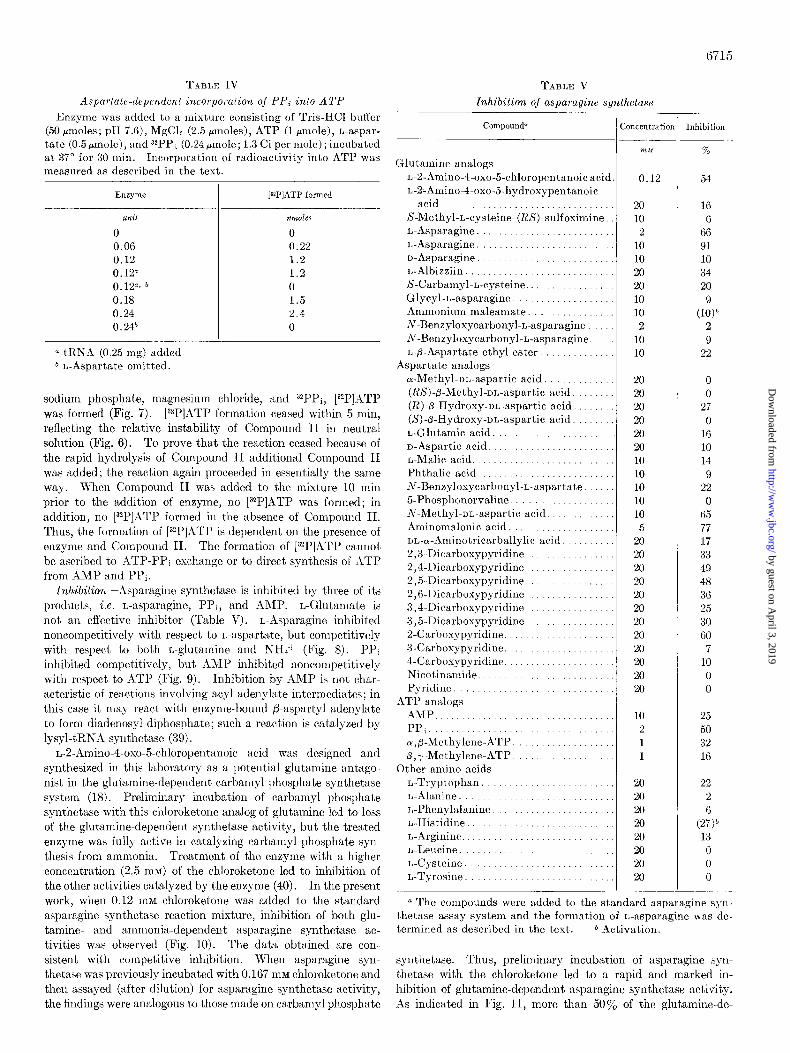
TARLE IV
Aspartate-dependent incorporation of PPi into Al’P
Enzyme was added to a mixture consisting of Tris-IICl buffer (50 pmoles; pH 7.6), MgCln (2.5 pmoles), ATP (1 Hmole), L-aspar-
tate (0.5 pmole), and 3ZPP, (0.24 pmole; 1.3 Ci per mole) ; incubated at 37” for 30 min. Incorporation of radioactivity into ATP was measured as described in the text.
[=P]ATP formed
unit mldes
0 0
0.06 0.22 0.12 1.2 0.12a 1.2 0.12”. b 0 0.18 1.5 0.24 2.4 0.24h 0
a tRNA (0.25 mg) added. b L-Aspartxte omitted.
sodium phosphate, magnesium chloride, and “PPi, [32P]Xl’P was formed (Fig. 7). [3”P]ATP formation ceased within 5 min, reflecting the relative instability of Compound II in neutral solution (Fig. 6). To prove that the reaction ceased because of the rapid hydrolysis of Compound II additional Compound II was added; the reaction again proceeded in essentially the same way. When Compound II was added to the mixture 10 min prior to the addition of enzyme, no [V]ATP was formed; in addition, no [32P]ATP formed in the absence of Compound II. Thus, the formation of [32P]ATP is dependent on the presence of enzyme and Compound II. The formation of [32P]ATP cannot be ascribed to ITP-PPi exchange or to direct synthesis of ATP from AMP and PPi.
Inhibition--,4sparagille synthetase is inhibited by three of its products, i.e. L-asparagine, PPi, and BMP. L-Glutamate is not an effective inhibitor (Table V). L-Asparagine inhibited noncompetitively with respect to L-aspartate, but competitively with respect to both L-glutamine and NHd+ (Fig. 8). 1’Pi inhibited competitively, but AMP inhibited noncompetitively with respect to ATP (Fig. 9). Inhibition by AMP is not char- acteristic of reactions involving acyl adenylate intermediates; in this case it may react with enzyme-bound fi-aspartyl adenylate to form diadenosyl diphosphate; such a reaction is catnlyzed by lysyl-tRNA synthetase (39).
L-2-Amino-4-oxo-5-chloropentanoic acid was designed and synthesized in this laboratory as a potential glutamine antago- nist in the glutamine-dependent carbamyl phosphate synthetase system (18). Preliminary incubation of carbamyl phosphate synthetase with this chloroketone analog of glutamine led to loss of the glutamine-dependent synthetase activity, but the treated enzyme was fully active in catalyzing carbamyl phosphate syn- thesis from ammonia. Treatment of the enzyme with a higher concentration (2.5 mM) of the chloroketone led to inhibition of the other activities catalyzed by the enzyme (40). In the present work, when 0.12 mM chloroketone was added to the standard asparagine synthetase reaction mixture, inhibition of both glu- tamine- and arnrnonia-dependent asparagine synthetase ac- tivities was observed (Fig. 10). The data obtained are con- sistent with competitive inhibition. When asparagine syn- thetase was previously incubated with 0.167 mM chloroketone and then assayed (after dilution) for asparagine synthetase activity, the findings were analogous to those made on carbamyl phosphate
Ta~m V
Znhibition of asparagine synthetase
Compounda ( kmcentration Inhibition
1. Glutamine analogs
L-P-Amino-4-oxo-5.chloropentanoic acic L-2-Amino-4.oxo-5-hydroxypentanoic
acid .............................. S-Methyl-L-cysteine-(ES)-sulfoximine, L-Asparagine ........................
r,-Asparagine ......................... n-Asparagine ........................ L-Albizziin ............ ... .......... S-Carbamyl-L-cysteine ...............
Glycyl-L-asparagine .................. Ammonium maleamate. .. ........... N-Benzyloxycarbonyl-L-asparagine ....
N-Benzyloxycarbonyl-L-asparagine. .. L-fl-Aspartate ethyl ester. ............
Aspartate analogs
or-Methyl-oL-aspart,ic acid. .......... (k%‘-p-Methyl-or,-aspartic acid. ...... (K)#-Hydroxy-nL-aspartic acid. .....
(S)-fl-Hydroxy-nL-aspartic acid. ...... L-Glutamic acid. .......... .......... n-Aspartic acid. ...................... L-Malic acid. ....................... Phthalic acid. ..................... N-Benzyloxycarbonyl-L-aspartate ..... 5-Phosphonorvaline. ...............
iv-Methyl-DL-aspartic acid ............ Aminomalonic acid. .......... ...... uL-wAminotricarballylic acid. ......
2,3-Dicarboxypyridine ......... 2,4-Dicarboxypyridine .......
2,5-Dicarboxypyridine. .......... 2,6-Dicarboxypyridine ............. 3,4-Dicarboxypyridine ........... 3,5-Dicarboxypyridine ............
2-Carboxypyridine. ................ 3-Carboxypyridine. ............ 4Xarboxypyridine. ............
Nicotinamide. .... ................ Pyridine ............. .........
ATP analogs
AMP ............................ PPi. .................... ...... a,@-Methylene-ATP ............... p,r-Mcthylene-ATP ................
Other amino acids L-Tryptophan ..................... L-Alanine ................ ....... r,-Phenylalanine ......... ..... L-Histidine .................. ......
L-Arginine .......................... 1,.Leucine ................. ......... L-Cysteine ........................ L-Tyrosine .........................
??&.+I
0.12
20 10
2
10 10 20
20 10 10
2 10 10
20 20
20 20 20
20 10 10 10
10 10 5
20
20 20 20
20 20 20
20 20 20
20 20
10
2 1 1
20 20
20 20 20
20 20 20
%
54
16
6 66 91 10
34 20
(1:)” 2 9
22
0 0
27 0
16 10 14
9
22 0
65
77 17 33
49 48 36 25
30 60
7 10
0 0
25 50
32 16
22 2
(2& 13
0 0 0
a The compounds were added to the standard asparagine syn- thetase assay system and the formation of L-asparagine was de- termined as described in the text. b Activation.
synthetase. Thus, preliminary incubation of asparagine syn- thetase with the chloroketone led to a rapid and marked in- hibition of glutamine-dependent asparagine synthetase activity. As indicat’ed in Fig. 11, more than 50% of the glutamine-de-
by guest on April 3, 2019
http://ww
w.jbc.org/
Dow
nloaded from

6716
8 z?) 3
$8 R
8 0 -
2 mM L-ASPARAGINE
I 2 3 L-ASPARTATE (mM)
I I I I I I
6 12 18 L- GLUTAMINE (mM)
20 mM L-ASPARAGINE
20 40 60 80 100
NH, Cl (mM)
Fro. 8. Inhibition of asparagine synthetase by L-asparagine. Ji:nzyme was added to the standard synthetase assay mixture con- taining varying amounts of 1,.aspartate (A), L-glutamine (H), or NH,Cl (C). r,-Asparagine was added at t,he given concentrations. Odinale, L-[‘%]asparagine formed (coLmts per min).
I > F 2;
-&
; z 0.5 m m PPi
0.2 0.4 0.2 0.4 0.6
ATP (mM)
FIG. 9. Inhibition of asparagine synthetase by PPi and AMP. Ia:nzyme was added to the standard asparagine synthetase assay mixtllre containing varying amounts of ATP. PPi (A) and AMP (IZ) were added at the concentrations indicated. Ordi~~ate, I,- [‘4C]asparagine formed (counts per min).
l’elldeilt asparagine synthetase activity disappeared after 1
min of preliminary incubation aud about 90% of the activity
disappeared within 20 min (Curve 2); the glutaminase activity
followed a virtually parallel course (Curve 3). On the other
hand, the ammonia-depelrdellt asparagiue synthetase activity
was inhibited ouly slightly under these conditions. Wheu
preliminary incubation with chloroketone was carried out in the
presence of L-glutamine, much less inhibition of the glutamine-
depeudeut asparagine sgnthetnse activity was observed (Fig. 11,
sing/e point). The f?ildillgs ilrdicate that preliminary incubatiou
of the enzyme with the chloroketone leads to irreversible in-
activation of the enzyme aud therefore suggest that the chlo-
roketone biuds to the enzyme as has been shown for carbamyl
phosphate synthetase (40). To investigat.e such binding as-
paragine syuthet,ase was previously iucubated with LLamino-
6 12 I6 L-GLUTAMINE (mM)
I 1 I I I
50 100’ %
NH, Cl (mM1
0
FIN;. 10. Inhibition of asparagine synthetase by 1,.2.amino-4- oxo-5-chloropent,anoate. Enzyme was added to the standard synthetase assay mixtlu-e containing varying concentrations of L-glutamine (A) or NH,Cl (11). The chloroketone was added as indicated. Ord+rlale, L-[‘%]asparagine formed (co\mts per min).
4-oxo-5-chloro[5-14C]pentanoic acid and the reaction mixture
was then subjected to gel filtration on a column of Sephadex G-
25. ~1s indicated in Fig. 12, a substantial amount of radio-
activity emerged from the column with the protein. In a corn-
parable experiment with an enzyme l)rel)aration that exhibited
a specific activity of 2.2 units per mg, 7.7 nmoles of chloroketone
were bound per mg of protein; when glutsmiue (0.1 M) was
present during preliminary iucubatiou, only 4.4 nmoles of chlo-
roketoue were bound. The data therefore indicate that about
3.3 runoles of chloroketone are bound l)er mg of ljrotein to glu-
tamine-protectable sites on the enzyme.
Substantial inhibition of the enzyme was found with amino-
malouate; thus, when 1 mM aminomnlonate was added to the
standard ghltamine-deperlderlt asparagitle syuthetase assay
by guest on April 3, 2019
http://ww
w.jbc.org/
Dow
nloaded from

6717
5 IO 15 20 15 20 25 30 35 40 5 IO
MINUTES FRACTION INHIBITOR CONCENTRATION ImM)
FIG. 11 (left). Effect of preincubating enzyme with L-2.amino- 4.oxo&chloropentanoate (0.167 mM). Aliquots (0.15 ml) were removed at the times indicated and added to the synthetase assay mixture (final volume, 0.5 ml) containing either L-glutamine (Curve 2) or NH&l (Curve 1). Enzyme was also added to the standard glutaminase assay mixture (Curve 3). Asparagine (Curves 1 and 2) and glutamate (Curve 3) were determined. The effect of including L-glutamine (33.3 mM) during the preliminary incubation period on the inactivation of the L-glutamine-depend- ent synthetase was determined (single point, control).
FIG. 12 (center). Binding of L-2-amino-4-oxo-5-chloropentano- ate to enzyme. Enzyme (4.16 mg of protein; 2.4 units synthetase) was previously incubated at 20” with [5J4C]chloroketone (0.35 pmole; 1.13 Ci per mole) in a final volume of 2.2 ml. After 8 min
L-ASPARTATE (mMJ
FIG. 14. Inhibition of asparagine synthetase by aminomalonnte (12) and by N-methyl-nz-aspartate (B). Enzyme was added to the standard synthetase assay mixture containing varying amounts of I,-aspartate. Asparagine formation was determined.
system the activity was inhibited about 40% (Fig. 13). The
data given in Fig. 14 indicate that aminomalonate acts as a
competitive inhibitor with respect to L-aspartate. When amino- malonate was substituted for L-aspartate in the system used for
the study of the ATP-l’l’i exchange reaction, it was found to be
12% as effective as L-aspartate in stimulating the incorporation
of pyrophosphate into ATI?. This observation suggested that
aminomalonic acid might serve as a substrate in the synthetase
reaction and therefore that aminomalonamic acid might be
synthesized. To test this possibility, [2-14C]nn~inomalonate
was incubated with the enzyme in the standard glutamine-
dependent asparagine synthetase system (lacking aspartate)
and the reaction mixture was examined by paper electrophoresis
for the formation of [14C]aminomalonamate. No evidence for
the synthesis of this amide was obtained.
;\nother effective inhibitor of asparagine synthetase activit?
is N-methyl-uL-aspartate; at a concentration of 2 m&f, about 24%
this solution was quickly chilled to 4” and passed through a Seph- adex G-25 column (37 X 1.1 cm) eqnilihrated and eluted with O.OG M Tris-HCI buffer (pH 7.6) containing 1 mM l>TT and 0.5 rnM EDTA. The flow rate was 15 ml per hour. One-milliliter frac- tions were collected. The rllso (0) was determined. illiqllots (10 kl) of the fractions were monitored for radioactivity by counting in 5 ml of Bray’s solution (25) with a scintillation counter (0). The data were corrected for background.
FIG. 13 (right). Inhibition of asparagine synthetase by amino- malonate (Curve 1) and by i\r-methyl-nL-aspartate (Curve 2). Enzyme was added to the standard synthetase assay mixture con- taining aminomalonate or N-methyl-nr,-aspartate. Asparagine formation was determined.
inhibition was observed (Fig. 13). In view of the specificity of
the eiizyme toward L-aspartate, it seems probable that only the
L isomer of N-methyl-nL-aspartate inhibits. The data given in
Fig. 14 indicate that this compourld acts as a competitive it]-
hibitor with respect to L-aspartate. N-1Llethyl-nL-asl)art~~te did not promote incorporatiol~ of pyrophosphate into -1Tl’ under
the conditions described in Table IV.
A number of other compounds were tested for their ability to inhibit asparagine syilthetase activity; n summary of this surve)
is given in Table V. Inhibitioll by quinolinate may reflect the structural resernblanre between this compound and aspartate.
However, substxntial inhibition was also observed with the other dicarboxypyridine derivatives. 2-Carboxypyridine proved to be a much better inhibitor than were the other carboxgppridine
derivatives tested. Several of these compounds were also ex- amined as possible inhibitors of the glutaminase activity of the
enzyme (Table VI). It seems notable that the effects of the coml)ounds tested were essent.ially parallel to the data obtailred on glutamille-dependent asparagine synthetase activity.
I~IHCUSSION
The findings indicate that aspsragine synthetase from R.\ I ),il cells is a glutamine amidotransferase that catalyzes the coul)led synthesis of asparagine and the cleavage of hT1’ to hM1’ and
iiiorganic p.yropliosphate. While it was suggested previously ill
studies 011 bacterial (5-7) alld tumor (9) asparagine synthetases that P-L-aspartyl adenylate is an intermediate in the reaction, direct evidence for such an intermediate has been lacking. AS
shown here, the purified asparagine synthetase catalyzes the synthesis of ATl’ front chemically synthesized p-L-asparty
adenylate and pyrophosphate. ‘This observation, which is analogous to earlier firrdiilgs on the a-aminoacyl-RNh syl~-
thetases (41-43), offers strong support for the formation of ellzyme-bound /!-L-aspartyl adellglate in the reaction.
hsparagine synthetase from R.WAl cells exhibits two rather
by guest on April 3, 2019
http://ww
w.jbc.org/
Dow
nloaded from

6715
TIHLC V1
Inhibition oj asparagine synthetase and glulaminasc by ghtamine analogs
Thr compounds were added to the standard glutnminase and asparngine synthetasc assay mixtures and t,he format,ion of I,- gllltamate and of rA-asparagine was determined as described in the text. -
Compound
I‘-Asparagine. L-2-Amino-4-oxo-5-chloro-
pentanoic acid. n-Asparagine L-Albizziin. S-Carhamyl-L-cysteine. Glycyl-I-asparagine
Concentration
iii I T
2
10 10 20 20
10
Inhibition
Asparagine synthetase
%
66
91 10 34 20
9
Xutaminase
~~__ %
72
94 10 48
18 10
interesting characteristics. First, it catalyzes the transfer of the amide nitrogen atom of L-glutamine to water as well as to @-L-aspartyl adenylate. The data presented here provide strong evidence that the glutnminase activity exhibited by the enzyme preparation is a function of asparagine synthetase itself. It seems possible that other rnammalian asparagine synthetases also exhibit glutaminase activity; the asparagiue synthetdse isolated from Novikoff hepatoma (9) was reported to catalyze the formation of equimolar quantities of asparagine, pyrophos- phate, and AMP, but studies 011 the formation of glutamate or ammonia were not reported. A number of the other glutamine amidotransferases that have been studied, for example, authra- nilate synthetase (44, 45)) carbamyl phosphate synthetase (46, 47)) cytidine triphosphdte synthetase (48)) and formylglycin- amidine ribonucleotide synthetase (49), have been found to exhibit glutaminase activity. However, it is notable that in each case stoichiometry was observed between the formation of L-glutamate and the other products. The present findings on asparagine synthetase are unique in that the step involving transfer of the amide group of the glutamine is apparently UKI- coupled from the rest of the reaction. It is possible that such uncoupling is associated with modification of the enzyme during isolation. A somewhat similar phenomenon has been observed with carbamyl phosphate synthetase; when this enzyme is stored at $1 9 at 0” the glutaminase activity increases markedly and the sgnthetidse activity decreases.* It is also conceivable that the uncoupling effect observed wit.11 asparagine synthetase reflects a novel type of cellular regulatory mechanism; however, such a mechanism for the control of asparagine synthesis would seem to be relatively costly in terms of energy utilization.
.1 second interesting property of asparagine synthetase re- vealed by the present studies is that the transfer of the amide nitrogen atom of L-glutamine to acceptor requires chloride or, with somewhat less activity, bromide or iodide. Since the ammonium-dependent asparagine sgnthetase activity does not require chloride, and since the glutaminase activity exhib- ited by the enzyme requires chloride it appears that only the amidotransferase function is anioll-mediated. Dependence of enzyme activity on anions does not seem to be a common ob- servation, although it is not clear from an examination of the
4 V. 1’. Wellner and A. Meister, unpublished (see also lteference 47).
literature that this proljerty has been thoroughly studied. The effect of anions on hydrolytic enzymes has been examined; thus, amylanes (50@53), certairl peptidases (54&57), cyclophospho- diesterase (58), sulfataae (59), fumarase (60), AMP deaminase (61), and a bacterial glutaminase (62), have been reported to be stimulated by chloride. ‘fhere is considerable literature on the stimulation of glutaminases by phosphate (see example, References 63-66). However we have found no reports sug- gesting that other glutamille amidotransferases may be signifi- cantly stimulated by anions. It may be noted that our finding that asparagine synthetase is aniorl-mediated was serendipitous, and occurred only after our study of the glutaminase was initiated when experirnents in which MgCls was omitted were carried out. The possibility camlot be excluded that chloride may be required for other related enzymes. The mechanism by which chloride acts is not clear; there is evidence that anions can stimulate enzymatic activity by illcreasing their stability under the con- ditions of assay (52-65)) by shifting the pH dependence to a rnore basic region (51, 52, 58, 67-69), and by increasing the affinity for substrate (58, 66, 68). It has bee11 known for some time that chloride ion exerts a specific effect in protecting snake venom L-
amino acid oxidase against reversible inactivation (70). Several substrate analogs have been found to be particularly
effective as inhibitors of asparagine synthetase. One of these, L-2-amino-4-oxo-5-chloropentanoic acid, acts as a glutamine antagonist. The data are ill accord with the view that this chloroketone derivative binds to the enzyme by forming a cova- lent linkage. In analogous studies on carbamyl phosphate synthetase, the chloroketone was shown to bind to a cysteme residue at the glutamine binding site of the enzyme (40). Pre- liminary incubation of asparagine synthetase and of carbamyl phosphate synthetase with the chloroketone yields an enzyme which car1 110 longer function with glutamine but which can still use ammonia for synthesis. The chloroketone may be useful in structural studies of asparagine synthetase. Several other compounds which seem to have some promise as inhibitors of the enzyme are listed in Table V. These, or similar compounds may be useful when administered with asparaginase in the ther- apy of tumors; such a11 approach may lead to a therapy for asparaginase-resistant tumors.
Aclcnowledgmenls-We wish to thank Xtr. Robert Young for his valuable help in preparing the tumor cells and Mrs. Susan Lord Lundt for her assistance in a number of the experiments.
1. LESS, E. M., FARNDEN, K. J. F., .IND I~LI,IOTT, W. I-T. (19G8) Arch. Biochem. Biophys. 126, 539-546
2. KESSLER. C.. N2\~.l~.~~2~~, G. R., .YNI) L.~UINGI:R, C. (1!)69) Biochik. kophys. Ada i84, 578-582
3. NAIR. P. IV. (1969) Arch. Biochem. Biovhus. 133. 208 4. lto~N&s, S. Ed. (19iO) Fed. Eur. Biochek. >oc. L&t. 10, G2 5. I~VEL, J. M., NORTON, S. J., HUMPHRISYS, J. S., AND SJUVE,
W. (1962) J. Biol. Chem. 237, 2845-2849 6. BUR~HALL, J. J., RIECHELT, E. C., I~~~ WOLIN, R/l. J. (1964)
J. Biol. Chem. 239, 1794-1798 7. CEDAR, H., AND SCHWAR.~Z, J. H. (1969) J. Biol. Chem. 244, 4112 8. ARFIN, 8. M. (1967) Biochim. Biophys. Acta 136, 233-244 9. PATTERSON, M. K., AND ORR, G. It. (1968) J. Biol. Chem. 243,
376 10. HOIEENBERG, J. S. (1969) B&him. Biophys. Ada 186, 228-238 11. CIIOU, T. C., .~;uD HINDSCHUMACHER, li. 15. (1970) Fed. Proc.
29, 407 12. H~SKELL, C. M., .SND C.INIX,J,OS, G. P. (1970) Cancer Kes. 30,
1081 13. LKVINTO~-, L. (1957) Science 126, 011
by guest on April 3, 2019
http://ww
w.jbc.org/
Dow
nloaded from
14. HOROWITZ, B., MADRAS, B. K., M~ISIXH, A., OLD, L. J., BOYSE, E. A., AND STOCKERT, E. (1968) Science 160, 533-535
15. PATTERSON, M. K., JR., AND ORR, G. (1967) Biochem. Biophys. Res. Commun. 26, 228
16. BROOME, J. D., AND SCHWARTZ, J. H. (1967) Biochim. Biophys. Acta 138, 637-639
17. PRAGER, M. D., AND BACHYNSKY, H. (1968) Arch. Biochem. Biophys. 127, 645-654
18. KHEDOURI, E., ANDERSON, P. M., AND MEISTER, A. (1966) Biochemistry’6, 3552
19. MATTHB:W. M.. AND NIZUBI<RUF;R. A. (1963) Biochem. J.87.601-612 26. MEISTER,'A.,'LX~INTOW, L., G&E'NF&LD, R. E., AND'ABEND-
SCHEIN, P. A. (1955) J. Biol. Chem. 216, 441 21. TATE:, S., AND MXISTFX, A. (1968) Biochemistry 7, 3240 22. MOLDAVE, K. (1963) Methods Enzymol. 6, 757 23. OLD, L. J., BOYSI,:, 1~~. A., AND STOCKERT, E. (1963) J. Nut.
Cancer Inst. 31, 977 24. LOWRY, 0. H., ROSEBROUGH, N. J., FARR, A. L., AND RANDALL,
It. J. (1951) J. Biol. Chem. 193, 265-275 25. BRAY, G. A. (1960) Anal. Biochem. 1, 279-285 26. BERNT, E., AND BERGMEYER, H. U. (1965) in Methods of Enzy-
matic Analysis (BERGMKYER, H. U., ed) p. 384, Academic Press, New York
27. ADAM, H. (1965) in Methods of Enzymatic Analysis (BERG- MEYER, H. U., ed) p. 573, Academic Press, New York
28. DRYER, R. L., TAMMIZS, A. It., AND ROUTH, J. H. (1957) J. Biol. Chem. 226, 177
29. SATO, T. R., THOMSON, J. F., AND DANFORTH, W. F. (1963) Anal. Biochem. 6, 542
30. CHAYKIN, S. (1969) Anal. Biochem. 31, 375 31. DAVIS, B. J. (1964) Ann. N. Y. Acad. Sci. 121, 404 32. ANDREWS, P. (1964) Biochem. J. 91, 222-233 33. HWANG, L. N., .IND ZALKIN, H. (1971) J. Biol. Chem. 246,2338-
2345 34. HOGEBOOM, G. H. (1955) Methods Enzymol. 1, 16 35. BUSCH, H. (1967) Methods Enzymol. 12A, 421 36. MOLDAVE, K., CASTELFRANCO, P. , AND MI<:ISTER, A. (1959) J.
Biol. Chem. 234, 841 37. COLES, N., BUKENBERGER, M. W., AND MF:IST*:R, A. (1962)
Biochemistry 1, 317 38. STADTMAN, E. It., AA-D BARKER, H. A. (1950) J. Biol. Chem.
184, 769 39. ZAMECNX, P. C., STICPHENSON, M. L., JANI)~AY, C. M., AND
RANDVRATH, K. (1966) Biochewz. Biophys. Res. Commun. 24, 91
40. PINKUS. L M.. AND M>:~srr~:a A. (1972) J. Biol. Chem. 247,6119- 6127 ’ ’
41. DeMoss, J. A., GENUTH, S. M., BND NOVELLI, G. D. (1956) Proc. Nat. Acad. 42, 325
6719
42. BXXG, 1’. (1958) J. Biol. Chem. 233, 601 43. KRISHNASWAMY. P. I~.. AND MINISTER. A. (1960) J. Biol. Chetn.
236, 408 ’ ’ ,
44. NAGANO, I-I., ZALKIN, H., AND HENDI~RSON, E. J. (1970) J. Biol. Chem. 246. 3810-3820
45. TAMIR, H., AND SRINIVASAN, P. R. (1971) J. Biol. Chew 246, 3024-3029
46. TATIBANA, M., .~ND ITO, K. (1969) J. Biol. Chem. 244, 540% 5413
47. PINICUS, L., W~LLNER, V. P., .~XD M~:IsTI~:R, A. (1972) Fed. Proc. 31, 474
48. LEVITZKI, A., .~ND KOSHLAND, D. E., JIX. (1971) Biochemistry 10, 3365
49. LI, H.-C., AND BUCHANAN, J. M. (1971) J. Biol. Chem. 246, 4713-4719
50. COLE, S. W. (1904) J. Physiol30, 202 51. MYRB~CK. K. (1926) Howwe-Seuler’s Z. Phusiol. Chem. 169. 1 52. Muus, J.; BR&K&T, F: P.,“AND CONNOLLY, C. C. (lb56)
Arch. Biochem. Biophys. 66, 268 53. illuus, J. (1953) C. R. l’rav. Lab. Carlsberg Ser. Chim. 23, 317 54. KAKIUCHI, S., AND TOMIZAWA, H. H. (1964) J. Biol. Chem. 239,
2160-2164 55. SKEGGS, L. T., JR., MARSH, W. H., K.?HN, J. It., AND SHUWU-.\Y,
N. P. (1954) J. Exp. Med. 99, 275 56. JOHNSON, M. J. (1941) J. Biol. Chem. 137, 575 57. MCDONALD, J. K., ELLIS, S., AND REILLY, T. J. (1966) J. Biol.
Chem. 241, 1494-1501 58. UNIXMOTO, T., BND HAYASHI, M. (1969) Biohcim. Biophlys. Acta
171, 89-102 59. WEBB, E. C., AND MORROW, P. F. W. (1959) Biochem. J. 73, 7 60. Mass~c~, V. (1953) Biochewz. J. 63, 67 61. ASKAHI, A. (1966) Mol. Pharmacol. 2, 518 62. HUGHXS, 1). E., AND WILLIAMSON, D. H. (1952) Biochem. J.
61, 45 63. CARTEIS, C. E., AND GRI<I,:NSTRIN, J. P. (1947) J. Nat. Cancer
Inst. 7, 433 64. ERRERA, M., AND GREENSTXIN, J. P. (1949) J. Biol. Chem. 178,
495 65. KLINGMAN, J. D., AND H,~NDIZI~, P. (1958) J. Biol. Chews.
232, 369 66. SAYRIC, T. W., AND ROIJIYXTS, E. (1958) J. Biol. Chem. 233 1128 67. CALDWELL, M. L., AND KUNG, J. T. (1953) J. Amer. Chews.
Sot. 76, 3132 68. SCIIUBIXTH, J. (1966) Biochim. Biophys. Acta 122, 470 69. REYES, P., AND HUENNISKI:NS, F. M. (1967) Biochemistry 6,
3519 70. KEARNXY, 13. B., AND SINGER, T. P. (1951) Arch. Biochewb.
Biophys. 33, 397-413
by guest on April 3, 2019
http://ww
w.jbc.org/
Dow
nloaded from

Bernard Horowitz and Alton MeisterDEPENDENCE, MECHANISM OF ACTION, AND INHIBITION
Glutamine-dependent Asparagine Synthetase from Leukemia Cells: CHLORIDE
1972, 247:6708-6719.J. Biol. Chem.
http://www.jbc.org/content/247/20/6708Access the most updated version of this article at
Alerts:
When a correction for this article is posted•
When this article is cited•
to choose from all of JBC's e-mail alertsClick here
http://www.jbc.org/content/247/20/6708.full.html#ref-list-1
This article cites 0 references, 0 of which can be accessed free at
by guest on April 3, 2019
http://ww
w.jbc.org/
Dow
nloaded from