Embed Size (px)
Citation preview
REVIEWS
Glutathione and the response of malignant ,cells to chemotherapy Pamela R. Kearns and Andrew G. Hall
In the past, the role of glutathione (GSH) in the
production of drug resistance was assumed to be
limited to electrophilic agents such as the bifunctional
alkylating agents and cisptatin. Recently, however,
evidence has emerged that links GSH homeostasis with
regulation of the multidrug-resistance-related protein
and the control of apoptotic cell death. These findings
open up new avenues for the discovery of drugs that
augment anticancer agents already n use or that have
cytotoxic potential in their own right
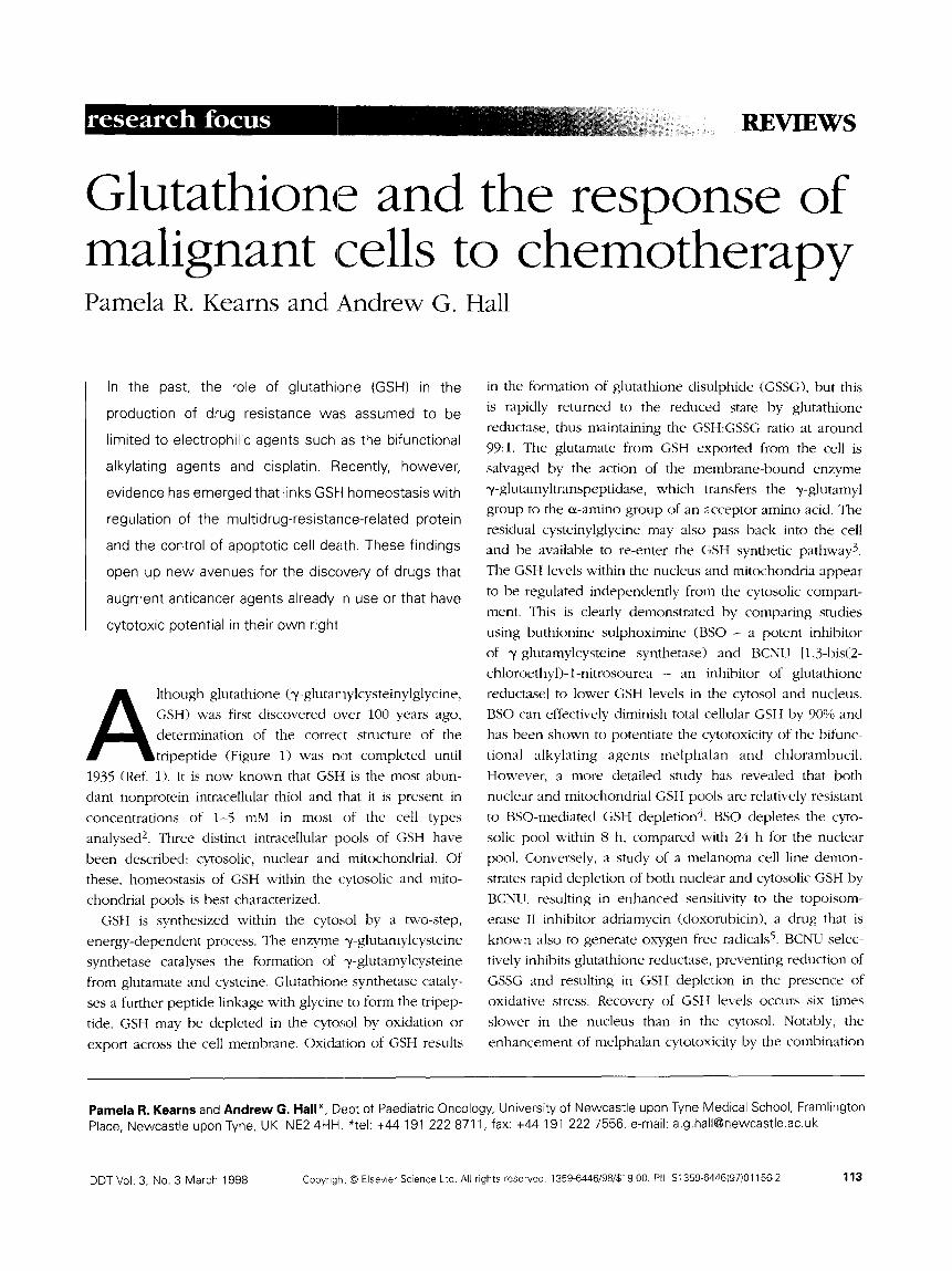
A lthough glutathione (y-glutamylcysteinylglycine,
GSH) was first discovered over 100 years ago,
determination of the correct structure of the
tripeptide (Figure 1) was not completed until
1935 (Ref. 1). It is now known that GSH is the m o s t abun-
dant nonprotein intracellular thiol and that it is present in
concentrations of 1-5 mM in most of the cell types
analysed 2. Three distinct intracellular pools of GSH have
been described: cytosolic, nuclear and mitochondrial. Of
these, homeostasis of GSH within the cytosolic and mito-
chondrial pools is best characterized.
GSH is synthesized within the cytosol by a two-step,
energy-depenctent process. The enzyme y-glutamylcysteine
synthetase catalyses the formation of y-glutamylcysteine
from glutamate and cysteine. Glutathione synthetase cataly-
ses a further peptide linkage with glycine to form the tripep-
tide. GSH may be depleted in the cytosol by oxidation or
export across the cell membrane. Oxidation of GSH results
in the formation of glutathione disulphide (GSSG), but this
is rapidly returned to the reduced state by glutathione
reductase, thus maintaining the GSH:GSSG ratio at around
99:1. The glutamate from GSH exported from the cell is
salvaged by the action of the membrane-bound enzyme
y-glutamyltranspeptidase, which transfers the y-glutamyl
group to the e~-amino group of an acceptor amino acid. The
residual cysteinylglycine may also pass back into tile cell
and be available to re-enter the GSH synthetic pathway 3.
The GSH levels within the nucleus and mitochondria appear
to be regulated independently from the cytosolic compart-
ment. This is clearly demonstrated by comparing studies
using buthionine sulphoximine (BSO - a potent inhibitor
of y-glutamylcysteine synthetase) and BCNU [1,3-bis(2-
chloroethyl)-l-nitrosourea - an inhibitor of glutathione
reductase] to lower GSH levels in the cytosol and nucleus.
BSO can effectively diminish total cellular GSH by 90% and
has been shown to potentiate the ck~totoxicity of the bifunc-
tional alkylating agents melphalan and chlorambucil.
However, a more detailed study has revealed that both
nuclear and mitochondrial GSH pools are relatively resistant
to BSO-mediated GSH depletion 4. BSO depletes the cyto-
solic pool within 8 11, compared with 24 h for the nuclear
pool. Conversely, a study of a melanoma cell line demon-
strates rapid depletion of both nuclear and cytosolic GSH by
BCNU, resulting in enhanced sensitivity" to the topoisom-
erase II inhibitor adriamycin (doxorubicin), a drug that is
known also to generate oxygen free radicals 5. BCNU selec-
tively inhibits glutathione reductase, preventing reduction of
GSSG and resulting in GSH depletion in the presence of
oxidative stress. Recove W of GSH levels occurs six times
slower in the nucleus than in the cytosol. Notably, the
enhancement of melphalan cytotoxicity by the combination
Pamela R. Kearns and Andrew G. Hall*, Dept of Paediatric Oncology, University of Newcastle upon Tyne Medical School, Framlington Place, Newcastle upon Tyne, UK NE2 4HH. *tel: +44 191 222 871 1, fax: +44 191 222 7556, e-mail: [email protected]
DDT Vol. 3, No. 3 March 1998 Copyrigh: © Elsevier Science Ltd. All rights reserved. 1359-6446/98/$19.00. Pit: $1359-6446(97)01156-2 113
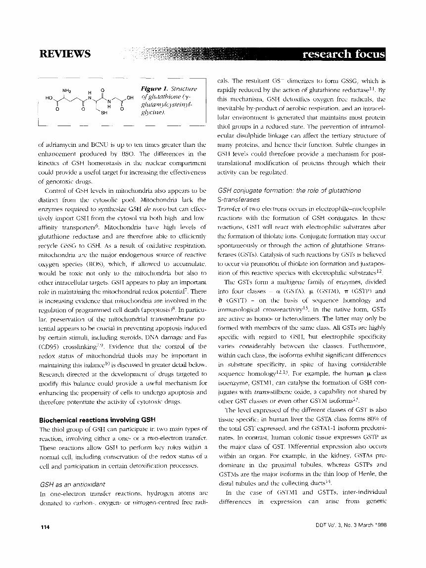
REVIEVCS
NH2 O F i g u r e 1. Structure H
HO ]-- ~ N . J L - - - . -OH ofgh, tathione (T-
gO.'cine).
of adriamycin and BCNU is tip to ten times greater than the
enhancement produced by BSO. The differences in the
kinetics of GSH homeostasis in the nuclear compartment
could provide a useful target for increasing the effectiveness
of genotoxic drugs.
Control of GSH levels in mitochondria also appears to be
distinct from the cytosolic pool. Mitochondria lack the
enzymes required to synthesize GSH de ,aovo but can effec-
tively import GSH from the cytosol via both high- and low-
affinity transporters 6. Mitochondria have high levels of
glutathione reductase and are therefore able to efficiently
recycle GSSG to GSH. As a result of oxidative respiration,
mitochondria are the major endogenous source of reactive
oxygen species (ROS), which, if allowed to accumulate,
would be toxic: not only to the mitochondria but also to
other intracellular targets. GSH appears to play an important
role in maintaining the mitochondrial redox potential 7, There
is increasing evidence that mitochondria are involved in the
regulation of programmed cell death (apoptosis) 8. In particu-
lar, preservation of the mitochondrial t ransmembrane po-
tential appears to be crucial in preventing apoptosis induced
by certain stimuli, including steroids, DNA damage and Fas
(CD95) crosslinking 7'9. Evidence that the control of the
redox status of mitochondrial thiols may be important in
maintaining this balance l° is discussed in greater detail txelow.
Research directed at the development o1: drugs targeted to
modify this balance could provide a useful mechanism for
enhancing the propensity of cells to undergo apoptosis and
therefore potentiate the activity of cytotoxic drugs.
Biochemical reactions involving GSH The thiol group of GSH can participate ir two main types of
reaction, involving either a one- or a two-electron transfer.
These reactions allow GSH to perform key roles within a
normal cell, including conservation of the redox status of a
cell and participation in certain detoxification processes.
GSH as an antioxidant In one-electron transfer reactions, hydrogen atoms are
donated to carbon-, oxygen- or nitrogen-centred free radi-
cals. The resultant GS dimerizes to form GSSG, which is
rapidly redtlced by the action of glutathione reductase li. By
this mechanism, GSH detoxifies oxygen free radicals, the
inevitable by-product of aerobic respiration, and an intracel-
lular environment is generated that maintains most protein
thiol groups in a reduced state. The prevention of intramol-
ecular disulphide linkage can affect the tertiary structure of
many proteins, and hence their function. Subtle changes in
GSH levels could therefore provide a mechanism for post-
translational modification of proteins through which their
activity can be regulated.
GSH conjugate formation. the role of glutathione
S-transferases Transfer o f two electrons occurs in electrophile-nucleophile
reactions with the tormation of GSH conjugates. In these
reactions, GSH will react with electrophilic substrates after
the formation of thiolate ions. Conjugate formation may occur
spontaneously or through the action of glutathione S-trans-
ferases (GSTs). Catalysis of such reactions by GSTs is believed
to occur via promotion of thiolate ion formation and juxtapos-
ition of this reactive species with electrophilic substrates 12.
The GSTs form a multigene family of enzymes, divided
into four classes - ot (GSTA), I* (GSTM), = (GSTP) and
O (GSTT) - on the basis of sequence homolo#" and
immunological crossreactivity 13. In the native form, GSTs
are active as homo- or heterodimers. The latter may only be
formed with members of the same class. All GSTs are highly
specific with regard to GSH, but electrophile specificity
varies considerably between the classes. Furthermore,
within each class, the isoforms exhibit significant differences
in substrate specificity, in spite of having considerable
sequence homology 12,13. For example, the human Ix-class
isoenzyme, GSTM1, can catalyse the formation of GSH con-
iugates with trans-stilbene oxide, a capability not shared by
other GST classes or even other GSTM isoforms 13.
The level expressed of the different classes of GST is also
tissue specific; in human liver the GSTA class forms 80% of
the total GST expressed, and the GSTAI-1 isoform predomi-
nates. In contrast, human colonic tissue expresses GSTP as
the major class of GST. Differential expression also occurs
within an organ. For example, in the kidney, GSTAs pre-
dominate in the proximal tubules, whereas GSTPs and
GSTMs are the maior isoforms in the thin loop of Henle, the
distal tubules and the collecting ducts 14.
In the case of GSTM1 and GSTTs, inter-individual
differences in expression can arise from genetic
114 DDT Vol. 3, No. 3 March 1998
REVIEWS
polymorphism 15,16. In approximately 50Y0 of the Caucasian
population the gene encoding GSTM1 is deleted from both
alleles on chromosome 1. Several studies have emphasized
the importance of this genetic polymorphism in susceptibil-
ity to carcinogenesis, particularly in smokers 17, but no cor-
relation with drug resistance has been reported.
Differences in expression both at the tissue level and
between individuals is of particular sigmficance when con-
sidering the role of GSTs in detoxiFying xenobiotics.
Furthermore, it is important to consider differences in GST
isoform substrate specificity in the interpretation of studies
measuring GST enzyme activity. This i,s exemplified in a
study by Evans and coworkers 18 in which GST denitrozation
of BCNU was studied in two rat glioma cell lines with differ-
ing drug sensitivity. The resistant line had lower GST activ-
ity than the sensitive cell line when the total enzyme activity
was measured with the most commonly used GST substrate,
1-chloro-2,4-dinitrobenzene (CDNB). However, in a subse-
quent study 19, denitrozation was measured directly, and the
resistant cell line was shown to have an elevation in a spe-
cific I~-class isoenzyme responsible for inactivation of
BCNU. This was associated with a decrease in expression of
other GST isoforms with a high specific activity against
CDNB. This emphasizes the importance of establishing the
exact pattern of isoform expression and the substrate speci-
ficity of each isoform before valid interpretation can be
made from studies of GST-mediated drug resistance.
GSH and cytotoxic drugs Correlation between GSH, GST levels and cellular
resistance Over the past three decades, increasing evidence has accu-
mulated supporting the role of GSH and GSTs in cellular
resistance to cytotoxic drugs. Studies have used cell lines,
with comparisons between drug-sensitive and drug-resistant
clones, and samples obtained from a varie D" of tumour
types. The majority of human tumours display increased
levels of certain GST isoenzymes, in particular those of the
GSTM class 2°. Many, but not all, studies have demonstrated
an elevation of GSH levels and GST activity in sublines resist-
ant to bifunctional alkylating agents or cisplatin 2°-22. For
example, analysis of the chlorambucil-resistant Chinese
hamster ovarian carcinoma cell line (CHO-Chl r) showed a
1.8-fold increase in GSH levels and expression of an ~x-form
of GST that was not detectable in parental CHO-K1 cells 23.
In another study, a breast cell line, MCF7, exhibiting adri-
amycin resistance in vitro, demonstrated a 45-fold increase
in GST activity compared with the parent line 24. GST and
GSH levels have also been compared in cell lines derived
from the ovarian adenocarcinoma of a patient before and after
treatment with cisplatin, chlorambucil and 5-fluorouracil.
Elevation of both parameters was associated with the devel-
opment of clinical and in vitro resistance 25.
Further evidence that alterations in GSH and GST levels
are related not only to in vitro drug resistance but also to
clinical response to chemotherapy, comes from a study in
which GST expression in lymphoblasts was examined in 70
children with acute lymphoblastic leukaemia 26. A clear rela-
tionship was demonstrated between expression of GSTMs
and disease-free survival. More recently, in a study of
patients with advanced ovarian carcinoma, GSH levels in
peripheral blood lymphocytes have been found to be sup-
pressed following ifosfamide therapy, and the degree of
suppression appears to correlate with clinical outcome 27.
Evidence correlating increased levels of GSH and GST
with drug resistance is not necessarily evidence of a causal
relationship but may simply represent coexpression with
other proteins responsible for drug resistance. More-direct
evidence for the involvement of GSH/GST in drug resistance
has been obtained from experiments in which either GST
expression is increased by transfection of cDNA or in which
drug sensitivity is assessed following exposure to agents that
reduce GSH levels or inhibit GST activity.
Several groups have successfully transfected plasmids
carrying specific GST genes into several cell lines but have
produced conflicting results. For example, Miyazaki and
coworkers 28 have demonstrated that transfection of human
GSTP into CHO cells is associated with a 2-3-fold increase
in resistance to cisplatin and carboplatin. They also demon-
strated that transfection of GSTA increases resistance to
bleomycin. Conversely, Leyland Jones and coworkers 29
have reported that MCF7 breast carcinoma cells, also trans-
fected with a- and "n-class genes, do not develop resistance
to cisplatin or bifunctional alkylating agents. These results
imply that transfection of GST alone is inadequate to convey
drug resistance in some cell types and that usually the devel-
opment of drug resistance arises from several coordinated
changes in both this and other enzyme systems.
Further information on the specific involvement of GSTs
in drug resistance is derived from inhibition studies. This has
been achieved through the use of agents such as ethacrynic
acid and indomethacin, although interpretation of the
results of these studies is complicated by the fact that these
agents may also affect GSH levels. In a study of CHO-Chl r
DDT Vol. 3, No. 3 March 1998 115
REVIEWS
cells, known to have increased expression of an cx-form of
GST, it was demonstrated that 1 mM indomethacin causes a
partial, but significant, reversal of chlorar, lbucil resistance 3°.
In both rat and human tumour cell lines, sensitization to
alkylating agents has been reported following exposure to
ethacrynic acid 22,31. This was associated with decreased
GST activity towards CDNB and a fall in intraceltular GSH
(Ref. 32). Using immunodeficient mice bearing human
colonic carcinoma, it was shown that the combination of
melphalan and ethacwnic acid enhanced tumour growth
delay 33. Clinical trials of ethacrynic acid in combination with
alkylating agents have been carried out but unfortunately
were complicated by systemic toxicity. Notably, ethacrynic
acid is a potent diuretic and its use resulted in significant
depression of blood pressure. There was also an increase in
systemic toxicity induced by the alkylating agents. The anti-
GST activity of ethacrynic acid is not specific to tumour cells
and results in increased myelosuppression, which proved to
be the most dose-limiting toxicity 34. proliferating cells, it
Figure 2. X-ray c~,stallography of glu,~athione-S-transferase structure (from Web: http..//molbio.infiJ.nih.gov/cgi-bin/moldraw?l GSF).
X-ray crystallography studies of GSTs have clarified their
structure and have advanced the understanding of the
mechanism of their catalytic activity35,3c,. This in turn has
made the development of more isoform-specific GST modu-
lators possible. The GST protein exists in dimers, comprising
two 2.4-26-kDa subunits (206-221 amino acids) (Figure 2).
Each monomer contributes a kinetically independent active
site that has m~o binding regions: a G site for binding GSH
and an H site for binding the hydrophobic electrophile. The
pKfor the cysteine thiol in GSH is around 9.6; however, the
local environment of the active site favours a more neutral
pK, facilitating GSH conjugation with the electrophile.
Studies involving site-directed mutagenesis of key amino
acids within these regions have provided important infor-
mation about the mechanism of catalysis and the way the
(}ST isoforms achieve their different substrate specificities
(reviewed in Ref. 12). If the isoform profile of tumour cells
could be identified and shown to be different from normal
may then be possible to use isoform-
specific GST inhibitors with alkylating
agents to enhance tumour-specif ic
cytotoxicity. Studies are currently
being carried out to identify isoform-
specific inhibitors of GSTs. Exami-
nation of GST enzyme activity using
a series of GSH analogues has shown
that the binding-site shape and lipo-
philicity are key determinants of GST
isozyme selectivity 37. This information,
and improved knowledge of GST iso-
zyme crystal structure, has contributed
to the deve lopment of isozyme-
selective GST inhibitors that could
prove useful adjuvants to chemo-
therapy. The best characterized of
these inhibitors is the GSH analogue
-,/-glut a myl- S-(benzyl)cysteinyl-R( - )-
phenylglycine diethyl ester (TER199) 38.
TER199 is a GSTP-specific inhibitor 39
and has been shown to potentiate the
cytotoxicity of chlorambucil in several
carcinoma cell lines and, in combi-
nation with melphalan, to enhance the
growth delay of xenograft tumours in
severe combined immunodeficient
(SCID) mice 4°. In both these respects,
TER199 seems similar to ethacrynic
116 DDT Vol. 3, No. 3 March 1998
REVIEWS
acid as a modulator of GST activity; however, it also appears
to stimulate cytokine production and growch of bone mar-
row stem cells 38. If this proves to be true in a clinical setting,
TER199 could play an important role :n potentiating the
cytotoxicity of alkylating agents without dose-limiting
myelosuppression.
The specific overexpression of GSTs in tumour cells has
also been exploited in the development of prodrugs acti-
vated by GST Several compounds have been identified
that are activated on reaction with GSH to form cytotoxic
alkylating species: for example, 1,2-dibromoethane and
1 - b e n z o y l - 1 , 2 - b i s ( m e t h y l s u l p h o n y l ) - 2 - ( 2 - c h l o r o e t h y l ) -
hydrazine 12. These reactions are not isoform specific, how'-
ever, and in clinical use are likely to be associated with lim-
iting systemic toxicity. If turnout cells were to overexpress
specific GST isozymes, this would provide a mechanism by
which a drug could be designed to be selectively activated
by that isoform, hence targeting the cytotoxic effect to
tumour cells. It is well established that GSTP is frequently
overexpressed in tumour cells, and TER286 [y-glutamyl-
e~-amino-lg(2-chloroethyl)phosphodiamicLate sulphonylpro-
pionyl-(R)-(-)-phenylglycine] is a prodrug that comprises a
GSTP-specific GSH analogue linked to an analogue of
cyclophosphamide. The cytotoxicity of tlIle drug is masked
until the alkylating component is cleaved from the com-
pound by intracellular GSTP activity. In vitro studies using
recombinant GST proteins showed GSTA to have similar
efficiency as GSTP in cleaving this compound 38. Thus,
although TER286 is not exclusively activated by GSTP, in
view of the high expression of GSTP in tumour cells, this
compound could prove to be clinically advantageous.
Direct evidence for the spontaneous formation of
drug-GSH conjugates has been shown for cisplatin and
the bifunctional alkylating agents melphalan 41 and chloram-
bucil. The reaction of GSH with bifunctional allcylating
agents is accelerated by some forms of GST. This substrate
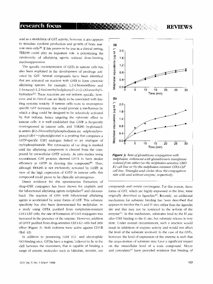
specificity has also been demonstrated for melphalan. In
a study using GSTA purified from melphalan-resistant
CHO-Chl r cells, the rate of formation of GSH conjugates was
increased in the presence of the enzyme. However, addition
of GSTP purified from drug-sensitive CHO-K1 cells had little
effect (Figure 3). Both isoforms were active against CDNB
(Ref. 42).
In addition to possessing GSH (G)- and electrophile
(H)-binding sites, GSTAs have a region, believed to lie in the
cleft between the monomers, that is capable of binding a
range of anionic molecules such as bilirubin, steroids, azo
(a)
2.0- E 03
1.6-
_~ 1.2- t~ >
"~ 0.8-
0.4
~ 0.0 0
(b)
E
._> g ID
t'- t~
..c
1'o l'S 2'o 2's 3'0 Time (min)
2.0-
1.6-
1.2
0.8
0.4
0 . 0 ~ / . . . . 0 5 10 15 20 2'5 3'0
Time (min)
Figure 3. Rate of glutathione conjugation with melphalan, enhanced with glutathione-S-transferase isolated from either (a) the melphalan-sensitive CHO- K1 cell line or (b) the melphalan-resistant CHO-ChP" cell line. Triangles and circles show the congugation rate with and without enzyme, respectively.
compounds and certain carcinogens. For this reason, these
forms of GST, which are highly expressed in the liver, were
originally described as ligandins 43. Recently, an additional
mechanism for substrate binding has been described that
appears to involve the G and H sites rather than the ligandin
site and that may not be restricted to the c~-form of the
enzyme 44. In this mechanism, substrates bind to the H site
after GSH binding to the G site, but substrate release is vew
slow. Under normal circumstances, such a reaction would
result in inhibition of enzyme activity and would not affect
the level of the substrate involved. ]in the case of the GSTs,
however, the level of expression of the enzyme is such that
the sequestration of substrate may have a significant impact
on the intracellular level of a toxic compound. Meyer
and coworkers 44 have provided evidence that binding of
DDT Vol. 3, No. 3 March 1998 117

REglIEW$
chlorambucil to GST may occur at the H site and suggest that
this may be an important factor in the development of drug
resistance. In the study described in Figure 3, measurement
of melphalan-GSH conjugates was performed after protein
precipitation with trifluoroacetic acid, a process that may
liberate sequestered molecules.
Elevation of intracellular GSH is most frequently associ-
ated with development of resistance to bi~unctional alkylat-
ing agents and anthracyclines. The imporl:ance of the intra-
cellular concentration of GSH to cancer therapy was first
recognized over 40 years ago when GSH deplet ion
was shown to sensitize cells to ionizing radiation 45.
Subsequently, studies using the murine leukaemic cell line
L1210 demonstrated that melphalan cytotoxicity could be
modified by changes in GSH levels 46. This was supported by
evidence from studies of human tumour cell lines and
patient samples. Increased GSH levels were shown to be
associated with decreased sensitivity to both platinum-
based drugs and alkylating agents in ova:dan, prostatic and
gastric carcinomas 45,47-5°.
As described above, depletion of GSH levels can be
achieved by inhibition of y-glutamylcysteine synthetase
activity by BSO. Treatment with BSO has been shown to
restore sensitivity to alkylating agents, anthracyclines 51 and
platinum-based drugs in resistant cell lines 52. Furthermore,
in v ivo studies in athymic nude mice bearing the human
ovarian cancer cell line NIH:OVCAR-3 intraperitoneally
demonstrated that the combination of BSO and melphalan
increased survival compared with melpkalan alone 53, sug-
gesting that GSH depletion may prove to be an effective
adjuvant treatment in resistant malignancies. Phase I clinical
trials have reported that BSO in combination with melpha-
lan is well tolerated, the main side effect being mild nausea
and vomiting 54,55. Treatment proved possible to reduce
GSH levels in peripheral mononuclear cells to 10% of con-
trol levels. In serial biopsies of ovariar,, lung and breast
tumours, similar reductions in GSH levels in the tumour
tissues were revealed. Phase II clinical :rials using combi-
nations of BSO with melphalan or platinum-based drugs in
ovarian carcinoma and in melanoma are in progress.
Studies investigating the mechanism by which GSH could
modify drug sensitivity have led to the s~ggestion that intra-
cellular GSH levels may be an important trigger in apop-
tosis. In steroid-induced apoptosis in murine thymocytes, the
percentage of cells undergoing apoptosis could be modified
by altering the GSH levels. Increased lewels of reduced GSH
inhibited dexamethasone-induced cell death, whereas treat-
ment with oxidized GSH increased the occurrence of apop-
tosis 56. It was suggested that the ratio of reduced and oxi-
dized GSH may be of particular importance in determining
the sensitivity of the cell to steroid-induced apoptosis.
Sensitivity,, to glucocorticoids has been well established as an
important indicator of prognosis in childhood acute lym-
phoblastic leukaemia. A recent study of 19 children and 13
adults with this disease demonstrated that higher GSH levels
in lymphoblasts were significantly correlated with a
decrease in sensitivity to prednisolone, daunorubicin and
melphalan i n Uitvo 57. Thus, although GSH appears to play
some role in steroid resistance, the mechanism requires
further investigation.
GSH, the multidrug-resistance phenotype and the multidrug-resistance-associated protein Alkylating agents and platinum drugs are strong electro-
philes that exert their cytotoxic effect through the formation
of DNA adducts. GSH has a higher affinity for these elec-
trophiles than the nucleophilic sites on target nucleotides
and therefore GSH conjugates that have reduced cytotoxic-
ity are formed. Glutathione conjugates are extruded from
the cell by a range of specific pumps 58. Sequential removal
of the glutamyl and glycine groups by y-glutamyltranspepti-
dase and surface-bound dipeptidases yields cysteine conju-
gates that are further processed to give mercapuric acids,
which can be excreted in the urine. Glutamate and glycine
are reabsorbed across the cell membrane to be resynthe-
sized into GSH as part of the ~/-glutamyl cycle, as described
above (Figure 4).
Recently there has been a resurgence of interest in the
role of GSH in drug resistance through the recognition that
there are links between the y-glutamyl cycle and the regu-
lation of the activity of the multidrug-resistance-associated
protein (MRP). The MRP is a 190-kDa glycoprotein expressed
at the cell surface and on vaculolar membranes. It is a mem-
ber of the ATP-binding cassette (ABC) superfamily of trans-
port proteins and has a 15% amino acid sequence homology
with p-glycoprotein (pgp)59. MRP expression was first
described in the Pgp-negative multidrug-resistant cell line
H69AR, a small-cell lung cancer cell line, and has subse-
quently been identified in a number of other types of tumour
tissue, including leukaemias, nonsmall-cell lung, breast,
cervix, prostate and bladder carcinomas (reviewed in Ref.
59). As with Pgp expression, MRP expression confers a
multidrug-resistance phenotype with decreased sensitivity to
certain groups of cytotoxic drugs, including anthracyclines,
118 DDT Vol. 3, No. 3 March 1998
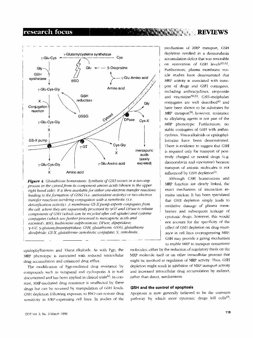
y-Glu-Cys
G l y ~
GSH ] synthetase ~
y-Glu-Cys-Gly
X Conjugatio~n
reaction
y-Glutamylcysteine synthetase Cys f' GhJ
BSO
~ d GSH uctase
GSSG y-Glu-Cys-Gly
I X
y-Glu-Cys-Gly I
X
GS-X pump
5-Oxoproline
~ y-Glu-Amino acid
Amino acid /
DPase
y-GT Cys-Gly I
y-Glu-Amino acid
Amino acid
Gly
Cys-X
1 mercapuric
acids (easily
excreted)
Figure 4. Glutathione homeostasis. S},nthesis of GSH occurs in a two-step process in the cytosol from its component amino acids (shown to the upper right hand side). It is then available for either one-electron transfer reactions leading to the formation of GSSG (i.e. antioxidant activity) or two-electron transfer reactions involving conjugation with a xenobiotic (:i.e. detox~'cation activity'). A membrane GS-X pump exports conjugates from the cell. u,'here thev are sequentially pr~ocessed by TGT and DPase to release components of GSH (which can be recycled after cell uptake) and cvsteine conjugates (which are fi, rther processed to mercapuric acids and excreted). BSO, buthionine sulphoxim,!ne; DPase, dipeptidase; T-GT, T-glutamvltranspeptidase; GSH, glutathione; GSSG, glutathione disulphide; GS-X. glutathione-xenobiotic conjugate,. X, xenobiotic.
epidophyllotoxins and Vinca alkaloids. As with Pgp, the
MRP phenotype is associated with reduced intracellular
drug accumulation and enhanced drug efflux. The modification of Pgp-mediated drug resistance by
compounds such as verapamil and cyclosporin A is well
documented and has been applied in clinical trials 6°. In con-
trast, MRP-mediated drug resistance is unaffected by these
drugs but can be reversed by manipulation of GSH levels.
GSH depletion following exposure to BSO can restore drug
sensitivity in MRP-expressing cell lines. In studies of the
mechanism of MRP transport, GSH
depletion resuhed in a daunorubicin
accumulation defect that was reversible on restoration of GSH levels 61,62.
Furthermore, plasma membrane ves-
icle studies have demonstrated that
MRP activity is associated with trans-
port of drugs and GSH conjugates,
including anthracyclines, etoposide and vincristine 58,63. GSH-melphalan
conjugates are well described 41 and
have been shown to be substrates for MRP transport58; however, resistance
to alkylating agents is not part of the
MRP phenotype. Furthermore, no
stable conjugates of GSH with anthra-
cyclines, Vinca alkaloids or epidophyl-
lotoxins have been demonstrated.
There is evidence to suggest that GSH is required only for transport of posi-
tively charged or neutral drugs (e.g.
daunorubicin and vincristine) because
transport of anionic molecules is not influenced by GSH depletion 64.
Although GSH homeostasis and
MRP function are clearly linked, the
exact mechanism of interaction re-
mains unclear. It has been speculated
that GSH depletion simply leads to
oxidative damage of plasma mem- branes and subsequent leakage of cytotoxic drugs;, however, this would
not account for the specificity of the
effect of GSH depletion on drug resist-
ance in cell lines overexpressing MRP.
GSH may provide a gating mechanism
to enable MRP to transport nonanionic
molecules, either by the reduction of regulatory thiols on the
MRP molecule itself or on other intracellular proteins that
might be involved in regulation of 1MRP activity. Thus, GSH
depletion might result in inhibition of MRP transport activity and increased intracellular drug accumulation by indirect,
rather than direct, mechanisms.
GSH and the control of apoptosis Apoptosis is now" generally believed to be the common pathway by which most cytotoxic drugs kill cells 65.
DDT Vol. 3, No. 3 March 1998 119
RE /IE-' S
Understanding the control of apoptosis is therefore crucial
to elucidate the many mechanisms of drug resistance.
Although several studies support the notion that cellular
GSH levels correlate to cytotoxic drug resistance, identifying
the specific role played by GSH in apoptosis is complicated
by the current incomplete knowledge of the mechanisms
regulating entry into the apoptotic pathway. Oxidative stress
has been proposed as a final common trigger for apop-
tosis 66. Evidence supporting the importance of ROS to apop-
tosis include the observations that addition of hydrogen per-
oxide or lipid peroxides, or the depletion of antioxidants
such as GSH, can induce apoptosis 67,68. Conversely, elev-
ation of levels of antioxidants 68-7° or overexpression of GSH-
dependent enzymes (e.g. glutathione peroxidase) 71 can
inhibit apoptosis. Several studies have clearly demonstrated
that the onset of apoptosis is associated with a fall in GSH
levels and, in some cases, an increase in ROS (Refs 72-74).
It has also been argued that ROS are not an essential trig-
ger for apoptosis because it can be induced in anaerobic
conditions, when the formation of ROS is minimized 75.
Studies of the Jurkatt leukaemia cell line demonstrated that
Fas/Fas-ligand-induced apoptosis was associated with spe-
cific transport of GSH from the cell rathe:r than an increase
in ROS - thus, there is a loss of protection against oxi-
dants 76,77. It is possible that the mitochondrial dysfunction
produced by culturing cells in low oxygen environments
results in increased cell susceptibility to oxidants, but it is
more probable that oxidative stress is not the final trigger
but only one of many different points of entry into the
apoptotic pathway.
GSH is frequently dismissed as a nonspecific antioxidant
that protects the cell by neutralizing potentially damaging
ROS. There is, however, compelling evidence to suggest that
GSH homeostasis has a more fundamental role in the acti-
vation or inhibition of proteins crucial to cell survival 76. Two
major groups of proteins have been defined as having key
roles in regulating apoptosis: the BCL-2 p, otein family (for a
recent review see Ref. 78) and the cell signalling cascade 79.
Many proteins have thiol moieties whose redox status is cru-
cial to their function 8°,81, and there is evidence that the
DNA-binding capacity of some transcription factors is redox
status dependent with sulphydryl groups on cysteine
residues as the target of regulation s2. This may have impo>
tant implications for the regulation of cell signalling path-
ways. Studies have shown that the DNA-binding function of
AP-1 (the Fos-Jun heterodimeric complex) is inhibited by
diamide and Nethylmaleimide, both of which are agents
that specifically modify sulphydryl groups 83. The influence
of oxidants and antioxidants on the function of AP-1 and the
nuclear factor (NF-KB) has been the subject of several stud-
ies. Interestingly, there is evidence of opposing effects of
redox status on induction of NF-KB compared to AP-1.
Meyer and coworkers 81 demonstrated that NF-KB activation
was p romoted by peroxides and hydrogen peroxide,
whereas antioxidants, including thiols, were strong
inhibitors 84. Conversely, antioxidants (N-acetylcysteine and
dithiocarbamates) efficiently induced AP-1 DNA binding
and hence transactivation. The AP-1 heterodimer appears to
be less sensitive to inhibition by oxidants. Droge and
coworkers 84 have demonstrated that increasing intracellular
GSSG can selectively inhibit DNA binding by NF-KB without
affecting AP-1 binding. Thiols, however, could enhance
DNA binding of both transcription factors. Thiol-induced
AP-I transactivation is dependent on induction of its com-
ponent proteins, Fos and Jun (Ref. 81), and reduction of a
single conserved cysteine residue in their DNA-binding
zone 83. The AP-1 complex is localized in the nucleus, in
contrast to NF-KB, which is primarily located in the cytosol.
NF-KB activation requires dissociation of an inhibitory pro-
tein IKB, followed by translocation of the transcription fac-
tor into the nucleus. It is interesting to note that although
oxidants promote the translocation activity" of NF-~B, NF-KB
DNA binding is enhanced by peroxides 81 and inhibition by
GSSG 84. As discussed previously, there are important kinetic
differences in the maintenance of GSH homeostasis in the
nuclear and cytosolic pools. This may have significant impli-
cations for the response of many cell signalling proteins and
transcription factors to the redox status within the different
intracellular compartments and may also prove to be cell-
type specific. Further evaluation of these pathways could
lead to more-focused targets in modulating apoptosis.
Clearly, the intracellular response to ROS and redox
homeostasis can no longer be considered in terms of non-
specific stress responses but appears to constitute important
mediators in the control of cell proliferation and cell sur-
vival. Further understanding of the activity of GSH and asso-
ciated enzymes within this framework may reveal valuable
targets by which cell survival could be manipulated.
Conclusion The involvement of GSH in detoxification of toxic com-
pounds by conjugation reactions and in the inactivation of
ROS is well established. However, recent advances in the
understanding of the importance of redox homeostasis to
120 DDT Vol. 3, No. 3 March 1998
REVIEWS
cell survival has led to renewed interest in the role of GSH,
as it appears to be important in both the control of apop-
tosis and the regulation of drug extrusion. Thus, after 40
years, the GSH pathway continues to offer attractive targets
for drug discovery.
Acknowledgements We thank the Leukaemia Research Fund and the North
of England Children's Cancer Research Fund for their
financial support.
REFERENCES 1 Fahey, R.C. and Sundquist, A.R. (1991) Adv. Enzymol. 64, 1-53
2 Tew, K.D. (1994) CancerRes. 54, 4313-4320
3 Meister, A. (1~)5) Meth. Enzymol. 251, 3-7
4 Thomas, M., Nlcklee, T. and Hedley, D.W. (1995) BtJ. Cancer72, 45-50
5 Jevtovic-Todorovic, V. and Guenttmer, T.M. (1992) ,giocbem. Pbarmacol. 44,
1383-1393
6 M~rtensson, J., Lai, J.CK and Meister, A. (1990) Proc. Natl. Acad. Sci. ~Z S. A.
87, 718%7189
7 Cossarizza, A. et al. (1995) Exp. Cell Res. 220, 232-240
8 Kroemer, G. et aL (1995) FASEBJ. 9, 12,-'7-1287
9 Macho, A. et al. (1997)J. Immunol 158, 4612-4619
10 Marchetti, E et aL (1997) EurJ. Immunol 27, 289-296
11 Meister, A. (1%~5) Meth. Enzymol. 252, 26-30
12 Gulick, A.M. and Fahl, W.E. (1995) PharmacoL The~ 66, 237-257
13 Commandeur, J.N.M., Stijntjes, G.J. and Vermeulen, N.EE. (1995) Pharmacol.
Rev. 47, 271-330
14 Campbell, J.AH. et al. (1991) Cancer67, 1608-1613
15 Pemble, S. el al. (1994) Biochem.J. 300, 271-276
16 Zhong, S. et al. (1993) Carcinogenesis 14, 1821-182.t
17 Nakajima, T. et al. (1995) Carcinogenesis 16, 707-711
18 Evans, C.G. el al. (1987) Cancer Res. 47, 252%2530
19 Smith, MT. etal. (1989) CancerRes. 49, 2621-2625
20 Tsuchida, S. and Sato, K. (1992) Crit. Rev. Biocbem. Mol. Biol. 27, 337-384
21 Chen, G. and Waxman, D.J. (1995) Biochem. Pharmacol. 49, 1691-1701
22 O'Brien, M.L. and Tew, K.D. (1996) Eu~L Cancer32, 967-978
23 Lewis, A.D. et al. (1988) Proc. Natl. Acad. Sci. U. S. . 4. 85, 8511~515
24 Batist, G. ei al. (1986)J. BioL Chem. 261,155/44-15549
25 Lewis, A.D., Hayes, J.D. and Wolf, C.R. (1988) Carci~zogenesis9, 1283--1287
26 Hall, A.G. et al. (1994) CancerRes. 54, 5251-5254
27 Malik, ].A. et al (1997) Cancer Chemother Pharmacol. 39, 561-565
28 Miyazaki, M. el al. (1990) Biochem. Biophy$. Res. Ccmmun. 166, 1358-1364
29 Leyland Jones, B.R. el al. (19-.)1) CancerRes. 51,587-594
30 Hall, A. el al. (1989) Cancer Res. 49, 626545268
31 Ciaccio, P.J., Tea,, KD. and LaCreta, EE (1991) Bioc~em. Pharmacol. 42,
1504-1507 32 Tew, K.D., Bomber, A.M. and Hoffman, SJ. (1988) CancerRes. 48, 3622-3625
33 Clapper, M.L., O'Dwyer, P.J. and Tew, K.D. (1990) in Glutathione
8-Transferases and Drug Resistance (Hayes, J.D., Pickett, C.B. and Mantle,
T.J., eds), pp. 451-459, Taylor and Francis
34 O'Dwyer, P.J. et al. (19-.)1) CancerRes. 51, 605945065
35 Ji, X. et al. (19-.)2) Biochemistry 31, 10169-10184
36 Rustm~ore, T.H. and Pickett, C.B. (1993) J. Biol. Chem. 268, 1147%11478
37 Koehler, R.T. et al. (1997) Proteins 28, 202-216
38 ,'k'hultz, M., Dutta, S. and Tew, K.D. (1997) Adv. Drug Deliv. Rev. 26, 91-104
39 Flatgaard, J~'E., Bauer, K.E. and Kauvar, L.M. (1993) Cancer Cbemother.
Pharmacol. 33, 63-70
40 Morgan, A.S. et aL (1996) Cancer Cbemotber Pbarmacol. 37, 363-370
41 Dulik, D.M., Fenselau, C. and Hilton, J. (1986) Biochem. Pharmacol. 35,
340%3409
42 Hall, A.G etal. (1994) CancerRes. 54, 3369--3372
43 Jakoby, WB. (1978) Adv. Enzk~oL 46, 383-414
44 Meyer, D.J. el aL (1992) BrJ. Cancer66, 433-438
45 Dreyfus, E et al. (19-)5) BoneMarrow Transplant. 15, 707-711
46 Seitz, D.E., Winter, M.A. and Pearce, H.L. (1989) CancerRes. 30, 497
47 Chen, G. and Waxman, D.J. (1994) Biochem Pharmacol. 47, 1079-1087
48 Medh, R.D., Gupta, V. and Awasthi, Y.C. (1991) Biocbem. Pharmacol. 42,
439--441
49 Meijer, C. etal. (1990) BrJ. Cancer62, 72-,-'7
50 Mistry, E etal. (1991) BrJ. Cancer64, 21%220
51 Kisara, S. et al. (19-)5) Oncol. Res. 7, 191-200
52 Ferrari, G., Yan, C.Y. and Greene, LA. (19-)5) J. Neurosci. 15, 2857-2.866
53 Ozols, R.E et al. (1987) Biochem. Pbarmacol. 36, 147-153
54 O'Dwyer, P.J. et al. (1992)J. Natl. CancerIm't. 84, 264-270
55 Bailey, H.H. etaL (1994)J. Ch'n. Oncol. 12, 194-205
56 Beaver, J.E and Warmg, P. (1995) EurJ. Cell BioL 68, 47-54
57 Maung, Z.T. etaL (1994) Leukemia8, 1487-1491
58 Jedlitschky, G. et aL (1996) CancerRes. 56, 988-994
59 Loe, D.W., Deeley, R.G. and Cole, S.EC. (1996) EurJ. Cancer32, 945-957
60 Hussey, A01. and Hayes, J.D. (1992) Biochem.J. 286, 929-935
61 Zaman, G.J.R. el al. (1995) Proc. Natl. Acad. &i. U. S. A. 92, 7690-7694
62 Versantvoort, C.H. et al. (1995) BrJ. Cancer72, 82-89
63 Paul, 8. et al. (1996) Proc. Natl. Acad. Sci. U. S. A. 93, 6929-6934
64 Feller, N. el aL (1995) FEBSLett. 368, 385-388
65 Hickman, J.A. (1996) Ew: J. Cancer32, 921-!)26
66 Payne, C.M., Bernstein, C. and Bernstein, H. (1995) Leuk. Lymphoma 19,
43-93 67 Burdon, R.H. et al. (1996) Free Radio. Res. 24, 81-93
68 81ater, A.EG. el al. (1995) Toxicol. Lett. 82-3, 149-153
69 Urn, H.D., Orenstein, J.M. and Wahl, S.M. (1996)J. lmmunoL 156, 3469-3477
70 Iwata, S. et al. (1997)./Immunol. 158, 3108-3117
71 Hockenbery, D.M. et al. (1993) Cell75, 241-251
72 Packham, G., &shmun, R.A. and Cleveland, J.L. (1996) J. lmmunol. 156,
2792-2800
73 Ghibelli, L. etaL (1995) Biochem. Biophys. Res. Commun. 216, 313-320
74 Beaver, J.E and Waring, P. (1995) Eur J. Cell BioL 68, 47-54
75 Jacobson, M.D. and Raff, M.C. (19-.)5) Nature 374, 814-816
76 Slater, A.EG. et al. (1996) Cell Death Dzffer. 3, 57-62
77 Vandendobbelsteen, D.J. et al. (1996) J. Biol. Chem. 271, 15420-15427
78 Kr{xemer, G. (1997)Nat. Med. 3, 614-620
79 Nehm6, A. el al. (1995) lnt.J. Cancer61, 643-648
80 tin, K.I. el al. (1995)J. CellBiol. 131, 1149-1161
81 Meyer, M., Schreck, R. and Baeuerle, P.A. (1993) EMBOJ. 12, 200%2015
82 Pognonec, P., Kato, H. and Roeder, R.G. (1992)./Biol. Chem. 267,
24563-24567
83 Abate, C. etal.(1990) Science249, 1157-1161
84 Droge, W. etaL (1994) FASEBJ. 8, 1131-1139
DDT Vol. 3, No. 3 March 1998 121


![Chemoimmunotherapy versus chemotherapy for metastatic ... · [Intervention Review] Chemoimmunotherapy versus chemotherapy for metastatic malignant melanoma Andre D Sasse 1, Emma C](https://img.pdfslide.net/doc/110x75/5ca3dc4888c99374538bc446/chemoimmunotherapy-versus-chemotherapy-for-metastatic-intervention-review.jpg)