Embed Size (px)
Citation preview
Biochem. J. (1990) 268, 401-407 (Printed in Great Britain)
Purification, characterization and partial amino acid sequence ofglycogen synthase from Saccharomyces cerevisiaeAssumpta CARABAZA,* Joaquin ARINO,* Jay W. FOX,t Carlos VILLAR-PALASIt and Joan J. GUINOVART*§*Departament de Bioquimica i Biologia Molecular, Facultat de Veterin'aria, Universitat Aut6noma de Barcelona,08193-Bellaterra, Barcelona, Spain, and tDepartment of Microbiology and $Department of Pharmacology,University of Virginia School of Medicine, Charlottesville, VA 22908, U.S.A.
Glycogen synthase from Saccharomyces cerevisiae was purified to homogeneity. The enzyme showed a subunit molecularmass of 80 kDa. The holoenzyme appears to be a tetramer. Antibodies developed against purified yeast glycogen synthaseinactivated the enzyme in yeast extracts and allowed the detection of the protein in Western blots. Amino acid analysisshowed that the enzyme is very rich in glutamate and/or glutamine residues. The N-terminal sequence (11 amino acidresidues) was determined. In addition, selected tryptic-digest peptides were purified by reverse-phase h.p.l.c. and submittedto gas-phase sequencing. Up to eight sequences (79 amino acid residues) could be aligned with the human muscle enzymesequence. Levels of identity range between 37 and 100 %, indicating that, although human and yeast glycogen synthasesprobably share some conserved regions, significant differences in their primary structure should be expected.
INTRODUCTION
Glycogen synthase (EC 2.4.1.1 1) is the enzyme responsible forthe synthesis of glycogen in a wide variety of species, from yeastto mammals. In contrast with mammalian glycogen synthase, theenzyme from yeast has not been extensively characterized. Mainlyfrom the work of Cabib and collaborators, it is known that yeastglycogen synthase exists as an oligomer that contains a singletype of subunit [1]. As observed for the mammalian enzyme,glycogen synthase appears in yeast in two forms, I and D, whichdiffer in their sensitivity to glucose 6-phosphate and areinterconvertible [2]. Interconversion is, most probably, producedby phosphorylation-dephosphorylation reactions [1,2]. Inaddition, the yeast enzyme appears to be very sensitive toproteolysis.
Recently our laboratory became interested in the regulation ofglycogen synthesis in yeast. Therefore we decided to characterizeyeast glycogen synthase in order to obtain structural informationto understand its regulation in yeast cells. In the present paper wedescribe a rapid purification method for yeast glycogen synthase,and our results indicate that, although yeast and mammalianglycogen synthases probably share regions with a high degree ofsequence identity, significant differences in their primary structureshould be expected.
MATERIALS AND METHODS
MaterialsTrypsin [tosylphenylalanylchloromethane- ('TPCK '-)treated],
rabbit liver glycogen (type III), phenylmethanesulphonylfluoride, leupeptin and benzamidine were from Sigma ChemicalCo. DEAE-cellulose (DE-32) was from Whatman. Sepharose 4Band Sephacryl S-300 came from Pharmacia. Other reagents werefrom Merck or Sigma Chemical Co.a-Amylase was purified from human saliva as described in ref.
[3]. Amylase activity was determined at 37 °C with maltotetraoseas a substrate with a kit from Roche.The Sepharose 4B-hexamethylenediamino-3-carboxypro-
pionylglucosamine 6-phosphate affinity system was prepared asfollows. Sepharose 4B was activated with CNBr and coupled to
1,6-diaminohexane as described by the manufacturer. Thehexamethylenediamino-Sepharose 4B matrix was resuspended inwater, succinic anhydride (I mmol/ml of gel) was added and thereaction was carried out at pH 6.0 for 5 h at 4 'C. Subsequently,glucosamine 6-phosphate was coupled at pH 4.5 in the presenceof l-(dimethylaminopropyl)-3-ethylcarbodi-imide.
Growth of yeastSaccharomyces cerevisiae ABYS- (a mutant lacking four major
proteinases [4]) was grown in medium containing glucose (2 %,w/v), peptone (1 %, w/v) and yeast extract (I %, w/v) at 30 'C.Cells were harvested shortly after the onset of the stationaryphase.
Purification of glycogen synthaseAll the purification steps were carried out between 0 and 4 'C
unless otherwise stated.
Buffers. Buffer A was 50 mM-Tris/HCI buffer, pH 7.4, con-taining 0.6 M-sucrose, 5 mM-EDTA, 2 mM-EGTA, 1 mm-dithiothreitol, 1 mM-phenylmethanesulphonyl fluoride, 0.25 ,ugof leupeptin/ml, 0.1 mM-tosyl-lysylchloromethane ('TLCK') and0.5 mM-benzamidine. Buffer B was the same as buffer A exceptthat the concentration of sucrose was lowered to 0.25 M and thatit contained in addition 2 mM-Na2SO4, 10 mM-MgCl2 and0.5 mM-glucose 6-phosphate. Buffer C was the same as buffer Aexcept that the concentration of sucrose was lowered to 0.25 Mand that tosyl-lysylchloromethane was omitted. Buffer D was50 mM-imidazole/HCI buffer, pH 7.4, containing 0.25 M-sucrose,5 mM-EDTA, 2 mM-EGTA, 10 mM-MgCl2, I mM-dithiothreitoland 1 mM-phenylmethanesulphonyl fluoride. Buffer E was50 mM-Tris/HCI buffer, pH 7.8, containing 25 % (v/v) glycerol,5 mM-EDTA, 2 mM-EGTA and 1 mM-dithiothreitol.
Extraction. Yeast cells were harvested by centrifugation (7000 gfor 10 min) and washed twice with cold distilled water. A 300 g(wet wt.) batch of yeast was resuspended in 600 ml of buffer A.Then 90 ml portions were shaken five times for 1 min with 270 gof glass beads (0.45 mm in diameter) in a pre-cooled Vibrogencell homogenizer. The temperature was kept below 8 'C. Thehomogenate was decanted, the glass beads were washed with
§ To whom correspondence should be addressed.
Vol. 268
401
A. Carabaza and others
300 ml of buffer A, and the washes and original homogenatewere pooled and centrifuged at 13 000 g for 30 min at 4 'C. Thesupernatant was filtered through glass-wool. The filtrate wascentrifuged at 117000 g for 90 min at 4 'C. The supernatant wasremoved and discarded. Pellets were resuspended in 70 ml ofbuffer B.
Treatment with oc-amylase. Human salivary amylase(4000 units) was added and the preparation was incubated at30 'C for 30 min. After the incubation, the preparation wascentrifuged at 170000 g for 30 min at 4 'C and the supernatantrecovered.
DEAE-cellulose chromatography. The supernatant was loaded(1 ml/min) on to a 70 ml DEAE-cellulose column equilibratedwith buffer C. The column was washed with 100 ml of buffer C,followed by 700 ml ofbuffer C containing 50 mM-NaCl. Glycogensynthase activity was eluted with buffer C containing 200 mm-NaCl. Fractions containing glycogen synthase activity werepooled and dialysed for 2 h against buffer C containing 10 mM-MgCl2.
Affinity column chromatography. The preparation was thenloaded on to a 4 ml Sepharose 4B-hexamethylenediamino-3-carboxypropionylglucosamine 6-phosphate column equi-librated with buffer D. The column was washed with 10 mlof buffer D including 25 mM-NaCl at 4 'C. The column was thenwarmed to room temperature, and glycogen synthase was elutedwith buffer D containing 25 mM-NaCl and 10 mM-glucose 6-phosphate. Fractions containing glycogen synthase activity werepooled, and rabbit liver glycogen was added to achieve a finalconcentration of 1 mg/ml. Then the enzyme was precipitated byadding cold'ethanol, to 20 % (v/v) final concentration. The pelletwas recovered by centrifugation at 18000 g for 15 min at -10 °C,resuspended in 1 ml of buffer E and dialysed against the samebuffer.
Determination of proteinProtein concentration was determined by the method of
Bradford [5], with BSA as standard.
Electrophoretic analysisSDS/PAGE was performed in 7 % polyacrylamide slab gels as
described by Laemmli [6].
Assay of enzyme activityThe glycogen synthase - glucose 6-phosphate/ + glucose 6-
phosphate activity ratio was determined by the method ofThomas et al. [7]. Glycogen phosphorylase activity was measuredby the method described in ref. [8] in the presence of 10 mm-caffeine and 10 mM-EDTA. One unit of enzyme activity is theamount of enzyme that incorporates 1 ,umol of [14C]glucose/minfrom UDP-[14C]glucose (glycogen synthase) or from [14C]glucose1-phosphate (glycogen phosphorylase) into glycogen at 30 'C.
Determination of molecular mass and subunit structure of nativeglycogen synthase
Purified glycogen synthase (400,g in 1 ml) was chromato-graphed on a Sephacryl S-300 column (1.5 cm x 90 cm) in 50 mm-Tris/HCI buffer, pH 7.8, containing 5 mM-EDTA, 2 mm-dithiothreitol and 1 M-KCI. Fractions were collected andglycogen synthase activity was measured. Under the sameconditions a linear calibration curve was obtained with standardproteins.
Immunochemical techniquesAntisera were raised in rabbits against homogeneous
preparations of yeast glycogen synthase. A subcutaneous in-jection (50 ,ug) in Freund's complete adjuvant was given, followed10 and 20 days later by subcutaneous injection of yeast glycogensynthase (25 ,ug) in Freund's incomplete adjuvant. Bleeding wascarried out 15 days later. Immunoglobulins were partially purifiedby precipitation with (NH4)2SO4 (final concentration 45 %saturation at 4° C) and stored at -20 'C.
Amino acid compositionGlycogen synthase (10,g)- was reduced and alkylated as
described in ref. [9]. Gas-phase hydrolysis was performedin vacuo at 110 'C in 6 M-HCI containing I % phenol for24 h. Phenylthiocarbamoyl derivatives of the amino acids wereidentified by h.p.l.c. on a Hypersil ODS (3 ,gm particle size)reverse-phase column (0.46 cm x 25 cm). The solvent A was55 mM-sodium acetate buffer, pH 5.95, containing 2% (v/v)acetonitrile and solvent B was acetonitrile. Peaks were monitoredat 254 nm and identified and quantified by comparison withknown standards.
Determination of N-terminal sequenceSDS/PAGE was performed on 1.5 mm-thick 7%
polyacrylamide gels as described by Laemmli [6]. Gels were pre-run for 15 min before loading of the sample (100,g of theglycogen synthase preparation). After electrophoresis, the proteinwas transferred to Immobilon [poly(vinylidene difluoride)]membranes (Millipore). The blotting was done according to themethod of Matsudaira [10]. Blots were stained as described inref. [11], and the 80 kDa glycogen synthase band was excised andsequenced directly with an Applied Biosystems model 470 A gas-phase sequencer equipped with a model 120 A phenyl-thiohydantoin analyser. The sequencing was carried out usingthe manufacturer's recommended programs and procedures.
Production of tryptic peptides and determination of peptidesequences
Glycogen synthase (200 ,ug in 300 ,ul of 0.4 M-Tris/HCl buffer,pH 8.1, containing 2 mM-EDTA and 6 M-guanidium chloride)was reduced at 37 'C for 2 h in the presence of 5 mM-dithiothreitoland then carboxymethylated by incubation with 90 ,l of 50 mm-iodo['4C]acetic acid (2200 c.p.m./nmol), pH 8.0, at 37 'C for 1 h.Carboxymethylated glycogen synthase was separated from unusediodoacetic acid by Sepharose G.25 (15 cm x 1 cm) gel filtration.The protein-containing fractions were evaporated in a Speed-Vac (Savant) and finally dissolved in 0.2 ml of 50 mM-ammoniumbicarbonate buffer, pH 8.0. Trypsin was added to a final ratioenzyme/trypsin of 20:1 (w/w) and the digestion was continuedfor 16 h at 30 'C. The addition of trypsin was repeated twice. Thedigest was applied to a Brownlee Aqua Pore C8 reverse-phaseh.p.l.c. column. The acetonitrile gradient was 0-100% (v/v)(100 min) in 0.1 % (v/v) trifluoroacetic acid at a flow rate of0.8 ml/min. Peaks were monitored at 214 nm. The peptides-collected from the column were purified to homogeneity by usinga shallower acetonitrile gradient, and their amino acid sequencewas determined by automated gas-phase Edman degradation.
RESULTS
Purification of yeast glycogen synthaseWe have developed a new method for the purification of yeast
glycogen synthase. Briefly, a glycogen--pellet is obtained andglycogen synthase released from the glycogen by incubation witha-amylase. After D-EAE-cellulose column chromatography,
1990
402
Characterization of yeast glycogen synthase
which removes glycogen phosphorylase, the enzyme is purifiedby affinity chromatography, with glucosamine 6-phosphate as aligand. This step is based on the fact that glucose 6-phosphate isa strong allosteric activator of the enzyme. This type of affinitychromatography was first used to purify glycogen synthase fromadipose tissue by Miller et al. [12]. However, in our case, ahexamethylenediamino-3-carboxypropionyl, instead of a 3-carboxypropionylaminodipropyl, spacer arm is included to avoidsteric interactions between the enzyme molecule and the gelmatrix. The enzyme is eluted at low salt concentration (25 mM)in the presence of 10 mM-glucose 6-phosphate, and at this steppreparations are essentially homogeneous (Fig. 1). Finally, thepreparation is concentrated by precipitation with ethanol at lowtemperature in the presence of added glycogen. The purifiedenzyme can be stored at -70 °C for at least 6 months withoutsignificant loss of activity. The whole procedure can be carriedout in 48 h, and therefore it is much faster than the protocoldescribed previously [1]. A summary of the purification isprovided in Table 1.SDS/PAGE of the final preparations (Fig. 2) shows a single
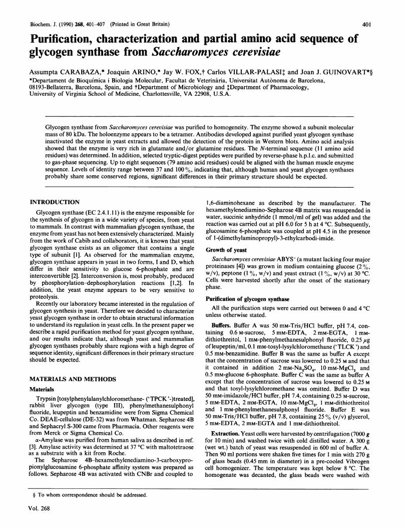
band corresponding to the glycogen synthase subunit. Thecalculated molecular mass was 80 kDa, which is somewhat higherthan the value described previously [1]. It is worth noting that inour case the source of the enzyme was a strain defective in four
97 kDa
66 kDa
45 kDa
36 kDa
Fig. 2. SDS/PAGE of purified yeast glycogen synthase
A typical preparation of pure yeast glycogen synthase (10 ,ug) wassubjected to SDS/PAGE (7% polyacrylamide) and protein wasstained with Coomassie Blue R-250. The mobility of molecular-massstandards is indicated on the left.
E204
-> 1.5
co5a)
cn
CD
o 0.5
0 5 10 15 20 25Fraction no.
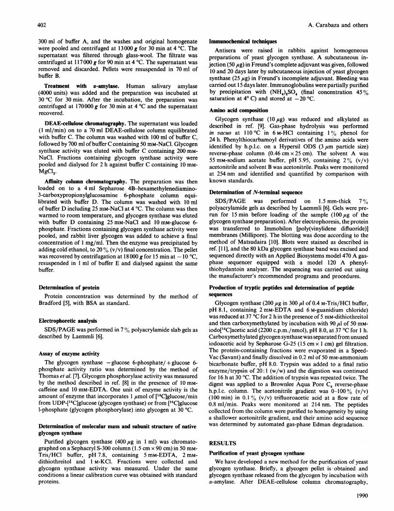
Fig. 1. Affinity chromatography of yeast glycogen synthase
Glycogen synthase preparations were loaded on to a Sepharose4B-hexamethylenediamino- 3-carboxypropionylglucosamine 6-phosphate column and 1 ml fractions were collected. Enzyme activitywas eluted with 25 mM-NaCl/10 mM-glucose 6-phosphate (*). Noadditional glycogen synthase activity was eluted at 100 mm-NaCl/ 15 mM-glucose 6-phosphate (*). The inset shows SDS/PAGEof fractions eluted from the affinity column. The apparent molecularmass is indicated on the left.
major proteinases. When the same procedure was carried outwith as starting material baker's yeast, purified preparationscontained variable amounts of lower-molecular-mass poly-peptides in the range 70-72 kDa, similar to those described inan earlier report [1]. Therefore yeast glycogen synthase may bevery sensitive to proteolysis, as is the case for the enzyme frommammalian sources, especially from liver tissue. In this regard,the inclusion in the buffers of several proteinase inhibitors, theuse of a strain deficient in proteinases as a starting material andthe development of a fast protocol for purification appear as keyfeatures in order to obtain a high-molecular-mass enzyme.
Native glycogen synthase shows a molecular mass of 310 kDa,as determined by gel filtration through a Sephacryl S-300 column.Therefore, assuming a molecular mass of 80 kDa for the subunit,the enzyme is probably a tetramer (results not shown).
Characterization of antibodies against yeast glycogen synthaseAntisera against yeast glycogen synthase were obtained as
described in the Materials and methods section. Antisera fromdifferent batches were tested for their ability to remove glycogensynthase activity from yeast extracts. Extracts were prepared asdescribed for purification of the enzyme, and samples incubatedfor 2 h at 4 °C with different amounts of antiserum. The mixturewas centrifuged at 15000 g for 5 min and glycogen synthase
Table 1. Summary of the purification of yeast glycogen synthase
For experimental details see the Materials and methods section.
Final Total Concn. of Specificvolume activity protein activity Recovery Purification
Fraction (ml) (units) (mg/ml) (units/mg) (%) (fold)
ExtractGlycogen pelletDEAE-cellulose chromatographyAffinity chromatography
875 13880 7550 19.81.1 17.6
/ / \ ~~~80 kDa-_^\ ~~~~~~~~~~~~~~~~~~~~~~~~~~~~~~~~~~~~~~~~~. ..........eI ; 357 9
* \I \I
* ._e. @ @_. . 0 0 S
Vol. 268
15.74.170.70.7
0.0100.220.56
22.85
1005414.312.7
2256
2300
403
go
iAi
F
A. Carabaza and others
80
-E
n 60
c-)
o 40
c
tnC
0) 20
21-UD
0
0 20 40 60Volume of antiserum (ail)
80
100
75
50
25
0
preparation of the samples, the 72 kDa band was alwaysdetected.
.t)
0
0
r-C
CL
0)
0)C-)
UD
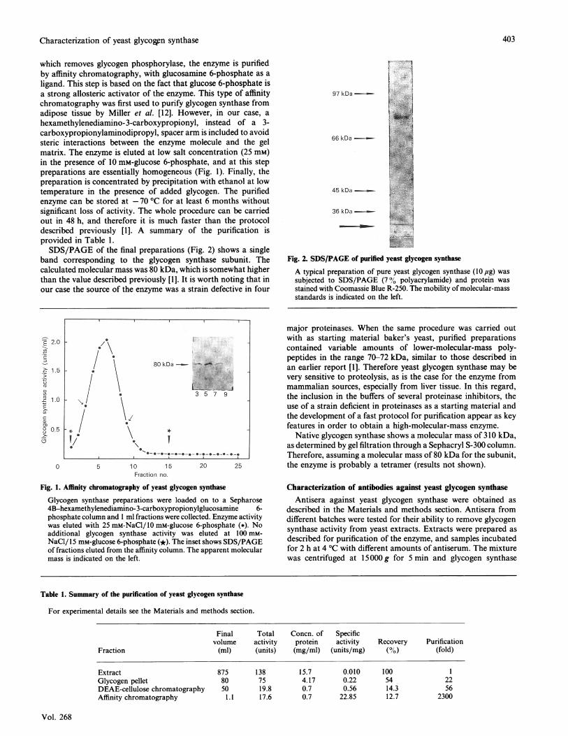
Fig. 3. Binding of antibodies to glycogen synthase from yeast extracts
Extracts from cultures of S. cerevisiae (ABYS-) were prepared, andto equal portions were added increasing volumes of control serum
(0) or anti-(glycogen synthase) serum (a) and the mixtures wereincubated for 2 h at 4 'C. Then, samples were centrifuged andglycogen synthase activity was measured in the supernatant (in thepresence of 6.6 mM-glucose 6-phosphate). Glycogen phosphorylaseactivity (A) was also determined in samples incubated with anti-(glycogen synthase) antibodies and is represented as percentage over
the controls. The inset shows Western-blot analysis of yeast extracts.Extracts were prepared, and then were subjected to SDS/PAGE(7% polyacrylamide) and the gels were blotted on to nylonmembranes. '25I-Protein G was used to detect the antigen-antibodyreaction. The mobility of molecular-mass standards is indicated on
the left.
activity was measured in the supernatant. As shown in Fig. 3, theantibodies were able to remove glycogen synthase activitycompletely from the extracts. Glycogen phosphorylase activitywas not recognized by the antibodies. The antibodies did notcross-react with purified glycogen synthase from rat liver or
rabbit muscle.Glycogen synthase was also detected in yeast extracts by
Western-blot techniques. Extracts from S. cerevisiae ABYS-cultures were prepared and immediately placed in 1% (w/v)SDS/350 mM-2-mercaptoethanol (final concentrations) andboiled for 5 min. Electrophoresis was performed in 7% poly-acrylamide gels. The proteins were blotted on to Immobilonmembranes. 125I-Protein G was used to detect proteins recognizedby the antibody. As shown in Fig. 3 inset, two bands were
detected, at 80 kDa and 72 kDa. The 80 kDa band exhibitsthe same mobility as the purified glycogen synthase subunit.Although precautions against proteolysis were taken during
N-Terminal sequence analysis of yeast glycogen synthasePurified yeast glycogen synthase (100,g) was subjected to
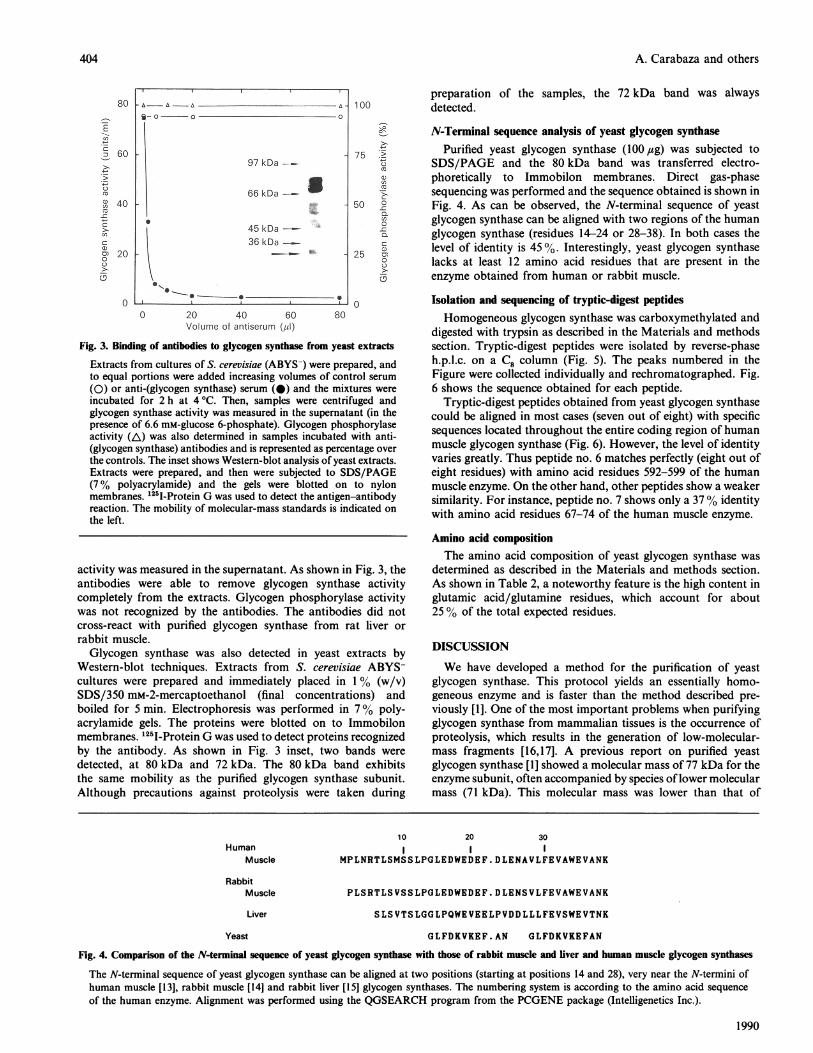
SDS/PAGE and the 80 kDa band was transferred electro-phoretically to Immobilon membranes. Direct gas-phasesequencing was performed and the sequence obtained is shown inFig. 4. As can be observed, the N-terminal sequence of yeastglycogen synthase can be aligned with two regions of the humanglycogen synthase (residues 14-24 or 28-38). In both cases thelevel of identity is 45 %. Interestingly, yeast glycogen synthaselacks at least 12 amino acid residues that are present in theenzyme obtained from human or rabbit muscle.
Isolation and sequencing of tryptic-digest peptidesHomogeneous glycogen synthase was carboxymethylated and
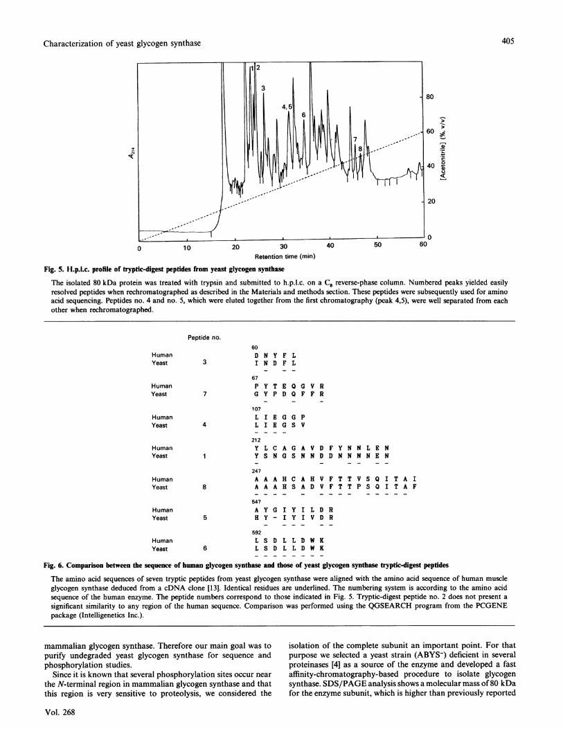
digested with trypsin as described in the Materials and methodssection. Tryptic-digest peptides were isolated by reverse-phaseh.p.l.c. on a C8 column (Fig. 5). The peaks numbered in theFigure were collected individually and rechromatographed. Fig.6 shows the sequence obtained for each peptide.
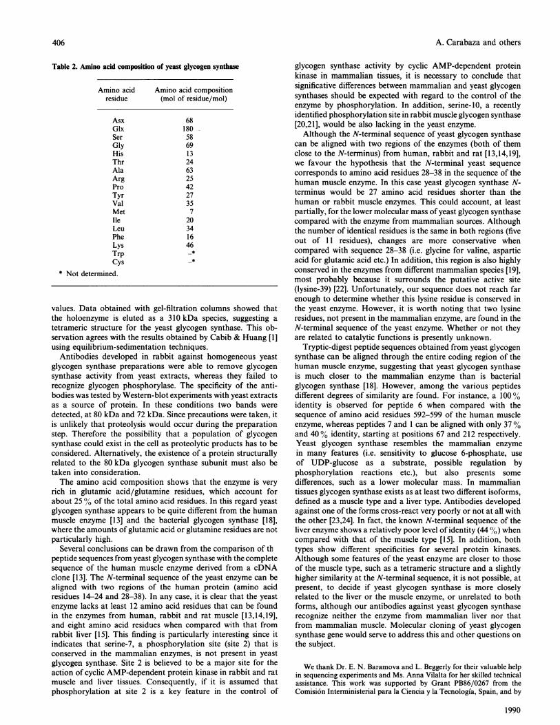
Tryptic-digest peptides obtained from yeast glycogen synthasecould be aligned in most cases (seven out of eight) with specificsequences located throughout the entire coding region of humanmuscle glycogen synthase (Fig. 6). However, the level of identityvaries greatly. Thus peptide no. 6 matches perfectly (eight out ofeight residues) with amino acid residues 592-599 of the humanmuscle enzyme. On the other hand, other peptides show a weakersimilarity. For instance, peptide no. 7 shows only a 37% identitywith amino acid residues 67-74 of the human muscle enzyme.
Amino acid compositionThe amino acid composition of yeast glycogen synthase was
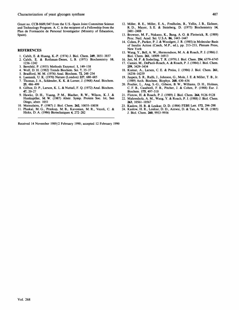
determined as described in the Materials and methods section.As shown in Table 2, a noteworthy feature is the high content inglutamic acid/glutamine residues, which account for about25 % of the total expected residues.
DISCUSSION
We have developed a method for the purification of yeastglycogen synthase. This protocol yields an essentially homo-geneous enzyme and is faster than the method described pre-
viously [1]. One of the most important problems when purifyingglycogen synthase from mammalian tissues is the occurrence ofproteolysis, which results in the generation of low-molecular-mass fragments [16,17]. A previous report on purified yeastglycogen synthase [1] showed a molecular mass of 77 kDa for theenzyme subunit, often accompanied by species oflower molecularmass (71 kDa). This molecular mass was lower than that of
10 20 30Human I I I
Muscle MPLNRTLSMSSLPGLEDWEDEF.DLENAVLFEVAWEVANK
RabbitMuscle PLSRTLSVSSLPGLEDWEDEF.DLENSVLFEVAWEVANK
Liver SLSVTSLGGLPQWEVEELPVDDLLLFEVSWEVTNK
Yeast GLFDKVKEF.AN GLFDKVKEFAN
Fig. 4. Comparison of the N-terminal sequence of yeast glycogen synthase with those of rabbit muscle and liver and human muscle glycogen synthases
The N-terminal sequence of yeast glycogen synthase can be aligned at two positions (starting at positions 14 and 28), very near the N-termini ofhuman muscle [13], rabbit muscle [14] and rabbit liver [15] glycogen synthases. The numbering system is according to the amino acid sequenceof the human enzyme. Alignment was performed using the QGSEARCH program from the PCGENE package (Intelligenetics Inc.).
1990
9- 0 ~~0 0
97 kDa--
66 kDa-
45 kDa--36 kDa
__
404
Characterization of yeast glycogen synthase
600 10 20 30 40 50
80
60 8ev_
._60
40 °U0
20
0
Retention time (min)
Fig. 5. H.p.l.c. profile of tryptic-digest peptides from yeast glycogen synthase
The isolated 80 kDa protein was treated with trypsin and submitted to h.p.l.c. on a C8 reverse-phase column. Numbered peaks yielded easilyresolved peptides when rechromatographed as described in the Materials and methods section. These peptides were subsequently used for aminoacid sequencing. Peptides no. 4 and no. 5, which were eluted together from the first chromatography (peak 4,5), were well separated from eachother when rechromatographed.
Peptide no.
3
60D N Y F LI N D F L
67P Y T E Q G V RG Y P D Q F F R
107L I E G G PL I E G S V
212Y L C A G A V D F Y N N L E NY S N G S N N D D N N N N E N
4
8
247A AA A
547
A H C A H V F T T V S Q I T A IA H S A D V F T T P S 0 I T A F
~~ ~
A Y G I Y I L D RH Y - I Y I V D R5
592
L SL S6
D L L D W KD L L D W K
Fig. 6. Comparison between the sequence of human glycogen synthase and those of yeast glycogen synthase tryptic-digest peptides
The amino acid sequences of seven tryptic peptides from yeast glycogen synthase were aligned with the amino acid sequence of human muscleglycogen synthase deduced from a cDNA clone [13]. Identical residues are underlined. The numbering system is according to the amino acidsequence of the human enzyme. The peptide numbers correspond to those indicated in Fig. 5. Tryptic-digest peptide no. 2 does not present a
significant similarity to any region of the human sequence. Comparison was performed using the QGSEARCH program from the PCGENEpackage (Intelligenetics Inc.).
mammalian glycogen synthase. Therefore our main goal was topurify undegraded yeast glycogen synthase for sequence andphosphorylation studies.
Since it is known that several phosphorylation sites occur near
the N-terminal region in mammalian glycogen synthase and thatthis region is very sensitive to proteolysis, we considered the
isolation of the complete subunit an important point. For thatpurpose we selected a yeast strain (ABYS-) deficient in severalproteinases [4] as a source of the enzyme and developed a fastaffinity-chromatography-based procedure to isolate glycogensynthase. SDS/PAGE analysis shows a molecular mass of80 kDafor the enzyme subunit, which is higher than previously reported
Vol. 268
HumanYeast
HumanYeast
HumanYeast
HumanYeast
HumanYeast
HumanYeast
HumanYeast
405
1
A. Carabaza and others
Table 2. Amino acid composition of yeast glycogen synthase
Amino acid Amino acid compositionresidue (mol of residue/mol)
AsxGlxSerGlyHisThrAlaArgProTyrValMetIleLeuPheLysTrpCys
* Not determined.
681805869132463254227357
20341646
values. Data obtained with gel-filtration columns showed thatthe holoenzyme is eluted as a 310 kDa species, suggesting atetrameric structure for the yeast glycogen synthase. This ob-servation agrees with the results obtained by Cabib & Huang [1]using equilibrium-sedimentation techniques.
Antibodies developed in rabbit against homogeneous yeastglycogen synthase preparations were able to remove glycogensynthase activity from yeast extracts, whereas they failed torecognize glycogen phosphorylase. The specificity of the anti-bodies was tested by Western-blot experiments with yeast extractsas a source of protein. In these conditions two bands weredetected, at 80 kDa and 72 kDa. Since precautions were taken, itis unlikely that proteolysis would occur during the preparationstep. Therefore the possibility that a population of glycogensynthase could exist in the cell as proteolytic products has to beconsidered. Alternatively, the existence of a protein structurallyrelated to the 80 kDa glycogen synthase subunit must also betaken into consideration.The amino acid composition shows that the enzyme is very
rich in glutamic acid/glutamine residues, which account forabout 25 % of the total amino acid residues. In this regard yeastglycogen synthase appears to be quite different from the humanmuscle enzyme [13] and the bacterial glycogen synthase [18],where the amounts of glutamic acid or glutamine residues are notparticularly high.
Several conclusions can be drawn from the comparison of thpeptide sequences from yeast glycogen synthase with the completesequence of the human muscle enzyme derived from a cDNAclone [13]. The N-terminal sequence of the yeast enzyme can bealigned with two regions of the human protein (amino acidresidues 14-24 and 28-38). In any case, it is clear that the yeastenzyme lacks at least 12 amino acid residues that can be foundin the enzymes from human, rabbit and rat muscle [13,14,19],and eight amino acid residues when compared with that fromrabbit liver [15]. This finding is particularly interesting since itindicates that serine-7, a phosphorylation site (site 2) that isconserved in the mammalian enzymes, is not present in yeastglycogen synthase. Site 2 is believed to be a major site for theaction of cyclic AMP-dependent protein kinase in rabbit and ratmuscle and liver tissues. Consequently, if it is assumed thatphosphorylation at site 2 is a key feature in the control of
glycogen synthase activity by cyclic AMP-dependent proteinkinase in mammalian tissues, it is necessary to conclude thatsignificative differences between mammalian and yeast glycogensynthases should be expected with regard to the control of theenzyme by phosphorylation. In addition, serine-10, a recentlyidentified phosphorylation site in rabbit muscle glycogen synthase[20,21], would be also lacking in the yeast enzyme.Although the N-terminal sequence of yeast glycogen synthase
can be aligned with two regions of the enzymes (both of themclose to the N-terminus) from human, rabbit and rat [13,14,19],we favour the hypothesis that the N-terminal yeast sequencecorresponds to amino acid residues 28-38 in the sequence of thehuman muscle enzyme. In this case yeast glycogen synthase N-terminus would be 27 amino acid residues shorter than thehuman or rabbit muscle enzymes. This could account, at leastpartially, for the lower molecular mass of yeast glycogen synthasecompared with the enzyme from mammalian sources. Althoughthe number of identical residues is the same in both regions (fiveout of 11 residues), changes are more conservative whencompared with sequence 28-38 (i.e. glycine for valine, asparticacid for glutamic acid etc.) In addition, this region is also highlyconserved in the enzymes from different mammalian species [19],most probably because it surrounds the putative active site(lysine-39) [22]. Unfortunately, our sequence does not reach farenough to determine whether this lysine residue is conserved inthe yeast enzyme. However, it is worth noting that two lysineresidues, not present in the mammalian enzyme, are found in theN-terminal sequence of the yeast enzyme. Whether or not theyare related to catalytic functions is presently unknown.
Tryptic-digest peptide sequences obtained from yeast glycogensynthase can be aligned through the entire coding region of thehuman muscle enzyme, suggesting that yeast glycogen synthaseis much closer to the mammalian enzyme than is bacterialglycogen synthase [18]. However, among the various peptidesdifferent degrees of similarity are found. For instance, a 1000%identity is observed for peptide 6 when compared with thesequence of amino acid residues 592-599 of the human muscleenzyme, whereas peptides 7 and 1 can be aligned with only 37%and 40 % identity, starting at positions 67 and 212 respectively.Yeast glycogen synthase resembles the mammalian enzymein many features (i.e. sensitivity to glucose 6-phosphate, useof UDP-glucose as a substrate, possible regulation byphosphorylation reactions etc.), but also presents somedifferences, such as a lower molecular mass. In mammaliantissues glycogen synthase exists as at least two different isoforms,defined as a muscle type and a liver type. Antibodies developedagainst one of the forms cross-react very poorly or not at all withthe other [23,24]. In fact, the known N-terminal sequence of theliver enzyme shows a relatively poor level of identity (44 %) whencompared with that of the muscle type [15]. In addition, bothtypes show different specificities for several protein kinases.Although some features of the yeast enzyme are closer to thoseof the muscle type, such as a tetrameric structure and a slightlyhigher similarity at the N-terminal sequence, it is not possible, atpresent, to decide if yeast glycogen synthase is more closelyrelated to the liver or the muscle enzyme, or unrelated to bothforms, although our antibodies against yeast glycogen synthaserecognize neither the enzyme from mammalian liver nor thatfrom mammalian muscle. Molecular cloning of yeast glycogensynthase gene would serve to address this and other questions onthe subject.
We thank Dr. E. N. Baramova and L. Beggerly for their valuable helpin sequencing experiments and Ms. Anna Vilalta for her skilled technicalassistance. This work was supported by Grant PB86/0267 from theComisi6n Interministerial para la Ciencia y la Tecnologia, Spain, and by
1990
406
Characterization of yeast glycogen synthase
Grant no. CCB-8609/047 from the U.S.-Spain Joint Committee Scienceand Technology Program. A. C. is the recipient of a Fellowship from thePlan de Formaci6n de Personal Investigador (Ministry of Education,Spain).
REFERENCES1. Cabib, E. & Huang, K.-P. (1974) J. Biol. Chem. 249, 3851-38572. Cabib, E. & Rothman-Denes, L. B. (1971) Biochemistry 10,
1236-12423. Bernfeld, P. (1955) Methods Enzymol. 1, 149-1584. Wolf, D. H. (1982) Trends Biochem. Sci. 7, 35-375. Bradford, M. M. (1976) Anal. Biochem. 72, 248-2546. Laemmli, U. K. (1970) Nature (London) 227, 680-6857. Thomas, J. A., Schlender, K. K. & Lamer, J. (1968) Anal. Biochem.
25, 486-4998. Gilboe, D. P., Larson, K. L. & Nuttall, F. Q. (1972) Anal. Biochem.
47, 20-279. Hawke, D. H., Yuang, P. M., Blacher, R. W., Wilson, K. J. &
Hunkapiller, M. W. (1987) Abstr. Symp. Protein Soc. 1st, SanDiego, abstr. 1051
10. Matsudaira, P. (1987) J. Biol. Chem. 262, 10035-1003811. Pluskal, M. G., Przekop, M. B., Kavonian, M. R., Vecoli, C. &
Hicks, D. A. (1986) Biotechniques 4, 272-282
12. Miller, R. E., Miller, E. A., Fredholm, B., Yellin, J. B., Eichner,R. D., Mayer, S. E. & Steinberg, D. (1975) Biochemistry 14,2481-2488
13. Browner, M. F., Nakano, K., Bang, A. G. & Fletterick, R. (1989)Proc. Natl. Acad. Sci. U.S.A. 86, 1443-1447
14. Cohen, P., Parker, P. J. & Woodgett, J. R. (1985) in Molecular Basisof Insulin Action (Czech, M.P., ed.), pp. 213-233, Plenum Press,New York
15. Wang, Y., Bell, A. W., Hermondson, M. A. & Roach, P. J. (1986) J.Biol. Chem. 261, 16909-16913
16. Jett, M. F. & Soderling, T. R. (1979) J. Biol. Chem. 254, 6379-674517. Camici, M., DePaoli-Roach, A. & Roach, P. J. (1984) J. Biol. Chem.
259, 3429-343418. Kumar, A., Larsen, C. E. & Preiss, J. (1986) J. Biol. Chem. 261,
16256-1625919. Jaspers, S. R., Rulfs, J., Johnson, G., Mole, J. E. & Miller, T. B., Jr.
(1989) Arch. Biochem. Biophys. 268, 630-63620. Poulter, L., Ang, S.-G., Gibson, B. W., Williams, D. H., Holmes,
C. F. B., Caudwell, F. B., Pitcher, J. & Cohen, P. (1988) Eur. J.Biochem. 175, 497-510
21. Flotow, H. & Roach, P. J. (1989) J. Biol. Chem. 264, 9126-912822. Mahrenholz, A. M., Wang, Y. & Roach, P. J. (1988) J. Biol. Chem.
263, 10561-1056723. Kaslow, H. R. & Lesikar, D. D. (1984) FEBS Lett. 172, 294-29824. Kaslow, H. R., Lesikar, D. D., Antwie, D. & Tan, A. W. H. (1985)
J. Biol. Chem. 260, 9953-9956
Received 14 November 1989/2 February 1990; accepted 12 February 1990
Vol. 268
407