Embed Size (px)
Citation preview

Golden Retriever Pigmentary Uveitis
What Every Owner Needs to Know
by
Terri L McCalla DVM MS DACVO
Animal Eye Care LLC
Bellingham WA
Note: This article is reproduced from the author’s website, www.animaleyecare.net . In the
online version of the article, there are many clickable links to items mentioned in the article.
This is “Chase”, who was one of my very special patients with GRPU that I will never forget.
This article is dedicated in memory of Chase, and for all of my other patients with GRPU.
Introduction
Did you know that there is a significant risk that your Golden Retriever will become blind in its
senior years from a genetic disease lurking in its eyes? And that if you take action now, there is
also a good chance that you can prevent your dog from going blind? This alarming disease is
called Golden Retriever Pigmentary Uveitis (GRPU), and it is spreading by leaps and bounds,
with the worst-affected dogs often ending up having both eyes removed. This is a fate that I hope
that I can help your Golden avoid.
The three top genetic eye diseases in Goldens are cataracts, progressive retinal atrophy (PRA)
and GRPU. But the worst of the three, and the most common, is GRPU. This disease also goes
by the names Golden Retriever Uveitis, and also Pigmentary Uveitis; the most recently embraced
term, however, is GRPU.
“Uveitis” is intraocular inflammation. Similar to “arthritis” (intra-articular inflammation),
“uveitis” is an easier term to say. And just as arthritis can be caused by many things, so can
uveitis. The hallmarks of ‘generic’ uveitis in an eye are redness of the conjunctiva, color change
to the iris, squinting, pupil constriction/abnormal pupil shape, light sensitivity (photophobia),
cloudiness of the eye, and low intraocular pressures. Some of these clinical signs are present in
dogs with GRPU.
This article is a comprehensive go-to source for both owners and for general practice
veterinarians (GPDVMs). Most GPDVMs have never heard of the disease, or if they have, their
knowledge is scant. This is because there is very little written about the condition (see
References) and the available information is largely in specialty journals that GPDVMs would
not have easy access to. I have attempted to provide all of the need-to-know information re: our
current understanding of GRPU. Out of necessity, some of the information presented here is a
summary of the author’s 20+ years of clinical experience treating dogs with GRPU, and thus is
inherently biased.
GRPU must be properly diagnosed by Veterinary Ophthalmologists—it is not a disease that
GPDVMs can accurately diagnose.
History
In the early 1990′s, veterinary ophthalmologists (VOs) in the Northeastern United States started
to see a new eye disease that only affected Golden Retrievers. Dogs (most typically 8–9 years of
age, but dogs as young as 2 years and as old as 13 years have been diagnosed with GRPU) were
presenting with cloudy red eyes (usually both eyes) and varying degrees of vision loss. Many
dogs had glaucoma (increased intraocular pressure= IOP) and/or cataracts. Vision loss was
usually due to glaucoma.
Most affected dogs were not showing obvious signs of discomfort—they were just not seeing
well (or at all). Sadly, however, they actually were uncomfortable (they had migraine-like pain
from the glaucoma) but were not obviously showing it. Nothing could be done for the blind dogs
to help them see again.
Then over the years, the disease spread to other parts of the country and GRPU became a
common condition (especially the Pacific Northwest).
Fast-forward 20 + years: GRPU is the scourge of Golden Retrievers, and (in the author’s
opinion) is the leading cause of blindness in this breed in the United States.
Cause
A landmark article about GRPU was published in 2000 in which the authors theorized that the
disease is a genetic primary uveitis¹ The mode of inheritance is unknown, but suspected to be
either autosomal dominant with incomplete penetrance, or autosomal recessive. There is no sex
predilection, and only the eyes are affected. Investigation of pedigrees revealed a common
ancestry, as many Goldens in the Northeastern US have arisen from one common breeding
stock.1
Groundbreaking GRPU research at Purdue University College of Veterinary Medicine is
performed by Dr. Wendy Townsend, who is a veterinary ophthalmologist. This research, both
complex and time-consuming, is concentrated on identifying genetic markers which could then
be used to develop a DNA test for GRPU.
Prevalence
As you are reading this, GRPU is spreading across the U.S and probably across the globe. The
prevalence of the disease is difficult to pinpoint, as it is a moving target.
GRPU readily spreads because:
1. Affected dogs are not usually diagnosed until they are (on average) 8 to 9 years of age—
usually after they are finished breeding. Unless dogs can be diagnosed before they are
bred, GRPU will continue to spread.
2. There may exist a carrier state—unaffected dogs that carry the gene and then breed,
spreading the disease. If a carrier state exists, there is currently no way to identify these
dogs.
3. Often, affected dogs are not diagnosed correctly as having GRPU. It is likely that many
affected dogs are bred that are never examined by a VO, with only GPDVMs seeing these
dogs when they are in crisis. Again, GRPU must be properly diagnosed by VOs—it is not
a disease that GPDVMs can accurately diagnose.
4. Artificial insemination of prize male showdogs/ performance dogs is commonly
employed. It is possible that sperm could be used from male dogs affected with GRPU or
that carry the gene for GRPU. Frozen sperm can be mailed anywhere, making it possible
for one affected /carrier male to sire several litters, magnifying the genetic spread.
5. Dogs owned by breeders that are showdogs and/or breeding animals are not typically
presented for annual OFA ECR genetic eye screening examinations once they are past
breeding age. This means that older dogs are not regularly examined by veterinary
ophthalmologists, resulting in missed diagnoses of dogs with GRPU. (However, the
Golden Retriever Club of America now recommends lifetime annual OFA ECR
examinations for ALL Goldens. See http://www.grca.org/pdf/health/eyes-health.pdf).

The prevalence of GRPU is not well known. However, a recent study of GRPU in three
Midwestern states revealed a prevalence of 5.5% in all Golden Retrievers examined by a
veterinary ophthalmologist; indeed, in dogs examined that were at least 8 years of age, the
prevalence was 9.9%.2 In other words, nearly 10% of all Goldens at least 8 years of age were
found to be affected with GRPU. This is a very high incidence!
CERF data (genetic eye screening) of Golden Retrievers examined 2000-2008 yielded a
diagnosis of GRPU in 0.2% of dogs examined; in 2009, 1.5% of dogs examined were diagnosed
with GRPU.2 Compared with the report of 9.9% incidence, these numbers are low. However,
keep in mind that dogs submitted for CERF and OFA ECR exams are NOT the average Golden
Retriever—they are generally high-quality younger breeding and/or show animals, and do not
reflect the general pet population. OFA ECR examinations in the Pacific Northwest in one
private referral practice have yielded a GRPU incidence of 25% – 33% in dogs 4 years of age
and greater.3 Additionally, in my private referral practice in the Pacific Northwest,
approximately 36% of Golden Retrievers presented for an eye problem (not including OFA ECR
exams) have been diagnosed with GRPU. These dogs are mostly family pets and not showdogs
or breeding dogs.
NOTE: Just as with other dog breeds, it must be understood that many Golden Retriever breeders
are not professional breeders. If dogs are bred by nonprofessional breeders, these animals usually
do not have annual (or any) OFA ECR examinations. (AND—if your dog is AKC-registered, this
does not guarantee anything regarding its health!) If you have a Golden Retriever as a family pet,
do not assume that the breeder of your dog had annual OFA ECR examinations done on the
parents or other related dogs. A professional breeder likely would have done so, and as proof it is
routine for breeders to provide photocopies of both parents’ most recent OFA ECR exam results;
these exams should have been done within one year of the breeding of the parents.
A limiting factor of OFA ECR exams is the availability of board-certified veterinary
ophthalmologists to perform these exams; the ACVO (American College of Veterinary
Ophthalmologists) provides a search function to locate veterinary ophthalmologists at
www.acvo.org
Clinical Signs
Typically, dogs are presented in crisis to veterinary ophthalmologists in the late stage of GRPU
at 8 + years of age, with an average age of 8 to 10 years. “Crisis” means recent vision loss or
reduced vision, accompanied by cloudy and/or red eyes. From the owner’s perspective, these
clinical signs developed quite recently. However, from the veterinary ophthalmologist’s
perspective, the clinical signs had been “brewing” for years, and only recently were they severe
enough that the owner noticed a problem. This is very sad, because if these dogs had been
examined annually by an ophthalmologist, GRPU could have been detected much earlier, the
dogs then placed on lifetime medication, and vision likely saved.
.
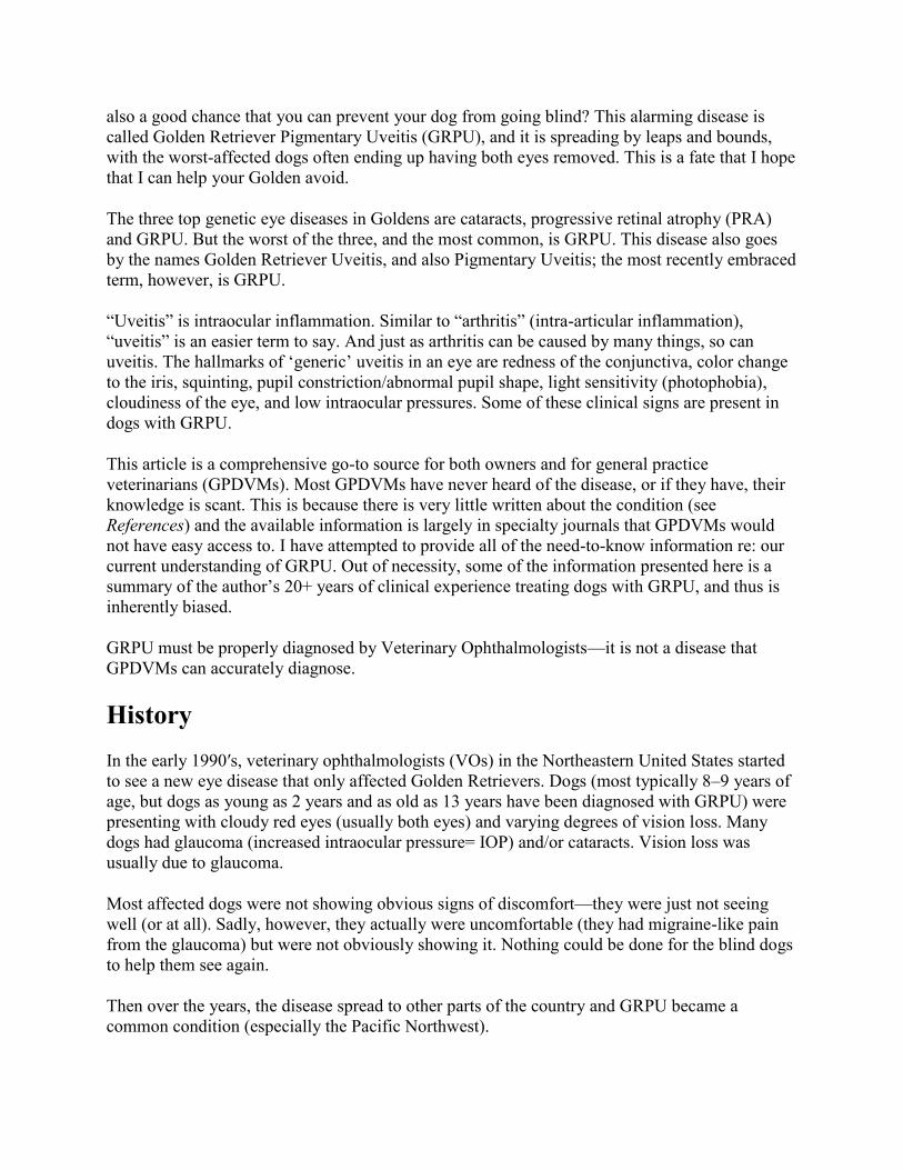
In order to understand the clinical signs, it is important to understand eye anatomy; please refer
to this diagram. Note: The Anterior Chamber is not specified in the diagram; it is the dome-
shaped, fluid-filled space between the cornea (clear “windshield” part of the eye) and the iris.
Both eyes are almost always affected. The cause of impaired vision/blindness is usually
glaucoma (increased intraocular pressure), but other conditions, such as cataracts and/or
intraocular hemorrhage, can impair vision. If GRPU is too advanced and the eye(s) are blinded
by glaucoma, vision cannot be recovered; sadly, in these cases both eyes usually need to be
removed.
In retrospect, many dogs presented in crisis have a history of intermittently red/inflamed eyes
that goes back several years. This redness is often mistakenly attributed to allergies (allergic
conjunctivitis), and it usually temporarily resolves with topical steroid ophthalmic medication
until the next episode of redness occurs. There is rarely any history of ocular discomfort or
vision loss until the time of crisis. Sometimes there is a history of gradual cloudiness occurring,
but usually owners will not present the dog to their family veterinarian for examination because
the dog is comfortable and (apparently) seeing well. It is important to understand that affected
dogs do not ‘complain’ or show outward signs that anything is wrong, until their vision declines.
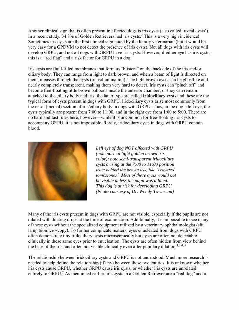
Another clinical sign that is often present in affected dogs is iris cysts (also called ‘uveal cysts’).
In a recent study, 34.8% of Golden Retrievers had iris cysts.2 This is a very high incidence!
Sometimes iris cysts are the first clinical sign noted by the family veterinarian (but it would be
very easy for a GPDVM to not detect the presence of iris cysts). Not all dogs with iris cysts will
develop GRPU, and not all dogs with GRPU have iris cysts. However, if either eye has iris cysts,
this is a “red flag” and a risk factor for GRPU in a dog.
Iris cysts are fluid-filled membranes that form as “blisters” on the backside of the iris and/or
ciliary body. They can range from light to dark brown, and when a beam of light is directed on
them, it passes through the cysts (transillumination). The light brown cysts can be ghostlike and
nearly completely transparent, making them very hard to detect. Iris cysts can “pinch off” and
become free-floating little brown balloons inside the anterior chamber, or they can remain
attached to the ciliary body and iris; the latter type are called iridociliary cysts and these are the
typical form of cysts present in dogs with GRPU. Iridociliary cysts arise most commonly from
the nasal (medial) section of iris/ciliary body in dogs with GRPU. Thus, in the dog’s left eye, the
cysts typically are present from 7:00 to 11:00, and in the right eye from 1:00 to 5:00. There are
no hard and fast rules here, however—while it is uncommon for free-floating iris cysts to
accompany GRPU, it is not impossible. Rarely, iridociliary cysts in dogs with GRPU contain
blood.
Many of the iris cysts present in dogs with GRPU are not visible, especially if the pupils are not
dilated with dilating drops at the time of examination. Additionally, it is impossible to see many
of these cysts without the specialized equipment utilized by a veterinary ophthalmologist (slit
lamp biomicroscopy). To further complicate matters, eyes enucleated from dogs with GRPU
often demonstrate tiny iridociliary cysts microscopically but cysts are often not detectable
clinically in these same eyes prior to enucleation. The cysts are often hidden from view behind
the base of the iris, and often not visible clinically even after pupillary dilation.1,3,4, 5
The relationship between iridociliary cysts and GRPU is not understood. Much more research is
needed to help define the relationship (if any) between these two entities. It is unknown whether
iris cysts cause GRPU, whether GRPU cause iris cysts, or whether iris cysts are unrelated
entirely to GRPU.2 As mentioned earlier, iris cysts in a Golden Retriever are a “red flag” and a
Left eye of dog NOT affected with GRPU
(note normal light golden brown iris
color); note semi-transparent iridociliary
cysts arising at the 7:00 to 11:00 position
from behind the brown iris, like ‘crowded
tombstones’. Most of these cysts would not
be visible unless the pupil was dilated.
This dog is at risk for developing GRPU
(Photo courtesy of Dr. Wendy Townsend)
risk factor for GRPU, and these dogs must be very carefully examined and regularly monitored
by an ophthalmologist for the presence of GRPU.
GRPU is a pigmenting disease. This means that dark brown microscopic flecks of pigment are
free-floating, like dust in the air, inside the anterior chamber and end up sticking to surfaces
inside the eye, especially the anterior lens capsule. The pigment can clog the drainage holes
inside the eye (iridocorneal drainage angle) that allow aqueous humor to exit the eye into the
body’s blood circulatory system. One theory is that iris cysts cause glaucoma,5 and another
theory is that complex auto-immune mechanisms trigger pigment dispersion inside the eye and
cause glaucoma.,
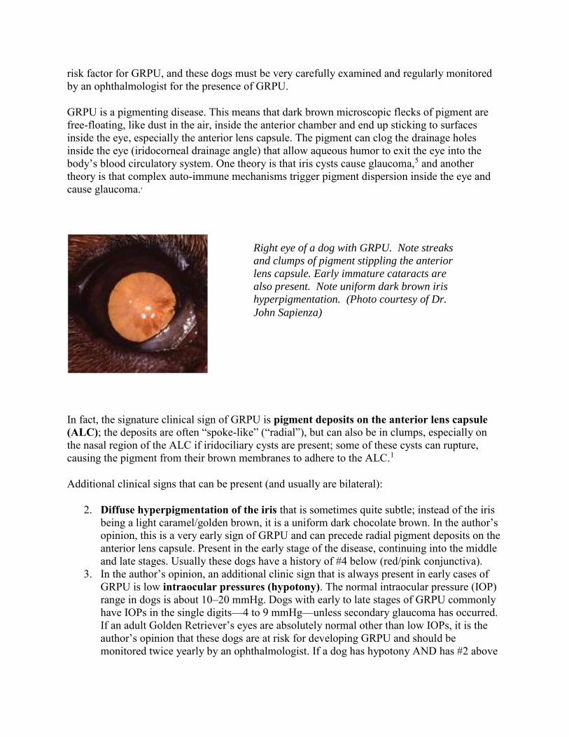
In fact, the signature clinical sign of GRPU is pigment deposits on the anterior lens capsule
(ALC); the deposits are often “spoke-like” (“radial”), but can also be in clumps, especially on
the nasal region of the ALC if iridociliary cysts are present; some of these cysts can rupture,
causing the pigment from their brown membranes to adhere to the ALC.1
Additional clinical signs that can be present (and usually are bilateral):
2. Diffuse hyperpigmentation of the iris that is sometimes quite subtle; instead of the iris
being a light caramel/golden brown, it is a uniform dark chocolate brown. In the author’s
opinion, this is a very early sign of GRPU and can precede radial pigment deposits on the
anterior lens capsule. Present in the early stage of the disease, continuing into the middle
and late stages. Usually these dogs have a history of #4 below (red/pink conjunctiva).
3. In the author’s opinion, an additional clinic sign that is always present in early cases of
GRPU is low intraocular pressures (hypotony). The normal intraocular pressure (IOP)
range in dogs is about 10–20 mmHg. Dogs with early to late stages of GRPU commonly
have IOPs in the single digits—4 to 9 mmHg—unless secondary glaucoma has occurred.
If an adult Golden Retriever’s eyes are absolutely normal other than low IOPs, it is the
author’s opinion that these dogs are at risk for developing GRPU and should be
monitored twice yearly by an ophthalmologist. If a dog has hypotony AND has #2 above
Right eye of a dog with GRPU. Note streaks
and clumps of pigment stippling the anterior
lens capsule. Early immature cataracts are
also present. Note uniform dark brown iris
hyperpigmentation. (Photo courtesy of Dr.
John Sapienza)

(diffuse iris hyperpigmentation), it is the author’s opinion that these dogs do have GRPU
and should be treated as such. Present in the early stage of the disease, continuing into the
middle and late stages.
4. A diffuse “flush” /reddening/pinkness to the conjunctiva (conjunctival hyperemia).
In retrospect, this is usually the very earliest clinical sign that the owner notices, years
before the dog is in crisis, but the owner and GPDVM do not realize that the redness of
the conjunctiva is due to GRPU; it is assumed to be due to allergies (allergic
conjunctivitis) because the redness resolves when topical steroid eyedrops or ointment is
applied to the eyes (—until the next flareup of redness occurs, which prompts a cycle of
another round of treatment).
5. Cloudiness of the eye, associated with a fibrinous “cotton wool” fluffy whitish plaque-
like infiltrate inside the eye, just in front of the pupil and sometimes nearly filling the
anterior chamber. This is termed fibrin (the “glue” of a blood clot), but the type of fibrin
that forms in dogs with GRPU is not typical fibriin: it does not readily break down and
dissolve, as typical fibrin does. The fibrin in GRPU dogs might not dissolve at all, or
dissolve only partially with medication. The fibrin often has tiny clots of blood admixed
with it. In the author’s opinion, eyes containing this fibrinous material are at high risk for
developing secondary glaucoma. This material promotes posterior synechiae (adhesions
of the iris to the ALC), which both “freezes” the pupil in place, and also increases the risk
of glaucoma occurring. Present in the middle to late stages of the disease.
6. Cloudiness of the eye, associated with cataract—( incipient or immature cataracts). It is
rare for the lens to develop a mature cataract and blind the eye, but cataracts can certainly
help impair vision. It is NOT recommended to perform cataract surgery on dogs with
GRPU, as postoperative inflammation is severe and usually cannot be adequately
controlled, leading to glaucoma and blindness. Present in the middle to late stages of the
disease.
Left eye of dog with GRPU. Note
cloudiness over pupil, with grey-white
streaks. This is a ‘cloud” of fibrin in
the anterior chamber, centering over
the pupil space. Note uniform dark
brown iris hyperpigmentation. This eye
also has late immature cataracts, and
the conjunctiva is inflamed. (Photo
courtesy of Dr. John Sapienza)

7. Bleeding inside the eye (hyphema). Sometimes it is more obvious that blood is inside
the eye, than #5 (fibrinous plaque). When blood is present inside the eye, it is always
accompanied by fibrinous material. Present in the middle to late stages of the disease.
8. Iris cysts. Usually nasal attached iridociliary cysts, but can be free-floating cysts in the
anterior chamber. Present in the early stage of the disease.
9. Secondary glaucoma in the late stage of the disease.
10. Blindness in the late stage of the disease (due to glaucoma).
GRPU is a disease with a spectrum of clinical signs, and not all dogs with GRPU will have all of
these clinical signs.
Treatment
It is obvious from this discussion that GRPU is a complex disease with no firm understanding
regarding genetics. Additionally, one study (Esson et al) has theorized that uveitis is not present
in GRPU, based on histopathologic evaluation of eyes enucleated due to glaucoma.4 However,
other authors believe that uveitis is indeed present, and that the eyes from the Esson study
showed little evidence of uveitis because the eyes had been treated with anti-inflammatory
medication prior to their removal. It is Dr. McCalla’s opinion that because dogs with GRPU
clinically respond favorably to anti-inflammatory medication, this is indirect evidence that
inflammation is a component of the disease. It is obvious that in order to better understand
GRPU, longterm studies of large numbers of dogs affected with GRPU are necessary.2 There is
no ‘best’ treatment regimen; all treatment regimens used by veterinary ophthalmologists are
based on their personal clinical experience with the disease and not on research findings. This
has evolved out of necessity, due to the lack of research regarding therapy of GRPU.
Treatment of GRPU depends on the stage of disease. It is the author’s opinion that if affected
dogs are identified in the early stage of the disease and placed on lifetime treatment and
examined regularly by an ophthalmologist, they will not progress to the middle and late stages of
the disease and their vision is saved.
Dogs in all stages of the disease may benefit from lifetime supplementation with the canine
antioxidant vision supplement, Ocu-GLO Rx™. An intriguing clinical study of dogs with GRPU
supplemented with Ocu-GLO Rx™ is currently being conducted by Dr. Wendy Townsend at
Purdue University to determine if supplementation would be beneficial. This research was
prompted by Dr. Townsend’s observation of one affected dog (that lost one eye to GRPU)
undergoing spontaneous resolution of GRPU (i.e. resolution of pigment deposition on the ALC)
in its remaining eye while on daily Ocu-GLO Rx™ supplementation. Ocu-GLO Rx™ also has
been shown to be neuroprotective in the presence of glaucoma, and this nutraceutical may thus
help preserve vision (and help preserve optic nerve function) in glaucomatous eyes.
In the early stage of GRPU, lifetime topical steroid and/or nonsteroidal anti-inflammatory
medication is prescribed (the author prefers topical steroid medication, specifically prednisolone
acetate 1% ophthalmic suspension) and the patients are reexamined every 6 months by the
ophthalmologist. Owners are cautioned to contact the ophthalmology clinic if either eye has

episodes of conjunctival redness or any signs of cloudiness. At each examination, tonometry is
performed to monitor the intraocular pressures.
In middle to late stages, treatment is as for the early stage, but some dogs also need systemic
anti-inflammatory medication. Many dogs respond with an oral non-steroidal anti-inflammatory
drug (NSAID), but some dogs need an oral steroid instead.
Late stage cases usually have glaucoma or IOPs that are starting to elevate. These dogs also need
topical glaucoma medications. Unfortunately, dogs initially presented to an ophthalmologist in
the late stages of the disease often end up losing their vision due to uncontrollable glaucoma, and
almost always, late-stage cases have both eyes affected, doubling the tragedy of this disease.
Surgical treatment for blind glaucomatous eyes must be performed if glaucoma cannot be
controlled medically, because these dogs are in constant discomfort (similar to a migraine
headache) and the eye(s) are enlarging because of the increased internal pressure. Because
Golden Retrievers are wonderful, uncomplaining dogs, they will not usually let their owners
know that their glaucomatous eyes are bothering them.
Many dogs with end-stage GRPU blinded by glaucoma end up having both eyes removed. Other
more cosmetic surgical choices are available that are discussed in the glaucoma article. However,
dogs with GRPU should not have the intravitreal injection of gentamicin (pharmacologic
ablation) procedure performed, because it would create additional uveitis and discomfort in these
dogs.
If a dog with GRPU blinded by glaucoma had high uncontrollable IOP(s) and was not healthy
enough to undergo surgery, a therapeutic dilemma occurs, and often these dogs end up being
euthanized because their quality of life with unrelenting migraine headaches is unacceptable,
which underscores the tragedy of this disease. It cannot be emphasized too often, that early
detection and lifetime treatment is the key to saving the vision in affected dogs.
Prevention
How can you help reduce the incidence of GRPU in Golden Retrievers?
1. Annual lifetime OFA ECR exams of all dogs 2 years of age and older that are
breeding/show animals. Dogs diagnosed with GRPU do not pass the exam, and should
not be bred.
2. Support the research efforts of Dr. Wendy Townsend (Purdue University College of
Veterinary Medicine, Dept. of Veterinary Clinical Sciences) to identify the genetic
marker for GRPU. To this end, she is requesting:
a. Blood samples from affected dogs, AND from normal-eyed elderly dogs 12 years
of age and older (these animals must have been examined by an ophthalmologist).
b. That tissues (enucleated eyes and also blood samples) be submitted from affected
dogs of all ages, AND from normal-eyed dogs (that are euthanized) 12 years of
age and older. You can access the submission form by accessing this GRPU
article on the author’s website www.animaleyecare.net . If you have any
questions, please contact Dr Townsend (her contact information is on the
paperwork).
3. Be aware of the danger of red/pink conjunctiva and the risk of GRPU being present
if iris cysts are present. If you have a dog with this clinical history, OR if you know that
your dog has iris cysts, seek out a veterinary ophthalmologist to examine your dog. To
locate a board-certified ophthalmologist, visit the www.acvo.org website.
4. If your dog has GRPU, PLEASE treat your dog as directed by the ophthalmologist
and bring it in as often as directed by the ophthalmologist. Tragically, dogs have had
their eyes removed, or been euthanized, because owners failed to follow directions.
5. If your dog has GRPU, inform your dog’s breeder of this diagnosis, so that the
breeder can make knowledgeable decisions about their dogs.
6. Support Dr. Wendy Townsend’s GRPU research by donating funds (tax-
deductible!) for her research, perhaps in honor of that special Golden Retriever in
your life. She is the only ophthalmologist performing research on this disease. Here’s
how you can help:
a. Online Giving (“Make a One Time Gift”) at https://donate.purdue.edu/
Select Designation Area: “College of Veterinary Medicine”
Fund: “College of Veterinary Medicine”
Other Instructions: Please indicate gift to be designated for “Dr. Townsend
Uveitis Research” You may also include tribute gift information (name of pet, in honor of, etc.)
Complete remaining pages including contact and payment information, and a
confirmation will be emailed to you.
b. By Mail: Make check payable to Purdue Foundation. In the memo line of check,
please indicate “Dr. Townsend Uveitis Research”
You can access the online gift form by accessing this GRPU article on the
author’s website www.animaleyecare.net .
c. By Phone: Please contact (765) 494-6304 and Purdue Foundation can place your
gift with a credit card.
Summary
It is critical for the general veterinary practitioner to be aware of GRPU as a disease, and to refer
suspected cases to an ophthalmologist ASAP.
GRPU is a very serious disease, in which early detection is critical—with early detection, dogs
can be placed on lifetime anti-inflammatory medications and a special canine antioxidant vision
supplement (OcuGLO Rx™—see www.ocuglo.com) to help control inflammation and also to
help support the health of all the cells of the eyes, and the immune system too. Some severely
affected dogs also need lifetime systemic medication (oral) to help control inflammation, and/or
glaucoma medication to attempt to control glaucoma. If GRU is caught early enough, and if the
dog is treated as prescribed by the ophthalmologist and is examined as often as needed by the
ophthalmologist, vision is almost always saved.
And finally—If a dog loses their vision from GRPU, it is important to understand that they
can still have a very acceptable quality of life without their sight, as long as they are not in

pain. A blind dog is not suffering, as long as the eyes are comfortable. If they are uncomfortable
(i.e. migraine headache from glaucoma) and this cannot be controlled medically, then surgery
(such as eye removal) must be done. Their general health needs to be acceptable in order to
perform surgery, and it is helpful if their environment is stable (don’t move furniture around in
your home) and it also helps (but is not necessary) if they can still hear.
It is relatively rare for my patients blinded by GRPU to be euthanized; I have had many GRPU
patients presented in the late stage of the disease, in which both eyes were removed. These dogs
have always thrived postoperatively. A dog’s job is to be with their family and share their life
with their family, and as long as that happens, they are happy!
This is “Chase” with a favorite toy—the same dog as shown in the beginning of this article.
GRPU robbed this kind and gentle dog of his vision. He developed secondary glaucoma in both
eyes, and both eyes were removed. Chase went on to live a full life, and even with no sight, he
continued catching tennis balls!
References
1. Sapienza JS, Simo FJ, Prades-Sapienza A. Golden Retriever uveitis: 75 cases (1994-
1999), Vet Ophthalmol 2009;3:241-246.
2. Townsend WM, Gornik KR. Prevalence of uveal cysts and pigmentary uveitis in Golden
Retrievers in three Midwestern states. J Am Vet Med Assoc 2013;9:1298-1301.
3. Golden Retriever Club of America, FAQ for Pigmentary Uveitis.
http://www.grca.org/pdf/health/eyes-health.pdf
4. Esson D, Armour M, Mundy P, et al. The histopathological and immunohistochemical
characteristics of pigmentary and cystic glaucoma in the Golden Retriever. Vet
Ophthalmol 2009;12:361-368.
5. Deehr AJ, Dubielzig RR. A histopathological study of iridociliary cysts and glaucoma in
Golden Retirevers. Vet Ophthalmol 1998;1:153-158.