Embed Size (px)
Citation preview

Yonsei Med J http://www.eymj.org Volume 55 Number 1 January 2014178
Gradual Lengthening of the Ulna in Patients with Multiple Hereditary Exostoses with a Dislocated Radial Head
Yong Jin Cho1 and Sung Taek Jung2
1Department of Orthopaedic Surgery, Yonsei University College of Medicine, Seoul;2Department of Orthopaedic Surgery, Chonnam National University College of Medicine, Gwangju, Korea.
Received: January 30, 2013Revised: April 8, 2013Accepted: April 11, 2013Corresponding author: Dr. Yong Jin Cho, Department of Orthopaedic Surgery,Yonsei University College of Medicine,50 Yonsei-ro, Seodaemun-gu, Seoul 120-752, Korea.Tel: 82-2-2228-2189, Fax: 82-2-363-1139E-mail: [email protected]
An abstract of this work was presented at Cairo, 6th International Meeting of the ASAMI 2010.
∙ The authors have no financial conflicts of interest.
© Copyright:Yonsei University College of Medicine 2014
This is an Open Access article distributed under the terms of the Creative Commons Attribution Non-Commercial License (http://creativecommons.org/ licenses/by-nc/3.0) which permits unrestricted non-commercial use, distribution, and reproduction in any medium, provided the original work is properly cited.
Purpose: Multiple hereditary exostoses of the forearm typically form in the distal ulna, causing disturbances in the growth of the ulna and functional disability. Mul-tiple hereditary exostoses inhibit the growth of the ulna, leading to an acquisition of a varus deformity in the radius, which sometimes leads to dislocation of the ra-dial head, the development of limitations in the pronation-supination of the fore-arm, and cosmetic problems. Materials and Methods: We retrospectively re-viewed the cases of four patients who had deformities of the forearm with radial head dislocation associated with multiple hereditary exostoses, and evaluated the radiologic and clinical results of excision of the osteochondromas from the distal ulna and gradual ulnar lengthening with an Ilizarov external fixator. Results: Good clinical and radiological results were obtained after a mean follow-up of 25 months. At the most recent follow-up, radial bowing, ulnar shortening, carpal slip, and the pronation/supination arch of the forearm had improved. There was little change in terms of preoperative radial articular angle and the flexion/extension arch of the el-bow by the most recent follow-up. Conclusion: Treatment of four forearms from four patients by excision of osteochondromas and gradual lengthening of the ulna with an Ilizarov external fixator spontaneously reduced dislocations of the radial heads without the need for any additional operative intervention. All patients were satisfied with the final results.
Key Words: Multiple hereditary exostosis, Ilizarov external fixator, gradual length-ening
INTRODUCTION
The first description of a patient with multiple exostosis has been attributed to Hunter in 1786. In 1814, Boyer published the first description of a family with multiple hereditary exostosis (MHE).1,2 Although exostoses are histological benign lesions, they can cause a variety of clinical problems. Patients with MHE most fre-quently report pain, cosmetic concerns and limitations to the adjacent joint. Defor-mities of the forearm and wrist occur in 39-59% of patients with multiple heredi-tary exostoses.3,4 Some commonly seen deformities in this condition are bowing of the radius, shortening of the ulna, ulna drift of the carpus, and occasionally dislo-
Original Article http://dx.doi.org/10.3349/ymj.2014.55.1.178pISSN: 0513-5796, eISSN: 1976-2437 Yonsei Med J 55(1):178-184, 2014
Gradual Lengthening of the Ulna
Yonsei Med J http://www.eymj.org Volume 55 Number 1 January 2014 179
Operative techniqueThe operative technique was as follows. First, we removed the osteochondromas of the distal ulna. Second, we took ul-nar transverse mid-diaphyseal subperiosteal osteotomy and positioned an Ilizarov external fixator. We used a temporary radial head reduction pin in all cases. The patients had a seven-day resting period and 1 mm/day of gradual length-ening with careful monitoring of neurovascular status, ad-justed joint pain and swelling, and pin tract infection. We lengthened the ulna by 5 mm plus variance, with the expec-tation of subsequent recurrence of ulnar shortening, except in one case that had already reached skeletal maturity, 24-year-old male. We removed the temporary radial head reduction pin when we were able to determine the radial head reduc-tion, using serial follow-up X-rays (Fig. 1).
Radiological and clinical evaluationRadiological assessment consisted of upper extremity antero-posterior radiograph with the hand and elbow in an anatomic position. A lateral radiograph of the forearm that included the hand, wrist, and elbow was obtained. We classified the fore-arm deformity using the Masada and Ono Classification system according to their morphological characteristics (Fig.
cation or subluxation of the radial head. These complex de-formities cause a loss of forearm pronation-supination.
Recent literature offers divergent views regarding treat-ment of these deformities. Some recommend aggressive early operative intervention to prevent or reduce the pro-gression of deformities and/or functional impairment, par-ticularly with regard to radial head dislocation. Others stat-ed that these surgical procedures do not produce any predictable functional improvement. Excision of the exos-toses may be performed for cosmetic reasons or to resolve local irritation alone.
Acute or gradual lengthening of the ulna, hemiphyseal stapling of the distal aspect of the radius, corrective osteoto-my of the radius or excision or open reduction of a dis-placed radial head may be included in corrective or recon-structive surgery of the forearm.5 However, there are no reports about the results of surgical treatment in patients with multiple hereditary exostoses who had a preoperative-ly dislocated radial head treated by excision of the osteo-chondroma and gradual lengthening. Our hypothesis was that patients who had a dislocated radial head would suffer limitations to the range of motion of the forearm, which would be improved by gradual lengthening of ulnar using the Ilizarov technique.
MATERIALS AND METHODS
PatientsWe retrospectively reviewed the cases of four patients who had deformities on one forearm each and radial head dislo-cation associated with multiple hereditary exostosis. The patients were treated at our hospital between 2006 and 2009. There were three girls and one boy, ranging in age from 5.2 to 24.0 years (average, 11.5 years). The follow-up period ranged from 20 to 29 months (mean: 25 months). The de-formations were present in three dominant side forearms and one non-dominant side forearm, and restriction of pro-nation/supination of the forearm before surgery was found in all cases (Table 1).
Table 1. Demographic Data of the Four Patients Treated for Forearm DeformityGender Affected side Age at operation Duration of follow up
Case I Female Dominant 7 yrs 10 months 20 monthsCase II Male Non-dominant 24 yrs 24 monthsCase III Female Dominant 5 yrs 2 months 27 monthsCase IV Female Dominant 8 yrs 10 months 29 months
Fig. 1. This patient was 7 years and 10 months old. (A) The preoperative X-ray shows dislocated radial head (arrow) and distal ulnar osteochondro-ma (arrow). (B) Immediate postoperative X-ray shows a temporary reduc-tion pin for the dislocated radial head. (C) 33 mm lengthening was done and we removed the temporary reduction pin six weeks after the operation. The arrow indicates the reduced radial head. (D) At the most recent follow-up X-ray, we found a reduced radial head.
A B C D

Yong Jin Cho and Sung Taek Jung
Yonsei Med J http://www.eymj.org Volume 55 Number 1 January 2014180
anterior-posterior plane radiography, and radial head stabili-ty at elbow lateral plane radiography (Fig. 3). We measured the amount of ulnar lengthening (AUL) at the most recent follow-up forearm anterior-posterior plane radiography and calculated the external fixation index (EFI). EFI was ob-tained by dividing the total duration of external fixation by the length gained.
We evaluated the range of pronation/supination, elbow flexion/extension at the preoperative and most recent visit to the outpatient department. Also, each patients was asked to rate the function of the extremity according to specific crite-ria (Table 2).6
Statistical evaluationData were analyzed using SPSS statistical analysis software program version 15.0 (SPSS Inc., Chicago, IL, USA). Test-ing for statistical significance was performed using the Wil-coxon signed rank test. Statistical significance was consid-ered at p≤0.05.
RESULTS
The presenting symptoms were a loss of range of motion of the forearm and cosmetic problems in all four patients. All cases were Class IIB type classified by the Masada and Ono Classification, which means that the radial head is dislocat-ed because of an osteochondroma in the distal portion of the ulna and there are small osteochondromas in the part of the radius. Dislocation of the radial head, which was ob-served preoperatively in all patients, was naturally reduced with a temporary reduction half pin that was located within 6 weeks. In Case 1, we could see radial head subluxation at 20 months follow up, but there was no further progression of the limitation of elbow range of motion. In other cases, stably reduced radial head was observed upon recent fol-low-up radiographic evaluation.
The average preoperative RL, RB, percentage of RB and RAA were 170.5 (133.0-222.0) mm, 16.0 (12.0-20.0) mm,
2).5 Radiographic review compared the preoperative and most recent follow-up radiographs. We measured the radial articular angle (RAA), radial length (RL), radial bowing (RB), percentage of RB, carpal slip (CS), ulnar length (UL), ulnar shortening, percentage of ulnar shortening at forearm
Table 2. Functional Assessment Criteria by the PatientScore Please check the one box that most closely describes the current condition of your hand and wrist.
5 I have no limitations of my activities and no pain.4 I have no pain. I have some limitation of my activities but have not had to change my lift (Sports activites or Job) because of it.3 I have no pain. I have had to change or limit my job or give up certain sports activities because of the condition of my hand.2 I have pain in my hand, wrist, or elbow, but I have no limitations because of it.1 I have pain in my hand, wrist, or elbow, which limits my activities.0 I have pain for which I take medications.
Fig. 2. Classification of MHE forearm deformities. Type 1: the main osteo-chondroma formation is in the distal portion of the ulna, but the radial head is not dislocated. Type IIA: the radial head is dislocated because of an os-teochondroma at the proximal metaphysic of the radius. Type IIB: in addi-tion to ulnar shortening the radial head is dislocated. Type III: the main os-teochondroma formation is in the metaphysic of the distal radius, and there is relative shortening of the radius. MHE, multiple hereditary exostosis.
Fig. 3. Radiologic assessment. (A) Ulnar shortening: measured by a line drawn from the distal end of the ulna to the linear axis. Radial bowing: the distance that the radial diaphysis deviates from the linear axis of the fore-arm divided by the length of the linear axis. (B) The ulna tilt of the distal radi-al articular surface was recorded as the radial articular angle. The ulna drift of the carpus was assessed by the carpal slip. RL, radial length; RB, radial bowing; CS, carpal slip; RAA, radial articular angle; US, ulna shortening.
A B
I IIA IIB III

Gradual Lengthening of the Ulna
Yonsei Med J http://www.eymj.org Volume 55 Number 1 January 2014 181
significant statistical improvement. Only one complication occurred in the forearm during the distraction osteogenesis procedure. This callus fracture was treated with a long arm splint for three weeks. Other common complications such as nerve palsies, tightness of the flexor tendons, and pin track sepsis were not found in this series.
DISCUSSION
The pathogenesis of forearm deformities in patients with multiple hereditary exostosis is complex and not yet fully understood. However, there have been several hypotheses to explain this situation. The forearm deformity caused by shortening of the ulnar relative to the radius in patients with multiple hereditary exostosis is related to four factors. First, the cross-sectional area of the distal ulnar physis is only one-quarter that of the distal radius. The distal ulnar physis being significantly smaller means that its growth can be more severely affected by the disease in the wrist.7 Second, the dis-tal ulna is more commonly involved in the condition than the distal radius.4 Third, there is more longitudinal growth at the
9.9 (5.4-12.0) % and 40.7 (34.0-45.0) degrees. At the time of most recent follow up, they were 174.2 (138.0-223.0) mm, 13.2 (12.0-16.0) mm, 7.8 (5.8-8.7) % and 37.5 (33.0-43.0) degree. When all of the patients were compared, there was a tendency towards improvement that was not significant in any of these four parameters (Fig. 4).
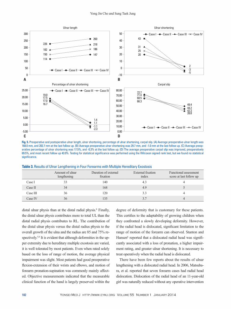
The average preoperative UL, ulnar shortening and per-centage of ulnar shortening were 168.0 (114.0-226.0) mm, 29.7 (20.0-43.0) mm and 17.5 (16.7-19.0) %, respectively. At the time of most recent follow, they were 202.7 (147.0-260.0) mm, -1.0 (-5.0-2.0) mm and -0.3 (-2.3-1.4) %, respec-tively. CS was improved, preoperatively 69.5 (65.3-72.7)% and most recent follow up 43.9 (39.8-49.4) % (Fig. 5).
The average AUL and duration of external fixation index were 34.5 (33.0-36.0) mm and 140.7 (120.0-168.0) days, respectively. Thus, the external fixation index was an aver-age of 4.1 (3.3-4.9) day/mm. All four patients were satisfied with the functional and cosmetic results. One patient scored 5 points and the others 4 points on the functional assess-ment score (Table 3). There was a tendency towards im-provement in forearm pronation/supination and elbow flex-ion/extension lag, however, we were unable to determine any
Fig. 4. Preoperative and postoperative radial length, radial bowing, percentage of radial bowing, radial articular angle. (A) Average preoperative radial length was 170.5 mm, and 174.2 mm at the last follow up. (B) Average preoperative radial bowing was 16.0 mm, and 13.2 mm at the last follow up. (C) Average preoperative percentage of radial bowing was 9.9%, and 7.8% at the last follow up. (D) The average preoperative radial articular angle was 40.7 degree, and 37.5 degree at the last follow up. Testing for statistical significance was performed using the Wilcoxon signed rank test, but we found no statistical signifi-cance.
0
0.00
2.00
4.00
6.00
8.00
10.00
12.00
14.00
0
0
50 5
105
100 10
2015
150 15
3025
200 20
4035
250 25
5045
Radial length
Percentage of radial bowing
Radial bowing
Radial articular angle
222
12.0
20
45
189
11.6
16
44
138
10.6
133
5.4
12
4034
223
8.7
16
43
193
8.4
143
8.3
13
38
138
5.8
12
3633
A
C
B
D
Case I Case II Case III Case IV
Case I Case II Case III Case IV
Case I Case II Case III Case IV
Case I Case II Case III Case IV
Yong Jin Cho and Sung Taek Jung
Yonsei Med J http://www.eymj.org Volume 55 Number 1 January 2014182
degree of deformity that is customary for these patients. This certifies to the adaptability of growing children when they confronted a slowly developing deformity. However, if the radial head is dislocated, significant limitation to the range of motion of the forearm can observed. Stanton and Hansen6 reported that a dislocated radial head was signifi-cantly associated with a loss of pronation, a higher impair-ment rating, and greater ulnar shortening. It is necessary to treat operatively when the radial head is dislocated.
There have been few reports about the results of ulnar lengthening with a dislocated radial head. In 2006, Matsuba-ra, et al. reported that seven forearm cases had radial head dislocation. Dislocation of the radial head of an 11-year-old girl was naturally reduced without any operative intervention
distal ulnar physis than at the distal radial physis.8 Finally, the distal ulnar physis contributes more to total UL than the distal radial physis contributes to RL. The contribution of the distal ulnar physis versus the distal radius physis to the overall growth of the ulna and the radius are 85 and 75% re-spectively.5,8 It is evident that although deformities in the up-per extremity due to hereditary multiple exostosis are varied, it is well tolerated by most patients. Even when rated solely based on the loss of range of motion, the average physical impairment was slight. Most patients had good preoperative flexion-extension of their wrists and elbows, and motion of forearm pronation-supination was commonly mainly affect-ed. Objective measurements indicated that the measurable clinical function of the hand is largely preserved within the
Table 3. Results of Ulnar Lengthening in Four Forearms with Multiple Hereditary ExostosisAmount of ulnar
lengtheningDuration of external
fixationExternal fixation
indexFunctional assessment score at last follow up
Case I 33 140 4.3 4Case II 34 168 4.9 5Case III 36 120 3.3 4Case IV 36 135 3.7 4
Fig. 5. Preoperative and postoperative ulnar length, ulnar shortening, percentage of ulnar shortening, carpal slip. (A) Average preoperative ulnar length was 168.0 mm, and 202.7 mm at the last follow up. (B) Average preoperative ulnar shortening was 29.7 mm, and -1.0 mm at the last follow up. (C) Average preop-erative percentage of ulnar shortening was 17.5%, and -0.3% at the last follow up. (D) The average preoperative carpal slip was improved, preoperatively 69.5%, and most recent follow up 43.9%. Testing for statistical significance was performed using the Wilcoxon signed rank test, but we found no statistical significance.
0
50
100
150
200
250
-5.00
0.00
5.00
10.00
15.00
20.00
25.00
-10
0
10
20
30
40
50
0.00
10.00
20.00
30.00
40.00
50.00
60.00
70.00
300
80.00
Ulnar length
Percentage of ulnar shortening
Ulnar shortening
Carpal slip
226
19.0
43
72.7
182
17.5
3125
71.4
150
17.0
114
16.7
20
68.566.3
260
1.4
49.4
218
1.1
186
-1.1
44.2
147
-2.3
42.339.8
A
C
B
D
Case I Case II Case III Case IV
Case I Case II Case III Case IV
Case I Case II Case III Case IV
Case I Case II Case III Case IV
-5-3 2

Gradual Lengthening of the Ulna
Yonsei Med J http://www.eymj.org Volume 55 Number 1 January 2014 183
observed in skeletally immature patients. Fortunately, no forearm has yet required a second ulnar lengthening in the four patients so treated.
Exostosis was removed if there was interference with joint movement or if the lesion was prominent, painful, and/or cos-metically undesirable. The indications for performing this procedures were a discrepancy of >5 mm between the lengths of the ulna and radius, a RAA of >30 degrees, a CS of >60%, bowing of the radius, or a combination of these changes.11 Most patients had a combination of these changes. Symp-tomatic dislocation of the head of the radius was defined as interfering with joint movement.
Ulnar lengthening should theoretically improve ulnar trans-location at the wrist, but no statistically significant clinical or radiological improvements were noted in our patients. Fo-gel, et al.7 performed ten ulnar lengthening procedures in eight patients, and found that relative shortening of the ulna usually recurred after the lengthening and also that, in agree-ment with our findings, ulnar lengthening did not result in a statistically significant improvement in rotation of the fore-arm, RAA or CS.
The timing of the surgery is extremely important, and there have been some reports regarding this issue. Some rec-ommend early intervention, which has more potential for re-modeling and leads to better surgical results.5,8,13,14 Masada, et al.5 noted recurrence of ulnar shortening after lengthen-
after gradual lengthening of the ulna. Ulnar shortening re-curred with the growth of four immature patients. In 2007, Akita, et al. reported dislocation of the head of the radius in five extremities/eight forearms. They performed immediate ulnar lengthening, corrective osteotomy of the radius, and an-nular ligament reconstruction, along with open reduction of the dislocated radial head. These treatments were not effec-tive in either case. Exicision of the dislocated radial head was performed in two patients (two forearms) after their skeletal maturity was reached. Other reports regarding the lengthen-ing of the ulna in patients with radial head dislocation and multiple hereditary exostoses are presented in Table 4.5,8-11
Although a number of techniques for the surgical treat-ment of forearm deformities have been described, the oper-ative treatment for such deformities in patients with multiple hereditary exostosis with radial head dislocation remains un-clear. Known surgical interventions include simple excision of the osteochondromas, acute or gradual ulnar lengthen-ing, corrective radial osteotomy, hemi-epiphyseal stapling of the distal radius, creation of a one-bone forearm and the Sauve-Kapandiji procedure.12 The rationale for ulnar length-ening is that the hypoplastic ulna, the keystone of the com-plex deformity, tethers the radial physis, theoretically di-minishing ulnar support of the carpus and increasing ulnar sided pressure on the radial epiphysis. In the present series, progression of the deformity during the growth period was
Table 4. Literature Review of Results of Preoperatively Dislocated Radial Head Treated by Excision of Osteochondroma and Ulnar Gradual Lengthening
Authors Number of cases Age (yrs) Follow-up duration Results
Masada, et al.5 Not available 15.3 31 monthsThe average gain in pronation and supination was 21.3 and 19.1 degrees. Postoperative improvement was dramatic. Temporary radial nerve paresis in 1 case.
Pritchett8
1 for radial head dislocation, 3 for radial head subluxation
9.1 41.8 months3 forearms showed improvement in pronation-supination. Ulnar shortening recurred with growth in all operated forearms in the skeletally immature patients.
Matsubara, et al.9 1 for radial head dislocation 10.8 7.1 yrs
Dislocation of the radial head of one patient was naturally reduced. Ulnar shortening recurred with growth of four immature patients. The overlength of 5 mm was negated by the recurrence of ulnar shortening.
Akita, et al.11 5 for radial head dislocation Not available Not available
Authors performed immediate ulnar lengthening, corrective osteotomy of the radius, and anular ligament reconstruction with palmaris longus tendon graft, but symptoms worsened. Excision of the dislocated radial head was performed in two patients (two forearms) after skeletal maturity. Nonunion in 2, fracture of callus at the site of lengthening in 2, and temporary radial.
Shin, et al.10 2 for radial head dislocation 8.8 Not available
Ulnar lengthening did not significantly affect the clinical outcome. The radiological parameters also did not change significantly with ulnar lengthening.

Yong Jin Cho and Sung Taek Jung
Yonsei Med J http://www.eymj.org Volume 55 Number 1 January 2014184
tem, Severence Hospital, Institutional Review Board (IRB# 4-2012-0192).
REFERENCES
1. Solomon L. Bone growth in diaphysial aclasis. J Bone Joint Surg Br 1961;43-B:700-16.
2. Wood VE, Sauser D, Mudge D. The treatment of hereditary multi-ple exostosis of the upper extremity. J Hand Surg Am 1985;10: 505-13.
3. Schmale GA, Conrad EU 3rd, Raskind WH. The natural history of hereditary multiple exostoses. J Bone Joint Surg Am 1994;76: 986-92.
4. Shapiro F, Simon S, Glimcher MJ. Hereditary multiple exostoses. Anthropometric, roentgenographic, and clinical aspects. J Bone Joint Surg Am 1979;61:815-24.
5. Masada K, Tsuyuguchi Y, Kawai H, Kawabata H, Noguchi K, Ono K. Operations for forearm deformity caused by multiple os-teochondromas. J Bone Joint Surg Br 1989;71:24-9.
6. Stanton RP, Hansen MO. Function of the upper extremities in he-reditary multiple exostoses. J Bone Joint Surg Am 1996;78:568-73.
7. Fogel GR, McElfresh EC, Peterson HA, Wicklund PT. Manage-ment of deformities of the forearm in multiple hereditary osteo-chondromas. J Bone Joint Surg Am 1984;66:670-80.
8. Pritchett JW. Lengthening the ulna in patients with hereditary multiple exostoses. J Bone Joint Surg Br 1986;68:561-5.
9. Matsubara H, Tsuchiya H, Sakurakichi K, Yamashiro T, Watanabe K, Tomita K. Correction and lengthening for deformities of the forearm in multiple cartilaginous exostoses. J Orthop Sci 2006;11:459-66.
10. Shin EK, Jones NF, Lawrence JF. Treatment of multiple hereditary osteochondromas of the forearm in children: a study of surgical procedures. J Bone Joint Surg Br 2006;88:255-60.
11. Akita S, Murase T, Yonenobu K, Shimada K, Masada K, Yoshika-wa H. Long-term results of surgery for forearm deformities in pa-tients with multiple cartilaginous exostoses. J Bone Joint Surg Am 2007;89:1993-9.
12. Rodgers WB, Hall JE. One-bone forearm as a salvage procedure for recalcitrant forearm deformity in hereditary multiple exostoses. J Pediatr Orthop 1993;13:587-91.
13. Peterson HA. Multiple hereditary osteochondromata. Clin Orthop Relat Res 1989:222-30.
14. Ip D, Li YH, Chow W, Leong JC. Reconstruction of forearm de-formities in multiple cartilaginous exostoses. J Pediatr Orthop B 2003;12:17-21.
15. Abe M, Shirai H, Okamoto M, Onomura T. Lengthening of the forearm by callus distraction. J Hand Surg Br 1996;21:151-63.
16. Arms DM, Strecker WB, Manske PR, Schoenecker PL. Manage-ment of forearm deformity in multiple hereditary osteochondro-matosis. J Pediatr Orthop 1997;17:450-4.
17. Dal Monte A, Andrisano A, Capanna R. Lengthening of the radius or ulna in asymmetrical hypoplasia of the forearm (report on 7 cases). Ital J Orthop Traumatol 1980;6:329-42.
18. Siffert RS, Levy RN. Correction of wrist deformity in diaphyseal aclasis by stapling. Report of a case. J Bone Joint Surg Am 1965; 47:1378-80.
ing in 13 forearms. Nine of the 13 forearms also underwent radial osteotomy; it is possible that these improvements were secondary to the osteotomy.
Intervention at a later age is sometimes recommended, because a recurrent operation can be avoided by postponing the procedure and good function can be acquired despite significant deformity after skeletal maturity.15,16
The use of an external fixator causes the interosseous membrane to drag the radius distally. In Type IIb deformi-ties, the use of an external fixator helps to reduce the dislo-cated radial head.5,8,17
The disadvantage of ulnar lengthening is that the defor-mity in young patients will recur and further lengthening may be needed. Some patients, particularly those with short arms or asymmetrical involvement, may accept the possible need for additional operations. Pritchett reported the results of ulnar lengthening in ten forearms (eight patients). He found that the deformity tended to recur in younger patients and that further lengthening was sometimes required. He recommended initial overcorrection in skeletally immature patients.8 We therefore over-lengthened by 5 mm in prepu-bescent patients. Initial overcorrection by 5 mm is well tol-erated and is recommended for skeletally immature patients. To prevent recurrence, it is better to over-lengthen the short-er ulna. Most patients required lengthening of between 20 and 40 mm, considering the recurrence. By lengthening gradually, patients can avoid neurovascular system prob-lems and the shorter radius can be lengthened as well. Sta-pling or hemi-epiphysiodesis of the distal radial physis, or shortening of the radius to prevent recurrence, was reported by Siffert and Levy.18 This procedure is not acceptable be-cause the affected limbs will be shorter. Furthermore, dislo-cation of the radial head can be reduced using gradual length-ening with an external fixator. Therefore, we believe that our method is a good way to treat forearm deformities caused by osteochondromas.
In conclusion, we treated four patients with dislocated ra-dial heads in the forearm with excision of osteochondromas and gradual lengthening of the ulna with an Ilizarov exter-nal fixator. Spontaneous reduction of dislocated radial head was observed in all four cases, and the radiological and clinical results of the procedure were satisfactory.
ACKNOWLEDGEMENTS
This study was approved by Yonsei University Health Sys-