Embed Size (px)
Citation preview

G protein expression and second messenger formation in humangranulosa cells
A. L\l=o'\pezBernal, J. Bellinger, J. M. Marshall, S. Phaneuf,G. N. Europe-Finner, G. Asb\l=o'\thand D. H. Barlow
Nuffield Department of Obstetrics and Gynaecology, John Radcliffe Hospital, Headinglon,Oxford, 0X3 9DU, UK
The expression of heterotrimeric (\g=a\\g=b\\g=g\subunits) GTP-binding regulatory proteins(G proteins) and the activation of G protein-linked receptors in human granulosa cells were
investigated. The cells were obtained from stimulated follicles in women undergoing in vitrofertilization and were cultured in serum-supplemented medium. Immunoblotting withspecific antibodies showed that granulosa cell membranes express \g=a\s, \g=a\i3\g=a\i1,2, \g=a\q,11 and \g=b\subunits. Three antibodies against \g=a\o failed to detect this protein. The cells responded tohCG and to prostaglandin E2 with a dose-dependent increase in cAMP formation,confirming the functional activation of G\g=a\s. The \g=a\2adrenoceptor agonist, clonidine,inhibited hCG-stimulated cAMP formation and this effect was blocked with pertussis toxin,thus involving a Gi-type protein, most likely G\g=a\i2.Oxytocin provoked an increase information of inositol phosphates and intracellular calcium concentration, which was partlypertussis toxin resistant, providing evidence of G\g=a\ q,11activation. However, a significantcomponent of the response to oxytocin could be blocked by pertussis toxin, indicatingGi-mediated phospholipase C activation (by either \g=a\ior \g=b\\g=g\subunits). These datademonstrate the presence of G proteins in granulosa cells and suggest a complex regulationof hormonal signalling. The concentration of cAMP in these cells depended on the balanceof G\g=a\s:G\g=a\iactivation, whereas activation of the inositol phospholipid pathway and rises inintracellular calcium involved both Gq,11and Gi pathways.
Introduction
Granulosa cells are essential for follicular development andcorpus luteum formation. During follicular development,granulosa and theca cells interact to produce steroids, predomi¬nantly oestrogens, and, after ovulation, luteinized granulosacells develop a large capacity to secrete progesterone (Steeleand Leung, 1992; Yong et al, 1992a). The functions of granu¬losa cells are controlled by gonadotrophins and other hormoneswhose mechanism of action usually involves binding to recep¬tors on the cell surface and the transmission of information toan effector system within the cells. GTP-binding proteins (Gproteins) mediate the transmission of the message from acti¬vated hormone receptors to effector systems such as adenylylcyclase or phospholipase C (Birnbaumer et al, 1990; Oíate andAllende, 1991; Hepler and Gilman, 1992). G proteins consist ofheterotrimers: ß subunits. The subunits vary considerablybetween G proteins and confer specificity for coupling toreceptors and effectors, whereas ß subunits are relativelycommon (Hepler and Gilman, 1992). There are at least twotypes of G protein that interact with adenylyl cyclase: Gas,which is stimulatory (increasing cyclic AMP formation); andGa¡, which is inhibitory (Gilman, 1990; Levitzki and Bar-Sinai,1991). Proteins of the G n family interact with phospholipase
C (PLC), resulting in the formation of two second messengers:diacylglycerol, which activates protein kinase C, and inositol1,4,5,-trisphosphate (InsP3), which mobilizes intracellular cal¬cium (Smrcka et al, 1991; Taylor et al, 1991). Moreover, ß subunits can interact directly with some types of PLC andadenylyl cyclase (Levitzki and Bar-Sinai, 1991; Hepler andGilman, 1992).
The purpose of this paper was to study G protein expressionin human granulosa cells, and to investigate the functionalinvolvement in receptor activation of G proteins by measuringsecond messenger formation in response to agonists that oper¬ate through G protein-linked receptors (Kimura et al, 1992; Luand Menon, 1994). Human chorionic gonadotrophin was usedto stimulate adenylyl cyclase (McFarland et al, 1989; Cookeet al, 1989), and oxytocin was used to activate PLC (Schreyet al, 1987; Phaneuf et al, 1993). In addition, the presence ofadenylyl cyclase inhibitory pathways was investigated usingclonidine, which activates a2-adrenergic receptors thought tobe coupled to G¡ (Osborne, 1991; Regunathan et al, 1991).
Materials and MethodsCollection and culture of cells
Granulosa cells were obtained with patients' consent andapproval of the Ethics Committee from women undergoingReceived 6 January 1995.

Table 1. G-protein antisera used in this study
Antibody Source Subunit Peptide sequence Binding site
AS/7EC/2GC/2IMIOClQLRM/1MS/1
NENNENNEN
MilliganMilligan
NENNENNEN
, aiI( ai2
a0«o
ß-common
KENLKDCGLFKNNLKECGLY
GCTLSAEERAALERSKNLKEDGISAAKDVK
ANNLRGCGLYQLNLKEYNLVRMHLRQYELLMSELDQLRQE
C-terminusC-terminusN-terminus
Residues 22—35C-terminusC-terminusC-terminusN-terminus
All antisera were raised in rabbits. Sources: NEN-Du Pont,of Biochemistry, University of Glasgow.
Boston, MA; G. Milligan, Molecular Pharmacology Group, Department
routine oocyte retrievals for in vitro fertilization. Folliculargrowth was stimulated with daily injections of gonadotrophinsin patients previously desensitized with a long course of a
GnRH analogue (Forman et al, 1991). Several follicles were
aspirated from each patient 35 h after administration of10 000 iu of hCG (Profasi, Serono Laboratories UK Ltd,Hertfordshire). The mean follicular diameter was 17 ± 2.4 mm
(mean ± sd, = 150). After removing any oocytes present,follicular fluids and flushings were pooled and centrifuged(340 g) at room temperature for 10 min through Ficoll-Paque(Pharmacia, St Albans) to remove red blood cells. Granulosacell layers were dispersed with collagenase (Sigma Ltd Co,Poole) diluted to 300 Uml"1 and Trasylol (Bayer UK Ltd,Newbury) diluted to 100 U ml"1 for 20 min at 37°C in1 mmol CaCl2 1~ . Cell viability after enzymatic dispersionwas 73 ± 4%, as assessed by Trypan blue exclusion, and theaverage yield of viable cells was 0.59 ± 0.44 IO6 per patient(mean ± sd, = 48).
Cells were routinely seeded at a density of 40 000 cellscm"2 in 24- or 6-well fibronectin-coated wells and werecultured in Hams F12:DMEM (1:1 by volume; GIBCO, LifeTechnologies, Uxbridge) medium containing 10% (v/v) fetalcalf serum, 10% (v/v) horse serum (Advanced Protein Products,Brierley Hill), 1 pmol vitamin E 1
~
\ 20 nmol selenium 1~
J andpenicillin/streptomycin 100 units ml
~
1 (McAllister et al, 1990).Under these conditions, there was cell growth in culture(doubling time approximately 3 days), as demonstrated byreduction of tetrazolium (3-[4,5-dimethylthiazol-2-yl]-2,5-diphenyltetrazolium bromide; Sigma) to formazan and by[3H]thymidine uptake.
SteroidogenesisOestradiol and progesterone outputs into cell culture super¬
natants were measured with radioimmunoassay kits (DPC, LosAngeles, CA) following the manufacturers' recommendations.The sensitivity of the progesterone assay was 0.1 pmol ml-and the mid-point of the standard curve (50% displacement)was obtained with 6.7 pmol ml" . The coefficient of variationwas 4.7% (intra-assay) and 9.2% (interassay). The sensitivity ofthe oestradiol assay was 0.03 pmol ml
~
: and the mid-point ofthe standard curve was 1.1 pmol ml-1. The intra- and inter¬assay coefficients of variation for this assay were 4.7% and7.9%, respectively.
ImmunoblottingCultured granulosa cells were lifted with trypsin—EDTA and
resuspended in 25 mmol Tris 1_1 buffer, pH 7.4, 250 mmolsucrose 1
~
1-1 mmol EDTA 1~
, and were disrupted with a
ground glass homogenizer. A membrane pellet was obtainedby centrifuging the homogenate at 13 000 g for 30 min, andthe protein content was estimated by the Bradford—BioRadmethod (Richmond, CA). The pellet was solubilized in 10%(w/v) SDS buffer containing 8 mol urea 1
~ , 0.5 mol di-tiothreitol 1 (DTT) and 0.05 mol Tris l"1, pH 6.8, and theproteins were resolved on a 12.5% polyacrylamide gel contain¬ing 0.0625% (w/v) bis-acrylamide for 6 h at 40 mA. Proteinsfrom gels were transferred to nitrocellulose using BioRadelectroblotting apparatus with 0.025 mol Tris 1
~-0.195 mol
glycine '1 and 20% ( / ) methanol, pH 8.6, at 90 V for 2 h.After transfer, nitrocellulose papers were incubated in PBS,pH 7.5, containing 3% (w/v) skimmed milk (Marvel, PremierBrands UK Ltd, Adbaston) for 1 h at room temperature. Thepapers were washed three times with PBS and incubated withprimary antibodies at dilutions ranging from 1:250 to 1:1000.The antibodies used are given in Table I. Primary antisera were
removed and the blots were washed thoroughly with PBS andincubated with goat anti-rabbit immunoglobulin G coupled tohorseradish peroxidase at 1:1000 dilution for 1 h at room
temperature, and washed again with PBS. Antibody complexeswere detected by enhanced chemiluminescence (ECL) usingX-Omat X-ray film (Europe-Finner et al, 1993).
Cyclic AMP estimationsCells were challenged in serum-free medium with appropri¬
ate agonists or drugs. Incubations were carried out at 37°C inthe presence of 5 mmol theophylline 1
~ (Sigma), and were
terminated with 5% (v/v) perchloric acid. Cyclic AMP was
extracted by the freon-octylamine method, and was measuredby competitive protein-binding using [2,8-3H]adenosine3',5'-cyclic phosphate (specific activity 1.85 TBq mmol-1,Amersham International, Bucks) as the labelled ligand (LópezBemal et al, 1991).
Inositol phosphate formationGranulosa cells were labelled for 48 h with 2.5 pCi per well
(0.23 MBq ml"
J) of [3H\myo inositol (TRK 912B30; Amersham

International) in inositol-free RPMI medium containing 2%(v/v) fetal calf serum. The cells were washed and challenged byagonists in the presence of lithium chloride (15 mmol 1~ *) for30 min at 37°C. The reactions were stopped with 1 ml acidifiedchloroform/methanol (chloroform/methanol/hydrochloric acid;50/100/1). Inositol phosphate formation was measured by ionexchange chromatography on Dowex columns as described byPhaneuf et al (1993).
Calcium estimations
Granulosa cells were grown onto 13 mm diameterfibronectin-coated borosilicate glass coverslips. Subconfluentcells were loaded with 5 pmol Fura-2 acetoxymethyl ester 1
"
and 0.1% (w/v) Pluronic (both from Molecular Probes,Cambridge Biosciences, Cambridge) in Hank's medium for60 min at 3 7°C. After washing thoroughly, the coverslips were
placed in 3 ml plastic PMMA cuvettes (Disposolab-Kartell,Fisons, Loughborough) inside the chamber of a ShimadzuRF-500 1PC fluorimeter (Howe, Banbury) and allowed to settlefor 50 s before being challenged with oxytocin. Excitationwavelengths were 340 nm and 380 nm, and emission was
measured at 510 nm. The mean intracellular calcium peakconcentration ([Ca2+]¡) was calculated from 340:380 nm ratios(Phaneuf et al, 1993).
Statislical analysisExperiments were carried out at least twice using cells from
different donors and all estimations were performed with a
minimum of three replicates. Statistical comparisons wereassessed by analysis of variance using the Bonferroni multiplecomparisons test or by Student's I test.
Results
Steroid outputProgesterone output by granulosa cells increased steadily
over several days in culture. Oestradiol output increased onlymoderately over the same period. This steroid profile isreminiscent of the early luteal phase changes that occur in theperipheral circulation in vivo, and indicates that the cellsmaintained their luteinization in vitro. Progesterone output was
stimulated by hCG with an EC50 value of 0.14 ± 0.10 iu ml" x
(mean ± sem, = 3). The cells could synthesize large amountsof progesterone and oestradiol from their immediate precursors(5-pregnen-3ß-ol-20-one and testosterone, respectively), andretained 3 ß-hydroxysteroid dehydrogenase and aromataseactivities for at least 15 days in culture (data not shown).
Expression of G proteinsGranulosa cell membranes expressed as, ai3, an2, 1 and ß
subunits (Fig. 1). Three antibodies against a0 failed to detectthis protein.
Fig. 1. Expression of G proteins in membranes prepared from humangranulosa cells. Granulosa cells were cultured in serum-supplementedmedium for 14 days and were then lifted with trypsin—EDTA, washedwith PBS and disrupted for membrane preparation. Membraneproteins were separated by gel electrophoresis.
Table 2. Effect of several compounds on cAMP production byhuman granulosa cells
Compound pmol cAMP mg~
' protein
Control 88 ± 19Forskolin (1 µ 1
~l) 797 ± 202
hCG (1 iu ml~
') 904 ± 149FSH (liuml-1) 157+ 27Prostaglandin E2 (10 pmol 1
"
x) 750 ± 128
Granulosa cells were cultured in serum-supplemented medium for 10—14 days.The cells were then washed with PBS and the medium was changed toserum-free Hank's solution with and without the appropriate compounds.Results are means ± sem from two to five separate experiments. All the drugsprovoked a significant increase in cAMP concentration after 2 h incubation(P < 0.001; < 0.02 for FSH).
Stimulation of cAMP production in granulosa cellsGranulosa cells responded to forskolin, which activates the
catalytic unit of adenylyl cyclase directly, with a significantincrease in cAMP concentrations, demonstrating an operatingcyclase system (Table 2). The functional involvement of Gas inthe cells was investigated with agonists, the receptors of whichare coupled to stimulation of adenylyl cyclase. When granulosacells were exposed to hCG, there was rapid accumulation ofcAMP, and the concentration of cAMP remained stable for atleast 4 h (Fig. 2). The effect of hCG was dose-dependent(Fig. 2) with an EC50 value of 0.37 ± 0.14 iu ml
~
I (mean ± sem, = 3) in the same range as that for stimulation of progesteroneproduction. FSH had only a moderate stimulatory effect (Table2). Prostaglandin E2 had a clear stimulatory effect, indicatingthe presence of EP receptor subtypes coupled to Gas (Bastienet al, 1994; Regan et al, 1994) in these cells (Table 2).
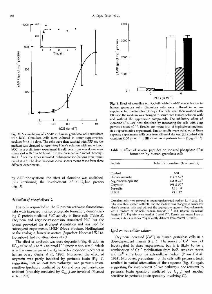
Inhibition of cAMP productionClonidine inhibited hCG-stimulated cAMP production,
providing evidence for an a2-adrenoceptor inhibitory path¬way opposed to the Gas pathway. Moreover, when the cellswere pretreated with pertussis toxin (which inactivates Ga¡
1200 - 400
200
900
OS.'enË. 600OEQ.
<
300
0 "-
0.01 0.1
hCG (iu ml"1)10
Fig. 2. Accumulation of cAMP in human granulosa cells stimulatedwith hCG. Granulosa cells were cultured in serum-supplementedmedium for 8—14 days. The cells were then washed with PBS and themedium was changed to serum-free Hank's solution with and withouthCG. In a preliminary experiment (inset), cells from one donor were
stimulated with 1 iu hCG ml"
' in the presence of 5 mmol theophyl-line 1
~
for the times indicated. Subsequent incubations were termi¬nated at 2 h. The dose—response curve shows means ± sem from threedifferent experiments.
by ADP-ribosylation), the effect of clonidine was abolished,thus confirming the involvement of a Grlike protein(Fig. 3).
Activation of phospholipase C
The cells responded to the G protein activator fluoroalumi-nate with increased inositol phosphate formation, demonstrat¬ing G protein-mediated PLC activity in these cells (Table 3).Oxytocin and arginine-vasopressin stimulated PLC, but theformer provoked the strongest stimulation and was used forsubsequent experiments. LHRH (Nova Biochem, Nottingham)or the analogue, buserelin acetate (Suprefact: Hoechst UK Ltd,Hounslow), had no stimulatory effect.
The effect of oxytocin was dose dependent (Fig. 4), with an
EC50 value of 3.46 ± 1.66 nmol 1 ~ : (mean ± sem, = 3), whichis in the same range as the Kd value for oxytocin receptors inhuman ovary (Fuchs et al, 1990). Moreover, the effect ofoxytocin was partly inhibited by pertussis toxin (Fig. 4),suggesting that at least two pathways, one pertussis toxin-sensitive (probably mediated by G¡) and one pertussis-toxin-resistant (probably mediated by G n) are involved (Phaneufet al, 1993).
4000
-ç 3000SoQ.
'oE 2000oEQ.
o 1000
^^ ^
hCG (¡u ml
Fig. 3. Effect of clonidine on hCG-stimulated cAMP concentration inhuman granulosa cells. Granulosa cells were cultured in serum-
supplemented medium for 14 days. The cells were then washed withPBS and the medium was changed to serum-free Hank's solution withand without the appropriate compounds. The inhibitory effect ofclonidine (P < 0.01) was abolished by incubating the cells with 1 pgpertussis toxin ml
~
\ Results are means ± sd of triplicate estimationsin a representative experiment. Similar results were obtained in threeseparate experiments with cells from different donors. (D) control; (0)clonidine (100 µ 1
~
J); ( ) clonidine + pertussis toxin (1 pg ml~
J).
Table 3. Effect of several peptides on inositol phosphate (IPs)formation by human granulosa cells
Peptide Total IPs formation (% of control)
ControlFluoroaluminateArginine/vasopressinOxytocinBuserelinLHRH
100317 + 52*246 + 31*498 ±57*
82 ± 993 ±12
Granulosa cells were cultured in serum-supplemented medium for 7 days. Thecells were then washed with PBS and the medium was changed to serum-freeHank's solution with and without the appropriate agonists. Fluoroaluminatewas a mixture of 20 mmol sodium fluoride 1 ~: and 10 µ aluminiumfluoride 1~ . Peptides were used at 1 µ 1~ . Results are means ± sem ofquadruplicate estimations. "Significantly different from control (P < 0.01).
Effect on intracellular calciumOxytocin increased [Ca2+]¡ in human granulosa cells in a
dose-dependent manner (Fig. 5). The source of Ca2+ was notinvestigated in these experiments, but it is likely to be a
combination of Ca2+ mobilization from InsP3-sensitive storesand Ca + entry from the extracellular medium (Phaneuf et al,1993). Moreover, pretreatment of the cells with pertussis toxinresulted in partial attenuation of the response (Fig. 5), againsuggesting the involvement of two pathways: one resistant topertussis toxin (possibly mediated by G ,,) and anothersensitive to pertussis toxin (possibly involving Gt).

450
co 300«ce-O
O
«
Em
150
o ·-
10"0 10 10 10 10
Oxytocin (mol 1)Fig. 4. Effect of oxytocin on inositol phosphate formation in humangranulosa cells. For these experiments, granulosa cells were cultured inserum-supplemented medium for 7-14 days. Results are means + sd ofquadruplicate estimations in cells from one donor. The inset is a
different experiment showing that cells incubated with 1 pg pertussistoxin ml
~
' for 24 h ( ) had a lower (*P < 0.01) response to oxytocin.Similar results were obtained in two other experiments.
DiscussionCultured human granulosa cells retain steroidogenic capacityand hormone responsiveness for at least two weeks andprovide a useful model to study second messenger pathways.The cells express a variety of G proteins. RM/l is a wellcharacterized antibody that has been used successfully todemonstrate Gas in a variety of animal and human tissues(Morris et al, 1990; Europe-Finner et al, 1993). There are foursplice variants of as, but human granulosa cells express a singleband, probably corresponding to the 'long form' of as (Europe-Finner et al, 1993). Strong functional evidence for an activeGas pathway is given by the finding that two unrelatedagonists, hCG and PGE2, the receptors of which belong to theG protein-linked family (Hipkin et al, 1993; Bastien et al, 1994;Lu and Menon, 1994; Regan et al, 1994), provoke a large andrapid increase in cAMP production by granulosa cells.
Of particular interest is the expression of Ga¡ subtypes, sincethis inhibitory pathway is often overlooked when studyingcAMP-mediated events in granulosa cells. The antibodies AS/7and EC/2 were raised against aitlai2 and ai3/a0, respectively,and the immunoblot data suggest that at least two differentmembers of the G¡ family (most likely ail2 and ai3) are presentin these cells. The absence of a0 was confirmed with threedifferent antibodies. Functional evidence for the existence of a
Ga¡ pathway was obtained with clonidine, which activates a2adrenoceptors linked to Goti2 (Osbome, 1991) and possibly toother ctrsubtypes (Raymond et al, 1994). Human granulosacells possess adrenoceptors that inhibit hCG-induced pro¬gesterone secretion (Fohr et al, 1993). Furthermore, theinhibitory effect of clonidine was blocked completely bypertussis toxin, confirming that it is a Garmediated event.
500-
400
oE f- 300
-
oro
200CD
100-
0 u
10O 10 ™ 10 10"Oxytocin (mol 1)
Fig. 5. Effect of oxytocin on [Ca2+]¡ concentration in human granulosacells. Cells were cultured onto fibronectin-coated glass coverslips inserum-supplemented medium for 7-14 days and were loaded withFura-2. Results are means ± sd of triplicate estimations in cells fromone donor. The inset is a different experiment showing that cellsincubated with 1 µg pertussis toxin ml" J for 24 h ( ) had a lower(*P < 0.01) response to oxytocin. Similar results were obtained in twoother experiments.
The Ga jj proteins are ubiquitous (Mitchell el al, 1991) andare expressed in granulosa cells. The oxytocin receptor isthought to be coupled to PLC activation via n and, whenthe cells were challenged with oxytocin, there was rapidaccumulation of inositol phosphates and an increase in intra¬cellular calcium, demonstrating an active PLC—InsP3 pathway.However, the mechanism of action of oxytocin is complex, asboth PLC activation and [Ca2+]¡ rises have a pertussis toxinsensitive component that cannot be accounted for by Gq. Thiscomponent may represent a¡ stimulation of PLC. Moreover,some PLC isozymes (e.g., PLCß3) can be activated directly byß subunits (Exton, 1993; Sternweis and Smrcka, 1993), and itis possible that the pertussis sensitive component of oxytocin-induced PLC activation is due to liberation of ß subunitsfrom Grlike proteins. Human granulosa cells express PLCßj (atarget for Gq), as well as PLCß2 and PLCß3 (C. H. Hamilton,S. Phaneuf and A. López Bemal unpublished observations);hence it is possible that there are multiple signalling pathwaysfor oxytocin. Oxytocin is produced by granulosa/luteal cells inruminants (Jones and Flint, 1988; Camier et al, 1991) andhumans (Guillou et al, 1992), and the oxytocin receptor isexpressed in the ovary (Fuchs et al, 1990; Zhang et al, 1991;Kimura et al, 1993). It is possible that oxytocin has a
paracrine/autocrine role in follicular steroidogenesis, modulat¬ing the effect of gonadotrophins (Pitzel et al, 1990; Voss andFortune, 1991; Clamagirand et al, 1991). The data presentedhere demonstrate that mature granulosa cells are a target tissuefor oxytocin, but its role and mechanism of action deservefurther investigation.

In conclusion, the data demonstrate the presence of Gas inhuman granulosa cells and reinforce the notion that this Gprotein mediates the stimulatory effect of hCG. ProstaglandinE2 is a luteotrophic agent in some species and may participatein the events leading to ovulation (Zelinski-Wooten andStouffer, 1990). PGE2 can interact with a variety of EP receptorsubtypes, and granulosa cells clearly respond via Gas. How¬ever, the presence of other signalling pathways for PGE2 in thistissue merits further investigation. There are at least twosubtypes of Ga¡ and the functional involvement of theseproteins is suggested by direct inhibition of hCG-stimulatedcAMP production (most likely an ct¡2-mediated effect) and bytheir contribution to oxytocin-mediated PLC activation (byeither a¡ or ß subunits). Granulosa cells play a vital role insteroidogenesis and oocyte maturation. The role of cAMP ingonadotrophin-mediated control of granulosa cell function iswell established (Yong et al, 1992b). Assuming a constant levelof catabolism by phosphodiesterase, cAMP concentrations ingranulosa cells are determined by the balance of stimulatory,Gas-mediated, and inhibitory, Ga¡ -mediated, pathways.Response to gonadotrophins, for example, may depend notonly on the amount of LH or FSH reaching the cells, but alsoon the concurrent amount of Ga¡ stimulation generated byother receptors. These findings apply to 'mature' granulosacells obtained from stimulated IVF cycles at the preovulatorystage and may not necessarily represent the characteristics ofthese cells at earlier stages of development. Nevertheless, theyillustrate the complexity of this endocrine tissue and thevarious potential sites for hormonal interaction.
The authors acknowledge the help of IVF laboratory staff and thesupport of Wellbeing Q. M. Marshall), MRC (S. Phaneuf) and Sparks(G. N. Europe-Finner).
References
Bastien L, Sawyer N, Grygorczyk R, Metters KM and Adam M (1994) Cloning,functional expression, and characterization of the human prostaglandin E2receptor EP2 subtype Journal of Biological Chemistry 269 11 873-11 877
Birnbaumer 1, Abramowitz J and Brown AM (1990) Receptor—effector couplingby G proteins Biochimica et Biophysica Ada 1031 163-224
Camier M, Benveniste D, Barre , Brakch and Cohen (1991) Synthesis andprocessing of pro-oxytocin in bovine corpus luteum and granulosa cellsMolecular and Cellular Endocrinology 77 141-147
Clamagirand C, Plevrakis I, Bussenot I, Parinaud J, Vieitez G and Grandjean H(1991) The effect of oxytocin on oestradiol-17ß and testosterone secretionby cultured human granulosa cells Human Reproduction 6 774-778
Cooke BA, Platts EA, Abayasekera R, Kurlak LO, Schulster D and Sullivan M(1989) Control of multiple transducing systems by LH which results inthe modulation of adenylate cyclase, protein kinase C, lipoxygenasesand cyclooxygenases Journal of Reproduction and Fertility Supplement 37139-145
Europe-Finner GN, Phaneuf S, Watson SP and López Bernai A (1993) Identifica¬tion and expression of G-proteins in human myometrium: up-regulation ofGas in pregnancy Endocrinology 132 2484-2490
Exton JH (1993) Role of G proteins in activation of phosphoinositide phos¬pholipase C Advances in Second Messengers and Phosphoproteins Research 2865-72
Fohr KJ, Mayerhofer A, Sterzik K, Rudolf M, Rosenbusch and Gratzl M (1993)Concerted action of human chorionic gonadotropin and norepinephrine onintracellular-free calcium in human granulosa-lutein cells: evidence for thepresence of a functional a-adrenergic receptor Journal of Clinical Endocrinologyand Metabolism 76 367-373
Forman RG, Robinson J, Egan D, Ross C, Gosden and Barlow DH (1991)Follicular monitoring and outcome of in vitro fertilization in gonado-tropin-releasing hormone-agonist-treated cycles Fertility and Sterility 55567-573
Fuchs AR, Behrens O, Helmer , Vangsted A, Ivanisevic M, Grifo J, Barros C andFields M (1990) Oxytocin and vasopressin binding sites in human andbovine ovaries American Journal of Obstetrics and Gynecology 163 61-67
Gilman AG (1990) Regulation of adenylyl cyclase by G proteins Advances inSecond Messengers and Phosphoproteins Research 24 51-57
Guillou MD, Barre , Bussenot I, Plevrakis I and Clamagirand C (1992) COOH-terminally-extended processing forms of oxytocin in human ovary Molecularand Cellular Endocrinology 83 233-238
Hepler JR and Gilman AG (1992) G proteins Trends in Biochemical Sciences 17383-387
Hipkin RW, Sánchez-Yagüe J and Ascoli M (1993) Agonist-induced phosphory-lation of the luteinizing hormone/chorionic gonadotropin receptor expressedin a stably transfected cell line Molecular Endocrinology 7 823-832
Jones DS and Flint AP (1988) Concentrations of oxytocin-neurophysin pro-hormone mRNA in corpora lutea of sheep during the oestrous cycle and inearly pregnancy Journal of Endocrinology 117 409—414
Kimura T, Tanizawa O, Mori K, Brownstein MJ and Okayama H (1992) Structureand expression of a human oxytocin receptor [published erratum appears inNature 1992 May 14; 357 6374: 176] Nature 356 526-529
Kimura T, Azuma C, Takemura M, Inoue T, Kikuchi T, Kubota Y, Ogita K, Saji Fand Tanizawa O (1993) Molecular cloning of a human oxytocin receptorRegulatory Peptides 45 73-77
Levitzki A and Bar-Sinai A (1991) The regulation of adenylyl cyclase byreceptor-operated G proteins Pharmacology and Therapeutics 50 271—283
López Bernai A, Buckley S, Rees CM and Marshall JM (1991) Meclofenamateinhibits prostaglandin E binding and adenylyl cyclase activation in humanmyometrium Journal of Endocrinology 129 439-445
Lu DL and Menon KM (1994) Molecular cloning of a novel luteinizing-hormone/human-chorionic-gonadotropin-receptor cDNA. Identification of a
long 3' untranslated region and cDNA sequence of the major transcript inrat ovary European Journal of Biochemistry 222 753—760
McAllister JM, Mason JI, Byrd W, Trant JM, Waterman MR and Simpson ER(1990) Proliferating human granulosa-lutein cells in long term monolayerculture: expression of aromatase, cholesterol side-chain cleavage, and3ß-hydroxysteroid dehydrogenase Journal of Clinical Endocrinology andMetabolism 71 26-33
McFarland KC, Sprengel R, Phillips HS, Kohler M, Rosemblit , Nikolics ,Segaloff DL and Seeburg PH (1989) Lutropin-choriogonadotropin receptor:an unusual member of the G protein-coupled receptor family Science 245494-499
Mitchell FM, Mullaney I, Godfrey PP, Arkinstall SJ, Wakelam MJ and Milligan G(1991) Widespread distribution of Gqu/GIla detected immunologically by an
antipeptide antiserum directed against the predicted C-terminal decapeptideFEBS Letters 287 171-174
Morris D, McHugh-Sutkowski E, Moos M, Jr, Simonds WF, Spiegel AM andSeamon KB (1990) Immunoprecipitation of adenylate cyclase with an anti¬body to a carboxyl-terminal peptide from Gsa Biochemistry 29 9079-9084
Oíate J and Allende JE (1991) Structure and function of G proteins Pharmacologyand Therapeutics 51 403—419
Osborne NN (1991) Inhibition of cAMP production by a2-adrenoceptorstimulation in rabbit retina Brain Research 553 84-88
Phaneuf S, Europe-Finner GN, Varney M, MacKenzie IZ, Watson SP and LópezBernal A (1993) Oxytocin-stimulated phosphoinositide hydrolysis in humanmyometrial cells: involvement of pertussis toxin-sensitive and -insensitiveG-proteins Journal of Endocrinology 136 497-509
Pitzel L, Jarry H and Wuttke W (1990) Effects of oxytocin on in vitro steroidrelease of midstage small and large porcine luteal cells Endocrinology 1262343-2349
Raymond JR, Arthur JM, Casanas SJ, Olsen CL, Gettys TW and Mortensen RM(1994) 2 Adrenergic receptors inhibit cAMP accumulation in embryonicstem cells which lack G¡„2 Journal of Biological Chemistry 269 13 073-13 075
Regan JW, Bailey TJ, Pepperl DJ, Pierce KL, Bogardus AM, Donello JE, FairbairnCE, Kedzie KM, Woodward DF and Gil DW (1994) Cloning of a novel humanprostaglandin receptor with characteristics of the pharmacologically definedEP2 subtype Molecular Pharmacology 46 213-220
Regunathan S, Evinger MJ, Meeley MP and Reis DJ (1991) Effects of clonidineand other imidazole-receptor binding agents on second messenger sys¬tems and calcium influx in bovine adrenal chromaffin cells BiochemicalPharmacology 42 2011-2018

Schrey MP, Read AM and Steer PJ (1987) Stimulation of phospholipid hy¬drolysis and arachidonic acid mobilization in human uterine decidua cells byphorbol ester Biochemical Journal 246 705—713
Smrcka AV, Hepler JR, Brown KO and Sternweis PC (1991) Regulation ofpolyphosphoinositide-specific phospholipase C activity by purified GqScience 251 804-807
Steele GL and Leung PC (1992) Intragonadal signalling mechanisms in thecontrol of steroid hormone production Journal of Steroid Biochemistry andMolecular Biology 41 515-522
Sternweis PC and Smrcka AV (1993) G proteins in signal transduction: theregulation of phospholipase C Ciba Foundation Symposium 176 96—11
Taylor SJ, Chae HZ, Rhee SG and Exton JH (1991) Activation of the ßl isozymeof phospholipase C by subunits of the G class of G proteins Nature 350516-518
Voss AK and Fortune JE (1991) Oxytocin stimulates progesterone productionby bovine granulosa cells isolated before, but not after, the luteinizinghormone surge Molecular and Cellular Endocrinology 78 17-24
Yong EL, Baird DT, Yates R, Reichert LE, Jr and Hillier SG (1992a) Hormonalregulation of the growth and steroidogenic function of human granulosacells Journal of Clinical Endocrinology and Metabolism 74 842—849
Yong EL, Baird DT and Hillier SG (1992b) Mediation of gonadotrophin-stimulated growth and differentiation of human granulosa cells byadenosine-3',5'-monophosphate: one molecule, two messages ClinicalEndocrinology 37 51-58
Zelinski-Wooten MB and Stouffer RL (1990) Intraluteal infusions of prosta¬glandins of the E, D, I, and A series prevent PGF2a-induced, but notspontaneous, luteal regression in rhesus monkeys Biology of Reproduction 43507-516
Zhang L, Dreifuss JJ, Dubois-Dauphin M and Tribollet E (1991) Autoradiographi-cal localization of oxytocin-binding sites in the guinea-pig ovary at differentstages of the oestrous cycle Journal of Endocrinology 131 421—426