Embed Size (px)
Citation preview
CentralBringing Excellence in Open Access
JSM Nanotechnology & Nanomedicine
Cite this article: Patil AG, Bafna HR, More MP, Deshmukh PK, Patil PO (2017) Green Synthesis of Graphene Based Silver Nanocomposite for Enhanced Anti-bacterial Activity against Dental Pathogens. JSM Nanotechnol Nanomed 5(3): 1058.
*Corresponding author
Pravin O. Patil, Department of Quality Assurance, H.R. Patel Institute of Pharmaceutical Education and Research, Shirpur, Dist- Dhule, 425405, India, Email:
Submitted: 24 October 2017
Accepted: 15 November 2017
Published: 18 November 2017
ISSN: 2334-1815
Copyright© 2017 Patil et al.
OPEN ACCESS
Keywords•Graphene oxide; Nanocomposite; Sapodilla peel
extract; Antibacterial; Dental pathogen; Dental caries
Research Article
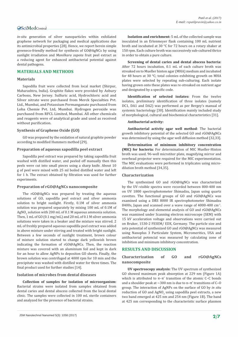
Green Synthesis of Graphene Based Silver Nanocomposite for Enhanced Antibacterial Activity against Dental PathogensAshwini G. Patil1, Harshada R. Bafna2, Mahesh P. More2, Prashant K. Deshmukh2, and Pravin O. Patil2*1Department of Microbiology, R. C. Patel Arts, Commerce and Science College, India2Department of Quality Assurance, H. R. Patel Institute of Pharmaceutical Education and Research, India
Abstract
In present research work graphene silver nanocomposite (rGO@AgNCs) was synthesized by using the sapodilla peel extract under the photochemical irradiation. Antibacterial potential GO@AgNCs was studied against dental pathogen viz. Bacillus pumilus, Pseudomonas aeruginosa, Enterococcus faecalis, Escherichia coli, and Staphylococcus aureus.The minimum inhibitory concentration of GO and rGO@AgNCs was studied against dental pathogen in which the fabricated rGO@AgNCs exhibited highest MIC of 7.81 µg/mL against Pseudomonas aeruginosa while the least MIC of 15.62 µg/mL was observed against Staphyloccous aureus and GO exhibited highest MIC of 62.5 µg/mL against Pseudomonas aeruginosa and Staphyloccous aureus. Thus rGO@AgNCs may show to be superior antibacterial agents against dental pathogens.
INTRODUCTIONRecently, graphene has involved in both academic and
industrial interest because graphene is a material made of carbon atoms that are bonded together in a repeating pattern of hexagons [1]. Evidently, graphene have been exposed in a wide range of applications, such as electronic, energy storage and supercapacitor, batteries (fuel cells, solar cells), and bioscience/biotechnologies because of its unique physicochemical properties: high surface area, excellent thermal conductivity and electric conductivity and strong mechanical strength [2,3].
In recent years graphene-based nanocomposites have great potential in various biomedical applications including drug/gene delivery, imaging, antibacterial and anti-cancer activities [4]. In which graphene acts either as a functional element or a substrate for immobilizing the other constituents such as metal likes silver, gold, zinc oxide, copper etc [5]. From the category of inorganic materials silver has unique optical, electronic, and catalytic properties, which are different from their bulk counterparts and hence lead to more attention in various areas of applications like catalysis [6], chemical sensing [7], antibacterial [8], and biosensing [9,10]. In recent years, various investigations have been reported for synthesis of GO based Ag nanocomposite by several methods like chemical [11], thermal [12], microwave [13], and green methods [14]. In chemicals reduction method chemical such as Hydrazine (N2H4) [15], Sodium borohydride (NaBH4) [16], Dimethyl hydrazine (C2H8N2) [17], Hydroquinone (C6H6O2) [18], Sulphuric acid (H2SO4) [19], and Aluminium
powder (Al) [20], involved in the reduction and functionalization of graphene oxide. These types of chemical reducing agent have highest number of toxicity which laid down during processing or in the final product. Green approach for reduction of graphene oxide is desirable from which natural reducing agents, plant extracts have been considerably exploited due to their low cost, bulk availability and biocompatibility for the synthesis of graphene metal nanocomposite [21]. In developed countries, 42% of food waste is produced by households, while 39% losses occur in the food manufacturing industry, 14% in food service sector and remaining 5% in retail and distribution. So, now days various scientists focused on utilization of raw material for new products and applications [22]. Manilkara zapota is commonly known as sapodilla/ chikoo and grown in huge quantities in India, Pakistan and Mexico. The fruit and its peel contain high amounts of saponin, which has astringent properties similar to tannin and in addition, ethanolic extract of peel exhibited highest amount of flavonoid and total phenol contents [23,24]. It has antioxidant activity because of compounds contained polyphenols catechin, epicatechin, leucocyanidin, leucodelphinidin, leucopelargonidin, chlorogenic acid, and gallic acid is in unripe sapodilla fruit and ripe fruit, the level of 5-caffeoyl quinic acid was measured recently in the peel and pulp [25].
Among the various metal likes cadmium, gold, platinum, zinc, etc silver showing strong antibacterial properties and been applied in various applications such as medical coating, water filtration and home appliances [26,27]. Nano silver incorporated graphene nanocomposites were prepared through
CentralBringing Excellence in Open Access
Patil et al. (2017)E-mail:
JSM Nanotechnol Nanomed 5(3): 1058 (2017) 2/7
in-situ generation of silver nanoparticles within exfoliated graphene network for packaging and medical applications due its antimicrobial properties [28]. Hence, we report herein simple greeneco-friendly method for synthesis of GO@AgNCs by using sunlight irradiation and Manilkara zapota fruit peel extract as a reducing agent for enhanced antibacterial potential against dental pathogens.
MATERIALS AND METHODS
Materials
Sapodilla fruit were collected from local market (Shirpur, Maharashtra, India). Graphite flakes were provided by Asbury Carbons, New Jersey. Sulfuric acid, Hydrochloric acid and Silver nitrate were purchased from Merck Specialties Pvt. Ltd., Mumbai, and Potassium Permanganate purchased from Loba Chemie Pvt. Ltd, Mumbai. Hydrogen peroxide was purchased from RFCL Limited, Mumbai. All other chemicals and reagents were of analytical grade and used as received without purification.
Synthesis of Graphene Oxide (GO)
GO was prepared by the oxidation of natural graphite powder according to modified Hummers method [29].
Preparation of aqueous sapodilla peel extract
Sapodilla peel extract was prepared by taking sapodilla fruit washed with distilled water, and peeled off manually then this peels were cut into small pieces using a sharp knife. About 10 g of peel were mixed with 25 ml boiled distilled water and left for 1 h. The extract obtained by filtration was used for further experiments.
Preparation of rGO@AgNCs nanocomposite
The rGO@AgNCs was prepared by treating the aqueous solutions of GO, sapodilla peel extract and silver ammonia solution to bright sunlight. Firstly, 0.1M of silver ammonia solution was prepared separately by mixing 100 mL of 0.1M of AgNO3 solution with 200 mL of 0.1 M aqueous ammonia solution. Then, 1 mL of GO (0.1 mg/mL) and 20 mL of 0.1 M silver ammonia solutions were taken in a beaker and the mixture was stirred. 2 mL of freshly prepared aqueous sapodilla peel extract was added in above mixture under stirring and treated with bright sunlight. Between a few seconds of sunlight treatment, brown colour of mixture solution started to change dark yellowish brown indicating the formation of rGO@AgNCs. Then, the reaction mixture was covered with an aluminium foil and kept in dark for an hour to allow AgNPs to depositon GO sheets. Finally, the brown solution was centrifuged at 4000 rpm for 10 min and the precipitate was washed with distilled water for three times. The final product used for further studies [14].
Isolation of microbes from dental diseases
Collection of samples for isolation of microorganism: Bacterial strains were isolated from samples obtained from dental caries and dental abscess collected from the local dental clinic. The samples were collected in 100 mL sterile containers and analyzed for the presence of bacterial strains.
Isolation and enrichment: 5 mL of the collected sample was inoculated in an Erlenmeyer flask containing 100 mL nutrient broth and incubated at 30 °C for 72 hours on a rotary shaker at 150 rpm. Each culture broth was successively sub-cultured thrice in order to obtain a pure culture.
Screening of dental caries and dental abscess bacteria: After 72 hours incubation, 0.1 mL of each culture broth was streaked on to Mueller hinton agar (MHA) medium and incubated for 48 hours at 30 °C; total colonies exhibiting growth on MHA plates were selected by repeating sub-culturing. Each isolate having grown onto these plates was re-streaked on nutrient agar and designated by a specific code.
Identification of selected isolates: From the twelve isolates, preliminary identification of three isolates (namely DC1, DA1 and DA2) was performed as per Bergey’s manual of systemic bacteriology [30]. Identification mainly included study of morphological, cultural and biochemical characteristics [31].
Antibacterial activity:
Antibacterial activity agar well method: The bacterial growth inhibitory potential of the selected GO and rGO@AgNCs was determined by using the agar well diffusion method [32,33].
Determination of minimum inhibitory concentration (MIC) for bacteria: For determination of MIC Mueller-Hinton broth was used. 96-well microtiter plate, magnifying mirror and overhead projector were required for the MIC experimentation. The MIC evaluations were performed in triplicates using micro-dilution broth method [34,35].
Characterization
The synthesized GO and rGO@AgNCs was characterized by the UV–visible spectra were recorded between 800-400 nm on UV 1800 spectrophotometer Shimadzu, Japan using quartz cuvettes. The functional groups of GO and rGO@AgNCs was examined using a DRS 8000 IR spectrophotometer Shimadzu 8400s, Japan and scanned over a wave range of 4000-400 cm-1. The morphology and elemental analysis of GO and rGO@AgNCs was examined under Scanning electron microscope (SEM) with 15 kV acceleration voltage and observations were carried out on Bruker, 1530-2 FESEM/ EDX, Germany. The particle size and zeta potential of synthesized GO and rGO@AgNCs was measured using Nanoplus 3 Particulate System, Micromeritics, USA and antibacterial potencial was measured by calculating zone of inhibition and minimum inhibitory concentration.
RESULTS AND DISCUSSION
Characterization of GO and rGO@AgNCs nanocomposite
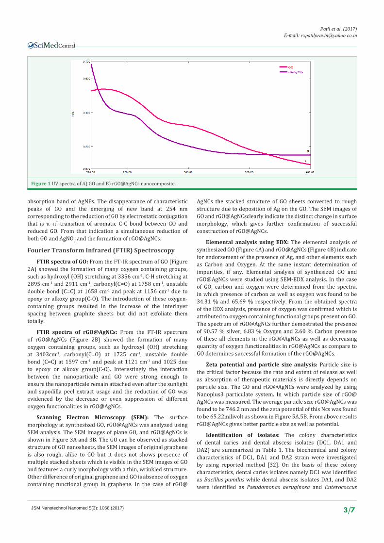
UV spectroscopy analysis: The UV spectrum of synthesized GO showed maximum peak absorption at 229 nm (Figure 1A) which is attributed to π–π* transition of the atomic C–C bonds and a shoulder peak at ~300 nm is due to n–π* transitions of C–O group. The interaction of AgNPs on the surface of GO by in situ reduction of GO and AgNO3 using sapodilla peel extracts, a new two band emerged at 425 nm and 254 nm (Figure 1B). The band at 425 nm corresponding to the characteristic surface plasmon
CentralBringing Excellence in Open Access
Patil et al. (2017)E-mail:
JSM Nanotechnol Nanomed 5(3): 1058 (2017) 3/7
absorption band of AgNPs. The disappearance of characteristic peaks of GO and the emerging of new band at 254 nm corresponding to the reduction of GO by electrostatic conjugation that is π–π* transition of aromatic C-C bond between GO and reduced GO. From that indication a simultaneous reduction of both GO and AgNO3 and the formation of rGO@AgNCs.
Fourier Transform Infrared (FTIR) Spectroscopy
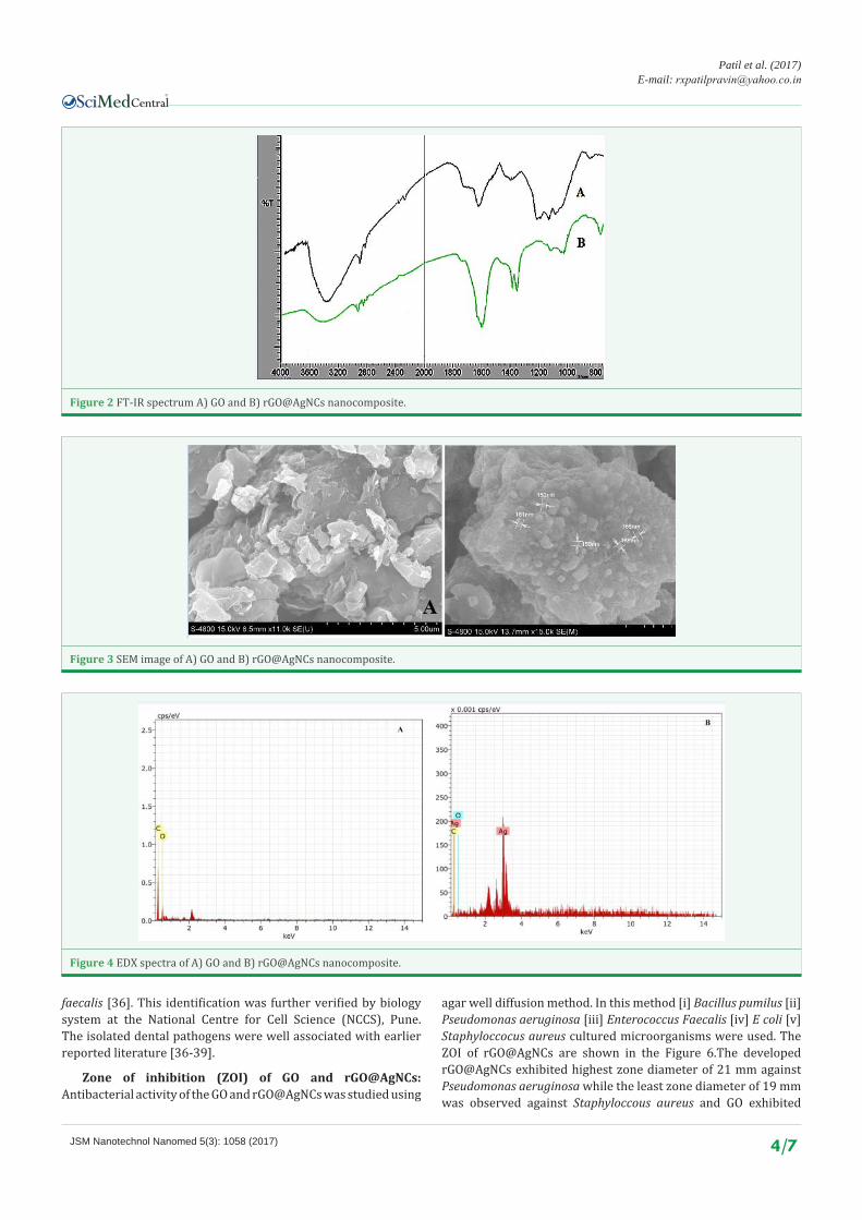
FTIR spectra of GO: From the FT-IR spectrum of GO (Figure 2A) showed the formation of many oxygen containing groups, such as hydroxyl (OH) stretching at 3356 cm-1, C-H stretching at 2895 cm-1 and 2911 cm-1, carbonyl(C=O) at 1758 cm-1, unstable double bond (C=C) at 1658 cm-1 and peak at 1156 cm-1 due to epoxy or alkoxy group(C-O). The introduction of these oxygen-containing groups resulted in the increase of the interlayer spacing between graphite sheets but did not exfoliate them totally.
FTIR spectra of rGO@AgNCs: From the FT-IR spectrum of rGO@AgNCs (Figure 2B) showed the formation of many oxygen containing groups, such as hydroxyl (OH) stretching at 3403cm-1, carbonyl(C=O) at 1725 cm-1, unstable double bond (C=C) at 1597 cm-1 and peak at 1121 cm-1 and 1025 due to epoxy or alkoxy group(C-O). Interestingly the interaction between the nanoparticale and GO were strong enough to ensure the nanoparticale remain attached even after the sunlight and sapodilla peel extract usage and the reduction of GO was evidenced by the decrease or even suppression of different oxygen functionalities in rGO@AgNCs.
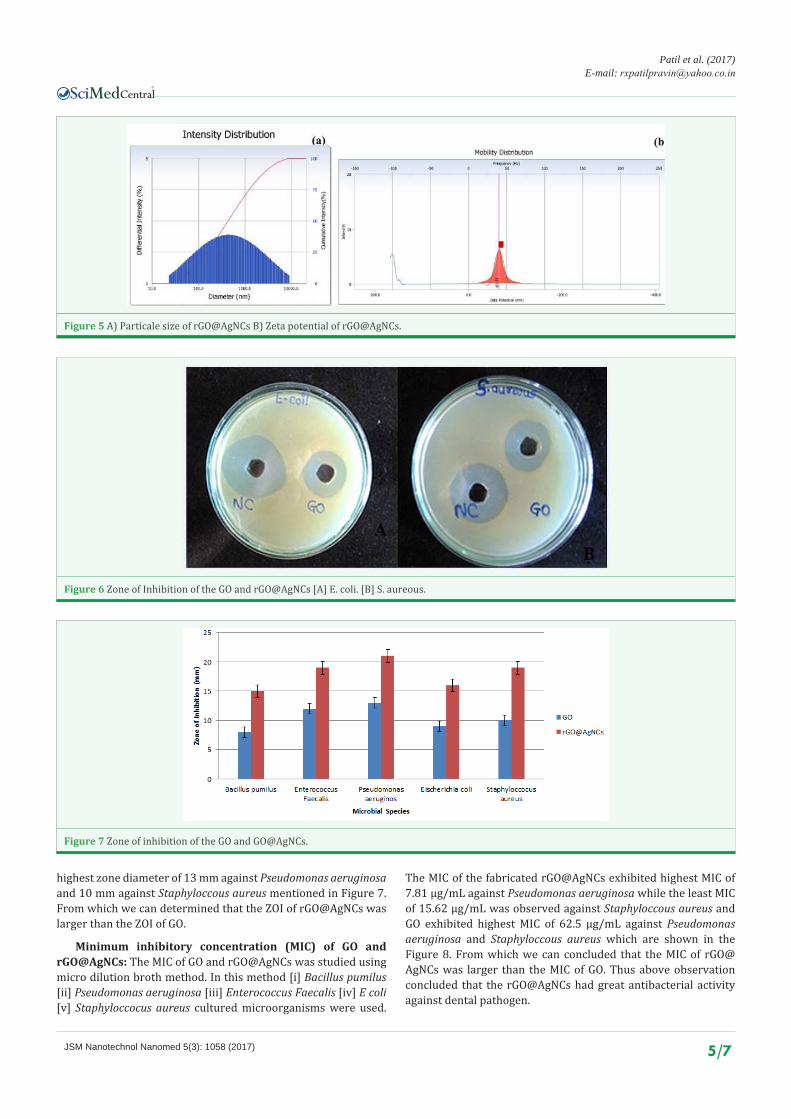
Scanning Electron Microscopy (SEM): The surface morphology at synthesized GO, rGO@AgNCs was analyzed using SEM analysis. The SEM images of plane GO, and rGO@AgNCs is shown in Figure 3A and 3B. The GO can be observed as stacked structure of GO nanosheets, the SEM images of original graphene is also rough, alike to GO but it does not shows presence of multiple stacked sheets which is visible in the SEM images of GO and features a curly morphology with a thin, wrinkled structure. Other difference of original graphene and GO is absence of oxygen containing functional group in graphene. In the case of rGO@
AgNCs the stacked structure of GO sheets converted to rough structure due to deposition of Ag on the GO. The SEM images of GO and rGO@AgNCsclearly indicate the distinct change in surface morphology, which gives further confirmation of successful construction of rGO@AgNCs.
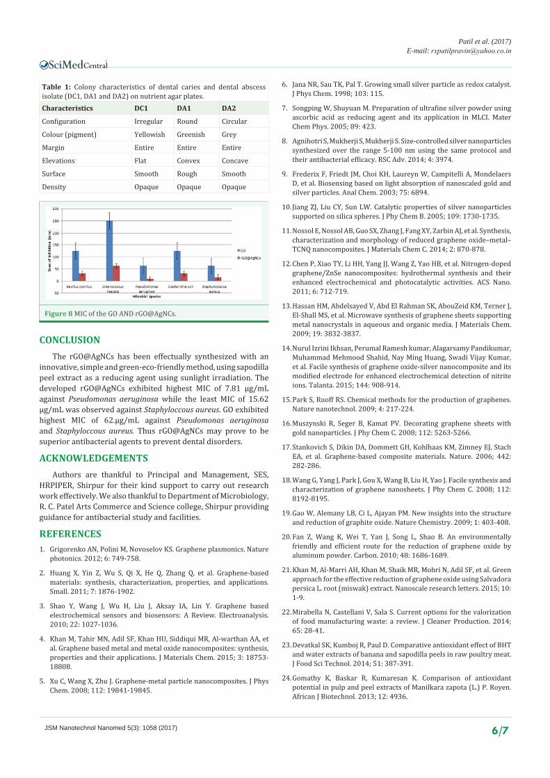
Elemental analysis using EDX: The elemental analysis of synthesized GO (Figure 4A) and rGO@AgNCs (Figure 4B) indicate for endorsement of the presence of Ag, and other elements such as Carbon and Oxygen. At the same instant determination of impurities, if any. Elemental analysis of synthesized GO and rGO@AgNCs were studied using SEM-EDX analysis. In the case of GO, carbon and oxygen were determined from the spectra, in which presence of carbon as well as oxygen was found to be 34.31 % and 65.69 % respectively. From the obtained spectra of the EDX analysis, presence of oxygen was confirmed which is attributed to oxygen containing functional groups present on GO. The spectrum of rGO@AgNCs further demostrated the presence of 90.57 % silver, 6.83 % Oxygen and 2.60 % Carbon presence of these all elements in the rGO@AgNCs as well as decreasing quantity of oxygen functionalities in rGO@AgNCs as compare to GO determines successful formation of the rGO@AgNCs.
Zeta potential and particle size analysis: Particle size is the critical factor because the rate and extent of release as well as absorption of therapeutic materials is directly depends on particle size. The GO and rGO@AgNCs were analyzed by using Nanoplus3 particulate system. In which particle size of rGO@AgNCs was measured. The average particle size rGO@AgNCs was found to be 746.2 nm and the zeta potential of this Ncs was found to be 65.22milivolt as shown in Figure 5A,5B. From above results rGO@AgNCs gives better particle size as well as potential.
Identification of isolates: The colony characteristics of dental caries and dental abscess isolates (DC1, DA1 and DA2) are summarized in Table 1. The biochemical and colony characteristics of DC1, DA1 and DA2 strain were investigated by using reported method [32]. On the basis of these colony characteristics, dental caries isolates namely DC1 was identified as Bacillus pumilus while dental abscess isolates DA1, and DA2 were identified as Pseudomonas aeruginosa and Enterococcus
Figure 1 UV spectra of A) GO and B) rGO@AgNCs nanocomposite.
CentralBringing Excellence in Open Access
Patil et al. (2017)E-mail:
JSM Nanotechnol Nanomed 5(3): 1058 (2017) 4/7
Figure 2 FT-IR spectrum A) GO and B) rGO@AgNCs nanocomposite.
Figure 3 SEM image of A) GO and B) rGO@AgNCs nanocomposite.
Figure 4 EDX spectra of A) GO and B) rGO@AgNCs nanocomposite.
faecalis [36]. This identification was further verified by biology system at the National Centre for Cell Science (NCCS), Pune. The isolated dental pathogens were well associated with earlier reported literature [36-39].
Zone of inhibition (ZOI) of GO and rGO@AgNCs: Antibacterial activity of the GO and rGO@AgNCs was studied using
agar well diffusion method. In this method [i] Bacillus pumilus [ii] Pseudomonas aeruginosa [iii] Enterococcus Faecalis [iv] E coli [v] Staphyloccocus aureus cultured microorganisms were used. The ZOI of rGO@AgNCs are shown in the Figure 6.The developed rGO@AgNCs exhibited highest zone diameter of 21 mm against Pseudomonas aeruginosa while the least zone diameter of 19 mm was observed against Staphyloccous aureus and GO exhibited
CentralBringing Excellence in Open Access
Patil et al. (2017)E-mail:
JSM Nanotechnol Nanomed 5(3): 1058 (2017) 5/7
Figure 5 A) Particale size of rGO@AgNCs B) Zeta potential of rGO@AgNCs.
Figure 6 Zone of Inhibition of the GO and rGO@AgNCs [A] E. coli. [B] S. aureous.
Figure 7 Zone of inhibition of the GO and GO@AgNCs.
highest zone diameter of 13 mm against Pseudomonas aeruginosa and 10 mm against Staphyloccous aureus mentioned in Figure 7. From which we can determined that the ZOI of rGO@AgNCs was larger than the ZOI of GO.
Minimum inhibitory concentration (MIC) of GO and rGO@AgNCs: The MIC of GO and rGO@AgNCs was studied using micro dilution broth method. In this method [i] Bacillus pumilus [ii] Pseudomonas aeruginosa [iii] Enterococcus Faecalis [iv] E coli [v] Staphyloccocus aureus cultured microorganisms were used.
The MIC of the fabricated rGO@AgNCs exhibited highest MIC of 7.81 µg/mL against Pseudomonas aeruginosa while the least MIC of 15.62 µg/mL was observed against Staphyloccous aureus and GO exhibited highest MIC of 62.5 µg/mL against Pseudomonas aeruginosa and Staphyloccous aureus which are shown in the Figure 8. From which we can concluded that the MIC of rGO@AgNCs was larger than the MIC of GO. Thus above observation concluded that the rGO@AgNCs had great antibacterial activity against dental pathogen.
CentralBringing Excellence in Open Access
Patil et al. (2017)E-mail:
JSM Nanotechnol Nanomed 5(3): 1058 (2017) 6/7
CONCLUSIONThe rGO@AgNCs has been effectually synthesized with an
innovative, simple and green-eco-friendly method, using sapodilla peel extract as a reducing agent using sunlight irradiation. The developed rGO@AgNCs exhibited highest MIC of 7.81 µg/mL against Pseudomonas aeruginosa while the least MIC of 15.62 µg/mL was observed against Staphyloccous aureus. GO exhibited highest MIC of 62.µg/mL against Pseudomonas aeruginosa and Staphyloccous aureus. Thus rGO@AgNCs may prove to be superior antibacterial agents to prevent dental disorders.
ACKNOWLEDGEMENTSAuthors are thankful to Principal and Management, SES,
HRPIPER, Shirpur for their kind support to carry out research work effectively. We also thankful to Department of Microbiology, R. C. Patel Arts Commerce and Science college, Shirpur providing guidance for antibacterial study and facilities.
REFERENCES1. Grigorenko AN, Polini M, Novoselov KS. Graphene plasmonics. Nature
photonics. 2012; 6: 749-758.
2. Huang X, Yin Z, Wu S, Qi X, He Q, Zhang Q, et al. Graphene-based materials: synthesis, characterization, properties, and applications. Small. 2011; 7: 1876-1902.
3. Shao Y, Wang J, Wu H, Liu J, Aksay IA, Lin Y. Graphene based electrochemical sensors and biosensors: A Review. Electroanalysis. 2010; 22: 1027-1036.
4. Khan M, Tahir MN, Adil SF, Khan HU, Siddiqui MR, Al-warthan AA, et al. Graphene based metal and metal oxide nanocomposites: synthesis, properties and their applications. J Materials Chem. 2015; 3: 18753-18808.
5. Xu C, Wang X, Zhu J. Graphene-metal particle nanocomposites. J Phys Chem. 2008; 112: 19841-19845.
6. Jana NR, Sau TK, Pal T. Growing small silver particle as redox catalyst. J Phys Chem. 1998; 103: 115.
7. Songping W, Shuyuan M. Preparation of ultrafine silver powder using ascorbic acid as reducing agent and its application in MLCI. Mater Chem Phys. 2005; 89: 423.
8. Agnihotri S, Mukherji S, Mukherji S. Size-controlled silver nanoparticles synthesized over the range 5-100 nm using the same protocol and their antibacterial efficacy. RSC Adv. 2014; 4: 3974.
9. Frederix F, Friedt JM, Choi KH, Laureyn W, Campitelli A, Mondelaers D, et al. Biosensing based on light absorption of nanoscaled gold and silver particles. Anal Chem. 2003; 75: 6894.
10. Jiang ZJ, Liu CY, Sun LW. Catalytic properties of silver nanoparticles supported on silica spheres. J Phy Chem B. 2005; 109: 1730-1735.
11. Nossol E, Nossol AB, Guo SX, Zhang J, Fang XY, Zarbin AJ, et al. Synthesis, characterization and morphology of reduced graphene oxide–metal–TCNQ nanocomposites. J Materials Chem C. 2014; 2: 870-878.
12. Chen P, Xiao TY, Li HH, Yang JJ, Wang Z, Yao HB, et al. Nitrogen-doped graphene/ZnSe nanocomposites: hydrothermal synthesis and their enhanced electrochemical and photocatalytic activities. ACS Nano. 2011; 6: 712-719.
13. Hassan HM, Abdelsayed V, Abd El Rahman SK, AbouZeid KM, Terner J, El-Shall MS, et al. Microwave synthesis of graphene sheets supporting metal nanocrystals in aqueous and organic media. J Materials Chem. 2009; 19: 3832-3837.
14. Nurul Izrini Ikhsan, Perumal Ramesh kumar, Alagarsamy Pandikumar, Muhammad Mehmood Shahid, Nay Ming Huang, Swadi Vijay Kumar, et al. Facile synthesis of graphene oxide-silver nanocomposite and its modified electrode for enhanced electrochemical detection of nitrite ions. Talanta. 2015; 144: 908-914.
15. Park S, Ruoff RS. Chemical methods for the production of graphenes. Nature nanotechnol. 2009; 4: 217-224.
16. Muszynski R, Seger B, Kamat PV. Decorating graphene sheets with gold nanoparticles. J Phy Chem C. 2008; 112: 5263-5266.
17. Stankovich S, Dikin DA, Dommett GH, Kohlhaas KM, Zimney EJ, Stach EA, et al. Graphene-based composite materials. Nature. 2006; 442: 282-286.
18. Wang G, Yang J, Park J, Gou X, Wang B, Liu H, Yao J. Facile synthesis and characterization of graphene nanosheets. J Phy Chem C. 2008; 112: 8192-8195.
19. Gao W, Alemany LB, Ci L, Ajayan PM. New insights into the structure and reduction of graphite oxide. Nature Chemistry. 2009; 1: 403-408.
20. Fan Z, Wang K, Wei T, Yan J, Song L, Shao B. An environmentally friendly and efficient route for the reduction of graphene oxide by aluminum powder. Carbon. 2010; 48: 1686-1689.
21. Khan M, Al-Marri AH, Khan M, Shaik MR, Mohri N, Adil SF, et al. Green approach for the effective reduction of graphene oxide using Salvadora persica L. root (miswak) extract. Nanoscale research letters. 2015; 10: 1-9.
22. Mirabella N, Castellani V, Sala S. Current options for the valorization of food manufacturing waste: a review. J Cleaner Production. 2014; 65: 28-41.
23. Devatkal SK, Kumboj R, Paul D. Comparative antioxidant effect of BHT and water extracts of banana and sapodilla peels in raw poultry meat. J Food Sci Technol. 2014; 51: 387-391.
24. Gomathy K, Baskar R, Kumaresan K. Comparison of antioxidant potential in pulp and peel extracts of Manilkara zapota (L.) P. Royen. African J Biotechnol. 2013; 12: 4936.
Table 1: Colony characteristics of dental caries and dental abscess isolate (DC1, DA1 and DA2) on nutrient agar plates.Characteristics DC1 DA1 DA2
Configuration Irregular Round Circular
Colour (pigment) Yellowish Greenish Grey
Margin Entire Entire Entire
Elevations Flat Convex Concave
Surface Smooth Rough Smooth
Density Opaque Opaque Opaque
Figure 8 MIC of the GO AND rGO@AgNCs.
CentralBringing Excellence in Open Access
Patil et al. (2017)E-mail:
JSM Nanotechnol Nanomed 5(3): 1058 (2017) 7/7
Patil AG, Bafna HR, More MP, Deshmukh PK, Patil PO (2017) Green Synthesis of Graphene Based Silver Nanocomposite for Enhanced Antibacterial Activity against Dental Pathogens. JSM Nanotechnol Nanomed 5(3): 1058.
Cite this article
25. Ma J, Luo XD, Protiva P, Yang H, Ma C, Basile MJ, Weinstein IB, Kennelly EJ. Bioactive novel polyphenols from the fruit of Manilkara zapota (Sapodilla). J Nat Prod. 2003; 66: 983-986.
26. Chook SW, Chia CH, Sarani Z, Ayob MK, Chee KL, Neoh HM, et al. Silver nanoparticles-Graphene oxide nanocomposite for antibacterial purpose. Int Adv Mat Res. 2012; 364: 439-443.
27. Wu M, Lu D, Zhao Y, Ju T. Facile synthesis of silver-modified functionalised graphene oxide nanocomposite with enhanced antibacterial property. IET Micro Nano Letters. 2013; 8: 82-85.
28. Sahu D, Sarkar N, Sahoo G, Mohapatra P, Swain SK. Silver Imprinted Graphene Nanocomposites: Synthesisis and Morphological Study. 2015.
29. Chen J, Yao B, Li C, Shi G. An improved Hummers method for eco-friendly synthesis of graphene oxide. Carbon. 2013; 64: 225-229.
30. Holt JG, Krieg NR, Sneath PHA, Staley JT, Williams ST. Bergey’s manual of determinative bacteriology. 9th edn. Baltimore (MD): Williams & Wilkins. 1994.
31. Brenner DJ, Krieg NR, Staley JT. Bergey’s Manual of Systematic Bacteriology. 2nd edn. New York: Springer. 2005; 323-328.
32. Wayne PA: Performance standards for antimicrobial susceptibility testing: nineteenth informational supplement. Clinical and laboratory standards institute; 2009, CLSI document M100-S19.
33. Perez C, Pauli M, Bazerque P. An antibiotic assay by the agar-well diffusion method. Acta Biol Med Exp. 1990; 113-115.
34. NCCLS. Methods for dilution antimicrobial susceptibility tests for bacteria that grow aerobically: Approved standard, 5th edition. NCCLS document M7- A5 (ISBN 1-56238-394-9). NCCLS, Pennsylvania, USA. 2000.
35. Eloff JN. A sensitive and quick microplate method to determine the minimal inhibitory concentration of plant extracts for bacteria. Planta Med. 1998; 64: 711-713.
36. Patil AG, Jobanputra AH. Rutin-chitosan nanoparticles: fabrication, characterization and application in dental disorders. Polymer-Plastics Technology and Engineering. 2015; 54: 202-208.
37. Riggio MP, Lennon A, Taylor DJ, Bennett D. Molecular identification of bacteria associated with canine periodontal disease. Vet Microbio. 2011; 150: 394-400.
38. Johnson BT, Shaw LN, Nelson DC, Mayo1 JA. Extracellular proteolytic activities expressed by Bacillus pumilus isolated from endodontic and periodontal lesions. J Med Micro. 2008; 57: 643-651.
39. Clinical and Laboratory Standards Institute (CLSI), Document M31-A3. Performance standards for antimicrobial disk and dilution susceptibility tests for bacteria isolated from animals, approved standard. 3rd edn. CLSI, 940 West Valley Road, Suite 1400, Wayne, Pennsylvania 19087-1898, USA. 2008.