Embed Size (px)
Citation preview

Green Synthesis of Magnetic Nanoparticles byMurraya Koenigii Leaves Extract for BiomedicalApplicationSivakami M ( [email protected] )
Bharathidasan UniversityRenuka Devi K
Government Arts College for women, PudukkottaiRenuka R
Government Arts College for women, Pudukkottai
Research Article
Keywords: Magnetic nanoparticles, spherical, Antimicrobial, Vibrating sample magnetometer, Anti-diabetic.
Posted Date: June 4th, 2021
DOI: https://doi.org/10.21203/rs.3.rs-583600/v1
License: This work is licensed under a Creative Commons Attribution 4.0 International License. Read Full License

Green synthesis of Magnetic nanoparticles by Murraya koenigii leaves extract for
Biomedical Application
M. Sivakami a*, K. Renuka Devi b, R. Renuka b
a H.H. The Rajah’s College, Pudukkottai-622001, India
b Government Arts College for women, Pudukkottai-622001, India
* Corresponding author: Assistant Professor, H.H. The Rajah’s College, Pudukkottai-622001, India,
[email protected], [email protected]
Abstract
Green synthesis of nanoparticles is the method with eco-friendly, cost-effectiveness, and
ease of resource availability approach. Nowadays, magnetic nanoparticles need increased due to
its use in magnetic sensing, medical imaging, waste water treatment, and antibiotic drugs. In this
report, the ecofriendly green synthesis of magnetic iron oxide nanoparticles were efficiently
synthesized by using Murraya koenigii leaves extract. UV-visible spectrum revealed the surface
Plasmon resonance band at 240 nm and analyzed the formation of iron oxide nanoparticles. X-
ray diffraction pattern determined its high crystalline nature with strong intense peaks. Fourier
Transform Infra-Red spectrum illustrated the Fe and O bonding stretching vibration. The particle
size distribution graph showed the formed particles are in nanometer range. Transmission
Electron Micrographs realized the spherical shaped iron oxide nanoparticles with its size at 10-
25 nm. The energy dispersive X-ray spectrum and mapping analysis revealed the purity of
prepared nanoparticles with only Fe and O presence. The vibrating sample magnetometer
analysis showed the paramagnetic behavior of prepared magnetic iron oxide nanoparticles. The
antimicrobial assay revealed potent inhibition of iron oxide nanoparticles on various human
pathogens. The antioxidant, anti-inflammatory and anti-diabetic assays revealed the biomedical
behavior of iron oxide nanoparticles.

Keywords: Magnetic nanoparticles; spherical; Antimicrobial; Vibrating sample magnetometer;
Anti-diabetic.
1. Introduction
Nanoparticles (NPs) with dimension in 1 to 100 nm have specific physical, chemical and
biological properties. Nanotechnology provides its tremendous growth on all fields such as optic,
electronic, textile, agricultural, environmental, and biomedical etc., [1] Nanoparticles have
enormous applications with peculiar large surface to volume ratio property and enhanced
efficiency in all fields in the form of catalysts, batteries, solar cells, gas sensors, LEDs,
semiconductors, glasses, sunscreen products, cosmetics, building materials, leathers, food
packaging, cloths and drugs etc. [2,3], The manipulation of nanoparticles are one of the
interesting research area for researchers which is necessitating field for the existence world.
Because, it formed simple to complicate networks that are imperative to materials science [4].
Nowadays, treating diseases and infections is becoming tough to the clinical field. The infectious
strains are easily spread and cause huge health crisis and problem to humans and animals. Some
pathogenic strains are not responding to antimicrobial agents and create huge damage to human
health system [5]. Bio nanotechnology involves the production, operation and implementation
process of drugs to several human and animal diseases. It can able to create antibiotic drugs
against human pathogens without producing any side effects. In clinical field, bio
nanotechnology plays a vital role for finding drugs to cure life threatening diseases [6].
In this peculiar nanotechnology, particles were synthesized by various physical, chemical
and biological methods. The physical and chemical methods produce nanoparticles with
polluting by-products. It can create harm to environment and organisms [7,8]. The nanoparticles
requiring toxic reducing agents must produce harmful by-products which create pollution to

environment and affect living organisms. But, in biosynthesis, it must be reduced by using plant
materials, bacteria or fungi, yeasts and agricultural wastes. It does not require high energy,
pressure, cost, and hazardous chemicals. It does not produce any toxic to environment [9].
Further, green synthesis of nanoparticles using plant extracts is an easy, cost-effectiveness, ease
of resource availability and large productive approach comparing to other biological methods.
The eco-friendly synthesis of nanoparticles has practical applications in clinical, biomedical,
agricultural, and drug delivery fields as drugs without producing any side effects [10].
Among green synthesized nanoparticles, inorganic nanoparticles have peculiar interest to
researchers due its ease availability and necessity [4]. It has many practical applications viz.,
sunscreen glasses and products, cosmetics, sensors, catalysts, food packaging, paints, ceramics,
batteries, and drugs [11]. With this, magnetic NPs attain interest towards research with their high
catalytic, intrinsic, and versatile properties. It is used in vast field’s viz., food, biosensing,
medical and magnetic separation, degradation of organic pollutants, and waste water treatment
[12]. Iron oxide NPs are mostly used in dye remover for preserving pollution free environment
[5]. Mostly, iron oxide NPs are used as biosensor and magnetic field assisted drug delivery
therapy in clinical field.
Reports in green synthesis of magnetic NPs using different plant extracts and some of
them are; Cymbopogon citratus [13], Carica papaya [14], Laurus nobilis [15], Platanus
orientalis [16], Amaranthus spinosus [17], Pinus pinaster [18], Moringa oleifera [19],
Eucalyptus [20], Rosemary [21], Mangifera indica [22], Camellia sinensis [23], Daphne
mezereum [5], Vitex negundo [24], Terminalia bellirica [19], and Psoralea corylifolia [25].
Murraya koenigii is native to India, and they are used for both medicinal and culinary
applications. They are highly aromatic and have a unique citrus flavor. Aside from being a

versatile culinary herb, they offer an abundance of health benefits due to the powerful plant
compounds they contain. Curry leaves are rich in alkaloids, glycosides, and phenolic compounds
that give potent health benefits. Most of them function as antioxidants in our body. Antioxidants
play an indispensable role in maintaining our body healthy and free from diseases. Curry leaves
are the good sources of iron and calcium. Due to this reason, they are used as medicine to treat
calcium deficiency. It is also used to reduce blood glucose levels effectively. They offer shield to
insulin producing cells and avoid harm caused by free radicals [26]. Traditionally, curry leaves
are assumed to have favorable effect for eyesight. It can be used to prevent early onset of
cataract. Most illness is initiated by infections or oxidative damage in the body. Curry leaves can
be used as an alternative and natural treatment for such infections. Curry leaves contains
carbazole alkaloids which are compounds with antibacterial, anti-inflammatory, antioxidant and
anticancer properties [26,27].
This is the first time reporting ecofriendly synthesis of magnetic iron oxide NPs by
Murraya koenigii leaves extract from the literature survey. The prepared magnetic iron oxide
NPs were studied by various analytical techniques for realizing its spectral, optical,
morphological, elemental, magnetic and biological properties.
2. Materials and Methods
2.1. Chemicals Used
Ferric chloride (FeCl3 (≥ 98.99%)), ethanol (≥ 99.92%), deionized water and Murraya
koenigii leaves collected from Pudukkottai were used. All the chemicals used were purchased
from Sigma Aldrich, India.
2.2. Murraya koenigii leaves extract preparation

5 g of Murraya koenigii fresh healthy leaves were taken and thoroughly cleaned with tap
water and deionized water. The leaves were immersed in 100 mL of ethanol and heated on a
magnetic stirrer to 80 oC for 30 min. After reaching the room temperature, the solution was
filtered with Whatman No.1 filter paper to remove the residual impurities. The filtered Murraya
koenigii leaves extract solution was preserved at 4 oC in a refrigerator for further use.
2.3. Synthesis of iron oxide NPs by Murraya koenigii leaves extract
About 0.1 M of FeCl3 was dissolved in 90 mL of deionized water by continuous stirring
on a magnetic stirrer. Then, 10 mL of Murraya koenigii leaves extract was taken in burette and
mixed into ferric chloride solution by drop wise. The solution was heated to 70 oC for 2 h with
continuous stir. The brown color solution turned into black. It indicates the formation of iron
oxide NPs. The dark brownish black precipitate was washed with deionized water and dried at 80
oC in a hot air oven. Then, the iron oxide NPs was preserved and analyzed by various analytical
techniques. The synthesis scheme was revealed in Figure (1).
2.4. Characterization of iron oxide NPs
The prepared iron oxide NPs were characterized using various analytical techniques. UV-
visible analysis was completed by UV-DRS spectrophotometer Thermofisher Evaluation 220. X-
ray powder diffraction (XRD) analysis was recorded with the help of PANalytical X'Pert PRO
powder X-ray diffractometer in a scan rate at 10o to 80o. Fourier Transform Infra-Red spectrum
was recorded using Thermo Nicolet 380 FTIR spectrometer at 4000-400 cm-1. The particle size
distribution analysis (Dynamic Light Scattering-DLS) was studied by Particle size analyzer. The
morphology of prepared sample was analyzed using JEOL-2100+ High-Resolution Transmission
Electron Microscope. The elemental and mapping analyses was taken from Energy Dispersive
X-ray Spectrometer Quantax 200 with X Flash® 6130. The phytochemical analysis was carried
out as in earlier report [28]. The magnetic property of prepared sample was studied from
vibrating sample magnetometer (VSM) by Microsense model ADE-EV9, applying 2.2 T
magnetic fields in a room temperature.
2.5. Antimicrobial Assay
Antimicrobial assay was completed through well plate method using microtitre plate [28].
LB broth and taken microbial cells (Pseudomonas aeruginosa, Acinetobacter baumannii,
Serratia marcescens, Chromobacterium violaceum, Enterobacter aerogenes, Klebsiella
penumonia (Gram-negative bacterial cells), Enterococcus faecalis, Staphylococcus aureus
(Gram-positive bacterial cells), Candida albicans, Candida tropicalis, Aspergillus niger, and
Aspergillus flavus (fungus cells)) were utilized as a growth medium. The analysis performed by
96 microtitre plates containing full of growth medium and taken microbial inoculums with
prepared sample in varied concentrations viz., 5, 10, 25, 50, 100, and 150 μg/mL. Similarly,
positive control as Chloramphenicol was treated. Without sample and positive control was
treated as control. Then, the plates were put at 37 oC for 24 h. The analysis was studied in
triplicate times. After, it was analyzed with UV-spectrophotometer. The changes in absorbance
were noted for calculating percentage of inhibition by,
Antimicrobial assay (%) = [(��-��)/��] X 100 ---- (1)
Where, Ac = Control absorbance, As = Sample absorbance.
2.6. Antioxidant Assay
Antioxidant assay was completed as in earlier report [29]. 0.5 mL of prepared NPs
solution was taken and separated into varied concentrations viz., 20, 40, 60, and 80 μg/mL. It
was mingled with 2 mL of DPPH methanol solution. The reaction mixture solution was
continuously stirred for 30 min. Then, it was put in a dark area at room temperature. After 30

min, it was analyzed by UV-visible spectrophotometer and the absorbance was recorded.
Similarly, the positive control of ascorbic acid was treated. The assay was performed in triplicate
times.
Antioxidant assay (%) = 100 - [(Ac – As) / Ac] x 100 ---- (2)
Where, Ac = Control absorbance, As = Sample absorbance.
2.7. Anti-inflammatory Assay
Anti-inflammatory assay was investigated by the method followed as in earlier report
[29]. At first, 2.8 mL of phosphate-buffered saline (PBS) (pH 6.4), 0.2 mL of bovine serum
albumin was taken and mixed with prepared NPs sample solution of 2 mL in varied
concentration viz., 100, 200, 300, 400, and 500 µg/ mL. After, the reaction mixture solution was
incubated at 37± 2 °C. After completing 15 min, it was heated to 70 oC. The absorbance was
recorded from UV-visible spectrophotometer, when the solution reached to room temperature.
Similarly, positive control of Diclofenac sodium was treated. The assay was performed in
triplicate times.
Anti-inflammatory assay (%) = 100 [ ��� - �� �/�� }] x 100 ---- (3)
Where, Vc = Control absorbance, Vt = Sample absorbance.
2.8. Anti-diabetic Assay
Anti-diabetic assay was investigated by the method followed as in earlier report [29]. At
first, 0.5 mg/mL of α-Amylase was maintained at 25 oC for 10 min. The prepared NPs solution of
2 mL was taken in varied concentrations viz., 100, 200, 300, 400, and 500 µg/ mL and mingled
to starch solution. 20 mM of sodium phosphate (pH 6.9) and 6 mM of sodium chloride were also
mingled to sample solution. Then, it was mixed to α-Amylase solution. The reaction mixture
solution was maintained at 25 oC for 30 min. In this solution, di-nitrosalicylic acid was mingled

in order to view the color change. Further, the reaction mixture solution was heated to 70 oC for
5 min. Then, the absorbance was recorded from UV-visible spectrophotometer to calculate the
percentage of inhibition. Similarly, positive control of acarbose was treated.
The assay was performed in triplicate times.
Anti-diabetic assay (%) = 100 [ ��� - �� �/�� }] x 100 ---- (4)
Where, Vc = Control absorbance, Vt = Sample absorbance.
3. Results and Discussion
In this report, the iron oxide NPs were green synthesized with Murraya koenigii leaves
extract. The green synthesis was the method with eco-friendly, cost-effectiveness and it avoided
the production of hazardous by-products. The formation of iron oxide NPs was confirmed with
visual observation of brownish black color change appeared in synthesis. The phytochemicals in
Murraya koenigii leaves extract played capping and stabilizing agents in the formation of iron
oxide NPs. The phytochemicals in Murraya koenigii leaves extract were analyzed via two ways
viz., alcoholic and aqueous extractive method. In this, alcoholic extract provided favorable
phytochemicals. Then, the synthesis was done in alcoholic extractive method. The
phytochemicals in Murraya koenigii leaves extract were listed in Table 1. The greener way
mediated iron oxide NPs were characterized by various analytical techniques for understanding
its optical, structural, elemental and biological properties.
3.1. Characterization of Iron Oxide NPs
UV-visible spectrum of prepared iron oxide NPs was exposed in Figure (2a). The surface
Plasmon resonance band was observed at 240 nm. It confirms the formation of iron oxide NPs
and it comparable with earlier report [30]. Rajendran et al. [30] reported iron oxide NPs using
Sesbania grandiflora leaf extract have its SPR band at 220 nm.

XRD pattern of prepared iron oxide NPs was shown in Figure (2b). The strong and
intense peaks at 36.09o, 41.87o, 60.72o, 72.74o, and 76.54o and their corresponding planes (111),
(200), (220), (311), and (222) represent the high crystalline formation of iron oxide NPs. The
pattern was harmonized with JCPDS no. 772355. The average crystallite size was calculated at
14 nm from Scherer’s formula. Kanagasubbulakshmi et al. reported iron oxide NPs using
Lagenaria Siceraria have its crystallite size at 14-18 nm [31]. The lattice strain calculated was at
0.0039. The dislocation density calculated was at 7.815 x 10¹⁵ m-2 by,
δ = 1/D2 ---- (5)
The microstrain found was at 5.030 x 10-3 by [32],
ε = β cos/4 ---- (6)
The high crystalline and small size of iron oxide NPs was attained by using Murraya
koenigii leaves extract.
FT-IR spectrum of prepared iron oxide NPs was revealed in Figure (2c). The bands
recorded at 3454, 2427, 1767, 1637, 1384, 1112, 831, 537, and 459 cm-1. The band at 3454 cm-1
may be due to OH stretching vibration of hydroxyl or phenolic group. The band at 2427 cm-1
corresponds to aromatic aldehyde group of C-H stretching vibration whereas the band at 1767
cm-1 may be due to ester group of C=O stretching vibration [33]. The band at 1637 cm-1 denotes
the amino acid stretching vibration whereas the band at 1384 cm-1 represents the germinal methyl
group stretching vibration [28]. The band at 1112 cm-1 corresponds to carboxylic group
stretching vibration [29]. The bands at 831, 537, and 459 cm-1 represents the Fe-O stretching
vibration. Generally, the bands below 1000 cm-1 denoted the metal oxygen bonding stretching
vibration [32]. The phytochemicals in Murraya koenigii leaves extract played a vital role in
capping and stabilizing process of prepared iron oxide NPs that present on the surface of free Fe

[12]. It was clearly understood from the other vibrational group presence in FT-IR rather than
Fe-O stretching vibration.
DLS analysis was carried out to observe the particle size distribution and it was shown in
Figure (2d). The particle size distribution spectrum gives the prepared iron oxide NPs was at 7-
15 nm. It was further confirmed by TEM particle size measurement. Comparing with TEM
analysis, the particle size observed in DLS was greater than it. It was appeared due to the
measurement in hydrodynamic size. The polydispersity index (PDI) was obtained at 0.298. It
intimates the prior indication of potential application in drug delivery and bio-medical fields
[34].
The magnetization curve of prepared iron oxide NPs was revealed in Figure (2e). The
paramagnetic behavior of prepared iron oxide NPs was found from no hysteresis loop containing
linear magnetization versus applied magnetic field (M-H) curve. The green synthesized iron
oxide NPs was weakly attracted by strong magnetic field (B) and formed internal induced
magnetic field in that applied direction [22,35].
TEM analysis was carried out to view the morphology of prepared iron oxide NPs and its
graphs were displayed in Figure (3a-b). The spherical shaped [36] iron oxide NPs was observed
and its size ranges from 8-12 nm. Some region seems to be agglomerated due to the usage of
Murraya koenigii leaves extract in synthesis (plant extract). Arsalani et al. synthesized iron oxide
NPs by natural rubber latex and reported its size at 7-15 nm with spherical shape [37]. Jamzad et
al. prepared iron oxide NPs by Laurus nobilis leaves extract and reported its size at 8-10 nm with
spherical shape [15]. Figure (3c) revealed the selected area electron diffraction (SAED) pattern
of iron oxide NPs. The polycrystalline formation of iron oxide NPs was understood from the
white spots in black area (SAED pattern). The d-spacing of (200) lattice plane in XRD was well

matched with SAED d-spacings measurement. It also further confirmed the formed NPs were
iron oxide. The EDS spectrum of iron oxide NPs was shown in Figure (4). It confirmed the
presence of iron (Fe) and oxygen (O) in the synthesized NPs. The low carbon (C) signal was
present due to the plant extract [28,32]. The atomic and weight percentages of Fe, O and C are
displayed in Figure (4). The EDS spectrum was further analyzed with mapping analysis which is
revealed in Figure (5). It also shows the iron, oxygen and carbon presence in NPs with red, green
and blue spots.
3.2. Biomedical Applications
3.2.1. Antimicrobial Assay
The antimicrobial analysis was a preliminary investigation in bio-medical field to
understand the antibiotic behavior against human pathogens with the reason of microbial
pathogens cause huge health crisis in the world. In this analysis, the prepared iron oxide NPs was
tested on various human pathogens such as Pseudomonas aeruginosa, Acinetobacter baumannii,
Serratia marcescens, Chromobacterium violaceum, Enterobacter aerogenes, Klebsiella
penumonia (Gram-negative bacterial cells), Enterococcus faecalis, Staphylococcus aureus
(Gram-positive bacterial cells), Candida albicans, Candida tropicalis, Aspergillus niger, and
Aspergillus flavus (fungus cells). The analysis was studied through Agar well diffusion method.
The various concentrations such as 5, 10, 25, 50, 100, and 150 μg/mL were applied for finding
antibacterial potency of prepared NPs. Chloramphenicol was set as positive control. The Gram-
negative bacterium was more pathogenic than Gram-positive bacterium [38]. Then, the analysis
was studied mostly on Gram-negative bacterial cells. The fungus cell has much stronger cell wall
structure than fungus cell. Therefore, analysis was also investigated in fungus cells to knowing
NPs potency. The tested microbes produced many dreadful infections and they present in soil,
water, and air medium. They mainly exist in dirt, hospital, drainage and unhygienic zones [29].
The inhibition of microbial cells by iron oxide NPs were expressed as percentage in Figure (6)
and taken in triplicate times.
On the tested microorganisms, Pseudomonas aeruginosa, Enterobacter aerogenes,
Klebsiella penumonia, Chromobacterium violaceum were more susceptible at 100 μg/mL.
Acinetobacter baumannii, Serratia marcescens, Enterococcus faecalis, Staphylococcus aureus,
Candida albicans, Candida tropicalis, Aspergillus niger, and Aspergillus flavus were susceptible
at 25 μg/mL. Gram-positive bacteria were much inhibited than Gram-negative and it comparable
with Premanathan et al [39]. The high concentration of iron oxide NPs used in analysis has
higher inhibition on microbial cells and it harmonized with Sangeetha et al [40]. These results
proved the synthesized iron oxide NPs has antibiotic behavior on the various human pathogens.
The process of inhibition of microbial cells by iron oxide NPs was as follows:
The generation of reactive oxygen species such as hydrogen peroxide (H2O2), superoxide
ions (O22- ions), free radicals, Fe2+ ions and its attachment on microbial cell wall leads to
rupturing of cell wall structure. Also, the electrostatic attraction between iron oxide NPs and
microbial cells prompt the cell wall damage. It caused the release of cytoplasmic ingredients
from the cell. Then, ROS easily penetrated into it and inhibit the DNA nuclei of cell. It leads to
cell decay and it prompts the cell damage and death [29,32,38].
3.2.2. Antioxidant Assay
The antioxidant assay of prepared iron oxide NPs is displayed in Figure (7a).
Antioxidants are the essential agents for human to attain stable functioning of health system.
They are rich in fruits, nuts, essential oils and vegetables. Free radicals contain unpaired
electrons and easily attach nearby molecule to attain stable. They are produced in human body to

systemize immune function, chemical signaling, energy supply and detoxification [17]. The
assay was carried out with various concentrations such as 20, 40, 60, 80 and 100 μg/mL.
Ascorbic acid was set as positive control. The results were expressed as percentage in Figure and
taken in triplicate times which values were listed in Table 2.
The percentage of inhibition by iron oxide NPs and ascorbic acid were 24.04, 45.17,
67.19, 80.06, 91.17% and 20.06, 40.15, 59.12, 75.27, 85.19% with 20, 40, 60, 80 and 100 μg/mL
concentrations respectively. The high concentration of NPs has higher inhibition percentage. In
all concentrations, NPs have higher inhibition percentage than positive control.
IC50 value represents the quantity of sample needed to attain 50 % inhibition. It denotes
the potency of sample. Low IC50 value symbolizes the efficient behavior of sample.
The IC50 value for iron oxide NPs and ascorbic acid was at 42 and 49 μg/mL respectively.
The results proved the potential behavior of prepared iron oxide NPs and comparable
with earlier reports [17,28].
3.2.3. Anti-inflammatory Assay
The anti-inflammatory assay of synthesized iron oxide NPs was revealed in Figure (7b).
It is an interesting analysis to show protective behavior of sample than other destroying analyses.
The fragmentation of secondary and tertiary protein structures caused loss of biological function
[41]. The assay was carried out with assorted concentrations such as 100, 200, 300, 400 and 500
μg/mL. Diclofenac sodium was set as positive control. The results were expressed as percentage
in Figure and taken in triplicate times which values were listed in Table 3.
The percentage of inhibition by iron oxide NPs and diclofenac sodium were 24.04, 45.27,
57.30, 70.04., 89.12% and 19.04, 39.47, 51.02., 64.17, 79.99% with 100, 200, 300, 400 and 500

μg/mL concentrations respectively. The high concentration of NPs has higher inhibition
percentage. In all concentrations, NPs have higher inhibition percentage than positive control.
The IC50 value for iron oxide NPs and diclofenac sodium was at 247 and 293 μg/mL
respectively.
The results proved the potential behavior of prepared iron oxide NPs and comparable
with earlier reports [29,41].
3.2.4. Anti-diabetic Assay
The anti-diabetic assay of prepared iron oxide NPs was revealed in Figure (7c). It is a
preliminary analysis to show the sample has anti-diabetic potency. Free radical caused the
oxidative damage and resists the enzyme such as α-Amylase, α-Glucosidase. These enzymes
cause diabetics. In this analysis, α-Amylase was used. It is an enzyme, which present in saliva
and pancreas. It played vital role in glycogen and starch conversion from carbohydrates. Further,
it rehabilitated into oligosaccharides from α-D(1,4) glycosidic bonds [28]. Earlier report [42]
said that, diabetes can be efficiently healed by plant material and green synthesized NPs have
efficient results in curing diabetes than chemically prepared NPs without any side effects. The
assay was carried out with various concentrations such as 100, 200, 300, 400 and 500 μg/mL.
Acarbose was set as positive control. The results were expressed as percentage in Figure and
taken in triplicate times which values were listed in Table 3.
The percentage of inhibition on α-Amylase by iron oxide NPs and acarbose were 24.93,
35.19, 45.06, 59.37, 75.06% and 19.12, 29.04, 37.17, 49.93, 69.99% with 100, 200, 300, 400 and
500 μg/mL concentrations respectively. The high concentration of NPs has higher inhibition
percentage. In all concentrations, NPs have higher inhibition percentage than positive control.

The IC50 value for iron oxide NPs and diclofenac sodium was at 330 and 397 μg/mL
respectively.
The results proved the potency of prepared iron oxide NPs and comparable with earlier
reports [28,42].
4. Conclusion
Conclusion of this article, the eco-friendly green synthesis of iron oxide NPs was
efficiently performed by using Murraya koenigii leaves extract. The synthesized iron oxide NPs
were characterized by various analytical techniques such as UV, XRD, FT-IR, DLS, TEM, and
EDS analyses. UV analysis confirmed the formation of iron oxide NPs with SPR band at 240
nm. XRD pattern proved the high crystalline formation of iron oxide NPs with strong and intense
peaks. FT-IR analysis confirmed the metal oxygen bonding functional group vibration. DLS
spectrum confirmed the formed particle was in nano meter range. TEM graphs confirmed the
formed NPs were in spherical shape with size in 10-25 nm. EDS spectrum with its mapping
proved the iron, oxygen presence in formed NPs. VSM analysis proved the paramagnetic
behavior of NPs. The biomedical applications such as antimicrobial, antioxidant, anti-
inflammatory and anti-diabetic assays were performed. The antimicrobial assay proved the
potency inhibition of iron oxide NPs on various human pathogens. The antioxidant, anti-
inflammatory and anti-diabetic assays proved the iron oxide NPs have biomedical behavior.
With these results, the prepared iron oxide NPs by using Murraya koenigii leaves extract will be
utilized as antibiotic drug and pesticides in biomedical and agricultural fields in future.
Conflict of Interest
The authors have no conflict of interest to declare.
References
1. S. Arokiyaraj, M. Saravanan, N.U. Prakash, M.V. Arasu, B. Vijayakumar, S.
Vincent, Mater. Res. 48(9), 3323 (2013)
2. K. Mathiyalagan, A. Ponnaiah, K. Karuppiah, S. Rengapillai, S. Marimuthu, Ionics 26(8),
3929 (2020)
3. M. Kouthaman, K. Kannan, P. Arjunan, T. Meenatchi, R. Subadevi, M. Sivakumar,
Mater. Lett. 276, 128181 (2020)
4. K.M. Kumar, B.K. Mandal, K.S. Kumar, P.S. Reddy, B. Sreedhar, Spectrochim. Acta
A. 102, 128 (2013)
5. N. Beheshtkhoo, M.A.J. Kouhbanani, A. Savardashtaki, A.M. Amani, S. Taghizadeh,
Appl. Phys. A 124(5), 1 (2018)
6. V.V. Makarov, S.S. Makarova, A.J. Love, O.V. Sinitsyna, A.O. Dudnik, I.V. Yaminsky,
N.O. Kalinina, Langmuir 30(20), 5982 (2014)
7. Y. Orooji, R. Mohassel, O. Amiri, A. Sobhani, M. Salavati-Niasari, J. Alloys Compd.
835, 155240 (2020)
8. M. Nasrollahzadeh, S.M. Sajadi, A. Rostami-Vartooni, J. Colloid Inter. Sci. 459, 183
(2015)
9. B. Khodadadi, M. Bordbar, M. Nasrollahzadeh, J. Colloid Inter. Sci. 493, 85 (2017)
10. M. Nasrollahzadeh, S.M. Sajadi, A. Rostami-Vartooni, S.M. Hussin, J. Colloid Inter.
Sci. 466, 113 (2016)
11. M. Sivakami, R. Renuka, T. Thilagavathi, J. Environ. Chem. Eng. 8(5), 104420 (2020)
12. S. Venkateswarlu, Y.S. Rao, T. Balaji, B. Prathima, N.V.V. Jyothi, Mater. Lett. 100 241
(2013)

13. D. Patiño-Ruiz, L. Sánchez-Botero, L. Tejeda-Benitez, J. Hinestroza, A.
Herrera, Environ. Nanotechnol. Monit. Manag. 14, 100377 (2020)
14. M.S.H. Bhuiyan, M.Y. Miah, S.C. Paul, T.D. Aka, O. Saha, M.M. Rahaman, M.
Ashaduzzaman, Heliyon 6(8), e04603 (2020)
15. M. Jamzad, M.K. Bidkorpeh, J. Nanostruct. Chem. 10(3), 193 (2020)
16. H.S. Devi, M.A. Boda, M.A. Shah, S. Parveen, A.H. Wani, Green Process. Synth. 8(1),
38 (2019)
17. H. Muthukumar, M. Matheswaran, ACS Sustain. Chem. Eng. 3(12), 3149 (2015)
18. M. Martínez-Cabanas, M. López-García, J.L. Barriada, R. Herrero, M.E.S. de Vicente,
Chem. Eng. J. 301, 83 (2016)
19. G.B. Jegadeesan, K. Srimathi, N.S. Srinivas, S. Manishkanna, D. Vignesh, Biocatal.
Agric. Biotechnol. 21, 101354 (2019)
20. T. Wang, X. Jin, Z. Chen, M. Megharaj, R. Naidu, Sci. Total Environ. 466, 210 (2014)
21. H.K. Farshchi, M. Azizi, M.R. Jaafari, S.H. Nemati, A. Fotovat, Biocatal. Agric.
Biotechnol. 16, 54 (2018)
22. B. Desalegn, M. Megharaj, Z. Chen, R. Naidu, Heliyon 5(5), e01750 (2019)
23. K.S.V. Gottimukkala, R.P. Harika, D. Zamare, J. Nanomed. Biother. Discov. 7, 151
(2017)
24. P. Karnan, A. Anbarasu, N. Deepa, R. Usha, Int. J. Curr. Pharm. Res. 10(3), 11 (2018)
25. P.C. Nagajyothi, M. Pandurangan, D.H. Kim, T.V.M. Sreekanth, J. Shim, J. Clust.
Sci. 28(1), 245 (2017)
26. H.K. Handral, A. Pandith, S.D. Shruthi, Asian J. Pharm. Clin. Res. 5(4), 5 (2012)

27. Y. Tachibana, H. Kikuzaki, N.H. Lajis, N. Nakatani, J. Agric. Food Chem. 49(11), 5589-
5594 (2001)
28. K. Velsankar, R. Preethi, P.S. Jeevan Ram, M. Ramesh, S. Sudhahar, Appl. Nanosci. 10,
3675 (2020)
29. K. Velsankar, V. Vinothini, S. Sudhahar, M. Krishna Kumar, S. Mohandoss, Appl.
Nanosci. 10(10), 3953 (2020)
30. S.P. Rajendran, K. Sengodan, J. Nanosci. 8348507, 7 (2017)
31. S. Kanagasubbulakshmi, K. Kadirvelu, Def. Life Sci. J, 2(4), 422 (2017)
32. K. Velsankar, S. Sudhahar, G. Parvathy, R. Kaliammal, Mater. Chem. Phys. 239, 121976
(2020)
33. J. Suresh, G. Pradheesh, V. Alexramani, M. Sundrarajan, S.I. Hong, Adv. Nat. Sci.
Nanosci. Nanotechnol. 9(1), 015008 (2018)
34. K. Velsankar, S. Sudhahar, G. Maheshwaran, M. Krishna Kumar J. Photoch. Photobio.
B. 200, 111650 (2019)
35. D.W. Park, K.S. Kim, J. Nanosci. Nanotechnol. 11(8), 7214 (2011)
36. H.S. Devi, M.A. Boda, M.A. Shah, S. Parveen, A.H. Wani, Green Process. Synth. 8(1),
38 (2019)
37. S. Arsalani, E.J. Guidelli, J.F. Araujo, A.C. Bruno, O. Baffa, ACS Sustain. Chem.
Eng. 6(11), 13756 (2018)
38. L. Panawala, , Epedıaa 3, 1 (2017)
39. M. Premanathan, K. Karthikeyan, K. Jeyasubramanian, G.
Manivannan, Nanomedicine 7(2), 184 (2011)
40. G. Sangeetha, S. Rajeshwari, R. Venckatesh, Mater. Res. Bull. 46(12), 2560 (2011)
41. K. Velsankar, R.M. Aswin Kumar, R. Preethi, V. Muthulakshmi, S. Sudhahar, J. Environ.
Chem. Eng. 8(5), 104123 (2020)
42. M. Thiruvengadam, I.M. Chung, T. Gomathi, M.A. Ansari, V.G. Khanna, V. Babu, G.
Rajakumar, Bioproc. Biosyst. Eng. 42(11), 1769 (2019)
Figure Captions
Figure: 1. Synthesis scheme of iron oxide NPs green synthesized by Murraya koenigii leaves
extract
Figure: 2. (a) UV-visible, (b) X-ray diffraction pattern, (c) Fourier Transform Infra-Red
spectrum, (d) Particle size distribution graph and (e) Magnetization curve of magnetic iron oxide
NPs green synthesized by Murraya koenigii leaves extract.
Figure: 3. (a-b) Transmission Electron Micrographs of magnetic iron oxide NPs green
synthesized by Murraya koenigii leaves extract at 20 nm, 50 nm scale range and (c) selected area
electron diffraction pattern at 1/10 nm scale range.
Figure: 4. Energy dispersive X-ray spectrum of magnetic iron oxide NPs green synthesized by
Murraya koenigii leaves extract.
Figure: 5. Energy dispersive X-ray spectrum mapping of magnetic iron oxide NPs green
synthesized by Murraya koenigii leaves extract.
Figure: 6. Antimicrobial assay of magnetic iron oxide nanoparticles green synthesized by
Murraya koenigii leaves extract on Pseudomonas aeruginosa, Acinetobacter baumannii, Serratia
marcescens, Chromobacterium violaceum, Enterobacter aerogenes, Klebsiella penumonia
(Gram-negative bacterial cells), Enterococcus faecalis, Staphylococcus aureus (Gram-positive
bacterial cells), Candida albicans, Candida tropicalis, Aspergillus niger, and Aspergillus flavus
(fungus cells).

Figure: 7. (a) Antioxidant, (b) Anti-inflammatory and (c) Anti-diabetic assays of magnetic iron
oxide nanoparticles green synthesized by Murraya koenigii leaves extract.
Table Captions
Table: 1. Phytochemical analysis of magnetic iron oxide nanoparticles green synthesized by
Murraya koenigii leaves extract.
Table: 2. Percentage of inhibition in antioxidant assay of magnetic iron oxide nanoparticles green
synthesized by Murraya koenigii leaves extract.
Table: 3. Percentage of inhibition in anti-inflammatory and anti-diabetic assays of magnetic iron
oxide nanoparticles green synthesized by Murraya koenigii leaves extract.

Table: 1. Phytochemical analysis of magnetic iron oxide NPs green synthesized by Murraya koenigii
leaves extract.
Phytochemicals analysis
Phytochemicals Aqueous
extract
Ethanolic
extract
Tannin + ++
Flavonoids - ++
Steroids - -
Saponin - -
Alkaloids + ++
Terpenoids + ++
Triterpenoids ++ +
Polyphenol + ++
Anthraquinone - -
Glycoside - ++
Coumarins - -
(-) Absent, (+) Present, (++) High concentration.

Table: 2. Percentage of inhibition in antioxidant assay of magnetic iron oxide NPs green synthesized
by Murraya koenigii leaves extract.
Samples Percentage of Inhibition (%)
Concentration (μg/mL)
20 40 60 80 100
Iron oxide NPs 24.04± 0.19 45.17± 0.97 67.19± 0.93 80.06± 0.06 91.17± 0.04
Ascorbic acid 20.06± 0.17 40.15± 0.12 59.12±0.27 75.27± 0.12 85.19± 0.06
± performed in triplicate times
Table: 3. Percentage of inhibition in anti-inflammatory and anti-diabetic assays of magnetic iron oxide
NPs green synthesized by Murraya koenigii leaves extract.
Samples Percentage of Inhibition (%)

Concentration (μg/mL)
100 200 300 400 500
Iron oxide NPs 24.04± 0.12 45.27± 0.93 57.30± 0.47 70.04± 0.19 89.12± 1.06
Diclofenac
sodium
19.04± 0.19 39.47± 0.27 51.02± 0.93 64.17± 0.33 79.99± 1.04
Iron oxide NPs 24.93± 0.33 35.19± 0.35 45.06± 1.12 59.37± 0.39 75.06± 0.99
Acarbose 19.12± 0.19 29.04± 0.37 37.17± 0.17 49.93± 0.93 69.99± 0.97
± performed in triplicate times

Figures
Figure 1
Synthesis scheme of iron oxide NPs green synthesized by Murraya koenigii leaves extract
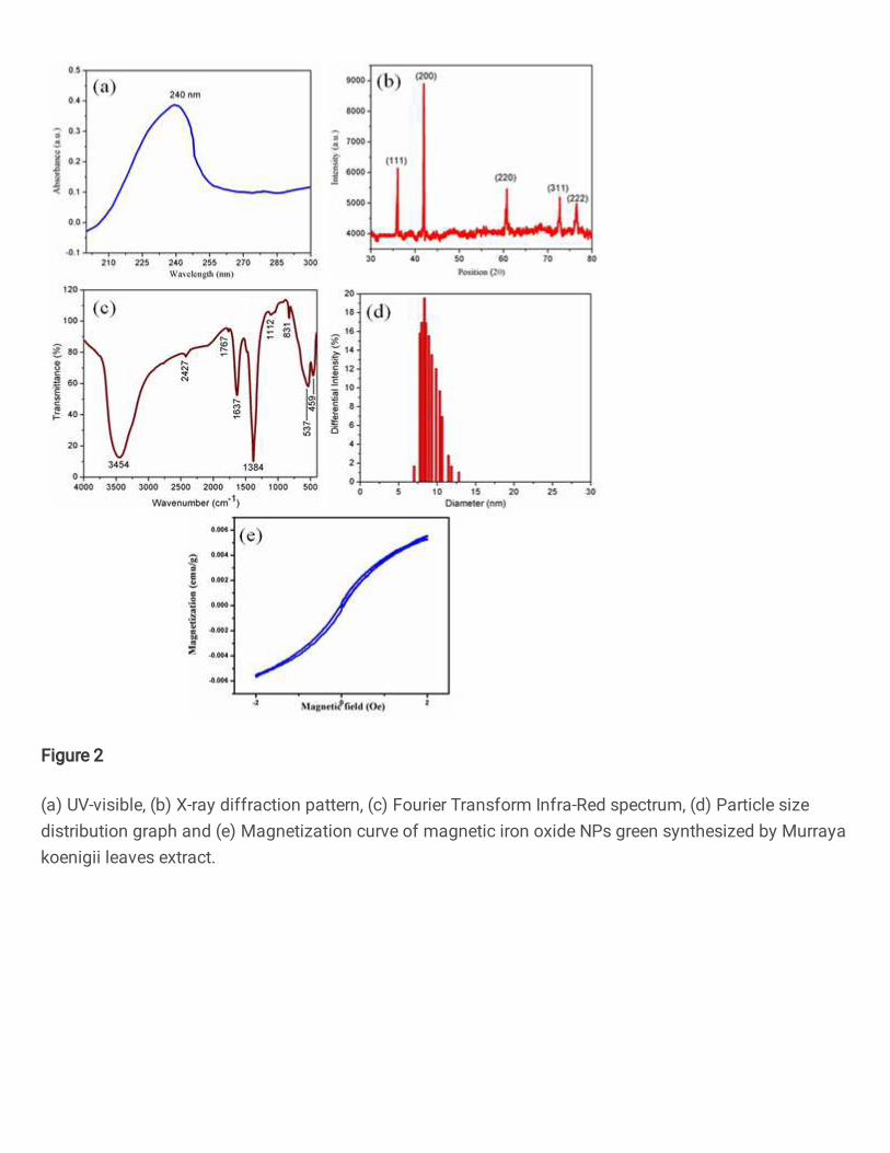
Figure 2
(a) UV-visible, (b) X-ray diffraction pattern, (c) Fourier Transform Infra-Red spectrum, (d) Particle sizedistribution graph and (e) Magnetization curve of magnetic iron oxide NPs green synthesized by Murrayakoenigii leaves extract.

Figure 3
(a-b) Transmission Electron Micrographs of magnetic iron oxide NPs green synthesized by Murrayakoenigii leaves extract at 20 nm, 50 nm scale range and (c) selected area electron diffraction pattern at1/10 nm scale range.

Figure 4
Energy dispersive X-ray spectrum of magnetic iron oxide NPs green synthesized by Murraya koenigiileaves extract.

Figure 5
Energy dispersive X-ray spectrum mapping of magnetic iron oxide NPs green synthesized by Murrayakoenigii leaves extract.

Figure 6
Antimicrobial assay of magnetic iron oxide nanoparticles green synthesized by Murraya koenigii leavesextract on Pseudomonas aeruginosa, Acinetobacter baumannii, Serratia marcescens, Chromobacteriumviolaceum, Enterobacter aerogenes, Klebsiella penumonia (Gram-negative bacterial cells), Enterococcusfaecalis, Staphylococcus aureus (Gram-positive bacterial cells), Candida albicans, Candida tropicalis,Aspergillus niger, and Aspergillus �avus (fungus cells).

Figure 7
(a) Antioxidant, (b) Anti-in�ammatory and (c) Anti-diabetic assays of magnetic iron oxide nanoparticlesgreen synthesized by Murraya koenigii leaves extract.