Embed Size (px)
Citation preview

Vol.:(0123456789)1 3
Environmental Chemistry Letters (2020) 18:703–727 https://doi.org/10.1007/s10311-020-00984-0
REVIEW
Green synthesis, biomedical and biotechnological applications of carbon and graphene quantum dots. A review
Siavash Iravani1 · Rajender S. Varma2
Received: 8 December 2019 / Accepted: 1 March 2020 / Published online: 10 March 2020 © Springer Nature Switzerland AG 2020
AbstractCarbon and graphene quantum dots are prepared using top-down and bottom-up methods. Sustainable synthesis of quantum dots has several advantages such as the use of low-cost and non-toxic raw materials, simple operations, expeditious reactions, renewable resources and straightforward post-processing steps. These nanomaterials are promising for clinical and biomedi-cal sciences, especially in bioimaging, diagnosis, bioanalytical assays and biosensors. Here we review green methods for the fabrication of quantum dots, and biomedical and biotechnological applications.
Keywords Quantum dots · Carbon dots · Graphene quantum dots · Biomedical applications · Cancer · Diagnosis · Bioimaging · Biotechnological applications · Sustainable synthesis
Introduction
Carbonaceous and carbon-based nanomaterials have been attracting a significant research interest because of their exclusive characteristics, including good biocompatibil-ity, non-toxicity, high mechanical/thermal properties, and relative ease of functionalization (Iravani 2011; Iravani and Varma 2019a, b; Mohammadinejad et al. 2016, 2019; Varma 2012, 2014a, b, 2016, 2019). Carbon dots are referred to as fluorescent carbon due to their strong characteristics which up on comparison with the traditional fluorescent dyes exhibit superior fluorescence including photostability, resistance to photobleaching and non-blinking (Xu et al. 2004). Additionally, carbon dots have promising character-istics, including water solubility, low toxicity, high chemi-cal stability, easy derivatization. Due to all these unique traits, carbon dots have found important and widespread
applications in biotechnology, energy, catalysis, biological labeling, bioimaging, and gene/drug delivery (Fig. 1 and Table 1) (Baker and Baker 2010; Shen et al. 2012; Cao et al. 2007; Yang et al. 2009a; Wang et al. 2013b). For instance, thermally reduced carbon dot-based fluorescence resonance energy transfer sensor was fabricated for sensitive, cost-effective, simple, and selective determination of vitamin B12 (~ 1–12 μg ml−1) in aqueous solutions (Wang et al. 2015a). Additionally, uniform oxygen and sulfur co-doped graphitic carbon nitride quantum dots were produced with significant blue photoluminescence, and good stability which can be applied for cellular imaging with acceptable biocompatibil-ity, and exhibited potential as fluorescent probe for bioimag-ing and biosensing purposes (Lu et al. 2015).
Carbon dots are traditionally defined as a class of core–shell composites comprising a carbon core and sur-face passivation with various functional groups, including hydroxyl, carboxyl, and amine, etc., which renders them hydrophilic and facilitate various surface functionalization and passivation. Surface passivation is usually attained by the production of a thin insulating layer of oligomeric poly-ethylene glycol on an acid-treated carbon dot surface; high fluorescence intensities and high quantum yield of carbon dots can be achieved with an effective surface passivation (Lim et al. 2015). For instance, carbon quantum dots were prepared with quantum yields as high as 60% via passivation with polyethylene glycol1500N (Wang et al. 2010). Addition-ally, polyethylenimine (Liu et al. 2012), boronic acid (Shen
* Siavash Iravani [email protected]
* Rajender S. Varma [email protected]
1 Faculty of Pharmacy and Pharmaceutical Sciences, Isfahan University of Medical Sciences, Isfahan, Iran
2 Department of Physical Chemistry, Faculty of Science, Regional Centre of Advanced Technologies and Materials, Palacký University in Olomouc, Šlechtitelů 27, 783 71 Olomouc, Czech Republic

704 Environmental Chemistry Letters (2020) 18:703–727
1 3
and Xia 2014), NH2-polyethylene-glycol and N-acetyl-l-cysteine (Gonçalves et al. 2010) have been applied for func-tionalizing carbon dots for application in gene delivery and bioimaging, blood sugar sensing and Hg(II) sensing (Liu et al. 2016).
Post-synthetic modifications of carbon dots are crucial as the introduction of functional groups, such as amines and carboxyl’s, can impose various defects on the carbon dot surface which operate as excitation energy traps and lead to large variations in fluorescence emissions. Surface pas-sivation with functional groups not only produces the sur-face defects from which the fluorescence originates but also provides potential reactive sites for modification reactions envisioned for specific tasks. By applying these reactive groups, a variety of specific organic, polymeric, inorganic and biomaterials can be affixed to the surfaces of carbon dots through covalent and hydrogen bonds and electrostatic interactions, serving as platforms for specific sensing, drug delivery and other explicit tasks (Liu et al. 2016).
Carbon and graphene quantum dots exhibit astounding attributes to current electrochemical biosensing because of their remarkable solubility in various solvents, intrinsic low toxicity, high electronic characteristics, strong chemical inertness, large specific surface area, availability of abundant edge sites for functionalization, significant biocompatibility, low cost, and versatility, as well as their ability to be altered with significant surface chemistries including nanostruc-tured materials (Campuzano et al. 2019). These quantum dots can be applied as signal tags or electrode surface mod-ifiers to produce electrochemical biosensing (Campuzano et al. 2019).
Various chemical precursors have been detected for gen-erating carbon dots, including ammonium citrate (Fang et al. 2017), ethylene glycol (Jaiswal et al. 2012), citric acid (Schneider et al. 2017), ethylene diamine tetra acetic acid (EDTA) (Liu et al. 2017a), phytic acid (Wang et al. 2013d), phenylenediamine (Vedamalai et al. 2014), thiourea (Wang et al. 2016a), carbon nanotube (Shinde and Pillai 2012) and graphite (Joseph and Anappara 2017). Meanwhile, diverse
green carbon precursors have been applied for the produc-tion of carbon dots including fruits, their juices and fruit peels (Mehta et al. 2015), animal and animal-derived materi-als such as chicken egg (Zhang et al. 2015c) and silkworm (Feng et al. 2016), vegetables and spices (Yin et al. 2013), waste kitchen materials like frying oil (Xu et al. 2015) or waste paper (Wei et al. 2014), plant leaves and derivatives (Feng et al. 2015b), among others. Additionally, graphite, nanodiamond, carbon nanotube and active-carbon can be applied as precursor for fabrication of carbon dots (Das et al. 2018).
Generally, carbon dots can be fabricated using top-down and bottom-up strategies (Fig. 2) (Sharma and Das 2019; Wang and Hu 2014). The top-down approaches, by which carbon dots are generally formed through the chemical or physical cutting procedures of relatively microscopic carbon structures, comprise arc discharge (Xu et al. 2004), laser ablation/passivation (Yang et al. 2009b; HU et al. 2009a, 2009b; Li et al. 2010b), electrochemical synthesis, (Ming et al. 2012; Zhou et al. 2007), and chemical oxidation (Qiao et al. 2010); most common representative sources for these techniques being carbon nanotube and graphite. Some draw-backs of top-down approaches for synthesizing carbon quan-tum dots are the requirements of expensive materials and setups, harsh reaction conditions, and longer reaction times (Sharma and Das 2019).
In bottom-up approaches, carbon dots are transformed from suitable molecular precursors under certain conditions exemplified by combustion/thermal/hydrothermal (Zhang et al. 2017; Liu et al. 2007) and microwave/ultrasonic irra-diation (Tang et al. 2012; Wang et al. 2011b; Li et al. 2011) wherein the precursors exhibit lower requirements of car-bon sources. Additionally, the other preparative strategies have been suggested, including plasma treatment (Jiang et al. 2009), supported synthesis (Bourlinos et al. 2008; Liu et al. 2009; Zong et al. 2011), solution chemistry approaches (Wang et al. 2011a; Hamilton et al. 2011; Mueller et al. 2010; Liu et al. 2011), and cage-opening of fullerenes (Lu et al. 2011). The produced carbon dots, regardless of their
Fig. 1 Sustainable and eco-friendly syntheses of quantum dots as well as their important biomedical and biotechnological applications. QDs: quantum dots, CQDs: Carbon quantum dots, GQDs: Graphene quantum dots

705Environmental Chemistry Letters (2020) 18:703–727
1 3
Table 1 Examples of carbon dots with key applications
Sources Synthetic approaches Size (nm) Applications References
Apple juice Hydrothermal 4.5 Imaging of mycobacterium and fungal cells
(Mehta et al. 2015)
Bee pollens Hydrothermal 1–2 Cellular imaging and catalysis (Zhang et al. 2015b)Beer Gel filtration chromatography 2.5 Breast cancer cell imaging and
drug delivery(Wang et al. 2015c)
Bloomed algae Microwave 8 In vitro imaging (Ramanan et al. 2016)Carrot Hydrothermal 2.3 Drug delivery (D’souza et al. 2018)Chionanthus retusus Hydrothermal 5 Metal ion sensing and imaging of
fungal cells(Atchudan et al. 2017)
Coriander leaves Hydrothermal 2.4 Sensoring of Fe3+ and cellular imaging
(Sachdev and Gopinath 2015)
Date kernel Hydrothermal 2.5 Sensing of drugs and cellular imaging
(Amin et al. 2018)
Food waste-derived Ultrasonic 4.6 In vitro bioimaging (Park et al. 2014)Garlic Hydrothermal 11 Cellular imaging and free radical
scavenging(Zhao et al. 2015)
Glycerin and polyethylene glycol Microwave 3–4 Nitrite sensing (Lin et al. 2011)Grape seed Microwave 1–8 Nucleus imaging and pH sensing (Kumawat et al. 2017a)Honey Solvothermal 2 Sensing Fe+3 and imaging of
Hep-2 and Hela cells(Yang et al. 2014)
Kidney beans Hydrothermal 20–30 Cellular imaging (Tripathi et al. 2017)Latex Microwave 2–8 Metal sensing and cellular imag-
ing(Balajia et al. 2018)
Lemon juice Hydrothermal 50 Optoelectronics and bioimaging (Hoan et al. 2019)Lignin biomass Ultrasonic and hydrothermal 2–6 Cellular imaging (Ding et al. 2018)Lotus root Microwave 9.41 Heavy metal ion detection and
cellular imaging(Gu et al. 2016)
Mango leaves Microwave 2–8 Cellular imaging and Temperature sensors
(Kumawat et al. 2017b)
Mangosteen pulp Hydrothermal 5 Sensoring of Fe3+ and cellular imaging
(Yang et al. 2017a)
Onion waste Hydrothermal 15 Sensoring of Fe3+ and cellular imaging
(Bandi et al. 2016)
Papaya juice Hydrothermal 3 Cellular imaging (Kasibabu et al. 2015)Prunus mume Hydrothermal 9 Cellular imaging (Atchudan et al. 2016b)Prunus persica Hydrothermal 8 Cellular imaging and oxygen
reduction reaction(Atchudan et al. 2016a)
Pseudo-stem of banana Hydrothermal 1–3 Sensing Fe+3, Imaging of Hela and MCF-7 cells
(Vandarkuzhali et al. 2017)
Saccharum officinarum Hydrothermal 3 Cellular imaging of bacteria and yeast
(Mehta et al. 2014)
Strawberry Hydrothermal 5.2 Fluorescent probes for mercury ions detection
(Huang et al. 2013)
Sugarcane molasses Hydrothermal 1.9 Sensoring of Fe3+ and cellular imaging
(Huang et al. 2017)
Sweet potato Hydrothermal 3.39 Fe+3 sensing and cellular imaging (Shen et al. 2017)Tissue paper Microwave 4.2 Determination of Glutathione (Sivasankaran et al. 2017)Trapa bispinosa peel Hydrothermal 5–10 Cellular imaging (Mewada et al. 2013)Walnut shell Hydrothermal 3.4 Cellular imaging (Cheng et al. 2017)Water Chestnut and onion Hydrothermal 3.5 Sensing of Cu (II) and Imaging of
Coenzyme A(Hu et al. 2017)
Winter melon Hydrothermal 4.5–5.2 Cellular imaging (Feng et al. 2015a)

706 Environmental Chemistry Letters (2020) 18:703–727
1 3
production techniques, have a mix of sizes, which needs complex separation techniques to obtain mono-dispersed carbon dots. Some of the explored post-synthesis separation techniques include dialysis (Liu et al. 2007), chromatogra-phy (Li et al. 2010a; Vinci et al. 2012), gel electrophoresis (Tao et al. 2012), and ultra-filtration (Zheng et al. 2013).
In this critical review, eco-friendly and sustainable fabri-cation of carbon and graphene quantum dots as well as their important biomedical and biotechnological applications are highlighted. The pros and cons and critical issues pertaining to the deployment of sustainable and eco-friendly strate-gies for manufacturing these quantum dots, with inherent advantages including non-toxicity, environmental friendli-ness, novelty, simplicity, greenness and cost-effectiveness, are discussed.
Carbon and graphene quantum dots with diverse unique properties
Generally, carbon dots can be classified in two main groups, carbon and graphene quantum dots. The graphene quantum dots are anisotropic, with lateral dimensions larger than the height, and possess the crystalline structure of single or a few graphene layer and connected via chemical groups on
the edges with circular or elliptical shape. In contrast, car-bon quantum dots are always spherical and carbon dots are divided into carbon nanoparticles without a crystal lattice; carbon quantum dots possessing an obvious crystal lattice with sp2/sp3 carbons (Lin and Zhang 2012; Nie et al. 2014).
All carbon dots have chemical groups on the surface, such as oxygen- or amino-based groups, and polymer chains and these functional groups play a pivotal role in the photolumi-nescence behavior. Both carbon and graphene quantum dots are effective in photon-harvesting in short-wavelength region because of π–π* transition of C = C bonds. They typically exhibit strong optical absorption in the ultraviolet region (~ 260–320 nm), with a tail extending into the visible range. The absorption located in 230–270 nm is typically attributed to π–π* transition of C = C bonds, and the peak/shoulder located around 300–390 nm is ascribed to n–π* transition of C = O bonds. Generally, carbon quantum dots are rela-tively more efficient in absorption of long wavelengths and have low quantum yield, thus requiring a surface passivation layer for improvement of their brightness. In comparison, graphene quantum dots usually offer higher quantum yield compared to carbon dots because of their layered structure and better crystallinity (Zheng et al. 2015; Zhu et al. 2015). Differently colored carbon dots have been synthesized, rang-ing from ultraviolet to red, and most commonly, blue and
Fig. 2 Carbon dots produced using top-down and bottom-up approaches. Adapted with permission from Sharma and Das (2019)

707Environmental Chemistry Letters (2020) 18:703–727
1 3
green. Undesirable for multi-color imaging, most of these carbon dots display broad emission spectra because of the large heterogeneity (in size and chemical composition) origi-nating from poorly controllable synthesis processes (Zheng et al. 2015).
Optical characteristics
Typically, carbon and graphene quantum dots with diverse structures have quite similar optical characteristics in terms of their optical fluorescence, absorption, chemilumines-cence, electrochemiluminescence, phosphorescence, up-conversion photoluminescence, and photo-induced electron transfer property. These materials have attracted immense attention due to their remarkable tunable optical characteris-tics, cost-effectiveness, simple fabrications and low toxicity, which makes them ideal candidates for optical sensors (Li et al. 2019).
Electrochemiluminescence activities are partly respon-sive to surface passivation, wherein the surface-passivated carbon quantum dots displayed weak electrochemilumines-cence activities but strong fluorescence (Dong et al. 2010). Methyl parathion sensors were generated from tyrosine methyl ester functionalized carbon dots via a hydrothermal reaction using citric acid as carbon resource; functionalized carbon dots display strong and stable photoluminescence with a quantum yield of 3.8% and could be deployed for the determination of assorted organophosphorus compounds (Hou et al. 2015a). Furthermore, alterations in fluorescence of carbon quantum dots were applied for detecting various numbers of inorganic molecules (Pooja et al. 2017). As an example, the detection of NO2 gas was achieved by using quantum dot-functionalized aerogels as the sensing probes wherein they revealed significant fluorescence activities in the solid state. The fluorescence was quenched by NO2 gas, while no quenching was detected when the carbon quantum dot-functionalized aerogels were exposed to a pure nitrogen atmosphere (Wang et al. 2013c).
Biocompatibility properties
Remarkable biocompatibility and low toxicity are critical for biological and biomedical appliances of carbon and graphene quantum dots, particularly in bioimaging and cellular imaging (Li et al. 2019). For instance, in vitro studies have revealed the low cytotoxicity of graphene quantum dots, which was credited to their significant oxy-gen content and ultra-small size. Additional in vivo biodis-tribution assay using mice showed no graphene quantum dots accumulations in the main organs and fast clearance of graphene quantum dot via kidney. Notably, injections with graphene quantum dot and graphene oxide showed
that graphene quantum dots had no noticeable influences on mice; differently, the graphene oxide revealed some toxicity, even initiating death because of the in vivo aggre-gations of graphene oxide. In another study, the effect of functional groups on the cytotoxicity of graphene quantum dots was ascertained by comparing their ability to generate reactive oxygen species. Consequently, it was revealed that ketonic carbonyl groups had noticeable effect on reactive oxygen species formation ability of graphene quantum dots. Thus, removing oxygen functional groups on gra-phene quantum dots could improve the photostability and reduce the cytotoxicity, which provided molecular level indication for the better design of biocompatible graphene quantum dots. Because of their remarkable biocompatibil-ity, graphene quantum dots with a large number of oxygen-containing groups were effectively applied as operative nano-radio-sensitizer for radiotherapy.
Graphene quantum dots could noticeably improve the sensitivity of colorectal carcinoma cells toward ion-izing irradiation. Additional investigations showed that the graphene quantum dots in combination with ionizing radiation could evidently suppress the cell proliferation, accelerate apoptosis, and increase the G2/M stage arrest of the cells. Principally, because of the overproduction of reactive oxygen species by graphene quantum dots in synergy with the ionizing radiation, they can activate the apoptosis-related regulation proteins culminating in tumor cell apoptosis (Zhou et al. 2017; Yuan et al. 2014).
Biosensors
There are a limited number of studies pertaining to bio-sensing via the deployment of carbon and graphene quan-tum dots, but future investigations are expected to increase in this arena (Shi et al. 2015b). As an example, graphene quantum dots in combination with gold nanoparticles were applied for developing the fluorescence resonance energy transfer biosensors for identifying a specific gene sequence from Staphylococcus aureus (Shi et al. 2015b). Further, well-defined modifications of graphene quantum dots have been applied in biosensors preparation. The resulting fluorescent intensity change produced a signal for target microRNA detection with satisfied discrimination abilities in a wide detection range from 0.1 to 200 nM, offering an approach for detection of microRNA based on graphene quantum dots (Zhang et al. 2015a). Additionally, carbon and graphene quantum dots can be applied for fabricat-ing of immuno-sensor. For instance, an immuno-sensor was prepared for 8-hydroxy-2′-deoxyguanosine (8-OHdG) where carbon quantum dots coated with gold/SiO2core-shell nanoparticles were immobilized onto a platinum electrode (Zhou et al. 2015).
708 Environmental Chemistry Letters (2020) 18:703–727
1 3
Carbon quantum dots
Carbon quantum dots are category of carbon nanostruc-tures which have received extensive consideration owing to their unique properties comprising significant pho-tostability, high solubility in water, non-toxicity, good biocompatibility, chemical inertness and high resistance to photobleaching. Furthermore, their optical properties can be fine-tuned by size control, chemical doping and functionalization for required applications, including gene delivery, bioimaging, optronics, drug delivery, sensing, and catalysis (Sachdev and Gopinath 2015; Yang et al. 2017a; Barras et al. 2016; Nekoueian et al. 2019; Li et al. 2015).
It has been shown that carbon quantum dots, which could serve as peroxidase mimetics, demonstrate an excel-lent catalytic activity for the decomposition of H2O2. Liu et al. reported that photoactive metal-free carbon quantum dot-carbon nitride nanocomposites break down the per-oxide (Liu et al. 2015a). These results provide a notion to help develop carbon quantum dot-containing hybrid semiconductors for resolving toxic issues during the water splitting process. In addition to bioimaging and biosensor applications, carbon quantum dots have been investigated for antibacterial, anti-inflammatory, and also for photo-dynamic therapy applications. In view of the increasing biomedical and clinical applications of carbon quantum dots, a better understanding of their safety when exposed to blood stream is the need of the hour. Determination of hemolytic properties is one of the fundamental tests in studies of nanoparticles’ interaction with blood compo-nents and cardiovascular system. Hemolysis may occur when the red blood cell membrane is ruptured and releases hemoglobin into blood plasma which may lead to anemia, hypertension, and renal toxicity. It is therefore imperative to study the hemotoxicity of all nanoparticles that would be administered intravenously. Recently, a special chemi-cal cleavage of layered N-doped carbon materials, carbon nitride quantum dots, was proposed (Cao et al. 2007; Yang et al. 2009a; Wang et al. 2013b).
The facile electrochemical fabrication of water-soluble carbon quantum dots has been achieved using an alkali-base electrolyte requiring chemicals/surface passive agents, and complex instrumental setups or post-treat-ments. Indeed, carbon quantum dots could be prepared by traditional approaches, including electrochemical oxida-tion, laser ablation, hydrothermal/solvothermal treatment, microwave irradiation and arc discharge, thermal cracking of organic compounds, pyrolysis and oxidation of candle soot. These protocols have disadvantages namely involve-ment of sophisticated instruments, strong acids for self-passivation and tedious procedures. Recently, synthesis
of self-passivated fluorescent carbon quantum dots by hydrothermal carbonization of materials has been reported in a single step using various biomass namely deploying ascorbic acid, gelatin, citric acid, egg albumin, bovine serum albumin, orange juice, honey, chitosan, cow milk, soy milk, banana, coffee seeds, watermelon peels, pomelo peel, grass, cellulose, starch, food caramels, and paper ash as carbon source (Cao et al. 2007; Yang et al. 2009a; Wang et al. 2013b). These carbon quantum dots display charac-teristic fluorescence depending upon the functionalities present on the surface. Moreover, hydrothermal carboniza-tion is a greener approach with the use of renewable bio-mass as a carbon source and at low reaction temperatures; both carbonization and functionalization can be achievable in a single step (Cao et al. 2007; Yang et al. 2009a; Wang et al. 2013b).
Eco‑friendly and sustainable synthesis of carbon quantum dots
Green chemistry is the engagement of important principles which reduces or eliminates the application or generation of hazardous substances in the design, manufacture, and application of chemical products; none or less hazardous chemical synthesis, applying safer and non-toxic chemicals, solvents and processes are some of them (Varma 2014b, 2016); as an example, bio wastes can be applied as sustain-able and cost-effective carbon sources for synthesizing car-bon quantum dots. In one study, spherical water-soluble car-bon quantum dots (about 1–3 nm) have been prepared from lemon peels waste deploying a cost-effective hydrothermal strategy; the ensuing stable carbon quantum dots were found to be oxygen-rich in surface functionalities and the water soluble and exhibited excellent photoluminescence proper-ties with quantum yield about 14%. The prepared carbon quantum dots were used to design an economic, green and highly sensitive fluorescent probe for the detection of Cr6+ ions with a detection limit of about 73 nM; water-soluble carbon quantum dots-based fluorescent probe could be used as a simple, rapid, convenient technique for the sensitive and selective detection of Cr6+ in water purification processes. Moreover, carbon quantum dots have been immobilized over electrospun TiO2 nanofibers and the emanating photocata-lytic activity for such a TiO2-water-soluble carbon quantum dots composite was reported using methylene blue dye as a model pollutant; the photocatalytic activity being about 2.5 times more than that of titanium dioxide nanofibers (Tyagi et al. 2016).
A greener synthesis of water-soluble fluorescent car-bon quantum dots (~ 260–400 nm) was reported via a sim-ple one-step hydrothermal treatment using Tamarindus indica leaves; these biocompatible carbon quantum dots can be potentially applied in sensing, bioimaging, disease

709Environmental Chemistry Letters (2020) 18:703–727
1 3
diagnostics, and other analytical applications (Bano et al. 2018). Another facile approach for producing fluorescent carbon quantum dots entails the hydrothermal treatment of pineapples (Ananas comosus) and calamansi (Citrofor-tunella microcarpa) wastes (de Yro et al. 2019). The Escher-ichia coli, fluoresced with carbon quantum dots as effective probe, have been applied for bioimaging applications. The generation of hydrogen bonds between the bacterial cells and carbon quantum dots facilitated the attachment of these quantum dots onto the bacteria (de Yro et al. 2019).
Well dispersed carbon quantum dots (~ 2–4 nm) have been synthesized by greener ozone oxidation of lignite coal, which is plentiful and cheap, and comprise oxygen-rich functional groups with suitable water-solubility and optical characteristics, and yield reaching 35% (Liu et al. 2018b). The prepared quantum dots had quenching effects for Fe3+ with remarkable sensitivity and selectivity as well as desir-able anti-interference performance wherein the fluorescence intensity of carbon quantum dots showed significant linear responses to the Fe3+ concentrations (10–150 µmol L−1) with the detection limit of 0.26 µmol L−1 (Liu et al. 2018b). Highly stable and luminescent multi-color carbon quantum dots (~ 4.85 ± 0.07 nm) were produced in one pot using Cydonia oblonga powder as carbon precursors via micro-wave irradiation which have maximum emission intensity at 450 nm if excited at 350 nm with a quantum yield of 8.55%. Carbon quantum dots produced through microwave heating technique in 30 min were compared with those synthesized hydrothermally in a Teflon-linen stainless steel body auto-clave at 200 °C (Ramezani et al. 2018).
Citrus lemon juice as a precursor was applied for the greener synthesis of fluorescent carbon quantum dots (~ 2–10 nm) using hydrothermal approach (Tadessea et al. 2018) which showed high photoluminescence of 10.20% quantum yield; the photoluminescence intensity being depended on pH of the solution and maximum intensity was obtained at pH of 6. It was shown that carbon quantum dots can be used as florescent probe in the cell imaging (Tadessea et al. 2018). In related research, amorphous fluorescent car-bon dots from orange waste peels were produced using the hydrothermal carbonization approach at a mild temperature (180 °C) (Prasannan and Imae 2013); their composite with zinc oxide was used as a photocatalyst for the degradation of naphthol blue–black azo dye under ultraviolet irradia-tion, and the superior photocatalytic activity was discerned (Prasannan and Imae 2013).
Bright green luminescent graphitic carbon nitride quan-tum dots doped with oxygen and sulfur were produced using microwave treatment of citric acid and thiourea; they dis-played excitation wavelength and pH-dependent lumines-cence behaviors in the visible range. Further, these quan-tum dots showed high fluorescence quantum yield (31.67%), strong resistance to the interference of high ionic strength
environment, and good biocompatibility as evaluated by the cell viability assay (Li et al. 2016). In another study, hydrothermal carbonization of Prunus avium fruit extracts was applied for fabricating fluorescent nitrogen-doped car-bon dots (~ 7 nm) which exhibited significant fluorescent characteristics and produced blue fluorescence; the green synthesized carbon dots were applied as fluorescent probes for detecting biologically important Fe3+ ions in water with remarkable selectivity and sensitivity, and for bioimaging of the examined cells (Edison et al. 2016b). An expeditious and facile microwave method was introduced for producing fluorescent nitrogen-doped carbon dots which were hydro-thermally prepared by applying L-ascorbic acid (as the car-bon precursor) and β-alanine (as the nitrogen dopant) with remarkable blue fluorescence and low cytotoxicity. These carbon dots can be applied as staining probes for confocal cellular imaging (Edison et al. 2016a).
Synthesis of mono-dispersed carbon quantum dots via a single-step thermal decomposition method was reported using the fennel seeds (Foeniculum vulgare) (Fig. 3) (Dager et al. 2019); the prepared quantum dots showed significant colloidal, photostability, environmental stability (pH), and did not need extra surface passivation step for improving their fluorescence. Additionally, these particles demon-strated remarkable photoluminescence activity and excita-tion-independent emission. The effect of some factors like reaction time and temperature on pyrolysis provided insight into the formation of carbon quantum dots; these quantum dots can be applied in biosensing, cellular imaging, light-emitting diode, solar cell, supercapacitor, printing, and sen-sors (Dager et al. 2019).
Highly photoluminescent carbon dots have been prepared, with a quantum yield of up to 35.3%, at room temperature which can be further separated out into green-emissive amorphous carbon nano-dots and yellow-emissive crystal-line graphene quantum dots through a silica gel column. The ensuing carbon dots, even with the same particle-size distribution and chemical groups, have different degrees of surface oxidation (Liu et al. 2017b). Additionally, highly fluorescent carbon quantum dots (~ 6 nm) were prepared by the pyrolysis of Eleusine coracana as a carbon source wherein Cu2+ strongly quenched the fluorescent intensity of carbon quantum dots compared to other metal ions; Cu2+ preferentially adsorbed on aromatic CC (π-bond) of carbon quantum dots, whereas other divalent metals form σ-bond(s) with the carbon quantum dots (Murugan et al. 2019).
Carbon quantum dots (~ 2.0–6.0 nm) with good biocom-patibility, photostability, and sustainability attributes were prepared from by-products derived from a biorefinery proce-dure using one-pot hydrothermal treatment (Fig. 4) (Huang et al. 2019a). The significant components of by-products were the degradation products (autohydrolyzate) of biomass pretreated by autohydrolysis. The carbon quantum dots

710 Environmental Chemistry Letters (2020) 18:703–727
1 3
Fig. 3 Carbon quantum dots formation from fennel seeds by pyroly-sis. a Fennel seeds, b ground fennel powder, c pyrolysis of fennel powder, d sonication of carbon quantum dots, e centrifugation of carbon quantum dots, f dialysis of carbon quantum dots, g carbon
quantum dots under ultraviolet, and h transmission electron micro-graph image of carbon quantum dots. Reprinted with permission from Dager et al. (2019)
Fig. 4 Green synthesis of carbon quantum dots from biorefinery by-products, as the precursor, with good biocompatibility, photostability and sustainability attributes. Reprinted with permission from Huang et al. (2019a)

711Environmental Chemistry Letters (2020) 18:703–727
1 3
showed blue–green fluorescence with a quantum yield of about 13%, and the fluorescence behaviors were reported to be stable with remarkable resistance to photobleaching and temperature alterations (Huang et al. 2019a).
Low-cost biomass of cyanobacteria was used as the car-bon source for producing water-soluble carbon quantum dots by a simple hydrothermal method (Fig. 5) (Wang et al. 2019); the ensuing carbon quantum dots were mono-dis-persed (about 2.48 nm), and showed excitation-dependent emission performance with a quantum yield of 9.24%. The cyanobacteria-derived carbon quantum dots had almost no photobleaching under long-time ultraviolet irradiation, and exhibited high photostability in the solutions with a wide range of pH and salinity. Since no additional chemical rea-gent was involved in their generation, the synthesized carbon quantum dots displayed low cytotoxicity for PC12 cells even at a high concentration. Moreover, the carbon quantum dots could be efficiently taken up by cells to illuminate the whole cell and create a clear distinction between cytoplasm and nucleus (Wang et al. 2019).
A greener hydrothermal synthesis for carbon quantum dots (~ 12 nm) was reported from walnut oil (Arkan E et al. 2018). The produced carbon quantum dots showed valuable fluorescent quantum yield of 14.5% with quinine sulfate (quantum yield 54%) as a reference and significant photo as
well as pH stabilities. The walnut carbon quantum dots were reported to be significantly potent cytotoxic agent, especially against MCF-7 and PC-3 cell lines. The apoptosis induction by carbon quantum dots was convoyed by acceleration in the activation of caspase-3, while caspase-9 activity did not hasten after exposure to the carbon quantum dots (Arkan E et al. 2018).
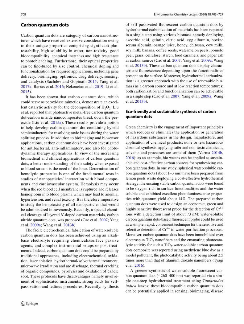
Novel types of N-doped chitosan-based carbon quantum dots have been achieved with simultaneous N-doping using biocompatible amino acids according to “green chemistry” principles (Fig. 6) (Janus et al. 2019). For the carbon quan-tum dots production, renewable chitosan has been applied with good biocompatibility. In this study, the cytotoxicity evaluations of the carbon quantum dots has been analyzed by 2,3-Bis-(2-methoxy-4-nitro5-sulfophenyl)-2H-tetrazolium-5-carboxanilide salt approach; the production of biocompat-ible quantum dots with carbon cores was recognized, which exhibited luminescence in visible range. It was reported that modifications with lysine (11.5%) and glutamic acid (7.4%) had significant effects on quantum yield, but amino acid functionalization did not remarkably influenced the fluores-cence characteristics (Janus et al. 2019). In another study, the synthesized spherical carbon quantum dots (with two-dimensional structures) from chitosan have been applied as corrosion inhibitors in BIS 2062 carbon steel (Keerthana and
Fig. 5 Synthesis of carbon quantum dots from cyanobacteria: a possible formation procedure of the cyanobacteria-derived carbon quantum dots; b fluorescence image of PC12 cells incubated with carbon quantum dots (c). Reprinted with permission from Wang et al. (2019)
712 Environmental Chemistry Letters (2020) 18:703–727
1 3
Ashraf 2019). It was revealed that the prepared carbon dots were rich in C = O and C–O functional groups (Keerthana and Ashraf 2019).
Biomedical and biotechnological applications
Carbon dots have been deployed for diverse biomedical and biotechnological applications due to their excellent photo-luminescence properties as well as the wavelength-tuneable emission property, unique electronic, mechanical, thermal, and chemical properties. In contrast to the traditional quan-tum dots, essentially comprising heavy metals which have known toxicity and are environmentally hazardous, carbon dots are much safer and non-toxic both in cells and at animal level, suggesting their good environmental and biological compatibility for biomedical and biotechnological applica-tions such as biosensing, bioimaging, gene and drug delivery (Wang et al. 2013a; Baker and Baker 2010).
Sensors
Various kind of sensors have been designed using carbon dots to detect assorted targets such as DNA (Wang et al. 2013e), heavy metals (Kumar et al. 2017), glucose (Shi et al. 2011), proteins (Dai et al. 2012), H2O2 (Zhao et al. 2013),
nitrite (Lin et al. 2011), and phosphate (Zhao et al. 2011), among others. For instance, carbon dots prepared from flour and electrochemical carbonization of sodium citrate and urea show the photoluminescence emission which could be selectively quenched by Hg2+ with a detection limit of 0.5 nM and 3.3 nM, respectively (Qin et al. 2013; Hou et al. 2015b); other heavy metals such as Sn2+, Fe3+, Pb2+, Cr6+, Mn2+ and Cu2+ were also detected (Gogoi et al. 2015; Yazid et al. 2013; Xu et al. 2014). The produced carbon quantum dots showed an excitation-dependent behavior with a high quantum yield of about 46.6% and could be applied as a very sensitive nanoprobe for the turn-off sensing of Hg2+ with a minimum limit of detection as low as 6 nM in the dynamic range from 0 to 0.1 μM. Furthermore, it can act as a turn-on sensor for glutathione detection with valuable selectivity (Bano et al. 2018).
Boronic acid modified carbon dots have been utilized for non-enzymatic blood glucose sensing wherein the glucose level can be quantified in the range of 9–900 µM with a detection limit of 1.5 µM; plasma glucose concentration determined by this method is in good agreement with the values measured by a commercial blood glucose monitor (Shen and Xia 2014). Carbon dot-based fluorescence turn-on sensors were instructed for hydrogen peroxide (H2O2) moni-toring in aqueous solutions through a photo-induced electron
Fig. 6 Green synthesis of N-doped chitosan-based carbon quantum dots using chitosan. The prepared quantum dots with carbon cores and good biocompatibility exhibited luminescence in visible range. Reprinted with permission from Janus et al. (2019)

713Environmental Chemistry Letters (2020) 18:703–727
1 3
transfer mechanism where the sensor showed good selectiv-ity, and sensitivity with a detection limit of 84 nM (Lan et al. 2015). In another study, N, S-co-doped fluorescent carbon nano-dots have been synthesized by applying thermal treat-ment of ammonium persulfate, ethylenediamine and glucose. They displayed bright blue emission with a remarkable fluo-rescent quantum yield of 21.6%, approved water solubility, uniform morphology, and significant chemical stability as compared to pure carbon dots. The fluorescence of the pro-duced carbon nano-dots could be remarkably quenched by methotrexate through fluorescence resonance energy transfer between carbon nano-dots and methotrexate, and has been applied for highly selective and sensitive identification of methotrexate (more than 50.0 μM) with a low detection limit of 0.33 nM; it can be useful for hands-on finding of metho-trexate in human serum (Wang et al. 2015b). Amino-func-tionalized fluorescent carbon dots have also been prepared by hydrothermal treatment of glucosamine and the ensued carbon dots exhibited stabilized green emission fluorescence at various excitation wavelengths and pH environments; they can be used to produce biosensors and for selective detection of hyaluronidase (Liu et al. 2015b).
Imaging and bioimaging
Carbon quantum dots can be applied for tumoral cells imag-ing, which is suitable for clinical and biological imaging and related diagnostic and therapeutic areas, including photo-therapies and diagnostic cancer imaging. For instance, the optical imaging of the carbon quantum dot-wheat straw has been evaluated by intravenous injection of 200 μL carbon quantum dot-wheat straw (0.2 μg mL−1) into the mouse through the tail vein. Optical images of the carbon quan-tum dot-wheat straw distributions in the tumor-bearing mouse over increasing times, as well as optical images of fluorescence intensities within the harvested organs can be observed in Fig. 7 (Huang et al. 2019a).
Application of carbon dots as fluorescent labels for cel-lular imaging has been documented (Sun et al. 2006). Car-bon dots are promising candidate for bioimaging purposes because of their low side effects and toxicity, excellent water solubility and visible-to-near infrared (NIR) emis-sion properties (Miao et al. 2015). Various types of cells have been imaged using carbon dots such as Ehrlich ascites carcinoma cells (EACs) (Ray et al. 2009), E. coli (Liu et al. 2009), HepG2 cells (Xu et al. 2013), NIH-3T3 fibroblast cells (Zhang et al. 2013), HeLa cells (Dong et al. 2013), and human lung cancer (A549) (Wu et al. 2013). (Jiang et al. 2014) reported the presence of photoluminescence carbon dots from commercial Nescafe instant coffee with the size of 4.4 nm and quantum yield about 5.5%; coffee-derived carbon dots have been directly applied in the imaging of carcinoma cells and small guppy fish without functionalization. Carbon
dots passivated with PPEI-EI for two-photon luminescence microscopy were used to image human breast cancer MCF-7 cells (Cao et al. 2007); carbon dots showed bright photolu-minescence both on the cell membrane and in the cytoplasm after 2 h incubation at 37 °C wherein the cellular uptake of carbon dots was temperature-dependent with no interna-tionalization observed at 4 °C. Zhu et al. (2014) prepared a two-photon “turn-on” fluorescent probe, which was then used for imaging hydrogen sulfide in live cells and tissues.
Ultra-small carbon dots (0.9 nm) with quantum yield of 47% have been produced via pyrolysis of anhydrous citric acid in organosilane at 240 °C for 1 min. The organosilane-functionalized carbon dots are stable in various non-aqueous solutions and can be directly fabricated into a hybrid fluo-rescent film or monolith through a simple heating proce-dure, in the absence of any additional polymers or inorganic compounds. Alternatively, they can be further assembled into silica-encapsulated nanoparticles by hydrolyzing and co-condensing the carbon dots with silica precursors for the bio-labeling and imaging applications (Zou et al. 2015).
Drug and gene delivery systems
Carbon quantum dots possess excellent drug/gene loading capability in view of their biocompatibility, non-toxicity and photoluminescence properties, where their small size and
Fig. 7 a In vivo fluorescence imaging of nude mice after intravenous injection of carbon quantum dot-wheat straw solution; b representa-tive fluorescence images of dissected organs of a mouse after intrave-nous injection of carbon quantum dot-wheat straw solution for 24 h. Reprinted with permission from Huang et al. (2019a)

714 Environmental Chemistry Letters (2020) 18:703–727
1 3
large surface area allow rapid cellular uptake with limited influence on drug activity (Gogoi and Chowdhury 2014; Karthik et al. 2013). Lee et al. (2014) confirmed the deliv-ery of doxorubicin in vitro and in vivo using carbon dots; doxorubicin was loaded on carbon dots via electrostatic interactions with 95% loading efficiency and induced death of HepG2 and MCF-7 cancer cells as well as tumor in mice. Further, pure carbon dots preferably labeled the nucleus, whereas carbon dots loaded with doxorubicin were mainly distributed in the cytoplasm. A smart stimuli-response drug delivery system comprising carbon dots and alginate was suggested where carbon dots, coated on the surface of algi-nate beads and garlic extract containing allicin, was taken as model drug system (Majumdar et al. 2016); the amount of garlic extract loaded on alginate beads coated with car-bon dots was 60% higher than uncoated alginate beads. The loaded system displayed pH-dependent controlled drug release, resulting in enhanced therapeutic efficiency depend-ing, interestingly, upon the amount of pathogen present in the target.
A smart carrier was developed for redox-responsive con-trolled drug delivery by grafting carboxyl-abundant car-bon dots to the surface of silica nanoparticles which was found to be pH-dependent (Jiao et al. 2016); doxorubicin loaded on the grafted silica had high drug loading (up to 13.1%) and showed a high cellular uptake and an excellent therapeutic effect on cancer cells by 3-(4,5-Dimethylthiazol-2-yl)-2,5-diphenyl tetrazolium bromide assay. Additionally, gene delivery has been successfully illustrated with posi-tively charged carbon dots which could be linked to plasmid DNA and efficiently transfect the therapeutic plasmid into cells with high efficiency and low cytotoxicity (Yang et al. 2017b). It has been shown that hyaluronate and polyethyl-enimine-functionalized carbon dots were internalized read-ily into the cytoplasm of cancer via hyaluronate-receptor-mediated endocytosis; the functionalized carbon dots have excellent gene condensation compatibility via electrostatic attraction and protective capacity by preventing nuclease degradation (Zhang et al. 2017).
Antimicrobial and antiviral effects
Carbon quantum dots are capable of effectively interacting with viruses and block-viral infectivity (Barras et al. 2016), as exemplified by carbon quantum dots functionalized with boronic acid and/or amine functions which interfere with the entry of the herpes simplex virus type 1 (Barras et al. 2016); carbon quantum dots prepared from 4-aminophe-nylboronic acid hydrochloride, via an altered hydrothermal carbonization procedure, prevent herpes simplex virus type 1 infection, whereas carbon quantum dots emanating from phenylboronic acid demonstrated no effects even at elevated concentrations. These carbon quantum dots can be applied as
therapeutic options for extremely pathogenic human corona-virus infections; mechanistically, it may be due to the human coronavirus-229E entrance inhibition, because of the inter-action of the boronic acid functions of carbon quantum dots with the HCoV-229E S protein through pseudo-lectin-based interactions. These findings inspired researchers to substi-tute presently applied antiviral agents (such as ribavirin and interferons), recognized to have major side effects, including short-term memory loss, deficits in decision-making functions, confusion, and extrapyramidal influences. More explorations should be undertaken for investigating the clinical effects of these nanostructures as alternative therapeutics and approaches to confront the severe and life-threatening diseases (Nekoueian et al. 2019).
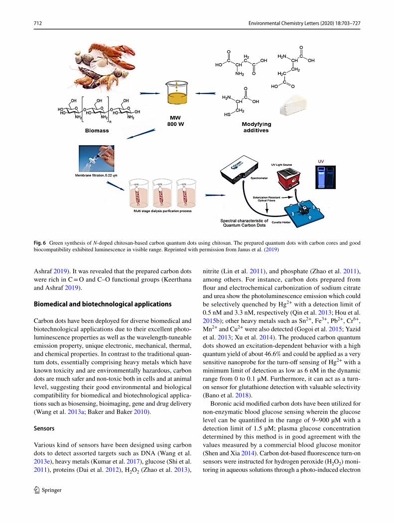
Carbon dots can be applied for microbial imaging and evaluations, as well as killing of infectious organisms like E. coli, S. aureus, Pseudomonas aeruginosa and Candida albi-cans. These carbon dots can be applied in microbial detection, microbial viability assessments, gram-type identification, and biofilm imaging (Priyadarshini et al. 2018; Habiba et al. 2015; Lin et al. 2019). Interestingly, the carbon dots platform has been harnessed fundamentally as a carrier for conventional disinfection agents to destroy bacteria, and also as a fluores-cence label for analyzing the dead bacterial cells. Apparently, these carbon dots could selectively interact with the Gram-positive bacteria, get adsorbed, and their fluorescence emis-sion being accelerated remarkably. The carbon dot adsorption could destruct the bacterial cell surface, as these carbon dots changed the charge balance of the bacterial surface and were inserted into the bacterial surface through the long alkyl chains leading to the bacterial inactivation. This carbon dot-based dif-ferentiation method has been fast, accurate, and easy to operate (Fig. 8) (Yang et al. 2019b).
Pharmaceutical formulations
Carbon quantum dots (~ 6 nm) which have been prepared from carbon powder by acid treatment at elevated temperatures were capable of effectively inhibiting insulin fibrillation in a concen-tration-dependent manner (about 0.2 μg mL−1). Additionally, carbon quantum dots (40 μg mL−1) prevented 0.2 μg mL−1 of human insulin from fibrillation for 5 days under 65 °C, whereas insulin denatures in 3 h under the same conditions without carbon quantum dots. Therefore, these carbon quan-tum dots have the potentials to inhibit insulin fibrillation in bio-systems and in the pharmaceutical industry for the pro-cessing and formulation of insulin (Li et al. 2015).
715Environmental Chemistry Letters (2020) 18:703–727
1 3
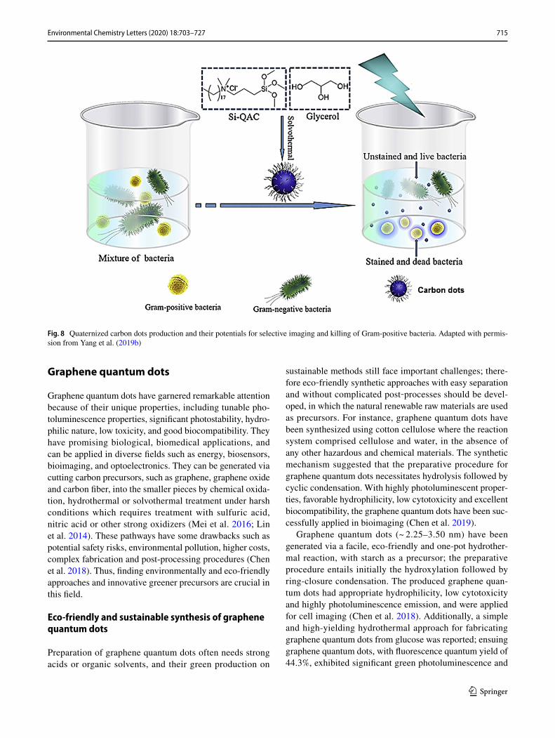
Graphene quantum dots
Graphene quantum dots have garnered remarkable attention because of their unique properties, including tunable pho-toluminescence properties, significant photostability, hydro-philic nature, low toxicity, and good biocompatibility. They have promising biological, biomedical applications, and can be applied in diverse fields such as energy, biosensors, bioimaging, and optoelectronics. They can be generated via cutting carbon precursors, such as graphene, graphene oxide and carbon fiber, into the smaller pieces by chemical oxida-tion, hydrothermal or solvothermal treatment under harsh conditions which requires treatment with sulfuric acid, nitric acid or other strong oxidizers (Mei et al. 2016; Lin et al. 2014). These pathways have some drawbacks such as potential safety risks, environmental pollution, higher costs, complex fabrication and post-processing procedures (Chen et al. 2018). Thus, finding environmentally and eco-friendly approaches and innovative greener precursors are crucial in this field.
Eco‑friendly and sustainable synthesis of graphene quantum dots
Preparation of graphene quantum dots often needs strong acids or organic solvents, and their green production on
sustainable methods still face important challenges; there-fore eco‐friendly synthetic approaches with easy separation and without complicated post‐processes should be devel-oped, in which the natural renewable raw materials are used as precursors. For instance, graphene quantum dots have been synthesized using cotton cellulose where the reaction system comprised cellulose and water, in the absence of any other hazardous and chemical materials. The synthetic mechanism suggested that the preparative procedure for graphene quantum dots necessitates hydrolysis followed by cyclic condensation. With highly photoluminescent proper-ties, favorable hydrophilicity, low cytotoxicity and excellent biocompatibility, the graphene quantum dots have been suc-cessfully applied in bioimaging (Chen et al. 2019).
Graphene quantum dots (~ 2.25–3.50 nm) have been generated via a facile, eco-friendly and one-pot hydrother-mal reaction, with starch as a precursor; the preparative procedure entails initially the hydroxylation followed by ring-closure condensation. The produced graphene quan-tum dots had appropriate hydrophilicity, low cytotoxicity and highly photoluminescence emission, and were applied for cell imaging (Chen et al. 2018). Additionally, a simple and high-yielding hydrothermal approach for fabricating graphene quantum dots from glucose was reported; ensuing graphene quantum dots, with fluorescence quantum yield of 44.3%, exhibited significant green photoluminescence and
Fig. 8 Quaternized carbon dots production and their potentials for selective imaging and killing of Gram-positive bacteria. Adapted with permis-sion from Yang et al. (2019b)

716 Environmental Chemistry Letters (2020) 18:703–727
1 3
excitation-independent photoluminescence emission proper-ties (Gu et al. 2014).
A facile one-pot solid-phase method was reported for synthesizing N-doped graphene quantum dots using citric acid as the carbon source and 3,4-dihydroxy-l-phenylalanine as the nitrogen source; ensuing N-graphene quantum dots with oxygen-rich functional groups, were uniform with an average diameter of 12.5 nm and served as highly efficient fluorosensor for detection of Hg2+ because of the effective quenching effect of metal ions via non-radiative electron transfer. This selective fluorosensor showed high sensitiv-ity toward Hg2+ with a detection limit of 8.6 nM and was practically used for the effective detection of Hg2+ in river-water samples (Shi et al. 2015a). An eco-friendly approach to prepare N-graphene quantum dots with a high quantum yield of 28.10% has been described which showed strong blue fluorescence emission with the maximum emission and excitation at 450 nm and 355 nm, respectively. Tak-ing advantage of the effective quenching effect of Hg2+ on N-graphene quantum dots, they were developed for the efficient and sensitive detection of Hg2+ with a relatively low detection limit of 0.032 μM. Based on the selective coordination of biothiols and Hg2+, the fluorescence of the N-graphene quantum dot/Hg system was recovered with the addition of biothiols. This fluorescent “Off–On” process displayed a sensitive response to biothiols with a detection limit of 0.036 μM for cysteine and 0.034 μM for glutathione. The N-graphene quantum dot-based fluorescence method has been successfully used to monitor Hg2+ in real water samples and biothiols in serum samples (Yan et al. 2016).
Silica supported silver nanoparticles and graphene quan-tum dots compounds have been designed and synthesized via a greener photochemical approach and an electrophore-sis deposition technique to provide a highly active surface-enhanced Raman scattering substrate. In this method, the electrochemically prepared aqueous solution of graphene
quantum dots was used both, as a solvent and a reducing agent to generate in situ silver-graphene quantum dots compounds under ultraviolet irradiation conditions. These compounds were collected on a SiO2 supported Si substrate through the electrophoresis deposition technique. Benefit-ing from their proper size (~ 1–4 nm) and distribution in the spatial gaps between adjacent silver nanoparticles, graphene quantum dots could act as “hot spot” sites for lighting up the Raman scattering signals. Together with the enhanced adsorption of Rhodamine 6G (R6G) molecules through π-π stacking, the electrostatic interactions from graphene quan-tum dots, and the enlarged specific surface area provided by the SiO2 template, the as-prepared substrate showed a strong surface-enhanced Raman scattering signal with excel-lent reproducibility, the detection limit of R6G being pushed to 8.0 × 10⁻¹⁴ M (Ge et al. 2016).
A by-product obtained from coking industries, coal tar pitch, has an exclusive molecule structure including an aromatic nucleus with various side chains bonding on this graphene-like nucleus, with a structural similarity to the structure of graphene quantum dots. An approach to con-vert coal tar pitch to, mono-dispersed graphene quantum dots (~ 1.7 ± 0.4 nm) by applying oxidation with hydrogen peroxide under mild conditions has been described with high yield more than 80 wt% (Liu et al. 2018a). These graphene quantum dots were significantly soluble and strongly fluores-cent in aqueous solution (Fig. 9) (Liu et al. 2018a).
A greener and economical method has been disclosed to synthesize water-soluble graphene quantum dots from cow’s milk for concurrent drug delivery and imaging in cancers. These spherical and multi-fluorescent quantum dots (~ 5 nm) were prepared via one-pot microwave-assisted heating approach. It was revealed that heating time and ionic strength significantly influence on photoluminescence char-acteristics of them. Berberine hydrochloride was attached on graphene quantum dots (88% efficacy in drug loading)
Fig. 9 Green synthesis of graphene quantum dots using coal tar pitch (CTP). Reprinted with permission from Liu et al. (2018a)

717Environmental Chemistry Letters (2020) 18:703–727
1 3
(Thakur et al. 2016). Consequently, the graphene quantum dots were biocompatible with L929 cells, but the produced theranostic complex exhibited influential cytotoxic effects on diverse examined cancerous cell lines. Because of drug delivery and bioimaging characteristics of the prepared com-plex, it showed in vitro theranostic applications in cancer therapies (Thakur et al. 2016).
An approach was described involving hydrazine hydrate-assisted hydrothermal cutting followed by functionalizations with polyethylenimine for producing remarkably fluorescent graphene quantum dots from coffee grounds. The polyeth-ylenimine-functionalized graphene quantum dots presented enhanced band-edge photoluminescence with single expo-nential decay, and their sensing and bioimaging applications were documented (Wang et al. 2016b).
Greener synthetic method for producing fluorescent gra-phene quantum dots without involving any harsh reagents has been documented wherein graphene oxide was applied used as a precursor, and hydrothermal synthesis (about 2 h) was accomplished with assistance from hydrogen peroxide. The prepared graphene quantum dots showed high photosta-bility and significant biocompatibility as demonstrated by cell viability evaluations. These quantum dots can be applied as fluorescent nano-probes, because of the cell-graphene quantum dots interactions as they exhibited high potentials for bioimaging, diagnostics and drug delivery (Halder et al. 2018).
Biomedical and biotechnological applications
Drug delivery and cancer therapy
Preparation of drug delivery systems based on graphene quantum dots can find diverse applications in bio- and nano-medicine (Dong et al. 2018) and multifunctional graphene quantum dots can be harnessed for targeted cellular imaging, which is useful for cancer therapy (Wang et al. 2014b). Drug delivery systems based on arginine-glycine-aspartic acid-conjugated graphene quantum dots were applied in load of doxorubicin (an antitumor drug) for targeted imaging and drug delivery. Indeed the characteristic stable fluorescence of graphene quantum dots permitted the real-time analyses of the cellular uptake from the produced nanoassembly and the following doxorubicin drug release. The doxorubicin releasement showed significant pH dependence and implied hydrogen-bonding interaction between graphene quantum dots and doxorubicin; the graphene quantum dots can be served as pH-sensitive drug carriers (Dong et al. 2018). Compared to free doxorubicin, the prepared conjugates revealed remarkable cytotoxicity effects to U251 glioma cells within diverse range of doxorubicin concentrations. Findings form the cellular uptake of these conjugates dem-onstrated that doxorubicin and some graphene quantum dots
have entered into cell nuclei after incubation (~ 16 h). These combinational enhancements with suitable nuclear delivery enhanced the doxorubicin cytotoxicity effects, noticeably (Dong et al. 2018). Additionally, the prepared folic acid-con-jugated graphene quantum dots could be applied for loading of doxorubicin, real-time evaluating of cellular uptake and the consequent drug release. This prepared nanoassembly was precisely internalized promptly by HeLa cells through receptor-mediated endocytosis, whereas doxorubicin release and accumulations were sustained. In vitro toxicity stud-ies revealed that the produced nanoassembly targeted HeLa cells differentially and efficiently while showing meaning-ful decrease in cytotoxicity on non-target cells (Wang et al. 2014b).
The engineered nanocarriers comprising biodegradable charged polyester vectors and graphene quantum dots were evaluated for their therapeutically influences on pancreatic cancers (MiaPaCa-2 cells) (Yang et al. 2019a). These nano-carriers were applied for co-loading doxorubicin and small interfering ribonucleic acids. Additionally, they showed significant physiological stabilities, outstanding K-ras downregulation activities, and operative bioactivity inhibi-tions for the examined cells. Further, laser light has been applied for generating heat for the nanocomplexes through the photothermal effects for cellular destruction; laser trig-gered the payloads release from the prepared nanostructured complexes and this activated release function significantly accelerated their anticancer activities of them. Moreover, the evaluation of initial colony generation showed that these complexes can be considered as promising carrier candidates for in vivo analyses (Yang et al. 2019a).
Cytotoxicity, autophagy and mutagenicity
Graphene quantum dots (~ 160–280 nm) were prepared and biologically assessed by employing two mice models to assess their in vitro mutagenicity. After radiolabeling experiment, it was shown that stable radiolabeled graphene quantum dots were prepared with a high yield (> 90%). The in vivo investigation displayed an ubiquitous behav-ior when used for healthy animals, with a high uptake by liver (> 26%) and small intestine (> 25%). Furthermore, in inflammation/VEGF hyper-expression animal model (endo-metriosis), a very peculiar behavior of graphene quantum dots was detected, with a high uptake by kidneys (over 85%) (de Menezes et al. 2019).
The cytotoxicity and autophagy induction of COOH–graphene quantum dots, OH–graphene quantum dots, and NH2–graphene quantum dots have been investi-gated (Xie et al. 2019). The OH–graphene quantum dots were found to be the most toxic, as substantial cell death was induced at the concentration of 100 μg mL−1, as indicated by WST-1 assay as well as Annexin-V-FITC/PI apoptosis

718 Environmental Chemistry Letters (2020) 18:703–727
1 3
assessment, whereas COOH– and OH–graphene quantum dots were non-cytotoxic within the analyzed concentra-tions. Additionally, OH– and NH2–graphene quantum dots stimulated autophagy of cells with different grades except for COOH–graphene quantum dots; it was revealed that all these quantum dots meaningfully activated p-p38MAPK, and p-ERK1/2 was obstructed by OH– and NH2–graphene quantum dots but activated by COOH-graphene quantum dots. Further, p-JNK was obstructed by COOH– and NH2 graphene quantum dots, while activated by OH-graphene quantum dots. Concurrently, Akt was activated by OH–gra-phene quantum dots, but inhibited by NH2–graphene quan-tum dots. The autophagy inhibition effects from 3-MA could meaningfully improve the cytotoxicity effects of graphene quantum dots, indicating that autophagy had a protecting effects against the examined quantum dot toxicity (Xie et al. 2019).
Imaging and bioimaging
Because of the stable photoluminescence, low cytotoxicity, excellent solubility and biocompatibility, the graphene quan-tum dots are especially eco-friendly and were illustrated to be excellent probes for bioimaging. In one study, synthesis of strongly green-photoluminescent graphene quantum dots on large scale was reported; the produced graphene quan-tum dots showed significant fluorescence with photolumi-nescence quantum yields as high as 11.4%. Additionally, the graphene quantum dots were readily soluble in water and most polar organic solvents with no further chemical modifications (Zhu et al. 2011).
Graphene quantum dots (~ 10 nm) have been synthesized by vigorous oxidation of graphite. The prepared graphene quantum dots showed good physiological solubility, high photostability, low cytotoxicity, and yellow-green fluores-cence with quantum yield of ~ 7%. They have been inves-tigated for cell imaging applications (Zhang et al. 2012).
In the field of bioimaging applications of graphene quan-tum dots, there are still some important issues which need to be resolved. Though graphene quantum dots with emission wavelengths ranging from ultraviolet to near infrared were prepared through different strategies, the described quantum yields of graphene quantum dots were considerably lower than conventional semiconductor quantum dots; thus the quantum yields improvement of graphene quantum dots is prerequisite. Graphene quantum dots with bright red/near infrared emissions can be applied as suitable nano-probes for bioimaging (Wang et al. 2014a; Namdari et al. 2017).
The multifunctional nano-probes with great advance-ments can be applied for diverse fields of imaging and bioimaging; the radioactive and optical characteristics of graphene quantum dots can be useful for simultaneously imaging and therapy. Important challenges for designing
graphene quantum dots-based nano-probes for optical imag-ing simultaneously, magnetic resonance imaging, and com-puted tomography evaluating should be perceived (Wang et al. 2014a; Namdari et al. 2017). Targeted tumors imaging based on graphene quantum dots has been reported in vivo, infrequently. The subsequent forms of cancer diagnostic agents applied for small animals require enhanced accumu-lations in tissues of tumors. The functionalization of gra-phene quantum dots with peptides or antibodies for imaging with high cancers targeting issues should be additionally investigated. The optimization of emission characteristics and multi-photon excitations of graphene quantum dots, their innovative neurobehavioral analyses applications, the brain gene therapy, and brain barrier penetration through blood capillaries, should be scrutinized (Dager et al. 2019; Sharma and Das 2019; Wang et al. 2014a; Namdari et al. 2017). The side effects, which possibly can be detected by applying graphene quantum dots, need to be evaluated. The gene expression and toxicity as well as cytotoxicity evalua-tions of graphene quantum dots with various surface coat-ings, morphologies and sizes should be assessed; the bio-distribution analyses of graphene quantum dots in different animal models are essential (Wang and Hu 2014; Huang et al. 2019b; Zhu et al. 2011; Zhang et al. 2012; Wang et al. 2014a; Namdari et al. 2017).
Graphene quantum dots modified with polyethyleneimine or (3-carboxyl) phenyl bromide phosphine have been pre-pared via eco-friendly, rapid, cost-effective and large-scale preparative strategy (Fan et al. 2019). The graphene quantum dots-polyethyleneimine was generated by a simple hydro-thermal procedure, and then (3-carboxyl) phenyl bromide phosphine was conjugated to the graphene quantum dots-polyethyleneimine through the amide linkage; average sizes of the prepared graphene quantum dots-polyethyleneimine and -(3-carboxyl) phenyl bromide phosphine being 3.75 and 3.25 nm, respectively. These two composites showed signifi-cant optical property, low cytotoxicity and selective targeting and imaging characteristics for cell nucleus or mitochondria, suggesting applications, including cell nucleus imaging or mitochondria imaging in vivo and in vitro for diagnosis and therapy (Fan et al. 2019).
Challenges and opportunities
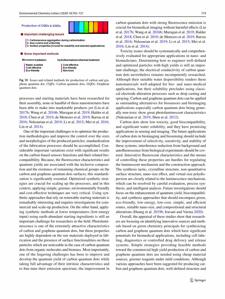
The optical and electrochemical detection of carbon and gra-phene quantum dots are of great interest to nanoresearchers in biomedical applications, but there are important chal-lenging issues regarding their production (Fig. 10); some of them are highlighted in this section. Expectantly, increasing interest in the fabrication of these quantum dots, in view of their straightforward production and their integration with other nanomaterials, is sustainable. Although various
719Environmental Chemistry Letters (2020) 18:703–727
1 3
processes and starting materials have been researched for their assembly, none or handful of these nanostructures have been able to make into marketable products yet (Liu et al. 2017b; Wang et al. 2016b; Murugan et al. 2019; Halder et al. 2018; Chen et al. 2019; de Menezes et al. 2019; Barras et al. 2016; Nekoueian et al. 2019; Li et al. 2015; Mei et al. 2016; Lin et al. 2014).
One of the important challenges is to optimize the produc-tion methodologies and improve the control over the sizes and morphologies of the produced particles; standardization of the fabrication processes should be accomplished. Con-siderable important variations exist with significant results on the carbon-based sensors functions and their related bio-compatibility. Because, the fluorescence characteristics and quantum yields are associated with the inclusive composi-tion and the existence of remaining chemical groups on the carbon and graphene quantum dots surfaces, this standardi-zation is significantly essential. Optimized synthetic strat-egies are crucial for scaling up the processes, and in this context, applying simple, greener, environmentally friendly and cost-effective techniques are very critical. Using syn-thetic approaches that rely on renewable starting materials is remarkably interesting and requires investigations for com-mercial and scale-up production. On the other hand, apply-ing synthetic methods at lower temperatures (low-energy input) using earth-abundant starting ingredients is still an important challenge for researchers in the field. Photolumi-nescence is one of the extremely attractive characteristics of carbon and graphene quantum dots, but these properties are highly dependent on the raw materials deployed in fab-rication and the presence of surface functionalities on these particles which are noticeable in the case of carbon quantum dots from organic molecules and biomass. Most importantly, one of the lingering challenges has been to improve and develop the quantum yield of carbon quantum dots while taking full advantage of their intrinsic characteristics and to fine-tune their emission spectrum; the improvement in
carbon quantum dots with strong fluorescence emission is crucial for biomedical imaging without harmful effects (Liu et al. 2017b; Wang et al. 2016b; Murugan et al. 2019; Halder et al. 2018; Chen et al. 2019; de Menezes et al. 2019; Barras et al. 2016; Nekoueian et al. 2019; Li et al. 2015; Mei et al. 2016; Lin et al. 2014).
Toxicity issues should be systematically and comprehen-sively evaluated for appropriate applications in nano- and biomedicines. Determining how to engineer well-defined and optimized particles with high yields is still an impor-tant challenge; the electrical conductivity of carbon quan-tum dots nevertheless remains incompetently researched. Although their suitable water dispersibility renders these nanomaterials well-adapted for bio- and nano-medical applications, but their solubility precludes using classi-cal electrode alteration processes such as drop casting and spraying. Carbon and graphene quantum dots can be applied as outstanding alternatives for biosensors and bioimaging applications especially carbon quantum dots being gener-ally non-toxic show great photoluminescent characteristics (Nekoueian et al. 2019; Shen et al. 2012).
Carbon dots show low toxicity, good biocompatibility, and significant water solubility, and they have promising applications in sensing and imaging. The future applications of carbon dots in bioimaging and biosensing should include the improvement of selectivity, sensitivity, and stability of these systems; interference reduction from background and autofluorescence from biological experiments should be cov-ered. Innovative fluorescent characteristics and the means of controlling these properties are hurdles for regulating the luminescent mechanism and the construction approach. The synthesis tactic, crystalline structure, non-quantitative surface structure, nano-size effect, and virtual size polydis-persion are closely related to the luminescence mechanism, which can be resolved by careful evaluation, precise syn-thesis, and intelligent analysis. Future investigations should focus on the enhancement of quantum yield, biocompatibil-ity, and synthesis approaches that should encompass green, eco-friendly, low-energy, low-cost, simple, and efficient routes, suitable nano-size, and compositional and structural alterations (Huang et al. 2019b; Iravani and Varma 2020).
Overall, the appraisal of these studies show that research-ers are focusing on identifying innovative sources and meth-ods based on green chemistry principals for synthesizing carbon and graphene quantum dots which have significant potentials for biomedical applications, including cell labe-ling, diagnostics or controlled drug delivery and release systems. Simple strategies providing feasible methods toward the commercial high yield production of carbon and graphene quantum dots are needed using cheap material sources, greener reagents under mild conditions. Although various approaches have been suggested for producing car-bon and graphene quantum dots, well-defined structure and
Fig. 10 Issues and related methods for production of carbon and gra-phene quantum dot. CQDs: Carbon quantum dots, GQDs: Graphene quantum dots

720 Environmental Chemistry Letters (2020) 18:703–727
1 3
precise sizes are barely available yet (D’souza et al. 2018; Kasibabu et al. 2015). It is vital to prepare quantum dots in a facile and greener manner with designed structure and size for proper investigations and biomedical applications. The synthetic routes and sources of carbon are important factors, which influence the sizes, morphologies and characteristics of the final product; the sizes for carbon dots are very crucial for understanding quantum phenomena, and also for bio-medical applications and optoelectronics (Li et al. 2019).
Recently, various pathways have been outlined for the fab-rication of both pure or doped carbon and graphene quantum dots with varied morphologies and characteristics. Though, discrete sustainable and greener synthetic approaches have been applied for synthesizing carbon quantum dots, inves-tigations into graphene quantum dots are still at a growing stage, and more elaborate and critical studies are needed. As an example, the well-defined and atom-precise structures have not been reported comprehensively, thus restricting the profound evaluation of the relationships between structures and characteristics, detailed properties regulations, and com-prehensive assessments of innovative synthetic approaches and applications. An all-inclusive consideration toward the optical characteristics of carbon dots is needed; additional theoretical evaluations and better understanding the mecha-nistic aspects are very critical.
Conclusion
Advancements in nanosciences for advanced and developed diagnosis and personalized therapy of various complex dis-eases (especially, cancers) are of contemporary interest. Par-ticularly, semiconductor quantum dots have been developed as innovative platforms for high-throughput quantitative analyses of multiple biomarkers in cells and clinical tissue samples (ex vivo), in vivo evaluations of cells with diseases, and potentially targeted and traceable drug delivery. Indeed, quantum dots show significant potentials in such biomedical, bioimaging, and photoluminescent applications, and they can be harnessed as promising fluorescent probes for imag-ing with low cytotoxicity as they show remarkable potentials in bioanalysis and related fields. Carbon quantum dots have received widespread attentions owing to their suitable bio-compatibility, robust chemical inertness and higher resist-ance to photobleaching. Additionally, their optical charac-teristics can be adjusted via size control, chemical doping and functionalization for featured and specific applications.
Biomass-based carbon quantum dots have become sub-stantial carbon materials because of their cost-effectiveness, ease of fabrication and lower environmental impact. But, it appears that there are not many investigations regard-ing using organisms (especially, cyanobacteria) as carbon sources for production of fluorescent carbon quantum dots.
Additionally, graphene quantum dots are attractive fluo-rophores because of their outstanding photoluminescence characteristics, water solubility, low cost, and toxicity. For instance, in some cases, COOH–graphene quantum dots demonstrated significant biocompatibility, and may be con-sidered for biological and medical applications. It appears that considerable efforts should be expanded to find greener, simple, efficient, and eco-friendly fabrication methodolo-gies for carbon and graphene quantum dots as well as their effective use in bio- and nano-medical applications. More scientific investigations need to be undertaken for scaling up and the commercial production of quantum dots by applying simple, efficient, economic and sustainable strategies with focus on biocompatibility and low cytotoxicity.
Acknowledgements RSV was supported, in part, from ERDF project “Development of pre-applied research in nanotechnology and biotech-nology” (No. CZ.02.1.01/0.0/0.0/17_048/0007323).
Compliance with ethical standards
Conflict of interest Authors declare no conflict of interest.
References
Amin N, Afkhami A, Hosseinzadeh L, Madrakian T (2018) Green and cost-effective synthesis of carbon dots from date kernel and their application as a novel switchable fluorescence probe for sensitive assay of Zoledronic acid drug in human serum and cellular imag-ing. Anal Chim Acta 1030:183–193. https ://doi.org/10.1016/j.aca.2018.05.014
Arkan E, Barati A, Rahmanpanah M, Hosseinzadeh L, Moradi S, Haji-alyani M (2018) Green synthesis of carbon dots derived from walnut oil and an investigation of their cytotoxic and apoptogenic activities toward cancer cells. Adv Pharm Bull 8:149–155. https ://doi.org/10.15171 /apb.2018.018
Atchudan R, Edison TNJI, Lee YR (2016a) Nitrogen-doped carbon dots originating from unripe peach for fluorescent bioimaging and electrocatalytic oxygen reduction reaction. J Colloid Inter-face Sci 482:8–18. https ://doi.org/10.1016/j.jcis.2016.07.058
Atchudan R, Edison TNJI, Sethuraman MG, Lee YR (2016b) Effi-cient synthesis of highly fluorescent nitrogen-doped carbon dots for cell imaging using unripe fruit extract of Prunus mume. Appl Surf Sci 384:432–441. https ://doi.org/10.1016/j.apsus c.2016.05.054
Atchudan R, Edison TNJI, Chakradhar D, Perumal S, Shim J-J, Lee YR (2017) Facile green synthesis of nitrogen-doped carbon dots using Chionanthus retusus fruit extract and investigation of their suitability for metal ion sensing and biological applications. Sens Actuators B Chem 246:497–509. https ://doi.org/10.1016/j.snb.2017.02.119
Baker SN, Baker GA (2010) Luminescent carbon nanodots: emergent nanolights. Angew Chem Int Ed 49(38):6726–6744. https ://doi.org/10.1002/anie.20090 6623
Balajia M, Jegatheeswarana S, Nithyaa P, Boomib P, Selvamc S, Sun-drarajana M (2018) Photoluminescent reduced graphene oxide quantum dots from latex of Calotropis gigantea for metal sens-ing, radical scavenging, cytotoxicity, and bioimaging in Artemia

721Environmental Chemistry Letters (2020) 18:703–727
1 3
salina: a greener route. J Photochem Photobiol 178:371–379. https ://doi.org/10.1016/j.jphot obiol .2017.11.031
Bandi R, Reddy Gangapuram BR, Dadigala R, Eslavath R, Singh SS, Guttena V (2016) Facile and green synthesis of fluorescent car-bon dots from onion waste and their potential applications as sen-sor and multicolour imaging agents. RSC Adv 6:28633–28639. https ://doi.org/10.1039/C6RA0 1669C
Bano D, Kumar V, Singha VK, Hasan SH (2018) Green synthesis of fluorescent carbon quantum dots for the detection of mercury(II) and glutathione. New J Chem 42:5814–5821. https ://doi.org/10.1039/C8NJ0 0432C
Barras A, Pagneux Q, Sane F, Wang Q, Boukherroub R, Hober D, Szunerits S (2016) High efficiency of functional carbon nanodots as entry inhibitors of herpes simplex virus type 1. ACS Appl Mater Interfaces 8:9004–9013. https ://doi.org/10.1021/acsam i.6b016 81
Bourlinos AB, Stassinopoulos A, Anglos D, Zboril R, Georgakilas V, Giannelis EP (2008) Photoluminescent carbogenic dots. Chem Mater 20(14):4539–4541. https ://doi.org/10.1021/cm800 506r
Campuzano S, Yáñez-Sedeño P, Pingarrón JM (2019) Carbon dots and graphene quantum dots in electrochemical biosensing. Nanoma-terials (Basel) 9:634. https ://doi.org/10.3390/nano9 04063 4
Cao L, Wang X, Meziani MJ, Lu F, Wang H, Luo PG, Lin Y, Harruff BA, Veca LM, Murray D (2007) Carbon dots for multiphoton bioimaging. J Am Chem Soc 129(37):11318–11319. https ://doi.org/10.1021/ja073 527l
Chen W, Li D, Tian L, Xiang W, Wang T, Hu W, Hu Y, Chen S, Chen J, Dai Z (2018) Synthesis of graphene quantum dots from natural polymer starch for cell imaging. Green Chem 20:4438–4442. https ://doi.org/10.1039/C8GC0 2106F
Chen W, Shen J, Lv G, Li D, Hu Y, Zhou C, Liu X, Dai Z (2019) Green synthesis of graphene quantum dots from cotton cellulose. Chem Sel 4:2898–2902. https ://doi.org/10.1002/slct.20180 3512
Cheng C, Shi Y, Li M, Xing M, Wu Q (2017) Carbon quantum dots from carbonized walnut shells: structural evolution, fluorescence characteristics, and intracellular bioimaging. Mater Sci Eng, C 79:473–480. https ://doi.org/10.1016/j.msec.2017.05.094
D’souza SL, Chettiar SS, Koduru JR, Kailasaa SK (2018) Synthesis of fluorescent carbon dots using Daucus carota subsp. sativus roots for mitomycin drug delivery. Optik 158:893–900. https ://doi.org/10.1016/j.ijleo .2017.12.200
Dager A, Uchida T, Maekawa T, Tachibana M (2019) Synthesis and characterization of mono-disperse carbon quantum dots from fen-nel seeds: photoluminescence analysis using machine learning. Sci Rep 9:14004. https ://doi.org/10.1038/s4159 8-019-50397 -5
Dai H, Yang C, Tong Y, Xu G, Ma X, Lin Y, Chen G (2012) Label-free electrochemiluminescent immunosensor for α-fetoprotein: performance of Nafion–carbon nanodots nanocomposite films as antibody carriers. Chem Commun 48(25):3055–3057. https ://doi.org/10.1039/C1CC1 6571B
Das R, Bandyopadhyay R, Pramanik P (2018) Carbon quantum dots from natural resource: a review. Mater Today Chem 8:96–109. https ://doi.org/10.1016/j.mtche m.2018.03.003
de Menezes FD, Dos Reis SRR, Pinto SR, Portilho FL, do Vale Chaves E, Mello F, Helal-Neto E, da Silva de Barros AO, Alencar LMR, de Menezes AS, Dos Santos CC, Saraiva-Souza A, Perini JA, Machado DE, Felzenswalb I, Araujo-Lima CF, Sukhanova A, Nabiev I, Santos-Oliveira R (2019) Graphene quantum dots unraveling: green synthesis, characterization, radiolabeling with 99mTc, in vivo behavior and mutagenicity. Mater Sci Eng C Mater Biol Appl 102:405–414. https ://doi.org/10.1016/j.msec.2019.04.058
de Yro PAN, Quaichon GMO, Cruz RAT, Emolaga CS, Que MCO, Magdaluyo Jr ER, Basilia BA (2019) Hydrothermal synthesis of carbon quantum dots from biowaste for bio-imaging. In: AIP con-ference proceedings, vol 2083. https ://doi.org/10.1063/1.50943 10
Ding Z, Li F, Wen J, Wang X, Sun R (2018) Gram-scale synthesis of single-crystalline graphene quantum dots derived from lignin biomass. Green Chem 20:1383–1390. https ://doi.org/10.1039/C7GC0 3218H
Dong Y, Zhou N, Lin X, Lin J, Chi Y, Chen G (2010) Extraction of electrochemiluminescent oxidized carbon quantum dots from activated carbon. Chem Mater 22(21):5895–5899. https ://doi.org/10.1021/cm101 8844
Dong Y, Pang H, Yang HB, Guo C, Shao J, Chi Y, Li CM, Yu T (2013) Carbon-based dots co-doped with nitrogen and sulfur for high quantum yield and excitation-independent emission. Angew Chem Int Ed 52(30):7800–7804. https ://doi.org/10.1002/anie.20130 1114
Dong J, Wang K, Sun L, Sun B, Yang M, Chen H, Wang Y, Sun J, Dong L (2018) Application of graphene quantum dots for simul-taneous fluorescence imaging and tumor-targeted drug delivery. Sens Actuators B Chem 256:616–623. https ://doi.org/10.1016/j.snb.2017.09.200
Edison TN, Atchudan R, Sethuraman MG, Shim JJ, Lee YR (2016a) Microwave assisted green synthesis of fluorescent N-doped car-bon dots: cytotoxicity and bio-imaging applications. J Photochem Photobiol, B 161:154–161. https ://doi.org/10.1016/j.jphot obiol .2016.05.017
Edison TN, Atchudan R, Shim JJ, Kalimuthu S, Ahn BC, Lee YR (2016b) Turn-off fluorescence sensor for the detection of ferric ion in water using green synthesized N-doped carbon dots and its bio-imaging. J Photochem Photobiol, B 158:235–242. https ://doi.org/10.1016/j.jphot obiol .2016.03.010
Fan Z, Nie Y, Wei Y, Zhao J, Liao X, Zhang J (2019) Facile and large-scale synthesis of graphene quantum dots for selective targeting and imaging of cell nucleus and mitochondria. Mater Sci Eng, C 103:109824. https ://doi.org/10.1016/j.msec.2019.10982 4
Fang L, Zhang L, Chen Z, Zhu C, Liu J, Zheng J (2017) Ammonium citrate derived carbon quantum dot as on-off-on fluorescent sensor for detection of chromium (VI) and sulfites. Mater Lett 191:1–4. https ://doi.org/10.1016/j.matle t.2016.12.098
Feng X, Jiang Y, Zhao J, Miao M, Cao S, Fang J, Shi L (2015a) Easy synthesis of photoluminescent N-doped carbon dots from winter melon for bio-imaging. RSC Adv 5:31250–31254. https ://doi.org/10.1039/C5RA0 2271A
Feng Y, Zhong D, Miao H, Yang X (2015b) Carbon dots derived from rose flowers for tetracycline sensing. Talanta 140:128–133. https ://doi.org/10.1016/j.talan ta.2015.03.038
Feng J, Wang W-J, Hai X, Yu Y-L, Wang J-H (2016) Green preparation of nitrogen-doped carbon dots derived from silkworm chrysa-lis for cell imaging. J Mater Chem B 4(3):387–393. https ://doi.org/10.1039/C5TB0 1999K
Ge J, Li Y, Wang J, Pu Y, Xue W, Liu X (2016) Green synthesis of graphene quantum dots and silver nanoparticles compounds with excellent surface enhanced Raman scattering performance. J Alloys Compd 663:166–171. https ://doi.org/10.1016/j.jallc om.2015.12.055
Gogoi N, Chowdhury D (2014) Novel carbon dot coated alginate beads with superior stability, swelling and pH responsive drug deliv-ery. J Mater Chem B 2(26):4089–4099. https ://doi.org/10.1039/C3TB2 1835J
Gogoi N, Barooah M, Majumdar G, Chowdhury D (2015) Carbon dots rooted agarose hydrogel hybrid platform for optical detection and separation of heavy metal ions. ACS Appl Mater Interfaces 7(5):3058–3067. https ://doi.org/10.1021/am506 558d
Gonçalves H, Jorge PA, Fernandes J, da Silva JCE (2010) Hg (II) sens-ing based on functionalized carbon dots obtained by direct laser ablation. Sens Actuators B Chem 145(2):702–707. https ://doi.org/10.1016/j.snb.2010.01.031
Gu J, Hu MJ, Guo QQ, Ding ZF, Sun XL, Yang J (2014) High-yield synthesis of graphene quantum dots with strong green

722 Environmental Chemistry Letters (2020) 18:703–727
1 3
photoluminescence. RSC Adv 4:50141–50144. https ://doi.org/10.1039/C4RA1 0011E
Gu D, Shang S, Yu Q, Shen J (2016) Green synthesis of nitrogen-doped carbon dots from lotus root for Hg(II) ions detec-tion and cell imaging. Appl Surf Sci 390:38–42. https ://doi.org/10.1016/j.apsus c.2016.08.012
Habiba K, Bracho-Rincon DP, Gonzalez-Feliciano JA, Villalobos-Santos JC, Makarov VI, Ortiz D, Avalos JA, Gonzalez CI, Weiner BR, Morell G (2015) Synergistic antibacterial activ-ity of PEGylated silver–graphene quantum dots nanocompos-ites. Appl Mater Today 1:80–87. https ://doi.org/10.1016/j.apmt.2015.10.001
Halder A, Godoy-Gallardo M, Ashley J, Feng X, Zhou T, Hosta-Rigau L, Sun Y (2018) One-pot green synthesis of biocom-patible graphene quantum dots and their cell uptake studies. ACS Appl Bio Mater 1:452–461. https ://doi.org/10.1021/acsab m.8b001 70
Hamilton IP, Li B, Yan X, Li L-s (2011) Alignment of colloidal gra-phene quantum dots on polar surfaces. Nano Lett 11(4):1524–1529. https ://doi.org/10.1021/nl200 298c
Hoan BT, Tam PD, Pham V-H (2019) Green synthesis of highly lumi-nescent carbon quantum dots from lemon juice. J Nanotechnol 2019:2852816. https ://doi.org/10.1155/2019/28528 16
Hou J, Dong J, Zhu H, Teng X, Ai S, Mang M (2015a) A simple and sensitive fluorescent sensor for methyl parathion based on l-tyrosine methyl ester functionalized carbon dots. Biosens Bio-electron 68:20–26. https ://doi.org/10.1016/j.bios.2014.12.037
Hou Y, Lu Q, Deng J, Li H, Zhang Y (2015b) One-pot electrochemi-cal synthesis of functionalized fluorescent carbon dots and their selective sensing for mercury ion. Anal Chim Acta 866:69–74. https ://doi.org/10.1016/j.aca.2015.01.039
HU S-L, BAI P-K, CAO S-R, SUN J (2009a) Preparation of fluores-cent carbon nanoparticles by pulsed laser. Chem J Chin Univ 8:1497–1500
Hu S-L, Niu K-Y, Sun J, Yang J, Zhao N-Q, Du X-W (2009b) One-step synthesis of fluorescent carbon nanoparticles by laser irradiation. J Mater Chem 19(4):484–488. https ://doi.org/10.1039/B8129 43F
Hu Y, Zhang L, Li X, Liu R, Lin L, Zhao S (2017) Green preparation of S and N Co-doped carbon dots from water chestnut and onion as well as their use as an off–on fluorescent probe for the quan-tification and imaging of coenzyme A. ACS Sustain Chem Eng 5:4992–5000. https ://doi.org/10.1021/acssu schem eng.7b003 93
Huang H, Lv J-J, Zhou D-L, Bao N, Xu Y, Wang A-J, Feng J-J (2013) One-pot green synthesis of nitrogen-doped carbon nanoparti-cles as fluorescent probes for mercury ions. RSC Adv 3:21691–21696. https ://doi.org/10.1039/C3RA4 3452D
Huang G, Chen X, Wang C, Zheng H, Huang Z, Chen D, Xie H (2017) Photoluminescent carbon dots derived from sugarcane molas-ses: synthesis, properties, and applications. RSC Adv 7:47840–47847. https ://doi.org/10.1039/C7RA0 9002A
Huang C, Dong H, Su Y, Wu Y, Narron R, Yong Q (2019a) Synthesis of carbon quantum dot nanoparticles derived from byproducts in bio-refinery process for cell imaging and in vivo bioimag-ing. Nanomaterials (Basel) 9:387. https ://doi.org/10.3390/nano9 03038 7
Huang S, Li W, Han P, Zhou X, Cheng J, Wena H, Xue W (2019b) Carbon quantum dots: synthesis, properties, and sensing appli-cations as a potential clinical analytical method. Anal Methods 11:2240–2258. https ://doi.org/10.1039/C9AY0 0068B
Iravani S (2011) Green synthesis of metal nanoparticles using plants. Green Chem 13:2638–2650. https ://doi.org/10.1039/C1GC1 5386B
Iravani S, Varma R (2019a) Plant-derived edible nanoparticles and miRNAs: emerging frontier for therapeutics and targeted drug-delivery. ACS Sustain Chem Eng 7:8055–8069. https ://doi.org/10.1021/acssu schem eng.9b009 54
Iravani S, Varma RS (2019b) Biofactories: engineered nanoparticles via genetically engineered organisms. Green Chem 21:4583–4603. https ://doi.org/10.1039/C9GC0 1759C
Iravani S, Varma RS (2020) Greener synthesis of lignin nanoparticles and their applications. Green Chem 22:612–636. https ://doi.org/10.1039/C9GC0 2835H
Jaiswal A, Ghosh SS, Chattopadhyay A (2012) One step synthesis of C-dots by microwave mediated caramelization of poly (ethylene glycol). Chem Commun 48(3):407–409. https ://doi.org/10.1039/C1CC1 5988G
Janus Ł, Piątkowski M, Radwan-Pragłowska J, Bogdał D, Matysek D (2019) Chitosan-based carbon quantum dots for biomedical applications: synthesis and characterization. Nanomaterials (Basel) 9:274. https ://doi.org/10.3390/nano9 02027 4
Jiang H, Chen F, Lagally MG, Denes FS (2009) New strategy for syn-thesis and functionalization of carbon nanoparticles. Langmuir 26(3):1991–1995. https ://doi.org/10.1021/la902 2163
Jiang C, Wu H, Song X, Ma X, Wang J, Tan M (2014) Presence of photoluminescent carbon dots in Nescafe® original instant cof-fee: applications to bioimaging. Talanta 127:68–74. https ://doi.org/10.1016/j.talan ta.2014.01.046
Jiao J, Liu C, Li X, Liu J, Di D, Zhang Y, Zhao Q, Wang S (2016) Fluorescent carbon dot modified mesoporous silica nanocarriers for redox-responsive controlled drug delivery and bioimaging. J Colloid Interface Sci 483:343–352. https ://doi.org/10.1016/j.jcis.2016.08.033
Joseph J, Anappara AA (2017) White-light-emitting carbon dots pre-pared by the electrochemical exfoliation of graphite. ChemPhy-sChem 18(3):292–298. https ://doi.org/10.1002/cphc.20160 1020
Karthik S, Saha B, Ghosh SK, Singh NP (2013) Photoresponsive qui-noline tethered fluorescent carbon dots for regulated anticancer drug delivery. Chem Commun 49(89):10471–10473. https ://doi.org/10.1039/C3CC4 6078A
Kasibabu BSB, D’souza SL, Jha S, Kailasa SK (2015) Imaging of bacterial and fungal cells using fluorescent carbon dots prepared from carica papaya juice. J Fluoresc 25:803–810. https ://doi.org/10.1007/s1089 5-015-1595-0
Keerthana AK, Ashraf PM (2019) Carbon nanodots synthesized from chitosan and its application as a corrosion inhibitor in boat-build-ing carbon steel BIS2062. Appl Nanosci. https ://doi.org/10.1007/s1320 4-019-01177 -0
Kumar A, Chowdhuri AR, Laha D, Mahto TK, Karmakar P, Sahu SK (2017) Green synthesis of carbon dots from Ocimum sanctum for effective fluorescent sensing of Pb2+ ions and live cell imaging. Sens Actuators B Chem 242:679–686. https ://doi.org/10.1016/j.snb.2016.11.109
Kumawat MK, Thakur M, Gurung RB, Srivastava R (2017a) Graphene quantum dots for cell proliferation, nucleus imaging, and pho-toluminescent sensing applications. Sci Rep 7:1–16. https ://doi.org/10.1038/s4159 8-017-16025 -w
Kumawat MK, Thakur M, Gurung RB, Srivastava R (2017b) Graphene quantum dots from mangifera indica: application in near-infrared bioimaging and intracellular nanothermometry. ACS Sustain Chem Eng 5:1382–1391. https ://doi.org/10.1021/acssu schem eng.6b018 93
Lan M, Di Y, Zhu X, Ng T-W, Xia J, Liu W, Meng X, Wang P, Lee C-S, Zhang W (2015) A carbon dot-based fluorescence turn-on sen-sor for hydrogen peroxide with a photo-induced electron transfer mechanism. Chem Commun 51(85):15574–15577. https ://doi.org/10.1039/C5CC0 5835J
Lee HU, Park SY, Park ES, Son B, Lee SC, Lee JW, Lee Y-C, Kang KS, Kim MI, Park HG (2014) Photoluminescent carbon nanotags from harmful cyanobacteria for drug delivery and imaging in cancer cells. Sci Rep. https ://doi.org/10.1038/srep0 4665
Li H, He X, Kang Z, Huang H, Liu Y, Liu J, Lian S, Tsang CHA, Yang X, Lee ST (2010a) Water-soluble fluorescent carbon

723Environmental Chemistry Letters (2020) 18:703–727
1 3
quantum dots and photocatalyst design. Angew Chem Int Ed 49(26):4430–4434. https ://doi.org/10.1002/anie.20090 6154
Li X, Wang H, Shimizu Y, Pyatenko A, Kawaguchi K, Koshizaki N (2010b) Preparation of carbon quantum dots with tunable photoluminescence by rapid laser passivation in ordinary organic solvents. Chem Commun 47(3):932–934. https ://doi.org/10.1039/C0CC0 3552A
Li H, He X, Liu Y, Yu H, Kang Z, Lee S-T (2011) Synthesis of fluo-rescent carbon nanoparticles directly from active carbon via a one-step ultrasonic treatment. Mater Res Bull 46(1):147–151. https ://doi.org/10.1016/j.mater resbu ll.2010.10.013
Li S, Wang L, Chusuei CC, Suarez VM, Blackwelder PL, Micic M, Orbulesu J, Leblanc RM (2015) Nontoxic carbon dots potently inhibit human insulin fibrillation. Chem Mater 27:1764–1771. https ://doi.org/10.1021/cm504 572b
Li H, Shao F-Q, Huang H, Feng J-J, Wang A-J (2016) Eco-friendly and rapid microwave synthesis of green fluorescent gra-phitic carbon nitride quantum dots for vitro bioimaging. Sens Actuators B Chem 226:506–511. https ://doi.org/10.1016/j.snb.2015.12.018
Li M, Chen T, Gooding JJ, Liu J (2019) Review of carbon and gra-phene quantum dots for sensing. ACS Sens 4:1732–1748. https ://doi.org/10.1021/acsse nsors .9b005 14
Lim SY, Shen W, Gao Z (2015) Carbon quantum dots and their appli-cations. Chem Soc Rev 44:362–381. https ://doi.org/10.1039/C4CS0 0269E
Lin LX, Zhang SW (2012) Creating high yield water soluble lumi-nescent graphene quantum dots via exfoliating and disinte-grating carbon nanotubes and graphite flakes. Chem Commun 48:10177–10179. https ://doi.org/10.1039/C2CC3 5559K
Lin Z, Xue W, Chen H, Lin J-M (2011) Peroxynitrous-acid-induced chemiluminescence of fluorescent carbon dots for nitrite sens-ing. Anal Chem 83(21):8245–8251. https ://doi.org/10.1021/ac202 039h
Lin L, Rong M, Luo F, Chen D, Wang Y, Chen X (2014) Lumines-cent graphene quantum dots as new fluorescent materials for environmental and biological applications. Trends Anal Chem 54:83–102. https ://doi.org/10.1016/j.trac.2013.11.001
Lin F, Bao Y-W, Wu F-G (2019) Carbon dots for sensing and killing microorganisms. C J Carbon Res 5:33. https ://doi.org/10.3390/c5020 033
Liu H, Ye T, Mao C (2007) Fluorescent carbon nanoparticles derived from candle soot. Angew Chem Int Ed 46(34):6473–6475. https ://doi.org/10.1002/anie.20070 1271
Liu R, Wu D, Liu S, Koynov K, Knoll W, Li Q (2009) An aqueous route to multicolor photoluminescent carbon dots using silica spheres as carriers. Angew Chem 121(25):4668–4671. https ://doi.org/10.1002/anie.20090 0652
Liu R, Wu D, Feng X, Müllen K (2011) Bottom-up fabrication of photoluminescent graphene quantum dots with uniform mor-phology. J Am Chem Soc 133(39):15221–15223. https ://doi.org/10.1021/ja204 953k
Liu C, Zhang P, Zhai X, Tian F, Li W, Yang J, Liu Y, Wang H, Wang W, Liu W (2012) Nano-carrier for gene delivery and bioim-aging based on carbon dots with PEI-passivation enhanced fluorescence. Biomaterials 33(13):3604–3613. https ://doi.org/10.1016/j.bioma teria ls.2012.01.052
Liu J, Liu Y, Liu N, Han Y, Zhang X, Huang H, Lifshitz Y, Lee S-T, Zhong J, Kang Z (2015a) Metal-free efficient photocatalyst for stable visible water splitting via a two-electron pathway. Science 347:970–974. https ://doi.org/10.1126/scien ce.aaa31 45
Liu S, Zhao N, Cheng Z, Liu H (2015b) Amino-functionalized green fluorescent carbon dots as surface energy transfer biosensors for hyaluronidase. Nanoscale 7(15):6836–6842. https ://doi.org/10.1039/C5NR0 0070J
Liu W, Li C, Ren Y, Sun X, Pan W, Li Y, Wang J, Wang W (2016) Carbon dots: surface engineering and applications. J Mater Chem B 4:5772–5788. https ://doi.org/10.1039/C6TB0 0976J
Liu F, Zhang W, Chen W, Wang J, Yang Q, Zhu W, Wang J (2017a) One-pot synthesis of NiFe2O4 integrated with EDTA-derived carbon dots for enhanced removal of tetracycline. Chem Eng J 310:187–196. https ://doi.org/10.1016/j.cej.2016.10.116
Liu ML, Lin Y, Li RS, Chen BB, Liu H, Huang CZ (2017b) Large-scale simultaneous synthesis of highly photoluminescent green amor-phous carbon nanodots and yellow crystalline graphene quantum dots at room temperature. Green Chem 19:3611–3617. https ://doi.org/10.1039/C7GC0 1236E
Liu Q, Zhang J, He H, Huang G, Xing B, Jia J, Zhang C (2018a) Green preparation of high yield fluorescent graphene quantum dots from coal-tar-pitch by mild oxidation. Nanomaterials (Basel) 8:844. https ://doi.org/10.3390/nano8 10084 4
Liu X, Hao J, Liu J, Tao H (2018b) Green synthesis of carbon quantum dots from lignite coal and the application in Fe3+ detection. In: IOP Conference Series: Earth Environmental Science vol 113, p 012063. https ://doi.org/10.1088/1755-1315/113/1/01206 3
Lu J, Yeo PSE, Gan CK, Wu P, Loh KP (2011) Transforming C60 mol-ecules into graphene quantum dots. Nat Nanotechnol 6(4):247–252. https ://doi.org/10.1038/nnano .2011.30
Lu Y-C, Chen J, Wang A-J, Bao N, Feng J-J, Wang W, Shao L (2015) Facile synthesis of oxygen and sulfur co-doped graphitic car-bon nitride fluorescent quantum dots and their application for mercury(ii) detection and bioimaging. J Mater Chem C 3:73–78. https ://doi.org/10.1039/C4TC0 2111H
Majumdar S, Krishnatreya G, Gogoi N, Thakur D, Chowdhury D (2016) Carbon-dot-coated alginate beads as a smart stimuli-responsive drug delivery system. ACS Appl Mater Interfaces 8(50):34179–34184. https ://doi.org/10.1021/acsam i.6b109 14
Mehta VN, Jha S, Kailasa SK (2014) One-pot green synthesis of car-bon dots by using Saccharum officinarum juice for fluorescent imaging of bacteria (Escherichia coli) and yeast (Saccharomy-ces cerevisiae) cells. Mater Sci Eng, C 38:20–27. https ://doi.org/10.1016/j.msec.2014.01.038
Mehta VN, Jha S, Basu H, Singhal RK, Kailasa SK (2015) One-step hydrothermal approach to fabricate carbon dots from apple juice for imaging of mycobacterium and fungal cells. Sens Actuators B Chem 213:434–443. https ://doi.org/10.1016/j.snb.2015.02.104
Mei Q, Chen J, Zhao J, Yang L, Liu B, Liu RY, Zhang ZP (2016) Atomic oxygen tailored graphene oxide nanosheets emissions for multicolor cellular imaging. ACS Appl Mater Interfaces 8:7390–7395. https ://doi.org/10.1021/acsam i.6b007 91
Mewada A, Pandey S, Shinde S, Mishra N, Oza G, Thakur M, Sha-ron M, Sharon M (2013) Green synthesis of biocompatible carbon dots using aqueous extract of Trapa bispinosa peel. Mater Sci Eng, C 33(5):2914–2917. https ://doi.org/10.1016/j.msec.2013.03.018
Miao P, Han K, Tang Y, Wang B, Lin T, Cheng W (2015) Recent advances in carbon nanodots: synthesis, properties and bio-medical applications. Nanoscale 7(5):1586–1595. https ://doi.org/10.1039/C4NR0 5712K
Ming H, Ma Z, Liu Y, Pan K, Yu H, Wang F, Kang Z (2012) Large scale electrochemical synthesis of high quality carbon nanodots and their photocatalytic property. Dalton Trans 41(31):9526–9531. https ://doi.org/10.1039/C2DT3 0985H
Mohammadinejad R, Karimi S, Iravani S, Varma RS (2016) Plant-derived nanostructures: types and applications. Green Chem 18:20–52. https ://doi.org/10.1039/C5GC0 1403D
Mohammadinejad R, Shavandi A, Raie DS, Sangeetha J, Soleimani M, Hajibehzad SS, Thangadurai D, Hospet R, Popoola JO, Arzani A, Gómez-Lim MA, Iravani S, Varma RS (2019) Plant molecular farming: production of metallic nanoparticles and therapeutic

724 Environmental Chemistry Letters (2020) 18:703–727
1 3
proteins using green factories. Green Chem 21:1845–1865. https ://doi.org/10.1039/C9GC0 0335E
Mueller ML, Yan X, Dragnea B, Li L-s (2010) Slow hot-carrier relaxa-tion in colloidal graphene quantum dots. Nano Lett 11(1):56–60. https ://doi.org/10.1021/nl102 712x
Murugan N, Prakash M, Jayakumar M, Sundaramurthy A, Sundra-moorthy AK (2019) Green synthesis of fluorescent carbon quantum dots from Eleusine coracana and their application as a fluorescence ‘turn-off’ sensor probe for selective detection of Cu2+. Appl Surf Sci 476:468–480. https ://doi.org/10.1016/j.apsus c.2019.01.090
Namdari P, Negahdari B, Eatemadi A (2017) Synthesis, properties and biomedical applications of carbon-based quantum dots: an updated review. Biomed Pharmacother 87:209–222. https ://doi.org/10.1016/j.bioph a.2016.12.108
Nekoueian K, Amiri M, Sillanpaa M, Marken F, Boukherroub R, Szunerits S (2019) Carbon-based quantum particles: an electro-analytical and biomedical perspective. Chem Soc Rev 48:4281–4316. https ://doi.org/10.1039/C8CS0 0445E
Nie H, Li MJ, Li QS, Liang SJ, Tan YY, Sheng L, Shi W, Zhang SX-A (2014) Carbon dots with continuously tunable full-color emis-sion and their application in ratiometric pH sensing. Chem Mater 26:3104–3112. https ://doi.org/10.1021/cm500 3669
Park SY, Lee HU, Park ES, Lee SC, Lee J-W, Jeong SW, Kim CH, Lee Y-C, Huh YS, Lee J (2014) Photoluminescent green carbon nanodots from food-waste-derived sources: large-scale synthe-sis, properties, and biomedical applications. ACS Appl Mater Interfaces 6(5):3365–3370. https ://doi.org/10.1021/am500 159p
Pooja D, Saini S, Thakur A, Kumar B, Tyagi S, Nayak MK (2017) A “Turn-On” thiol functionalized fluorescent carbon quan-tum dot based chemosensory system for arsenite detection. J Hazard Mater 328:117–126. https ://doi.org/10.1016/j.jhazm at.2017.01.015
Prasannan A, Imae T (2013) One-pot synthesis of fluorescent carbon dots from orange waste peels. Ind Eng Chem Res 52:15673–15678. https ://doi.org/10.1021/ie402 421s
Priyadarshini E, Rawat K, Prasad T, Bohidar HB (2018) Antifungal efficacy of Au@carbon dots nanoconjugates against opportunistic fungal pathogen, Candida albicans. Colloid Surf B Biointerfaces 163:355–361. https ://doi.org/10.1016/j.colsu rfb.2018.01.006
Qiao Z-A, Wang Y, Gao Y, Li H, Dai T, Liu Y, Huo Q (2010) Com-mercially activated carbon as the source for producing multicolor photoluminescent carbon dots by chemical oxidation. Chem Commun 46(46):8812–8814. https ://doi.org/10.1039/C0CC0 2724C
Qin X, Lu W, Asiri AM, Al-Youbi AO, Sun X (2013) Microwave-assisted rapid green synthesis of photoluminescent carbon nano-dots from flour and their applications for sensitive and selective detection of mercury (II) ions. Sens Actuators B Chem 184:156–162. https ://doi.org/10.1016/j.snb.2013.04.079
Ramanan V, Thiyagarajan SK, Raji K, Suresh R, Sekar R, Ramamur-thy P (2016) Outright green synthesis of fluorescent carbon dots from eutrophic algal blooms for in vitro imaging. ACS Sustain Chem Eng 4(9):4724–4731. https ://doi.org/10.1021/acssu schem eng.6b009 35
Ramezani Z, Qorbanpour M, Rahbar N (2018) Green synthesis of car-bon quantum dots using quince fruit (Cydonia oblonga) powder as carbon precursor: application in cell imaging and As3+ deter-mination. Colloids Surf A Physicochem Eng Asp 549:58–66. https ://doi.org/10.1016/j.colsu rfa.2018.04.006
Ray S, Saha A, Jana NR, Sarkar R (2009) Fluorescent carbon nanopar-ticles: synthesis, characterization, and bioimaging application. J Phys Chem C 113(43):18546–18551. https ://doi.org/10.1021/jp905 912n
Sachdev A, Gopinath P (2015) Green synthesis of multifunctional car-bon dots from coriander leaves and their potential application as
antioxidants, sensors and bioimaging agents. Analyst 140:4260–4269. https ://doi.org/10.1039/C5AN0 0454C
Schneider J, Reckmeier CJ, Xiong Y, von Seckendorff M, Susha AS, Kasák P, Rogach AL (2017) Molecular fluorescence in citric acid-based carbon dots. J Phys Chem C 121(3):2014–2022. https ://doi.org/10.1021/acs.jpcc.6b125 19
Sharma A, Das J (2019) Small molecules derived carbon dots: syn-thesis and applications in sensing, catalysis, imaging, and bio-medicine. J Nanobiotech 17:92. https ://doi.org/10.1186/s1295 1-019-0525-8
Shen P, Xia Y (2014) Synthesis-modification integration: one-step fab-rication of boronic acid functionalized carbon dots for fluorescent blood sugar sensing. Anal Chem 86(11):5323–5329. https ://doi.org/10.1021/ac500 1338
Shen J, Zhu Y, Yang X, Li C (2012) Graphene quantum dots: emer-gent nanolights for bioimaging, sensors, catalysis and photo-voltaic devices. Chem Commun 48(31):3686–3699. https ://doi.org/10.1039/C2CC0 0110A
Shen J, Shang S, Chen X, Wang D, Cai Y (2017) Facile synthesis of fluorescence carbon dots from sweet potato for Fe3+ sens-ing and cell imaging. Mater Sci Eng, C 76:856–864. https ://doi.org/10.1016/j.msec.2017.03.178
Shi W, Wang Q, Long Y, Cheng Z, Chen S, Zheng H, Huang Y (2011) Carbon nanodots as peroxidase mimetics and their applications to glucose detection. Chem Commun 47(23):6695–6697. https ://doi.org/10.1039/C1CC1 1943E
Shi B, Zhang L, Lan C, Zhao J, Su Y, Zhao S (2015a) One-pot green synthesis of oxygen-rich nitrogen-doped graphene quantum dots and their potential application in pH-sensitive photolumines-cence and detection of mercury(II) ions. Talanta 142:131–139. https ://doi.org/10.1016/j.talan ta.2015.04.059
Shi J, Chan C, Pang Y, Ye W, Tian F, Lyu J, Zhang Y, Yang M (2015b) A fluorescence resonance energy transfer (FRET) biosensor based on graphene quantum dots (GQDs) and gold nanoparticles (AuNPs) for the detection of mecA gene sequence of Staphy-lococcus aureus. Biosens Bioelectron 67:595–600. https ://doi.org/10.1016/j.bios.2014.09.059
Shinde DB, Pillai VK (2012) Electrochemical preparation of lumines-cent graphene quantum dots from multiwalled carbon nanotubes. Chem A Eur J 18(39):12522–12528. https ://doi.org/10.1002/chem.20120 1043
Sivasankaran U, Jesny S, Jose AR, Kumar KG (2017) Fluorescence determination of glutathione using tissue paper-derived car-bon dots as fluorophores. Anal Sci 33(3):281–285. https ://doi.org/10.2116/anals ci.33.281
Sun Y-P, Zhou B, Lin Y, Wang W, Fernando KS, Pathak P, Meziani MJ, Harruff BA, Wang X, Wang H (2006) Quantum-sized carbon dots for bright and colorful photoluminescence. J Am Chem Soc 128(24):7756–7757. https ://doi.org/10.1021/ja062 677d
Tadessea A, RamaDevid D, Hagosa M, Battud GR, Basavaiah K (2018) Facile green synthesis of fluorescent carbon quantum dots from citrus lemon juice for live cell imaging. Asian J Nanosci Mater 1:36–46. https ://doi.org/10.26655 /ajnan omat.2018.1.5
Tang L, Ji R, Cao X, Lin J, Jiang H, Li X, Teng KS, Luk CM, Zeng S, Hao J (2012) Deep ultraviolet photoluminescence of water-soluble self-passivated graphene quantum dots. ACS Nano 6(6):5102–5110. https ://doi.org/10.1021/nn300 760g
Tao H, Yang K, Ma Z, Wan J, Zhang Y, Kang Z, Liu Z (2012) In vivo NIR fluorescence imaging, biodistribution, and toxicology of photoluminescent carbon dots produced from carbon nanotubes and graphite. Small 8(2):281–290. https ://doi.org/10.1002/smll.20110 1706
Thakur M, Mewada A, Pandey S, Bhori M, Singh K, Sharon M, Sharon M (2016) Milk-derived multi-fluorescent graphene quantum dot-based cancer theranostic system. Mater Sci Eng C Mater Biol Appl 67:468–477. https ://doi.org/10.1016/j.msec.2016.05.007

725Environmental Chemistry Letters (2020) 18:703–727
1 3
Tripathi KM, Tran TS, Tung TT, Losic D, Kim T (2017) Water soluble fluorescent carbon nanodots from biosource for cells imaging. J Nanomater 2017:1–10. https ://doi.org/10.1155/2017/70297 31
Tyagi A, Tripathi KM, Singh N, Choudhary S, Gupta RK (2016) Green synthesis of carbon quantum dots from lemon peel waste: appli-cations in sensing and photocatalysis. RSC Adv 6:72423–72432. https ://doi.org/10.1039/C6RA1 0488F
Vandarkuzhali SAA, Jeyalakshmi V, Sivaraman G, Singaravadivel S, Krishnamurthy KR, Viswanathan B (2017) Highly fluorescent carbon dots from Pseudo-stem of banana plant: applications as nanosensor and bio-imaging agents. Sens Actuators B Chem 252:894–900. https ://doi.org/10.1016/j.snb.2017.06.088
Varma RS (2012) Greener approach to nanomaterials and their sustain-able applications. Curr Opin Chem Eng 1:123–128. https ://doi.org/10.1016/j.coche .2011.12.002
Varma RS (2014a) Greener and sustainable chemistry. Appl Sci 4:493–497. https ://doi.org/10.3390/app40 40493
Varma RS (2014b) Journey on greener pathways: from the use of alternate energy inputs and benign reaction media to sustain-able applications of nano-catalysts in synthesis and environ-mental remediation. Green Chem 16:2027–2041. https ://doi.org/10.1039/C3GC4 2640H
Varma RS (2016) Greener and sustainable trends in synthesis of organ-ics and nanomaterials. ACS Sustain Chem Eng 4:5866–5878. https ://doi.org/10.1021/acssu schem eng.6b016 23
Varma RS (2019) Biomass-derived renewable carbonaceous materials for sustainable chemical and environmental applications. ACS Sustain Chem Eng 7:6458–6470. https ://doi.org/10.1021/acssu schem eng.8b065 50
Vedamalai M, Periasamy AP, Wang CW, Tseng YT, Ho LC, Shih CC, Chang HT (2014) Carbon nanodots prepared from o-phe-nylenediamine for sensing of Cu2+ ions in cells. Nanoscale 6(21):13119–13125. https ://doi.org/10.1039/C4NR0 3213F
Vinci JC, Ferrer IM, Seedhouse SJ, Bourdon AK, Reynard JM, Fos-ter BA, Bright FV, Colón LA (2012) Hidden properties of car-bon dots revealed after HPLC fractionation. J Phys Chem Lett 4(2):239–243. https ://doi.org/10.1021/jz301 911y
Wang Y, Hu A (2014) Carbon quantum dots: synthesis, properties and applications. J Mater Chem C 2:6921–6939. https ://doi.org/10.1039/C4TC0 0988F
Wang X, Cao L, Yang ST, Lu F, Meziani MJ, Tian L, Sun KW, Blood-good MA, Sun YP (2010) Bandgap-Like strong fluorescence in functionalized carbon nanoparticles. Angew Chem Int Ed 49(31):5310–5314. https ://doi.org/10.1002/anie.20100 0982
Wang J, Xin X, Lin Z (2011a) Cu 2 ZnSnS 4 nanocrystals and graphene quantum dots for photovoltaics. Nanoscale 3(8):3040–3048. https ://doi.org/10.1039/C1NR1 0425J
Wang Q, Zheng H, Long Y, Zhang L, Gao M, Bai W (2011b) Micro-wave–hydrothermal synthesis of fluorescent carbon dots from graphite oxide. Carbon 49(9):3134–3140. https ://doi.org/10.1016/j.carbo n.2011.03.041
Wang K, Gao Z, Gao G, Wo Y, Wang Y, Shen G, Cui D (2013a) Systematic safety evaluation on photolumines-cent carbon dots. Nanoscale Res Lett 8(1):122. https ://doi.org/10.1186/1556-276X-8-122
Wang Q, Huang X, Long Y, Wang X, Zhang H, Zhu R, Liang L, Teng P, Zheng H (2013b) Hollow luminescent carbon dots for drug delivery. Carbon 59:192–199. https ://doi.org/10.1016/j.carbo n.2013.03.009
Wang R, Li G, Dong Y, Chi Y, Chen G (2013c) Carbon quantum dot-functionalized aerogels for NO2 gas sensing. Anal Chem 85:8065–8069. https ://doi.org/10.1021/ac401 880h
Wang W, Li Y, Cheng L, Cao Z, Liu W (2013d) Water-soluble and phosphorus-containing carbon dots with strong green fluores-cence for cell labeling. J Mater Chem B 2(1):46–48. https ://doi.org/10.1039/C3TB2 1370F
Wang Y, Wang S, Ge S, Wang S, Yan M, Zang D, Yu J (2013e) Facile and sensitive paper-based chemiluminescence DNA biosensor using carbon dots dotted nanoporous gold signal amplification label. Anal Methods 5(5):1328–1336. https ://doi.org/10.1039/C2AY2 6485D
Wang D, Chen JF, Dai L (2014a) Recent advances in graphene quan-tum dots for fluorescence bioimaging from cells through tis-sues to animals. Part Part Syst Charact 32:515–523. https ://doi.org/10.1002/ppsc.20140 0219
Wang X, Sun X, Lao J, He H, Cheng T, Wang M, Wang S, Huang F (2014b) Multifunctional graphene quantum dots for simultane-ous targeted cellular imaging and drug delivery. Colloids Surf B Biointerfaces 122:638–644. https ://doi.org/10.1016/j.colsu rfb.2014.07.043
Wang J, Wei J, Sua S, Qiu J (2015a) Novel fluorescence resonance energy transfer optical sensors for vitamin B12 detection using thermally reduced carbon dots. New J Chem 39:501–507. https ://doi.org/10.1039/C4NJ0 0538D
Wang W, Lu Y-C, Huang H, Wang A-J, Chen J-R, Feng J-J (2015b) Facile synthesis of N, S-codoped fluorescent carbon nanodots for fluorescent resonance energy transfer recognition of metho-trexate with high sensitivity and selectivity. Biosens Bioelec-tron 64:517–522. https ://doi.org/10.1016/j.bios.2014.09.066
Wang Z, Liao H, Wu H, Wang B, Zhao H, Tan M (2015c) Fluores-cent carbon dots from beer for breast cancer cell imaging and drug delivery. Anal Methods 7(20):8911–8917. https ://doi.org/10.1039/C5AY0 1978H
Wang L, Bi Y, Gao J, Li Y, Ding H, Ding L (2016a) Carbon dots based turn-on fluorescent probes for the sensitive determina-tion of glyphosate in environmental water samples. RSC Adv 6(89):85820–85828. https ://doi.org/10.1039/C6RA1 0115A
Wang L, Li W, Wu B, Li Z, Wang S, Liu Y, Pan D, Wu M (2016b) Facile synthesis of fluorescent graphene quantum dots from coffee grounds for bioimaging and sensing. Chem Eng J 300:75–82. https ://doi.org/10.1016/j.cej.2016.04.123
Wang X, Yang P, Feng Q, Meng T, Wei J, Xu C, Han J (2019) Green preparation of fluorescent carbon quantum dots from cyano-bacteria for biological imaging. Polymers (Basel) 11:616. https ://doi.org/10.3390/polym 11040 616
Wei J, Zhang X, Sheng Y, Shen J, Huang P, Guo S, Pan J, Liu B, Feng B (2014) Simple one-step synthesis of water-soluble fluo-rescent carbon dots from waste paper. New J Chem 38(3):906–909. https ://doi.org/10.1039/C3NJ0 1325A
Wu ZL, Zhang P, Gao MX, Liu CF, Wang W, Leng F, Huang CZ (2013) One-pot hydrothermal synthesis of highly lumines-cent nitrogen-doped amphoteric carbon dots for bioimaging from Bombyx mori silk–natural proteins. J Mater Chem B 1(22):2868–2873. https ://doi.org/10.1039/C3TB2 0418A
Xie Y, Wan B, Yang Y, Cui X, Xin Y, Guo L-H (2019) Cytotoxic-ity and autophagy induction by graphene quantum dots with different functional groups. J Environ Sci 77:198–209. https ://doi.org/10.1016/j.jes.2018.07.014
Xu X, Ray R, Gu Y, Ploehn HJ, Gearheart L, Raker K, Scrivens WA (2004) Electrophoretic analysis and purification of fluo-rescent single-walled carbon nanotube fragments. J Am Chem Soc 126(40):12736–12737. https ://doi.org/10.1021/ja040 082h
Xu Y, Wu M, Liu Y, Feng XZ, Yin XB, He XW, Zhang YK (2013) Nitrogen-doped carbon dots: a facile and general preparation method, photoluminescence investigation, and imaging appli-cations. Chem Eur J 19(7):2276–2283. https ://doi.org/10.1002/chem.20120 3641
Xu J, Zhou Y, Liu S, Dong M, Huang C (2014) Low-cost synthesis of carbon nanodots from natural products used as a fluorescent probe for the detection of ferrum (III) ions in lake water. Anal Methods 6(7):2086–2090. https ://doi.org/10.1039/C3AY4 1715H

726 Environmental Chemistry Letters (2020) 18:703–727
1 3
Xu J, Lai T, Feng Z, Weng X, Huang C (2015) Formation of fluores-cent carbon nanodots from kitchen wastes and their application for detection of Fe3+. Luminescence 30(4):420–424. https ://doi.org/10.1002/bio.2754
Yan Z, Qu X, Niu Q, Tian C, Fan C, Ye B (2016) A green synthesis of highly fluorescent nitrogen-doped graphene quantum dots for the highly sensitive and selective detection of mercury(II) ions and biothiols. Anal Methods 8:1565–1571. https ://doi.org/10.1039/C5AY0 3208C
Yang S-T, Cao L, Luo PG, Lu F, Wang X, Wang H, Meziani MJ, Liu Y, Qi G, Sun Y-P (2009a) Carbon dots for optical imaging in vivo. J Am Chem Soc 131(32):11308–11309. https ://doi.org/10.1021/ja904 843x
Yang S-T, Wang X, Wang H, Lu F, Luo PG, Cao L, Meziani MJ, Liu J-H, Liu Y, Chen M (2009b) Carbon dots as nontoxic and high-performance fluorescence imaging agents. J Phys Chem C 113(42):18110–18114. https ://doi.org/10.1021/jp908 5969
Yang X, Zhuo Y, Zhu S, Luo Y, Feng Y, Dou Y (2014) Novel and green synthesis of high-fluorescent carbon dots originated from honey for sensing and imaging. Biosens Bioelectron 60:292–298. https ://doi.org/10.1016/j.bios.2014.04.046
Yang R, Guoa X, Jia L, Zhang Y, Zhao Z, Lonshakov F (2017a) Green preparation of carbon dots with mangosteen pulp for the selective detection of Fe3+ ions and cell imaging. Appl Surf Sci 423:426–432. https ://doi.org/10.1016/j.apsus c.2017.05.252
Yang X, Wang Y, Shen X, Su C, Yang J, Piao M, Jia F, Gao G, Zhang L, Lin Q (2017b) One-step synthesis of photoluminescent carbon dots with excitation-independent emission for selective bioimag-ing and gene delivery. J Colloid Interface Sci 492:1–7. https ://doi.org/10.1016/j.jcis.2016.12.057
Yang C, Chan KK, Xu G, Yin M, Lin G, Wang X, Lin WJ, Birowo-suto MD, Zeng S, Ogi T, Okuyama K, Permatasari FA, Iskandar F, Chen CK, Yong KT (2019a) Biodegradable polymer-coated multifunctional graphene quantum dots for light-triggered syn-ergetic therapy of pancreatic cancer. ACS Appl Mater Interfaces 11:2768–2781. https ://doi.org/10.1021/acsam i.8b161 68
Yang J, Gao G, Zhang X, Ma Y-H, Chen X, Wu F-G (2019b) One-step synthesis of carbon dots with bacterial contact-enhanced fluo-rescence emission: fast Gram-type identification and selective Gram-positive bacterial inactivation. Carbon 146:827–839. https ://doi.org/10.1016/j.carbo n.2019.02.040
Yazid SNAM, Chin SF, Pang SC, Ng SM (2013) Detection of Sn (II) ions via quenching of the fluorescence of carbon nanodots. Microchim Acta 180(1–2):137–143. https ://doi.org/10.1007/s0060 4-012-0908-0
Yin B, Deng J, Peng X, Long Q, Zhao J, Lu Q, Chen Q, Li H, Tang H, Zhang Y (2013) Green synthesis of carbon dots with down-and up-conversion fluorescent properties for sensitive detection of hypochlorite with a dual-readout assay. Analyst 138(21):6551–6557. https ://doi.org/10.1039/C3AN0 1003A
Yuan X, Liu Z, Guo Z, Ji Y, Jin M, Wang X (2014) Cellular distri-bution and cytotoxicity of graphene quantum dots with differ-ent functional groups. Nanoscale Res Lett 9:108. https ://doi.org/10.1186/1556-276X-9-108
Zhang L, Xing Y, He N, Zhang Y, Lu Z, Zhang J, Zhang Z (2012) Prep-aration of graphene quantum dots for bioimaging application. J Nanosci Nanotechnol 12:2924–2928. https ://doi.org/10.1166/jnn.2012.5698
Zhang X, Wang S, Zhu C, Liu M, Ji Y, Feng L, Tao L, Wei Y (2013) Carbon-dots derived from nanodiamond: photoluminescence tunable nanoparticles for cell imaging. J Colloid Interface Sci 397:39–44. https ://doi.org/10.1016/j.jcis.2013.01.063
Zhang H, Wang Y, Zhao D, Zeng D, Xia J, Aldalbahi A, Wang C, San L, Fan C, Zuo X, Mi S (2015a) Universal fluorescence biosensor platform based on graphene quantum dots and pyrenefunctionalized molecular beacons for detection of
microRNAs. ACS Appl Mater Interfaces 7:16152–16156. https ://doi.org/10.1021/acsam i.5b047 73
Zhang J, Yuan Y, Liang G, Yu SH (2015b) Scale-Up synthesis of fragrant nitrogen-doped carbon dots from bee pollens for bio-imaging and catalysis. Adv Sci 2:1–6. https ://doi.org/10.1002/advs.20150 0002
Zhang Z, Sun W, Wu P (2015c) Highly photoluminescent carbon dots derived from egg white: facile and green synthesis, pho-toluminescence properties, and multiple applications. ACS Sustain Chem Eng 3(7):1412–1418. https ://doi.org/10.1021/acssu schem eng.5b001 56
Zhang M, Zhao X, Fang Z, Niu Y, Lou J, Wu Y, Zou S, Xia S, Sun M, Du F (2017) Fabrication of HA/PEI-functionalized car-bon dots for tumor targeting, intracellular imaging and gene delivery. RSC Adv 7(6):3369–3375. https ://doi.org/10.1039/C6RA2 6048A
Zhao HX, Liu LQ, De Liu Z, Wang Y, Zhao XJ, Huang CZ (2011) Highly selective detection of phosphate in very complicated matrixes with an off–on fluorescent probe of europium-adjusted carbon dots. Chem Commun 47(9):2604–2606. https ://doi.org/10.1039/C0CC0 4399K
Zhao J, Yan Y, Zhu L, Li X, Li G (2013) An amperometric biosen-sor for the detection of hydrogen peroxide released from human breast cancer cells. Biosens Bioelectron 41:815–819. https ://doi.org/10.1016/j.bios.2012.10.019
Zhao S, Lan M, Zhu X, Xue H, Ng T-W, Meng X, Lee C-S, Wang P, Zhang W (2015) Green synthesis of bifunctional fluorescent carbon dots from garlic for cellular imaging and free radical scav-enging. ACS Appl Mater Interfaces 7(31):17054–17060. https ://doi.org/10.1021/acsam i.5b032 28
Zheng XT, Than A, Ananthanaraya A, Kim D-H, Chen P (2013) Gra-phene quantum dots as universal fluorophores and their use in revealing regulated trafficking of insulin receptors in adipocytes. ACS Nano 7(7):6278–6286. https ://doi.org/10.1021/nn402 3137
Zheng XT, Ananthanarayanan A, Luo KQ, Chen P (2015) Glowing graphene quantum dots and carbon dots: properties, syntheses, and biological applications. Small 11(14):1620–1636. https ://doi.org/10.1002/smll.20140 2648
Zhou J, Booker C, Li R, Zhou X, Sham T-K, Sun X, Ding Z (2007) An electrochemical avenue to blue luminescent nanocrystals from multiwalled carbon nanotubes (MWCNTs). J Am Chem Soc 129(4):744–745. https ://doi.org/10.1021/ja066 9070
Zhou J, Han T, Ma H, Yan T, Pang X, Li Y, Wei Q (2015) A novel electrochemiluminescent immunosensor based on the quenching effect of aminated graphene on nitrogen-doped carbon quantum dots. Anal Chim Acta 889:82–89. https ://doi.org/10.1016/j.aca.2015.07.018
Zhou Y, Sun H, Wang F, Ren J, Qu X (2017) How functional groups influence the ROS generation and cytotoxicity of graphene quantum dots. Chem Commun 53:10588–10591. https ://doi.org/10.1039/C7CC0 4831A
Zhu S, Zhang J, Qiao C, Tang S, Li Y, Yuan W, Li B, Tian L, Liu F, Hu R, Gao H, Wei H, Zhang H, Sun H, Yang B (2011) Strongly green-photoluminescent graphene quantum dots for bioimaging applications. Chem Commun (Camb) 47:6858–6860. https ://doi.org/10.1039/C1CC1 1122A
Zhu A, Luo Z, Ding C, Li B, Zhou S, Wang R, Tian Y (2014) A two-photon “turn-on” fluorescent probe based on carbon nanodots for imaging and selective biosensing of hydrogen sulfide in live cells and tissues. Analyst 139(8):1945–1952. https ://doi.org/10.1039/C3AN0 2086J
Zhu S, Song Y, Zhao X, Shao J, Zhang J, Yang B (2015) The photolu-minescence mechanism in carbon dots (graphene quantum dots, carbon nanodots, and polymer dots): current state and future per-spective. Nano Res 8(2):355–381. https ://doi.org/10.1007/s1227 4-014-0644-3

727Environmental Chemistry Letters (2020) 18:703–727
1 3
Zong J, Zhu Y, Yang X, Shen J, Li C (2011) Synthesis of photolumines-cent carbogenic dots using mesoporous silica spheres as nanore-actors. Chem Commun 47(2):764–766. https ://doi.org/10.1039/C0CC0 3092A
Zou Y, Yan F, Zheng T, Shi D, Sun F, Yang N, Chen L (2015) Highly luminescent organosilane-functionalized carbon dots as a nanosensor for sensitive and selective detection of quercetin in aqueous solution. Talanta 135:145–148. https ://doi.org/10.1016/j.talan ta.2014.12.029
Publisher’s Note Springer Nature remains neutral with regard to jurisdictional claims in published maps and institutional affiliations.


![[Reddy, K. Rajender, M.D.] Los Requisitos en Gastr(BookSee.org)](https://img.pdfslide.net/doc/110x75/55cf8e6f550346703b921df9/reddy-k-rajender-md-los-requisitos-en-gastrbookseeorg.jpg)