Embed Size (px)
Citation preview

Gross Anatomy of the Urinary System

Lecture Objectives• Overview of the urinary system.• Describe the external and internal anatomical structure of
the kidney.• Describe the anatomical structure of the ureter and its
location in the body.• Describe the structure of the urinary bladder and its
relations to the peritoneum.• Discuss the anatomical structure of urethra and the
difference between males and females.• Understand the blood supply, venous drainage and
lymphatics of the urinary system.• Discuss the innervation of different parts of the urinary
system, with special attention to the nervous control of urinary bladder.

Urinary Tract
• Kidneys, ureters, urinary bladder & urethra
• Urine flows from each kidney, down its ureter to the bladder and to the outside via the urethra
• Filter the blood and return most of water and solutes to the bloodstream

Overview of Kidney Functions• Regulation of blood ionic composition
– Na+, K+, Ca+2, Cl‐ and phosphate ions• Regulation of blood pH, osmolarity & glucose• Regulation of blood volume
– conserving or eliminating water• Regulation of blood pressure
– secreting the enzyme renin– adjusting renal resistance
• Release of erythropoietin & calcitriol• Excretion of wastes & foreign substances

Kidneys: Shape & Location
• Paired kidney‐bean‐shaped organ
• 4‐5 in long, 2‐3 in wide,1 in thick
• Found on the upper part of the posterior abdominal wall – retroperitoneal along with
adrenal glands & ureters• Protected by 11th & 12th ribs
with right kidney at a lower level– Right kidney could be
palpable
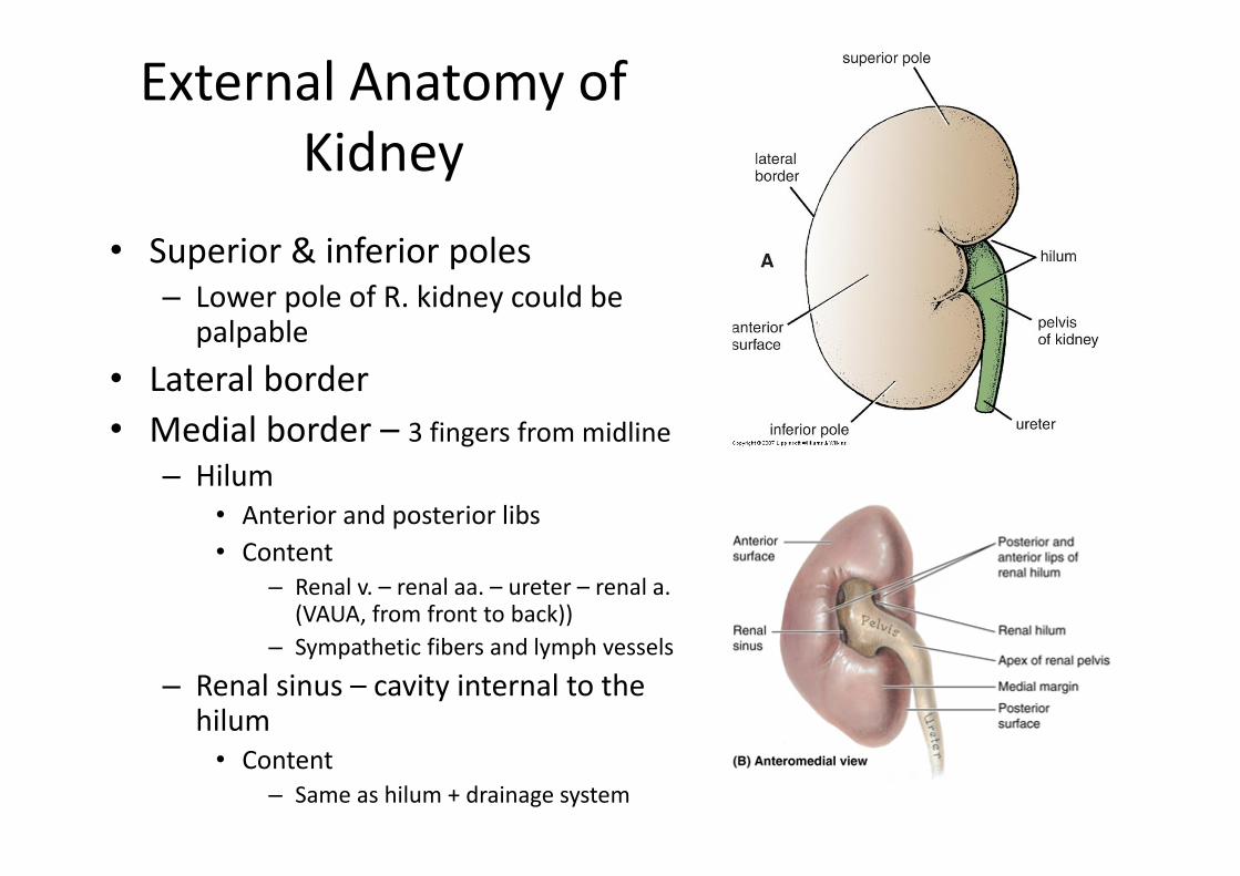
External Anatomy of Kidney
• Superior & inferior poles– Lower pole of R. kidney could be
palpable • Lateral border• Medial border – 3 fingers from midline
– Hilum• Anterior and posterior libs• Content
– Renal v. – renal aa. – ureter – renal a. (VAUA, from front to back))
– Sympathetic fibers and lymph vessels
– Renal sinus – cavity internal to the hilum
• Content– Same as hilum + drainage system

Kidney Coverings• Renal capsule (fibrous capsule) = transparent membrane maintains organ
shape• Perirenal fat = helps protect from trauma • Renal fascia = dense, irregular connective tissue that holds against back
body wall– Encloses the kidney & the suprarenal gland– Continuous with fascia transversalis
• Pararenal fat (paranephric fat) = protection– Part of retroperitoneal fat

Internal Anatomy of the Kidneys• Parenchyma of kidney
– renal cortex = superficial layer of kidney– renal medulla
• inner portion consisting of 8‐18 cone‐shaped renal pyramids separated by renal columns
• renal papilla point toward center of kidney– Apex of renal pyramid
• Drainage system fills renal sinus cavity– minor calyces ‐ cuplike structure
• collect urine from the papillary ducts of the papilla– One minor calyx for each renal papilla
– minor calyces empty into major calyces• Each major calyx empties 2‐3 minor calyces
– Major calyces empty into the renal pelvis, which empties into the ureter

Internal Anatomy of Kidney
• What is the difference between renal hilus & renal sinus?• Outline a major calyx & the border between cortex &
medulla.

Kidney‐ blood supply• Blood supply
– Renal aa. – aorta – L2– Renal vv. – IVC
• Lymph drainage– Lateral aortic lymph nodes
• Nerve supply– Sympathetic – renal plexus
– Afferents – T10‐T12 spinal segments

Kidney‐ relations• Anteriorly
– Viscera …
• Posteriorly– Ribs, muscles, nerves …
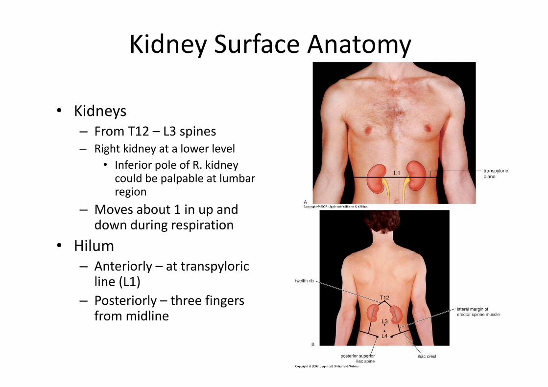
Kidney Surface Anatomy
• Kidneys– From T12 – L3 spines– Right kidney at a lower level
• Inferior pole of R. kidney could be palpable at lumbar region
– Moves about 1 in up and down during respiration
• Hilum – Anteriorly – at transpyloric
line (L1)– Posteriorly – three fingers
from midline

Ureters• 10 to 12 in long• Varies in diameter from 1‐10 mm• Extends from renal pelvis to
bladder• Retroperitoneal• Enters posterior wall of bladder• Three Constrictions (arrows)
– At junction with renal pelvis– At crossing the pelvic brim– At entering the urinary bladder
(oblique entrance)
• Physiological valve only– bladder wall compresses
ureteral opening as it expands during filling
– flow results from peristalsis, gravity & hydrostatic pressure

Ureters: Relations
• Anteriorly – Viscera, BVs, mesentery
• Posteriorly– Lumbar transverse processes, psoas m., bifurcation of common iliac a.

Ureters• Blood supply
– Upper end – renal vs.– Middle part – gonadal vs.– Lower end – superior vesical vs.
• Lymph drainage– Lateral aortic nodes & iliac nodes
• Nerve supply– Renal & gonadal plexuses in abdomen
– Hypogastric plexus in pelvis
– Afferents – L1‐L2 segments

Urinary Bladder
• Hollow, distensible muscular organ with capacity of about 500 ml
• In adults it is located in the pelvis behind the pubic symphysis– Upon distention, the
superior surface extend to the abdomen
– In infancy bladder have higher position
• Empty bladder lies within the abdomen
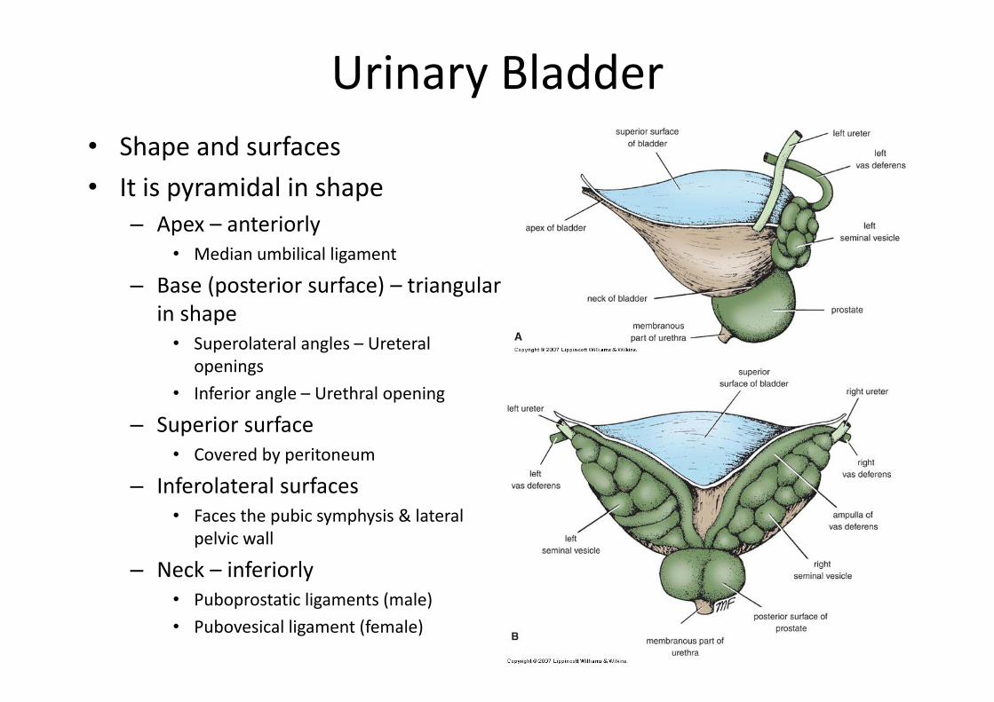
Urinary Bladder• Shape and surfaces • It is pyramidal in shape
– Apex – anteriorly • Median umbilical ligament
– Base (posterior surface) – triangular in shape
• Superolateral angles – Ureteral openings
• Inferior angle – Urethral opening
– Superior surface• Covered by peritoneum
– Inferolateral surfaces• Faces the pubic symphysis & lateral
pelvic wall
– Neck – inferiorly • Puboprostatic ligaments (male)• Pubovesical ligament (female)

Urinary Bladder: Internal Structure • Mucus membrane folds
– Disappear on distention
• Trigone is the mucus membrane of the bladder base– Always smooth flat area – Bordered by 2 ureteral openings (above) &
urethral opening (below)– Interureteric crest (superiorly)
• Uvula vesicae (in male) – Elevation behind the urethral opening– Caused by the median lobe of the prostate
• Detrusor muscle (bladder smooth m.)– Three layers
• Inner & outer longitudinal • Middle circular
– At neck – sphincter vesicae (internal urethral sphincter)

Urinary Bladder‐ Relations in Male
• Anteriorly – abdominal wall, retropubic pad of fat & pubic symphysis
• Laterally – obturator internus& levator anai mm.
• Inferiorly – prostate• Superiorly – peritoneal cavity
& parts of intestine • Posteriorly – rectovesical
pouch, vas deferens, seminal vesicles, rectovesical fascia & rectum

Urinary Bladder‐ Relations in Female
• Anteriorly – abdominal wall, retropubic pad of fat & pubic symphysis
• Laterally – obturatorinternus & levator anaimm.
• Inferiorly – urogenital diaphragm
• Superiorly – uterovesicalpouch & uterus
• Posteriorly – vagina

Urinary Bladder• Blood supply
– Superior and Inferior vesical aa. –internal iliac a.
– Vesical venous plexus – prostatic venous plexus – internal iliac v.
• Lymphatics– Internal & external iliac nodes

Urinary Bladder
• Nerve supply– Inferior hypogastric plexus
• Sympathetic: L1‐L2 ganglia (sympathetic trunk) –hypogastric plexus
– Contraction of sphincter vesicae
• Parasympathetic: S2‐S4 –pelvic splanchnic nn.
– Contraction of detrusor m.
– Afferent fibers• Parasympathetic (most) – S2‐S4 segments
• Sympathetic (some) – L1‐L2 segments
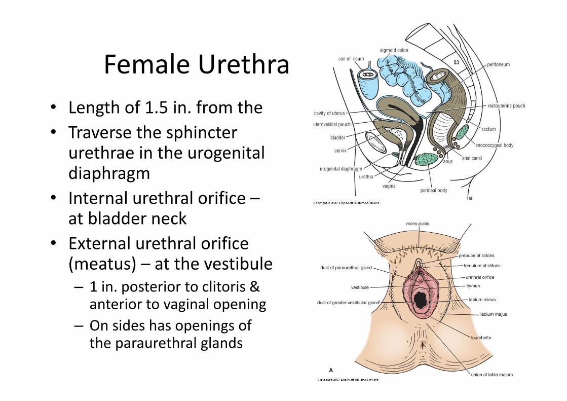
Female Urethra• Length of 1.5 in. from the • Traverse the sphincter urethrae in the urogenital diaphragm
• Internal urethral orifice –at bladder neck
• External urethral orifice (meatus) – at the vestibule– 1 in. posterior to clitoris & anterior to vaginal opening
– On sides has openings of the paraurethral glands

Male Urethra• Length 8 in. from bladder neck to
glans penis• Parts
– Prostatic urethra (1.25 in.) – widest part
• Urethral crest– Prostatic utricle
» On both sides has the openings of ejaculatory ducts
• Prostatic sinus– Membranous urethra (0.5 in. – in
urogenital diaphragm– Penile urethra (6 in.) – narrowest
part• Traverse the pulp & corpus spongiosum of the penis
• Receives the bulbourethral ducts –proximally
• Fossa terminalis – dilated distal part