Embed Size (px)
Citation preview
315
Forma, 14, 315–320, 1999Letter
Growth and Colony Patterning of Filamentous Fungi
Shu MATSUURA
School of High-Technology for Human Welfare, Tokai University,Numazu, Shizuoka 410-0395, Japan
E-mail: shum@wing. ncc. u-tokai.ac.jp
(Received November 19, 1999; Accepted December 20, 1999)
Keywords: Filamentous Fungi, Colony Patterning, Morphology Diagram
Abstract. A computer model of the fungal colony that grew consuming limited amountof nutrient with three control parameters, growth rate, and nutrient level and diffusion,was constructed to make a basic morphology diagram of mycelial colony. To apply thediagram with those parameters to the mycelial growth of Aspergillus nidulans wild andmutant strains, the relation between the colony expansion and the nutrient level wasmeasured. Differences in the colony patterns of these strains at low nutrient level wereattributed to the change in the above relation that caused a shift of the correspondingpatterns in the diagram.
1. Introduction
The shapes of fungal colonies exhibit striking diversity depending on the substrateconditions as well as on the fungal species. Although the shapes and the surface texturesof colonies provide useful information to determine the species or to monitor the state ofgrowth, colony patterning looks to be highly sensitive to the environmental factors.However, there might be underlying basic rules of pattern selection common for anyspecies.
Obviously, the nutrient level in the substrate is the main factor for the hyphalproduction. In addition, nutrient diffusion will be a significant factor, since it affects thenutrient flux into the colonized area and the distribution of the location in which uptake ofnutrient occurs intensely. As for the internal parameter of mycelium, the growth rate ofhyphae, or the rate of nutrient utilization, will largely contribute to determine where in thecolony the hyphal production occurs. In this letter, these three control parameters areconsidered independent with each other, in order to make general morphology diagram ofthe mycelial colonies by using a model.
This letter explores the colony morphology of wild and mutant strains of Aspergillusnidulans based on a morphology diagram that was made by a colony model. To apply themorphology diagram to the real colony patterns, the manner of colony expansion was
316 S. MATSUURA
examined for A. nidulans. It is suggested that the relationship between the nutrient level inthe substrate and the growth rate of the mycelium is important to relate the real patterns withthe morphology diagram.
2. Materials and Methods
The strains used in this study were A. nidulans wild type strain A4 (the index of strainin FGSC) and a mutant strain A583 supplied by the Fungal Genetic Stock Center (FGSC),Kansas City, USA. Spores of the strains were adsorbed to the silica particles for storage(TABOR and TABOR, 1970). The peptone agar media containing K2HPO4 (0.05%), KH2PO4
(0.05%), with Difco Neopeptone 0.01, 0.05, 0.1, 0.5, and 1%, and with Difco Bacto-agar0.5% were prepared, and 20 ml of sterile medium was poured into 9-cm Petri plates. Silicaparticles containing spores were directly put on the medium for inoculation, and the strainswere cultivated at 24°C. The plates were placed in closed vinyl bags to reduce drying of themedia. The colonies were photographed with a 35 mm camera with oblique illuminationfrom below.
3. Model
The colony patterns were developed on a 2-dimensional square lattice of 100 × 200lattice constant. Each lattice point is supplied with Ni nutrient particles initially forstorage. Each hyphal unit can occupy one lattice point. Seed hyphae are placed initiallyin a line of 100 lattice constants. A hyphal unit is chosen at random, and if the nutrientstorage remains in the lattice point that the hyphal unit occupies, it absorbs one nutrientparticle.
When a hyphal unit absorbs 3 nutrient particles, it creates one new hyphal unit ata randomly chosen unoccupied neighboring site and consumes the stored nutrient. Upto two daughter hyphal units are created by the parent one in the same mycelial planeto proceed dichotomous branching. When the neighboring sites in the same mycelialplane are already occupied, a higher or a lower plane is randomly chosen and the sameprocess is repeated for the new plane. In case that no vacant site remains for a hyphalunit to create its daughter hyphae, it accumulates up to 4 nutrient particles and ceasesabsorbing.
For the nutrient diffusion, any one of the lattice sites is chosen at random. Whenthe chosen site has at least one nutrient particle, a particle is released to walk at randomfor Rs steps. In the simulation, one sequence of random walk is regarded as the timeunit. Since the trajectory length Rs is proportional to the diffusion coefficient in oneunit time, let us regard the step length Rs as the diffusion coefficient of nutrientparticles.
The growth rate Gr is defined as the frequency of nutrient uptake for unit time.When the random walk of nutrient was repeated n times before one nutrient uptake, Gris equal to 1/n. In case that nutrient uptake occurs m times per one nutrient walk, Gr ism.
Growth and Colony Patterning of Filamentous Fungi 317
4. Results and Discussion
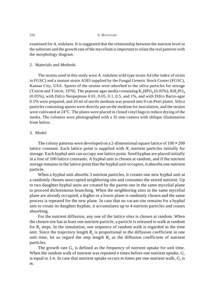
Examples of model colonies at a fixed Rs are shown in Fig. 1. Splitting andramified shapes appear with decreasing nutrient and growth rate. At high nutrient levelwith low growth rate, the colony forms thick layers due to the high nutrient influx.Under this condition, the growth is reaction-limited since the growth rate is the limitingfactor for colony expansion. At low nutrient level with high growth rate, the colony isenlarged with thin mycelial layers. The nutrient influx or the nutrient level is thelimiting factor. Since the nutrient particle diffuses into the colonized area duringrandom walk without being trapped by the hyphae, hyphae are created homogeneouslyinside the colonized area. Thus, a typical branched shape that is generally seen underdiffusion-limited growth does not appear.
Fig. 1. Morphology diagram of model colonies. Model colonies were grown up to 10000 hyphal units with thefixed nutrient diffusion Rs of 50 steps. Initial nutrient level indicates the number of nutrient particles initiallystored in each lattice site. Light color indicates higher mycelial layers.
318 S. MATSUURA
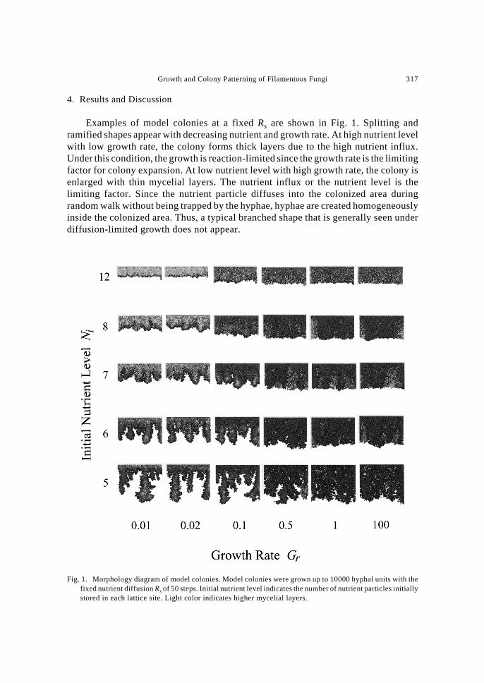
Figure 2 shows the photographs of A. nidulans wild type strain colonies cultivatedfor 10 days and the mutant strain colonies cultivated for 40 days. Although the wildtype strain forms circular colonies under all conditions tested, the size of the colony isslightly larger at lower nutrient levels. There appeared roughening in the colonyinterface at relatively high nutrient levels. At high nutrient level, hyphae wereproduced densely inside the colony. However, the colonies ceased to grow with largearea of the medium space left unoccupied. This is thought to be due to the accumulationof inhibitory metabolite inside the colonized area. In the previous study on Aspergillusoryzae (MATSUURA, 1998), the germination rate at the neighboring sites of the growingcolony was found reduced with time. This showed the decaying condition of themedium around the colony. Oppositely, colonies at low nutrient levels expand to coveralmost entire medium surface with far less hyphal density.
The growth of mutant strain was remarkably lowered at low nutrient levels, and
Fig. 2. Colonies of Aspergillus nidulans wild and mutant strains. Left row shows the colonies of a wild typestrain at various nutrient levels cultivated for 10 days, and right row shows those of a mutant strain cultivatedfor 40 days. The bar indicates 2 cm.
Growth and Colony Patterning of Filamentous Fungi 319
the colonies exhibited roughened or ramified morphologies at these conditions. Withincreasing nutrient or agar content, colony shapes became similar to the compactmorphology of the wild type strain.
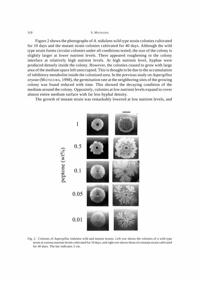
To relate the growth rate of colonies with the nutrient levels, changes of colonysizes estimated from the photograph of colonies were plotted against cultivation daysin Figs. 3a and 3b. The colony expansion of the wild strain was found lower as the initialnutrient level was raised. The colonies at high nutrient levels almost stopped expansionwithin 30 days, while those at low nutrient levels expanded to cover the entire mediumsurface.
The growth of mutant strain was quite similar to the wild strain at high nutrientlevels. However, at low nutrient, the hyphal growth was found suppressed for the first20 days. After this period, the colony began to expand forming complex shapes.
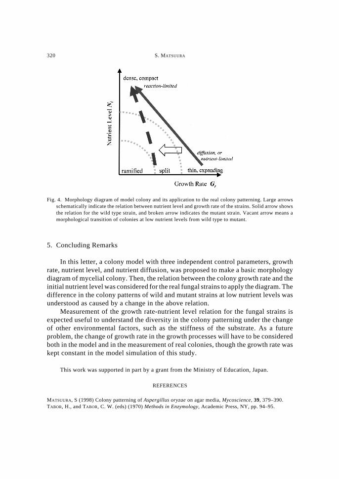
Let us now apply the model morphology diagram to the real colony patterns. InFig. 4, the growth rate-nutrient level relations for wild and mutant strains wereschematically drown on the morphology diagram of model colony. For the wild strain,the growth is diffusion-limited or nutrient level-limited at low nutrient level since thegrowth rate is maintained higher. As the nutrient level is raised, the growth rate islowered probably due to the accumulation of inhibitory metabolites, and then thecolony morphology approaches the roughening area in the diagram. Then, further raiseof nutrient level brings about reaction-limited colony formation with lowered growth.
For the mutant strain, the growth rate-nutrient level relation is changed into lowergrowth at low nutrient levels as compared with the wild type strain. Thus, the colonymorphology moves toward the ramified region at low nutrient levels as indicated in thefigure.
Fig. 3. Colony expansion of Aspergillus nidulans wild and mutant strains. Change of colony diameters wereplotted against days since inoculation for a, the wild type strain; and b, the mutant strain. Colony diameterswere calculated by approximating the colonized area as a circle.
320 S. MATSUURA
5. Concluding Remarks
In this letter, a colony model with three independent control parameters, growthrate, nutrient level, and nutrient diffusion, was proposed to make a basic morphologydiagram of mycelial colony. Then, the relation between the colony growth rate and theinitial nutrient level was considered for the real fungal strains to apply the diagram. Thedifference in the colony patterns of wild and mutant strains at low nutrient levels wasunderstood as caused by a change in the above relation.
Measurement of the growth rate-nutrient level relation for the fungal strains isexpected useful to understand the diversity in the colony patterning under the changeof other environmental factors, such as the stiffness of the substrate. As a futureproblem, the change of growth rate in the growth processes will have to be consideredboth in the model and in the measurement of real colonies, though the growth rate waskept constant in the model simulation of this study.
This work was supported in part by a grant from the Ministry of Education, Japan.
REFERENCES
MATSUURA, S (1998) Colony patterning of Aspergillus oryzae on agar media, Mycoscience, 39, 379–390.TABOR, H., and TABOR, C. W. (eds) (1970) Methods in Enzymology, Academic Press, NY, pp. 94–95.
Fig. 4. Morphology diagram of model colony and its application to the real colony patterning. Large arrowsschematically indicate the relation between nutrient level and growth rate of the strains. Solid arrow showsthe relation for the wild type strain, and broken arrow indicates the mutant strain. Vacant arrow means amorphological transition of colonies at low nutrient levels from wild type to mutant.