Embed Size (px)
Citation preview
/ . Embryol. exp. Morph. Vol. 65 {Supplement), pp. 225-241, 1981 2 2 5 Printed in Great Britain © Company of Biologists Limited 1981
Growth and development of pattern in the cranial neural epithelium of rat embryos
during neurulation
By G I L L I A N M. MORRISS-KAY 1
From the Department of Human Anatomy, Oxford
SUMMARY The pattern of growth and morphogenesis of the cranial neural epithelium of rat embryos
during neurulation is described. Transverse sections of the midbrain/hindbrain neural epithelium at different stages (0-14 somites) show a constant area and cell number throughout neurulation, even though there is a high level of mitosis. Mitotic spindles are orientated parallel to the long axis of the embryo, so that increase in cell number occurs in this direction only. Growth is expressed only as an increase in size of the forebrain, which projects rostrad to the tip of the notochord.
In the midbrain/upper hindbrain regions, cellular organization of the neural epithelium changes from columnar to cuboidal to pseudostratified, while its shape changes from flat to biconvex to V shaped. Closure is immediately preceded by neural crest cell emigration from the lateral edges. Throughout neurulation the cranial notochord develops an increasingly convex curvature in the rostrocaudal plane. The attached neural epithelium curves with the notochord (forming the primary cranial flexure) so that as its lateral edges move dorso-medially they form a more distant concentric arc with that of the notochord, and are hence stretched during the final closure period.
The whole rat embryo culture technique was used to investigate the morphogenetic role of proteoglycans during neurulation, neural crest cell emigration and other events in the lateral edge region prior to closure, and the importance of microfilament contraction during concave curvature of the neural epithelium.
I. INTRODUCTION
The process of neurulation has been most thoroughly studied in amphibian embryos, and in the absence of much direct information on neurulation in mammalian embryos the information gained from amphibian studies is frequently assumed to be relevant here also. Studies on neural induction have not yet been carried out in mammalian embryos, but studies on morphogenesis of the neural epithelium from neural plate to closed neural tube are beginning to reveal that differences between the brains of species belonging to different vertebrate classes are evident from the onset of neurulation.
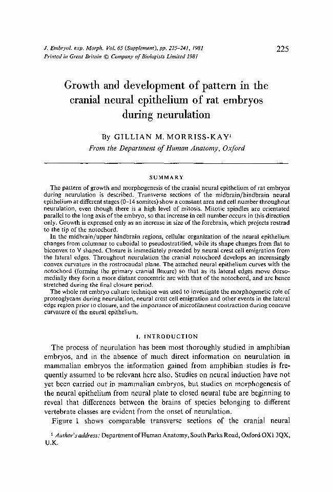
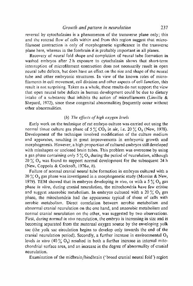
Figure 1 shows comparable transverse sections of the cranial neural 1 Author's address: Department of Human Anatomy, South Parks Road, Oxford 0X1 3QX,
U.K.
226 G. M. MORRISS-KAY
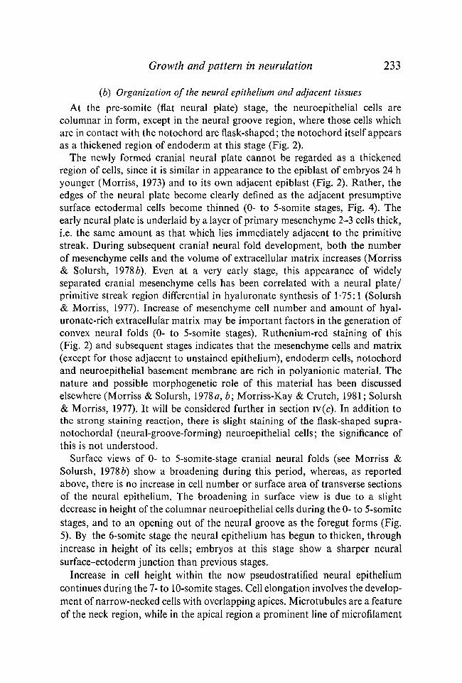
Fig. 1. Transverse sections of embryos at the time of cranial neural fold apposition. (a) Xenopus (stage 16), (b) chick (7-somite stage), (c) rat (13-somite stage). In the rat, the neural tube is sectioned at both forebrain (lower) and upper-hindbrain levels. a, 1st aortic arch; g, archenteron, foregut and buccal region in (a)-(c) respectively; n, notochord; nc, neural crest.
epithelium of Xenopus, chick and rat embryos at or just prior to the time of apposition of the neural folds. Many differences can be seen, including: neuroepithelial cell size, appearance, numbers and organization; notochordal size and form; timing of emigration of neural crest cells; overall shape, including the existence of a cranial flexure at this stage in the mammal only. These differences reflect not only the great differences of adult structure and function, but also the lack of close phylogenetic affinities. Modern amphibians are very distant relatives of the line which evolved into the first amniotes (cotylosaurs or stem reptiles), and the pre-mammalian and pre-avian reptile lines diverged very early in the radiation of reptilian types.
This article will examine in some detail the cranial neural epithelium and associated structures of the rat embryo during neurulation. Its aim is to establish a proper descriptive basis (a) for comparison with other vertebrate (including other mammalian) embryos and (b) for experimental studies aimed at elucidating the mechanisms underlying normal and abnormal morphogenesis during development of the mammalian brain tube.
Growth and pattern in neurulation 227
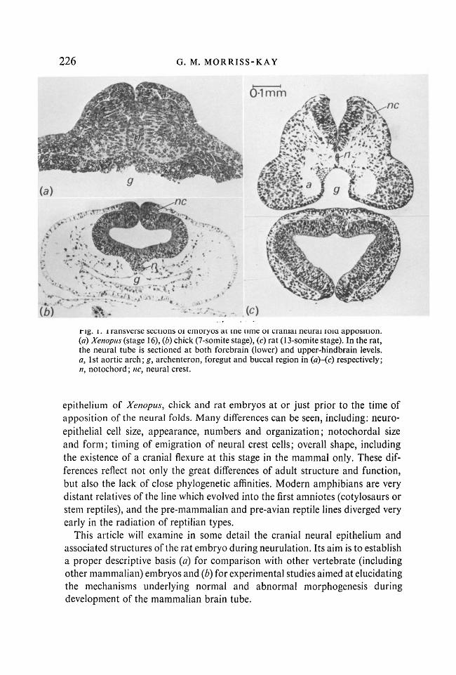
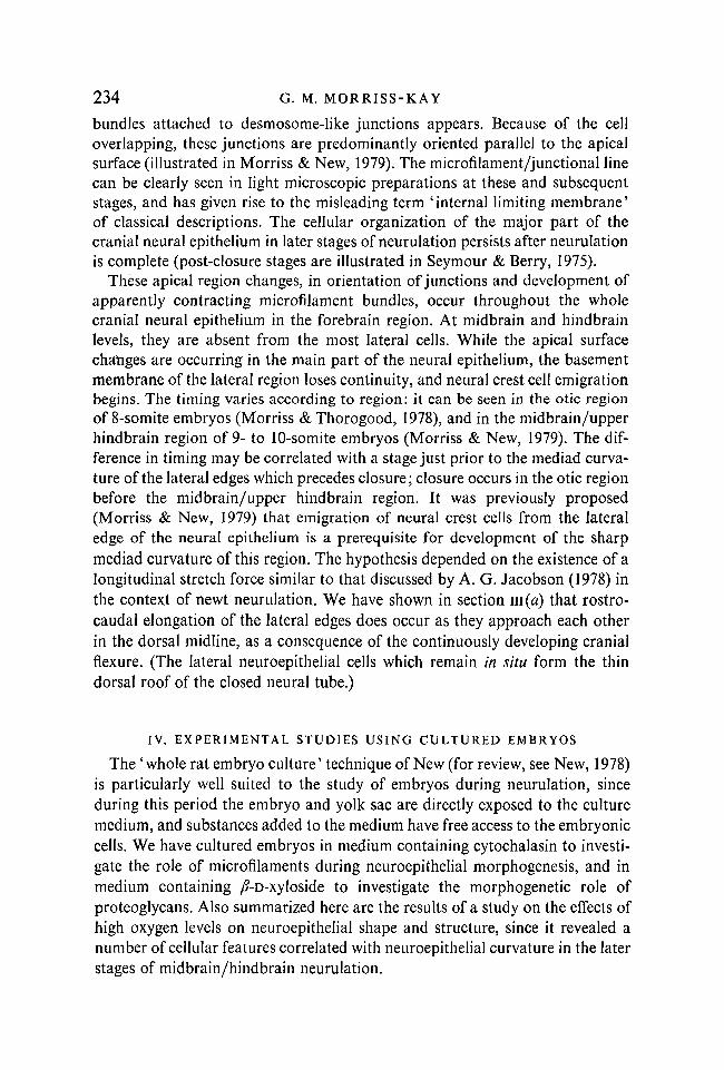
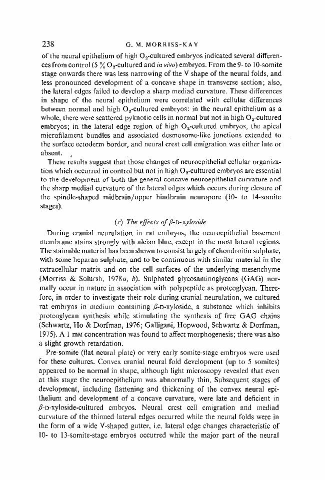
Fig. 2. Pre-somite-stage rat embryos, with flat neural plate, (a) One /im-thick section of embryo embedded in Spurr resin following fixation with glutaraldehyde (2-5%) containing cetyl pyridinium chloride (100mg/ml) and postfixation with Os04 containing ruthenium red (60 mg/ml), (b) SEM of embryo partially opened out by cutting the lateral extra-embryonic regions, ep, epiblast; h, hypoblast; m, primary mesenchyme; n, notochord ; np, neural plate; ng, neural groove;/?, primitive streak; s, presumptive surface ectoderm.
II. GROWTH (a) Growth of the whole embryo
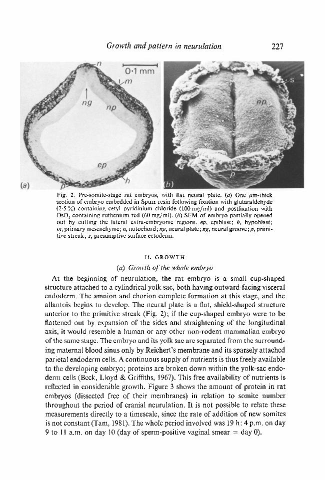
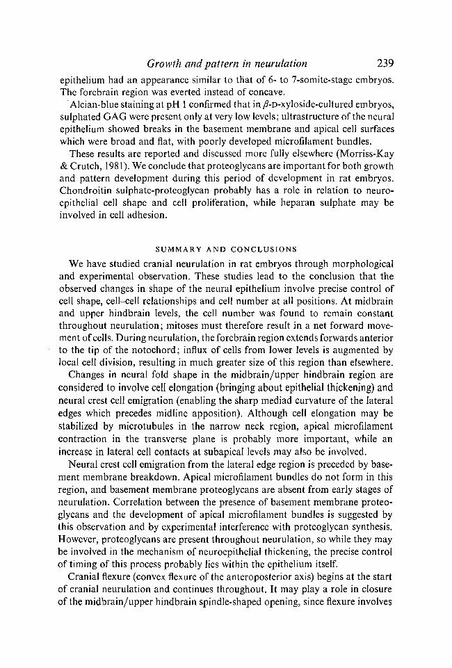
At the beginning of neurulation, the rat embryo is a small cup-shaped structure attached to a cylindrical yolk sac, both having outward-facing visceral endoderm. The amnion and chorion complete formation at this stage, and the allantois begins to develop. The neural plate is a flat, shield-shaped structure anterior to the primitive streak (Fig. 2); if the cup-shaped embryo were to be flattened out by expansion of the sides and straightening of the longitudinal axis, it would resemble a human or any other non-rodent mammalian embryo of the same stage. The embryo and its yolk sac are separated from the surrounding maternal blood sinus only by Reichert's membrane and its sparsely attached parietal endoderm cells. A continuous supply of nutrients is thus freely available to the developing embryo; proteins are broken down within the yolk-sac endoderm cells (Beck, Lloyd & Griffiths, 1967). This free availability of nutrients is reflected in considerable growth. Figure 3 shows the amount of protein in rat embryos (dissected free of their membranes) in relation to somite number throughout the period of cranial neurulation. It is not possible to relate these measurements directly to a timescale, since the rate of addition of new somites is not constant (Tarn, 1981). The whole period involved was 19 h: 4 p.m. on day 9 to 11 a.m. on day 10 (day of sperm-positive vaginal smear = day 0).
228 G. M. MORRISS-KAY
60
50
40
S 30 o
CL!
20
10 - , l l < II
II II
I ■ (16)(15)(6) (9)(11)(11)(8) (7) (5) (9) (4) (5) (5) (5) (3) (1)
—J 1 1 1 1 1 1 1 I I I I L _ J i l 0 1 2 3 4 5 6 7 8 9 10 11 12 13 14 15
Somite stage
Fig. 3. Protein content, ± 1 standard deviation, of rat embryos during the period of cranial neurulation. Numbers in parentheses above the somite stage indicate the number of embryos measured. For early stages some samples contained two or three embryos : no. of samples, 0 to 7-somite stages = 3, 3,2,4, 6,9,7 and 6 respectively. Technique according to Lowry, Rosebrough, Farr & Randall (1951), using bovine serum albumen standards.
A very small amount of this increase in embryonic protein content is presumably due to incorporation of part of the yolk sac into the foregut and hind-gut, which form during this period. The great majority of it, however, is real growth. There is a continuous lengthening of the body axis, and an increase in size of both head and heart.
(b) Growth of the neural epithelium There is as yet no exact calculation on the amount of growth which occurs in
the neural epithelium of amniote embryos during neurulation. However, in both the chick (Derrick, 1937) and the rat, there is a high level of mitosis in the neural epithelium, suggesting that growth of this tissue during neurulation is the rule in embryos which have a continuous supply of nutrients during this period. This is in contrast to amphibian embryos which have no extrinsic food source until hatching (stage 40 in the newt). During neurulation in Ambystoma, the cell number increases very little, from 113000 to 139000, from stages 13 to 19 (Gillette, 1944). In the newt Taricha torosa, the volume of the central nervous system does not increase from stage 13 to stage 33, i.e. during and for a considerable time after neurulation (A. G. Jacobson, 1978).
Projection drawings of transverse sections of the rat cranial neural epithelium
Growth and pattern in neurulation 229
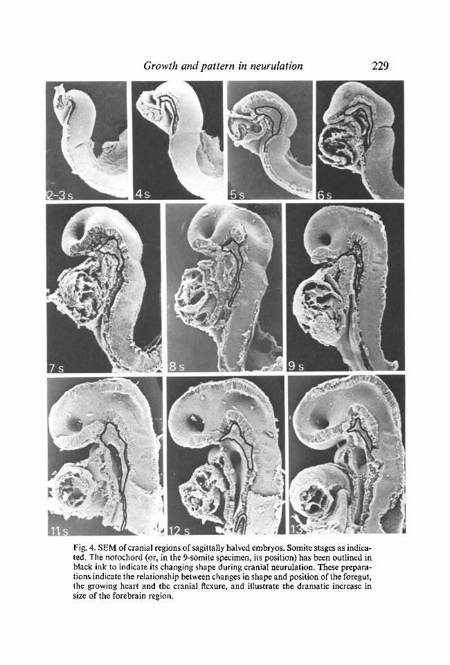
Fig. 4. SEM of cranial regions of sagittally halved embryos. Somite stages as indicated. The notochord (or, in the 9-somite specimen, its position) has been outlined in black ink to indicate its changing shape during cranial neurulation. These preparations indicate the relationship between changes in shape and position of the foregut, the growing heart and the cranial flexure, and illustrate the dramatic increase in size of the forebrain region.
230 G. M. M O R R I S S - K A Y
at various stages (pre-somite to 14 somites) reveal that except in the developing forebrain region, the cross-sectional area remains constant throughout this period. Cell counts from scanning electron micrographs of transversely cut neural folds at midbrain or upper hindbrain levels show that the cell number in each half of the cut neuroepithelial surface is 65-70 throughout neurulation. Counts from comparable sections of plastic-embedded embryos are similar. The length of the supranotochordal neural epithelium from the preotic sulcus to the tip of the notochord also remains constant throughout cranial neurulation. This was measured on the specimens illustrated in Fig. 4, using a piece of resin-cored solder. (The measurement was 220 /*m in all specimens, but due to shrinkage during preparation the actual length is likely to be slightly greater than this.)
This observed constancy of cross-sectional surface area, cross-sectional cell number, and supranotochordal neuroepithelial length is a surprising finding in view of the high level of mitosis observed. Examination of the spindle orientation in embryos cultured in medium containing colcemid (0-1 /*g/ml) and in untreated embryos revealed that in the midbrain/upper hindbrain region, the orientation was almost always parallel to the long axis of the embryo, so that each mitosis contributed only to longitudinal growth ; conversely, in the forebrain region the spindle orientation was predominantly in the transverse plane, allowing the great expansion which occurs in this region during neurulation (F. Tuckett, unpublished observations). Growth of the cranial neural epithelium therefore appears to be confined to the forebrain region, even though cell division occurs in all regions.
III. DEVELOPMENT OF PATTERN IN THE NEURAL EPITHELIUM
(a) Shape The overall changes in shape seen from surface views of the cranial neural
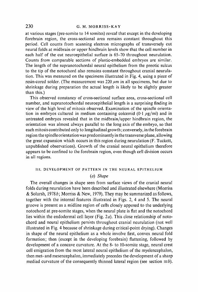
folds during neurulation have been described and illustrated elsewhere (Morriss & Solursh, 1978b\ Morriss & New, 1979). They may be summarized as follows, together with the internal features illustrated in Figs. 2, 4 and 5. The neural groove is present as a midline region of cells closely apposed to the underlying notochord at pre-somite stages, when the neural plate is flat and the notochord lies within the endodermal cell layer (Fig. 2d). This close relationship of notochord and neural epithelium persists throughout cranial neurulation (not well illustrated in Fig. 4 because of shrinkage during critical-point drying). Changes in shape of the neural epithelium as a whole involve first, convex neural fold formation; then (except in the developing forebrain) flattening, followed by development of a concave curvature. At the 8- to 10-somite stage, neural crest cell emigration from the most lateral neural epithelium of the myelencephalon, then met- and mesencephalon, immediately precedes the development of a sharp mediad curvature of the consequently thinned lateral region (see section mb).
Growth and pattern in neurulation 231
Fig. 5. SEM of embryos cut transversely across the midbrain/upper hindbrain region. Somite stages as indicated. (Four of these specimens have been illustrated in previous publications: Morriss & Solursh, 1978a, b; Morriss & New, 1979.)
232 G. M. MORRISS-KAY
Neural fold fusion begins in the upper cervical region at 7 somites, extending upwards into the hindbrain at 8 and 9 somites. At the early 10-somite stage a separate region of fusion begins, at the midbrain/forebrain junction, immediately above the developing cranial flexure. Final closure of the spindle-shaped opening between these two fusion areas is complete by 14 somites, and the apposed forebrain region (anterior neuropore) fuses soon afterwards. (This separate closure of two cranial neuropores also occurs in human embryos: see 10-somite embryo illustration, Hamilton, Boyd & Mossman (1972), p. 179. It is more pronounced in rodents, perhaps because of the more precocious development of the primary cranial flexure.)
Embryos bisected sagittally (Fig. 4) show very clearly that while the supra-notochordal neural epithelium of the midbrain/upper hindbrain region does not change in length during the 2- to 13-somite period, there is an enormous forwards extension of the anterior forebrain area, which increases greatly in size. The relative shapes and sizes of the forebrain and midbrain/hindbrain regions towards the end of cranial neurulation can be appreciated by reference to Fig. 1 (c).
In summary, the observations on changes in shape and size of the forebrain and midbrain/upper hindbrain regions, together with the observations on spindle orientation during mitosis, imply that there is a net forward movement of cells towards and into the developing forebrain. Mitoses in the midbrain/ hindbrain region contribute to this supply of forward-moving cells, while mitoses within the forebrain region contribute an intrinsic component to its growth.
A. G. Jacobson (1978, 1980) has assessed the degree to which the neural epithelium and notochord lengthen during neurulation in newt and chick embryos, using computer modelling. He considered that in newt embryos elongation provides a 'stretch force' which, together with microfilament-induced shrinkage of the neural plate, is responsible for neurulation. This theory is also applicable to his observations on stage-9 (9-somite) chick embryos, where elongation during a 4 h period was found to be 3-4 % in the closed region, 7 % in the open region, and 29 % in the closing region of the neural tube. Measurement of the length of the supranotochordal cranial neural epithelium on scanning electron micrographs of sagittally halved rat embryos (Fig. 4) indicates that there is no elongation of the cranial notochord and attached neural epithelium of the midbrain/hindbrain regions during neurulation (section ub). However, because of the continuous development of the cranial flexure while the lateral edges of the neural epithelium are moving dorsomedially, the lateral edges come to form an arc more distant from, but concentric with, the curved notochord. The resultant elongation of the dorsal surface of the midbrain/hindbrain region may be an important contributory factor in generating the dorsomediad movement of the lateral edges of the neural folds here in later stages of neurulation.
Growth and pattern in neurulation 233
(b) Organization of the neural epithelium and adjacent tissues At the pre-somite (flat neural plate) stage, the neuroepithelial cells are
columnar in form, except in the neural groove region, where those cells which are in contact with the notochord are flask-shaped ; the notochord itself appears as a thickened region of endoderm at this stage (Fig. 2).
The newly formed cranial neural plate cannot be regarded as a thickened region of cells, since it is similar in appearance to the epiblast of embryos 24 h younger (Morriss, 1973) and to its own adjacent epiblast (Fig. 2). Rather, the edges of the neural plate become clearly defined as the adjacent presumptive surface ectodermal cells become thinned (0- to 5-somite stages, Fig. 4). The early neural plate is underlaid by a layer of primary mesenchyme 2-3 cells thick, i.e. the same amount as that which lies immediately adjacent to the primitive streak. During subsequent cranial neural fold development, both the number of mesenchyme cells and the volume of extracellular matrix increases (Morriss & Solursh, 19786). Even at a very early stage, this appearance of widely separated cranial mesenchyme cells has been correlated with a neural plate/ primitive streak region differential in hyaluronate synthesis of 1-75:1 (Solursh & Morriss, 1977). Increase of mesenchyme cell number and amount of hyal-uronate-rich extracellular matrix may be important factors in the generation of convex neural folds (0- to 5-somite stages). Ruthenium-red staining of this (Fig. 2) and subsequent stages indicates that the mesenchyme cells and matrix (except for those adjacent to unstained epithelium), endoderm cells, notochord and neuroepithelial basement membrane are rich in polyanionic material. The nature and possible morphogenetic role of this material has been discussed elsewhere (Morriss & Solursh, 1978Ö, b; Morriss-Kay & Crutch, 1981 ; Solursh & Morriss, 1977). It will be considered further in section iv(c). In addition to the strong staining reaction, there is slight staining of the flask-shaped supra-notochordal (neural-groove-forming) neuroepithelial cells; the significance of this is not understood.
Surface views of 0- to 5-somite-stage cranial neural folds (see Morriss & Solursh, 19786) show a broadening during this period, whereas, as reported above, there is no increase in cell number or surface area of transverse sections of the neural epithelium. The broadening in surface view is due to a slight decrease in height of the columnar neuroepithelial cells during the 0- to 5-somite stages, and to an opening out of the neural groove as the foregut forms (Fig. 5). By the 6-somite stage the neural epithelium has begun to thicken, through increase in height of its cells; embryos at this stage show a sharper neural surface-ectoderm junction than previous stages.
Increase in cell height within the now pseudostratified neural epithelium continues during the 7- to 10-somite stages. Cell elongation involves the development of narrow-necked cells with overlapping apices. Microtubules are a feature of the neck region, while in the apical region a prominent line of microfilament
234 G. M. M O R R I S S - K A Y
bundles attached to desmosome-like junctions appears. Because of the cell overlapping, these junctions are predominantly oriented parallel to the apical surface (illustrated in Morriss & New, 1979). The microfilament/junctional line can be clearly seen in light microscopic preparations at these and subsequent stages, and has given rise to the misleading term 'internal limiting membrane' of classical descriptions. The cellular organization of the major part of the cranial neural epithelium in later stages of neurulation persists after neurulation is complete (post-closure stages are illustrated in Seymour & Berry, 1975).
These apical region changes, in orientation of junctions and development of apparently contracting microfilament bundles, occur throughout the whole cranial neural epithelium in the forebrain region. At midbrain and hindbrain levels, they are absent from the most lateral cells. While the apical surface chatiges are occurring in the main part of the neural epithelium, the basement membrane of the lateral region loses continuity, and neural crest cell emigration begins. The timing varies according to region: it can be seen in the otic region of 8-somite embryos (Morriss & Thorogood, 1978), and in the midbrain/upper hindbrain region of 9- to 10-somite embryos (Morriss & New, 1979). The difference in timing may be correlated with a stage just prior to the mediad curvature of the lateral edges which precedes closure; closure occurs in the otic region before the midbrain/upper hindbrain region. It was previously proposed (Morriss & New, 1979) that emigration of neural crest cells from the lateral edge of the neural epithelium is a prerequisite for development of the sharp mediad curvature of this region. The hypothesis depended on the existence of a longitudinal stretch force similar to that discussed by A. G. Jacobson (1978) in the context of newt neurulation. We have shown in section III(Ö) that rostro-caudal elongation of the lateral edges does occur as they approach each other in the dorsal midline, as a consequence of the continuously developing cranial flexure. (The lateral neuroepithelial cells which remain in situ form the thin dorsal roof of the closed neural tube.)
IV. EXPERIMENTAL STUDIES USING CULTURED EMBRYOS
The 'whole rat embryo culture' technique of New (for review, see New, 1978) is particularly well suited to the study of embryos during neurulation, since during this period the embryo and yolk sac are directly exposed to the culture medium, and substances added to the medium have free access to the embryonic cells. We have cultured embryos in medium containing cytochalasin to investigate the role of microfilaments during neuroepithelial morphogenesis, and in medium containing /?-D-xyloside to investigate the morphogenetic role of proteoglycans. Also summarized here are the results of a study on the effects of high oxygen levels on neuroepithelial shape and structure, since it revealed a number of cellular features correlated with neuroepithelial curvature in the later stages of midbrain/hindbrain neurulation.
Growth and pattern in neurulation 235 Experimental details not provided here may be found in Morriss & New
(1979) and Morriss-Kay & Crutch (1981).
(a) The effect of cytochalasins M.Jacobson (1978) writes: 'It is now safe to conclude that changes in
shape of neural epithelial cells during neurulation are due to intracellular microtubules which produce cell elongation and to intracellular microfilaments which produce apical constriction of the neuroectodermal cells.' Since the observations of Baker & Schroeder (1967) on the shape and position of apical microfilament bundles in the neuroepithelium of Xenopus embryos during neurulation, it has become accepted that microfilament contraction is directly responsible for generating neuroepithelial curvature. However, A. G. Jacobson (1978) found that isolated newt neural plates will undergo shrinkage in culture, as microfilament contraction occurs, but in the absence of underlying notochord they do not become curved to form neural tubes however long they are cultured. His observations suggest that in the newt, microfilament contraction brings about narrowing of the necks of the elongating neural plate cells, but does not itself bring about curvature of the neural epithelium.
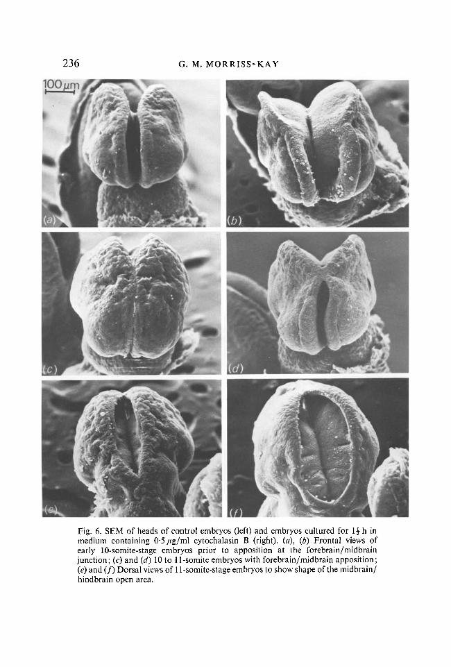
In order to investigate the role of microfilament contraction in mammalian neurulation, we cultured rat embryos in medium containing 0-5 /*g/ml cyto-chalasin B. (This concentration was found by transmission electron microscopy (TEM) to disrupt microfilament bundles while having a minimal effect on other aspects of cell structure.) Embryos of 9 to 11 somites were selected for these experiments, and were separated into two groups - those with and those without midbrain apposition (Figs 6a and c). At this stage, the whole cranial neural epithelium is developing an increasingly concave curvature in transverse section (see section ma); at the ultrastructural level microfilament bundles, associated with desmosome-like junctions, form a prominent line close to and parallel with the apical surface.
Changes in neural fold shape were evident within \ h of culture; the appearance after \\ h is illustrated by scanning electron micrographs in Fig. 6. In all embryos the neural folds splayed outwards, but the results were more pronounced in those which had not achieved midbrain apposition at the start of culture.
Similar results have been achieved using cytochalasin D (0-15 and 0-2 /tg/ml), and in these experiments embryos were washed and transferred to fresh medium after a 2 h exposure period. All of these embryos recovered neural fold shape and continued neurulation to form a closed brain tube, although it was of an abnormal shape, and the whole embryo was reduced in size compared with co-cultured control embryos (results to be reported in detail elsewhere).
These results suggest that microfilament contraction is essential for developing and maintaining the concave curvature of the neural epithelium. However, in the midbrain/hindbrain region, development of concave curvature and its
236 G. M. MORRISS-KAY
100 jum
Fig. 6. SEM of heads of control embryos (left) and embryos cultured for \\ h in medium containing 0-5/tg/ml cytochalasin B (right), (a), (b) Frontal views of early 10-somite-stage embryos prior to apposition at the forebrain/midbrain junction; (c) and (d) 10 to 11-somite embryos with forebrain/midbrain apposition; (e) and (ƒ) Dorsal views of 11-somite-stage embryos to show shape of the midbrain/ hindbrain open area.
Growth and pattern in neurulation 237 reversal by cytochalasins is a phenomenon of the transverse plane only; this and the rostrad flow of cells within and from this region suggest that microfilament contraction is only of morphogenetic significance in the transverse plane here, whereas in the forebrain it is probably important in all planes.
Recovery of neural fold shape and completion of neural tube formation in washed embryos after 2 h exposure to cytochalasin shows that short-term interruption of microfilament contraction does not necessarily result in open neural tube defects, but does have an effect on the size and shape of the neural tube and other embryonic structures. In view of the known roles of microfilaments in cell movement, cell division and other aspects of cell function, this result is not surprising. Taken as a whole, these results do not support the view that open neural tube defects in human development could be due to dietary intake of a substance that inhibits the action of microfilaments (Linville & Shepard, 1972), since these congenital abnormalitiesjrequently occur without other abnormalities.
(b) The effects of high oxygen levels Early work on the technique of rat embryo culture was carried out using the
normal tissue culture gas phase of 5 % C0 2 in air, i.e. 20 % 0 2 (New, 1978). Development of the technique involved modification of the culture medium and apparatus, resulting in great improvements in embryonic growth and morphogenesis. However, a high proportion of cultured embryos still developed with misshapen or unclosed brain tubes. This problem was overcome by using a gas phase containing only 5 % Oa during the period of neurulation, although 20 % O2 was found to support normal development for the subsequent 24 h (New, Coppola & Cockroft, 1976Ö, b).
Failure of normal cranial neural tube formation in embryos cultured with a 20 % 0 2 gas phase was investigated in a morphogenetic study (Morriss & New, 1979). TEM showed that in embryos developing in vivo, or with a 5 % 0 2 gas phase in vitro, during cranial neurulation, the mitochondria have few cristae and suggest anaerobic metabolism. In embryos cultured with a 20 % 0 2 gas phase, the mitochondria had the appearance typical of those of cells with aerobic metabolism. Direct correlation between aerobic metabolism and abnormal cranial neurulation on the one hand, and anaerobic metabolism and normal cranial neurulation on the other, was suggested by two observations. First, during normal in vivo neurulation, the embryo is increasing in size and is becoming separated from the maternal oxygen source by the enveloping yolk sac (the yolk sac circulation begins to develop only towards the end of the cranial neurulation period). Secondly, a further increase in environmental 0 2
levels in vitro (40 % 02) resulted in both a further increase in internal mitochondrial surface area, and an increase in the degree of abnormality of cranial neurulation.
Examination of the midbrain/hindbrain ('broad cranial neural fold') region
238 G. M. MORRISS-KAY
of the neural epithelium of high 02-cultured embryos indicated several differences from control (5 % 02-cultured and in vivo) embryos. From the 9- to 10-somite stage onwards there was less narrowing of the V shape of the neural folds, and less pronounced development of a concave shape in transverse section; also, the lateral edges failed to develop a sharp mediad curvature. These differences in shape of the neural epithelium were correlated with cellular differences between normal and high 02-cultured embryos: in the neural epithelium as a whole, there were scattered pyknotic cells in normal but not in high 02-cultured embryos; in the lateral edge region of high 02-cultured embryos, the apical microfilament bundles and associated desmosome-like junctions extended to the surface ectoderm border, and neural crest cell emigration was either late or absent. ...
These results suggest that those changes of neuroepithelial cellular organization which occurred in control but not in high 02-cultured embryos are essential to the development of both the general concave neuroepithelial curvature and the sharp mediad curvature of the lateral edges which occurs during closure of the spindle-shaped midbrain/upper hindbrain neuropore (10- to 14-somite stages).
(c) The effects of ß-D-xyloside During cranial neurulation in rat embryos, the neuroepithelial basement
membrane stains strongly with alcian blue, except in the most lateral regions. The stainable material has been shown to consist largely of chondroitin sulphate, with some heparan sulphate, and to be continuous with similar material in the extracellular matrix and on the cell surfaces of the underlying mesenchyme (Morriss & Solursh, 1978a, b). Sulphated glycosaminoglycans (GAG) normally occur in nature in association with polypeptide as proteoglycan. Therefore, in order to investigate their role during cranial neurulation, we cultured rat embryos in medium containing /?-D-xyloside, a substance which inhibits proteoglycan synthesis while stimulating the synthesis of free GAG chains (Schwartz, Ho & Dorfman, 1976; Galligani, Hopwood, Schwartz & Dorfman, 1975). A 1 mM concentration was found to affect morphogenesis; there was also a slight growth retardation.
Pre-somite (flat neural plate) or very early somite-stage embryos were used for these cultures. Convex cranial neural fold development (up to 5 somites) appeared to be normal in shape, although light microscopy revealed that even at this stage the neuroepithehum was abnormally thin. Subsequent stages of development, including flattening and thickening of the convex neural epithelium and development of a concave curvature, were late and déficient in /?-D-xyloside-cultured embryos. Neural crest cell emigration and mediad curvature of the thinned lateral edges occurred while the neural folds were in the form of a wide V-shaped gutter, i.e. lateral edge changes characteristic of 10- to 13-somite-stage embryos occurred while the major part of the neural
Growth and pattern in neurulation 239 epithelium had an appearance similar to that of 6- to 7-somite-stage embryos. The forebrain region was everted instead of concave.
Alcian-blue staining at pH 1 confirmed that in/?-D-xyloside-cultured embryos, sulphated GAG were present only at very low levels; ultrastructure of the neural epithelium showed breaks in the basement membrane and apical cell surfaces which were broad and flat, with poorly developed microfilament bundles.
These results are reported and discussed more fully elsewhere (Morriss-Kay & Crutch, 1981). We conclude that proteoglycans are important for both growth and pattern development during this period of development in rat embryos. Chondroitin sulphate-proteoglycan probably has a role in relation to neuroepithelial cell shape and cell proliferation, while heparan sulphate may be involved in cell adhesion.
SUMMARY AND CONCLUSIONS
We have studied cranial neurulation in rat embryos through morphological and experimental observation. These studies lead to the conclusion that the observed changes in shape of the neural epithelium involve precise control of cell shape, cell-cell relationships and cell number at all positions. At midbrain and upper hindbrain levels, the cell number was found to remain constant throughout neurulation; mitoses must therefore result in a net forward movement of cells. During neurulation, the forebrain region extends forwards anterior to the tip of the notochord ; influx of cells from lower levels is augmented by local cell division, resulting in much greater size of this region than elsewhere.
Changes in neural fold shape in the midbrain/upper hindbrain region are considered to involve cell elongation (bringing about epithelial thickening) and neural crest cell emigration (enabling the sharp mediad curvature of the lateral edges which precedes midline apposition). Although cell elongation may be stabilized by microtubules in the narrow neck region, apical microfilament contraction in the transverse plane is probably more important, while an increase in lateral cell contacts at subapical levels may also be involved.
Neural crest cell emigration from the lateral edge region is preceded by basement membrane breakdown. Apical microfilament bundles do not form in this region, and basement membrane proteoglycans are absent from early stages of neurulation. Correlation between the presence of basement membrane proteoglycans and the development of apical microfilament bundles is suggested by this observation and by experimental interference with proteoglycan synthesis. However, proteoglycans are present throughout neurulation, so while they may be involved in the mechanism of neuroepithelial thickening, the precise control of timing of this process probably lies within the epithelium itself.
Cranial flexure (convex flexure of the anteroposterior axis) begins at the start of cranial neurulation and continues throughout. It may play a role in closure of the midbrain/upper hindbrain spindle-shaped opening, since flexure involves
240 G. M. MORRISS-KAY
elongation of the dorsal part of the closing neural tube. In contrast to observations in amphibian embryos and in the closure region of chick embryos, the notochord does not appear to elongate.
Closure of the spinal neural tube of mammalian embryos may well be found to be closely comparable to that of other amniotes, while all amniotes differ from amphibian embryos in that they form new axial structures continuously throughout neurulation. However, the observations and conclusions presented here indicate that in the cranial region, neurulation in a mammalian embryo is much more complex in both morphogenesis and morphogenetic mechanisms than in all non-mammalian vertebrates.
I wish to thank the M.R.C. for financial support throughout the period during which the work described here was carried out, Mr M. Barker for technical assistance, Mr A. Barclay for photographic assistance, and Mr P. Entwistle for use of the JEOL SEM. Experimental work described in sections na, iv a and iv c was carried out in collaboration with Miss B. Crutch.
Miss F. Tuckett is an M.R.C. scholar.
REFERENCES BAKER, P. C. & SCHROEDER, T. E. (1967). Cytoplasmic filaments and morphogenetic move
ment in the amphibian neural tube. Devi Biol. 15, 432-450. BECK, F., LLOYD, J. B. & GRIFFITHS, A. (1967). A histochemical and biochemical study of
some aspects of placental function in the rat using maternal injection of horseradish peroxidase. / . Anat. 101, 461-478.
BURNSIDE, B. (1971). Microtubules and microfilaments in newt neurulation. Devi Biol. 26, 416-441.
DERRICK, G. E. (1937). An analysis of the early development of the chick by means of the mitotic index. / . Morph. 61, 257-284.
GALLIGANI, L., HOPWOOD, J., SCHWARTZ, N. B. & DORFMAN, A. (1975). Stimulation of synthesis of free chondroitin sulfate chains by /?-D-xylosides in cultured cells. / . biol. Chem. 250, 5400-5406.
GILLETTE, R. (1944). Cell number and cell size in the ectoderm during neurulation. J. exp. Zool. 96, 201-222.
HAMILTON, W. J. & MOSSMAN, H. W. (1972). Hamilton, Boyd and Mossmarfs Human Embryology, 4th ed., p. 179. Cambridge: Heffer.
JACOBSON, A. G. (1978). Some forces that shape the nervous system. Zoon 6, 13-21. JACOBSON, A. G. (1980). Computer modelling in morphogenesis. Am. Zool. 20, 669-677. JACOBSON, M. (1978). Developmental Neurobiology, 2nd ed. New York & London: Plenum
Press. LINVILLE, G. P. & SHEPARD, T. H. (1972). Neural tube closure defects caused by cytochalasin
B. Nature New Biology 236, 246-247. LOWRY, O. H., ROSEBROUGH, N. J., FARR, A. L. & RANDALL, R. F. (1951). Protein measure
ment with the folin phenol reagent. J. biol. Chem. 193, 265-269. MORRISS, G. M. (1973). The ultrastructural effects of excess maternal vitamin A on the
primitive streak stage rat embryo. J. Embryol. exp. Morph. 30, 219-242. MORRISS, G. M. & NEW, D. A. T. (1979). Effect of oxygen concentration on morphogenesis
of cranial neural folds and neural crest in cultured rat embryos. J. Embryol. exp. Morph. 54, 17-35.
MORRISS, G. M. & THOROGOOD, P. V. (1978). An approach to cranial neural crest cell migration and differentiation in mammalian embryos. In Development in Mammals, vol. 3 (ed. M. H. Johnson), pp. 363-412. Amsterdam: North-Holland.
Growth and pattern in neurulalion 241 MORRISS, G. M. & SOLURSH, M. (1978a). Regional differences in mesenchymal cell morpho
logy and glycosaminoglycans in early neural-fold stage rat embryos. / . Embryol. exp. Morph. 46, 37-52.
MORRISS, G. M. & SOLURSH, M. (19786). The role of primary mesenchyme in normal and abnormal morphogenesis of mammalian neural folds. Zoon 6, 33-38.
MORRISS-KAY, G. M. & CRUTCH, B. (1981). Culture of rat embryos with /?-D-xyloside: evidence of a role for proteoglycans in neurulation. / . Anat. (in press).
NEW, D. A. T. (1978). Whole-embryo culture and the study of mammalian embryos during organogenesis. Biol. Rev. 53, 81-122.
NEW, D. A. T., COPPOLA, P. T. & COCKROFT, D. L. (1976a). Improved development of head-fold rat embryos in culture resulting from low oxygen and modifications of the culture serum. J. Reprod. Fert. 48, 219-222.
NEW, D. A. T., COPPOLA, P. T. & COCKROFT, D. L. (19766). Comparison of growth in vitro and in vivo of post-implantation rat embryos. J. Embryol. exp. Morph. 36, 133-144.
SCHWARTZ, N .B . , Ho, P-L. & DORFMAN, A. (1976). Effect of/?-xylosides on synthesis of cartilage-specific proteoglycan in chondrocyte cultures. Biochem. biophys. Res. Commun. 71, 851-856.
SEYMOUR, R. M. & BERRY, M. (1975). Scanning and transmission electron microscope studies of interkinetic nuclear migration in the cerebral vesicles of the rat. J. Comp. Neurol. 160, 105-126.
SNOW, M. H. L. (1976). Embryo growth during the immediate postimplantation period. Ciba Fnd. Symp. 40, 53-66.
SOLURSH, M. & MORRISS, G. M. (1977). Glycosaminoglycan synthesis in rat embryos during the formation of the primary mesenchyme and neural folds. Devi Biol. 57, 75-86.
TAM, P. P. L. (1981). Control of somitogenesis in mouse embryos. J. Embryol. exp. Morph. 65 {Supplement), 103-128.