Embed Size (px)
Citation preview
CLINICAL REPORT
Growth Retardation, Intellectual Disability, FacialAnomalies, Cataract, Thoracic Hypoplasia, andSkeletal Abnormalities: A Novel PhenotypeHitesh Shah,1 Susanne Bens,2 Almuth Caliebe,2 John M. Graham Jr,3 and Katta Mohan Girisha4*1Pediatric Orthopedics Service, Department of Orthopedics, Kasturba Medical College, Manipal University, Manipal, India2Institute of Human Genetics, Christian-Albrechts-University Kiel & University Hospital Schleswig-Holstein, Kiel, Germany3Cedars Sinai Medical Center, Medical Genetics Institute, David Geffen School of Medicine at UCLA, Los Angeles, California4Genetics Clinic, Department of Pediatrics, Kasturba Medical College, Manipal University, Manipal, India
Manuscript Received: 13 February 2012; Manuscript Accepted: 16 July 2012
We report on a 14-year-old girl with growth deficiency, micro-
cephaly, intellectual disability, distinctive dysmorphic features
(bulbous nose with wide nasal base, hypotelorism, deeply set
eyes, protruding cupped ears, and thick lower lip), cataract,
pigmentary retinopathy, hypoplastic thorax, kyphoscoliosis,
and unusual skeletal changes but without chromosomal imbal-
ances detected by array-CGH who probably represents a novel
phenotype. � 2012 Wiley Periodicals, Inc.
Key words: short stature; intellectual disability; retinitis pigmen-
tosa; cataract; narrow thorax; microcephaly; hypotonia; laxity;
kyphosis; scoliosis; dislocation of hip; slender bones
INTRODUCTION
Identification of amultiple congenital anomaly/intellectual disabil-
ity (MCA/ID) syndrome requires documentation of two or more
cases, preferably from different families. Because of the rarity of
some of these conditions, it may often be difficult to collect several
cases by a single observer. We report on a child with MCA/ID
syndrome presumably a new phenotype. These features are
distinctive enough to suggest a specific dysmorphic syndrome.
Editor’s NoteThe article by Shah and colleagues reports on a girl with adistinctive
and provisionally unique pattern of malformation presumably
a newly recognized syndrome. The Journal rarely publishes a
single observation of this nature—even in the New Syndrome
category—so it is fair to ask: What is different about this
report?
AJMG commonly includes reports of single cases of a ‘‘new’’
skeletal dysplasia, usually in the chondrodystophy or dwarfism
category, but this patient does not clearly fit that grouping, and the
radiographic findings are relatively nonspecific.What was compel-
ling about this report was that the constellation of findings,
especially the degree of growth and developmental delay with
the distinctiveness of her craniofacial manifestations, was so dis-
crete that drawing the inference that the findings all joined together
as a syndromic pattern was effortless.
John C. Carey
Editor-in-Chief
Grant sponsor: NIH/NICHD program project grant; Grant number:
HD22657; Grant sponsor: Medical Genetics NIH/NIGMS training
program grant; Grant number: 5-T32-GM08243.
Conflict of interest: none to declare.
*Correspondence to:
Dr.KattaMohanGirisha,AssociateProfessor,GeneticsClinic,Department
of Pediatrics, Kasturba Medical College, Manipal University, Manipal
576104, India. E-mail: [email protected]
Article first published online in Wiley Online Library
(wileyonlinelibrary.com): 00 Month 2012
DOI 10.1002/ajmg.a.35618
How to Cite this Article:Shah H, Bens S, Caliebe A, Graham JM,
Girisha KM. 2012. Growth retardation,
intellectual disability, facial anomalies,
cataract, thoracic hypoplasia, and skeletal
abnormalities: A novel phenotype.
Am J Med Genet Part A.
� 2012 Wiley Periodicals, Inc. 1
CLINICAL REPORT
A 14-year-old girl was referred for evaluation of facial anomalies,
intellectual disability and hip dislocations. She is the only child
of a nonconsanguineous healthy couple of average stature. Her
gestation was unremarkable except for pregnancy-induced hyper-
tension near term and she was delivered by cesarean. Her birth
weight was 3.5 kg. Her parents noted neonatal feeding difficulties.
She was admitted for 22 days for suspected intracranial infection
and septicemia. Though exact details are not available, she had
undergone a lumbar puncture and treatment with intravenous
antibiotics and dexamethasone. Her parents noticed severe devel-
opmental delay. She achieved head control at 1 year, sitting with
support at 1.5 years, standing at 2 years, walking at 2.5 years. She
spoke her first word at about 2 years and at age 14 she had a
vocabulary of about 20 words and could not form sentences. At the
time of presentation, shewas dry by day but had nocturnal enuresis.
Her parents noticed unusual facial features by age 6 months and
reported floppiness in infancy and early childhood. She never had
seizures and her hearing was normal.
Her height was 118 cm, weight 17.4 kg (both between 5 and 6 SD
below the mean), and head circumference 46 cm (between 6 and
7 SD below themean for age 14 years). Her bone age was 10.3 years.
Her arm spanwas 113 cm. She had upper segment to lower segment
ratio of 54:64 (normal for her age) and decreased chest circumfer-
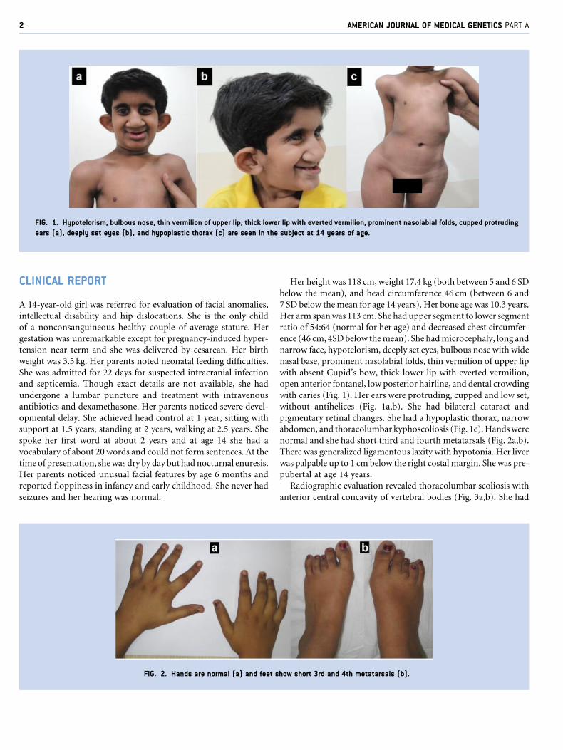
ence (46 cm, 4SDbelow themean). She hadmicrocephaly, long and
narrow face, hypotelorism, deeply set eyes, bulbous nose with wide
nasal base, prominent nasolabial folds, thin vermilion of upper lip
with absent Cupid’s bow, thick lower lip with everted vermilion,
open anterior fontanel, low posterior hairline, and dental crowding
with caries (Fig. 1). Her ears were protruding, cupped and low set,
without antihelices (Fig. 1a,b). She had bilateral cataract and
pigmentary retinal changes. She had a hypoplastic thorax, narrow
abdomen, and thoracolumbar kyphoscoliosis (Fig. 1c).Handswere
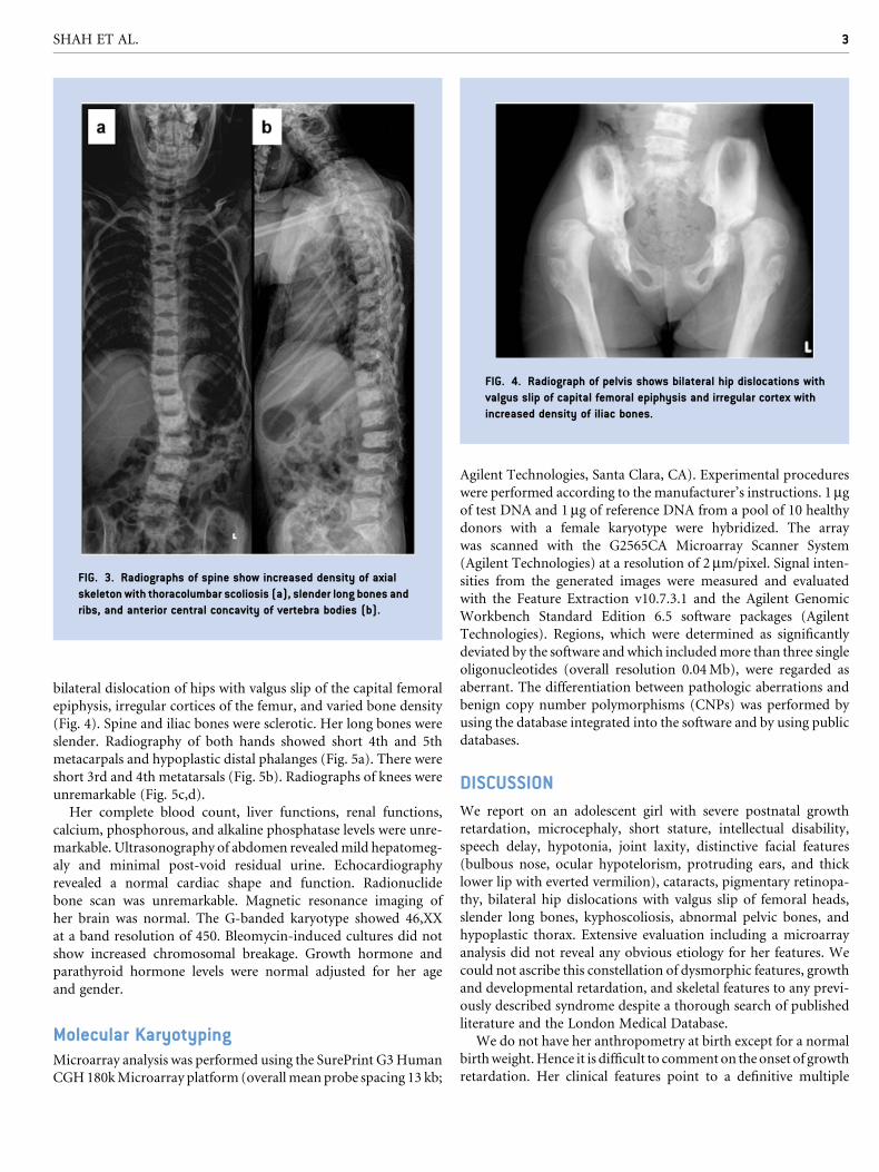
normal and she had short third and fourth metatarsals (Fig. 2a,b).
There was generalized ligamentous laxity with hypotonia. Her liver
was palpable up to 1 cm below the right costal margin. She was pre-
pubertal at age 14 years.
Radiographic evaluation revealed thoracolumbar scoliosis with
anterior central concavity of vertebral bodies (Fig. 3a,b). She had
FIG. 1. Hypotelorism, bulbous nose, thin vermilion of upper lip, thick lower lip with everted vermilion, prominent nasolabial folds, cupped protruding
ears (a), deeply set eyes (b), and hypoplastic thorax (c) are seen in the subject at 14 years of age.
FIG. 2. Hands are normal (a) and feet show short 3rd and 4th metatarsals (b).
2 AMERICAN JOURNAL OF MEDICAL GENETICS PART A
bilateral dislocation of hips with valgus slip of the capital femoral
epiphysis, irregular cortices of the femur, and varied bone density
(Fig. 4). Spine and iliac bones were sclerotic. Her long bones were
slender. Radiography of both hands showed short 4th and 5th
metacarpals and hypoplastic distal phalanges (Fig. 5a). There were
short 3rd and 4th metatarsals (Fig. 5b). Radiographs of knees were
unremarkable (Fig. 5c,d).
Her complete blood count, liver functions, renal functions,
calcium, phosphorous, and alkaline phosphatase levels were unre-
markable. Ultrasonography of abdomen revealedmild hepatomeg-
aly and minimal post-void residual urine. Echocardiography
revealed a normal cardiac shape and function. Radionuclide
bone scan was unremarkable. Magnetic resonance imaging of
her brain was normal. The G-banded karyotype showed 46,XX
at a band resolution of 450. Bleomycin-induced cultures did not
show increased chromosomal breakage. Growth hormone and
parathyroid hormone levels were normal adjusted for her age
and gender.
Molecular KaryotypingMicroarray analysis was performed using the SurePrint G3Human
CGH180kMicroarray platform (overallmean probe spacing 13 kb;
Agilent Technologies, Santa Clara, CA). Experimental procedures
were performed according to the manufacturer’s instructions. 1mgof test DNA and 1mg of reference DNA from a pool of 10 healthy
donors with a female karyotype were hybridized. The array
was scanned with the G2565CA Microarray Scanner System
(Agilent Technologies) at a resolution of 2mm/pixel. Signal inten-
sities from the generated images were measured and evaluated
with the Feature Extraction v10.7.3.1 and the Agilent Genomic
Workbench Standard Edition 6.5 software packages (Agilent
Technologies). Regions, which were determined as significantly
deviated by the software andwhich includedmore than three single
oligonucleotides (overall resolution 0.04Mb), were regarded as
aberrant. The differentiation between pathologic aberrations and
benign copy number polymorphisms (CNPs) was performed by
using the database integrated into the software and by using public
databases.
DISCUSSION
We report on an adolescent girl with severe postnatal growth
retardation, microcephaly, short stature, intellectual disability,
speech delay, hypotonia, joint laxity, distinctive facial features
(bulbous nose, ocular hypotelorism, protruding ears, and thick
lower lip with everted vermilion), cataracts, pigmentary retinopa-
thy, bilateral hip dislocations with valgus slip of femoral heads,
slender long bones, kyphoscoliosis, abnormal pelvic bones, and
hypoplastic thorax. Extensive evaluation including a microarray
analysis did not reveal any obvious etiology for her features. We
could not ascribe this constellation of dysmorphic features, growth
and developmental retardation, and skeletal features to any previ-
ously described syndrome despite a thorough search of published
literature and the London Medical Database.
We do not have her anthropometry at birth except for a normal
birthweight.Hence it is difficult to commenton theonset of growth
retardation. Her clinical features point to a definitive multiple
FIG. 3. Radiographs of spine show increased density of axial
skeleton with thoracolumbar scoliosis (a), slender long bones and
ribs, and anterior central concavity of vertebra bodies (b).
FIG. 4. Radiograph of pelvis shows bilateral hip dislocations with
valgus slip of capital femoral epiphysis and irregular cortex with
increased density of iliac bones.
SHAH ET AL. 3

congenital anomaly/mental retardation syndrome rather than any
acquired etiology.
We found Gurrieri syndrome (OMIM 601187) to be the closest
syndrome sharing growth retardation, intellectual disability, dental
anomalies, deep set eyes, brachydactyly, kyphosis, joint laxity,
hypoplastic iliac alae, thick-everted lower lips, and delayed speech
[Gurrieri et al., 1992a,b; Orrico et al., 1999]. However, absence of
seizures, osteoporosis, advanced bone age, prognathism, sparse
eyebrows, presence of microcephaly, cataracts, pigmentary reti-
nopathy, hip dislocations, valgus slip of femoral heads, and hypo-
plastic thorax differentiate the phenotype of our subject from this
possible autosomal recessive condition. As no gene is yet identified
for this condition, this condition was excluded on clinical basis
only.
The other conditions we considered were: geroderma osteodys-
plasticum (OMIM231070)with premature ageing of skin [Hennies
t al., 2008; Skidmore et al., 2011], microcephalic osteodysplastic
primordial dwarfisms (OMIM 210710, 210720, 210730) [Hall
et al., 2004; Abdel-Salam et al., 2012], filamin A related disorders
(OMIM 300017) [Robertson, 2007], and osteomesopyknosis
(OMIM 166450) [Proschek et al., 1985]. However, normal skin,
unique facial features, cataracts, pigmentary retinopathy, and
hypoplastic thorax differentiate these conditions. Since this is
an isolated case with normal microarray results, it is difficult to
postulate on the etiology of this condition.
ACKNOWLEDGMENTS
The authors gratefully acknowledge the cooperation of the child
and her parents for this study. The authors thank Dr. Benjamin
Joseph, Professor, Pediatric Orthopedics Service, Department of
Orthopedics, Kasturba Medical College, Manipal for his valuable
inputs and comments in preparation of this manuscript and
Dr. Reiner Siebert, Professor, Institute of Human Genetics, Kiel,
for continuous support. J.M.G. is supported by NIH/NICHD
program project grant (HD22657), and the Medical Genetics
NIH/NIGMS training program grant (5-T32-GM08243). K.M.G.
is a recipient of Hargobind Foundation Medical Fellowship.
FIG. 5. Radiographs of both hands (a) and feet (b) show short 4th and 5thmetacarpals, and 3rd, 4th, and 5thmetatarsals. Knee radiographs (c and d)
are normal.
4 AMERICAN JOURNAL OF MEDICAL GENETICS PART A

REFERENCES
Abdel-SalamGM,Abdel-HamidMS, IssaM,MagdyA, El-Kotoury A, AmrK. 2012. Expanding the phenotypic and mutational spectrum in micro-cephalic osteodysplastic primordial dwarfism type IAmJMedGenet PartA 158A:1455–1461.
Gurrieri F, SammitoV, Ricci B, LossaM, Bellussi A,Neri G. 1992a. Possiblenew type of oral-facial-digital syndrome with retinal abnormalities:OFDS type (VIII). Am J Med Genet 42:789–792.
Gurrieri F, Sammito V, Bellussi A, Neri G. 1992b. New autosomal recessivesyndrome of mental retardation, epilepsy, short stature, and skeletaldysplasia. Am J Med Genet 44:315–320.
Hall JG, Flora C, Scott CI Jr, Pauli RM, Tanaka KI. 2004. Majewskiosteodysplastic primordial dwarfism type II (MOPD II): Natural historyand clinical findings. Am J Med Genet Part A 130A:55–72.
HenniesHC,KornakU, ZhangH, Egerer J, ZhangX, SeifertW,K€uhnisch J,BuddeB,N€atebusM,Brancati F,WilcoxWR,M€ullerD,KaplanPB,RajabA, Zampino G, Fodale V, Dallapiccola B, Newman W, Metcalfe K,
Clayton-Smith J, Tassabehji M, Steinmann B, Barr FA, N€urnberg P,Wieacker P, Mundlos S. 2008. Gerodermia osteodysplastica is causedby mutations in SCYL1BP1, a Rab-6 interacting golgin. Nat Genet40:1410–1412.
Orrico A, Hayek G, Burroni L. 1999. Autosomal recessive syndrome ofgrowth and mental retardation, seizures, retinal abnormalities, andosteodysplasia with similarity to the Gurrieri syndrome. Am J MedGenet 82:84–87.
Proschek R, Labelle H, Bard C, Marton D. 1985. Osteomesopycnosis: Casereport. J Bone Joint Surg Am 67:652–653.
Robertson SP. 2007. Otopalatodigital syndrome spectrum disorders:Otopalatodigital syndrome types 1 and 2, frontometaphyseal dysplasiaand Melnick-Needles syndrome. Eur J Hum Genet 15:3–9.
Skidmore DL, Chitayat D, Morgan T, Hinek A, Fischer B, Dimopoulou A,Somers G, Halliday W, Blaser S, Diambomba Y, Lemire EG, Kornak U,Robertson SP. 2011. Further expansion of the phenotypic spectrumassociated with mutations in ALDH18A1, encoding D1-pyrroline-5-carboxylate synthase (P5CS). Am J Med Genet Part A 155A:1848–1856.
SHAH ET AL. 5
![Cellular/Molecular Ankyrin-Dependentand ...gia, hydrocephalus)] that presents with varying penetrance of hydrocephalus, mental retardation, and hypoplasia/absence of the corticospinal](https://img.pdfslide.net/doc/110x75/5e3d1f77f3a67e2bd53358e2/cellularmolecular-ankyrin-dependentand-gia-hydrocephalus-that-presents-with.jpg)