Embed Size (px)
Citation preview
1
GUIDANCE ON A STRATEGY FOR GENOTOXICITY TESTING OF CHEMICAL SUBSTANCES CONTENTS PARAGRAPH I. Preface 1-5 II. Introduction 6-10 III. Significance of Chemical Induced Mutation for Human Health 11-12 IV. General Principles of Testing Strategy 13-17 V Genotoxicity Testing Strategy 18 Stage 0: Preliminary Considerations Prior to Genotoxicity Testing (Figure 1) 19 Physico-chemical and Toxicological Properties 20
Structure Activity Relationships 21-23 Screening Tests 24-26 Stage 1: In Vitro Genotoxicity Testing (Figure 2)
Overview of Strategy 27-33 Discussion of Stage 1 Tests - General aspects 34-45 Discussion of Stage 1 Tests - Core tests In Vitro Bacterial Tests for Gene Mutation 46-47 In Vitro Mammalian Cell Micronucleus Test for Clastogenicity and Aneuploidy (MNvit) 48-53 Discussion of Stage 1 Tests- Non Core tests In Vitro Chromosomal Aberration Assay in Mammalian Cells (Metaphase Analysis) for Clastogenicity and Aneuploidy 54 In Vitro Mouse Lymphoma Assay for Gene Mutation
and Clastogenicity 55-56 In Vitro CHO/HPRT assay for Gene Mutation 57
In Vitro Assays using Human Reconstructed Skin 58 In Vitro Alkaline Comet Assay for DNA Damage 59 Summary Stage 1: (In Vitro Genotoxicity Testing) 60 Stage 2: In Vivo Genotoxicity Tests (Figure 3) Overview of Strategy 61-73 Discussion Stage 2-Initial Testing Strategy-General Aspects 74 Discussion Stage 2-Recommended Core In Vivo Genotoxicity Tests 75 Rodent Micronucleus and Chromosome Aberration Assays for Clastogenicity and Aneuploidy 76-77 Transgenic Rodent Mutation (TGR) for Gene Mutation 78 Rodent Comet Assay for DNA Damage 79 Non-Core In Vivo Test-Rat Liver UDS Assay for DNA Damage 80 Discussion Stage 2-Supplementary Tests 81-82 Summary Stage 2: (In Vivo Genotoxicity Testing) 83 VI: Possible Future Developments 84 Annex 1: Sensitivity and Specificity Data Considered by COM Annex 2: Tabulation of Genotoxicity Tests in Stages 1 and 2 and Mutagenicity and Genotoxicity End Points Detected Annex 3: Rationale for Selection of Ames Test and In Vitro
Micronucleus Assays as the Two Principal In Vitro Tests
2
GUIDANCE ON A STRATEGY FOR GENOTOXICITY TESTING
OF CHEMICAL SUBSTANCES
Executive Summary
The Committee on Mutagenicity of Chemicals in Food, Consumer Products and the
Environment (COM) has a remit to provide UK Government Departments and
Agencies with advice on the most suitable approaches to testing chemical
substances for genotoxicity. The COM published guidance in 1981, 1989 and again
in 2000. This document, incorporating some significant changes, reports on the
COM views regarding the most appropriate strategy for genotoxicity testing reached
in 2011.
The COM recommends a staged approach to testing:
Stage 0 consists of preliminary considerations which include physico-chemical
properties of the test chemical substance, Structure Activity Relationships (SAR), and
information from screening tests. However, data from SAR and screening tests
should not overrule test data from adequately designed and conducted genotoxicity
tests.
Stage 1 consists of in vitro genotoxicity tests. The COM recommends a core-test
battery of the Ames test combined with the in vitro micronucleus test. This
combination provides information on three types of genetic damage for which data
are required (namely, gene mutation, chromosomal damage and aneuploidy) and
gives appropriate sensitivity to detect chemical mutagens. There is no need to
independently replicate adequately designed and conducted core in vitro tests which
are either clearly negative or clearly positive. The strategy document also considers
the value which can be attributed to a number of non-core in vitro tests.
Stage 2 consists of in vivo genotoxicity tests. A case-by-case strategy should be
developed to answer one or more of the following specific queries;
1) Investigation of mutagenic end point(s) identified in Stage 1,
2) Investigation of genotoxicity in tumour target tissue(s),
3) Investigation of potential for germ cell genotoxicity,
4) Investigation of in vivo mutagenicity for chemicals, which were negative in
Stage 1 but where there is high or moderate and prolonged exposure,
5) Investigation of genotoxicity in site of contact tissues.
3
The core tests in Stage 2 are the rodent micronucleus/chromosome aberration
assays for aneuploidy and clastogenicity, the transgenic rodent gene mutation assay
and the rodent Comet assay for DNA damage.
Usually negative results obtained in a carefully selected in vivo test (possibly
studying more than one endpoint and tissue) will be sufficient to address positive
results found in vitro. However, a further test(s) may be needed if some of the
genotoxic effects seen in Stage 1 in vitro tests had not been adequately studied in
vivo (e.g. the chemical affects multiple mutagenic end-points), or other aspects of the
genotoxic potential of the chemical had not been fully resolved (e.g. in the case
where an investigation of heritable effects was required). The strategy document
also considers the value which can be attributed to a number of non-core in vivo
tests. In most instances information from core in vivo tests is sufficient to evaluate
the in vivo mutagenicity of chemical substances. A supplementary in vivo test
strategy can provide additional information on a case-by-case basis, to investigate
aspects such as further characterisation of germ cell genotoxicity, and DNA adduct
data which can provide information to elucidate the mode of genotoxic action of
carcinogenic chemicals.
It is acknowledged that the field of genotoxicology and genotoxicity testing is rapidly
developing. A short overview of possible future developments and techniques such
as toxicogenomics is provided.
4
I. Preface
1. The Committee on Mutagenicity of Chemicals in Food, Consumer Products
and the Environment (COM) is an independent expert advisory committee
whose members are appointed by the Chief Medical Officer for England and
the Chair of the Food Standards Agency (FSA) following an appointments
exercise involving public advertisement. Members serve in their own capacity
as independent experts and observe a published code of practice including
principles relating to the declaration of possible conflicting interests.
2. The remit of the COM is to advise all U.K. government departments and
agencies with an interest in the safety of chemicals across various sectors on
the human health aspects of the mutagenicity and genotoxicity of chemicals.
(These terms are defined for the purposes of this guidance document in
paragraphs 7-8 below.) The Secretariat is provided by the Health Protection
Agency (HPA), who lead, and the FSA. Other government departments with
an interest provide assessors to the COM; these are specifically from the
Department of Health (DH), the Department of Environment, Food and Rural
Affairs (Defra), the Chemicals Regulation Directorate (CRD) of the Health and
Safety Executive (HSE) (responsible for legislation regulating chemicals,
pesticides, biocides and detergents), the Environment Agency, the Veterinary
Medicines Directorate (VMD: a Defra agency responsible for the licensing of
veterinary drugs) and the Medicines and Healthcare products Regulatory
Agency (MHRA; a DH agency responsible for the licensing of human
medicines). In addition there are assessors from the Scottish Government,
the Welsh Assembly Government and the Northern Ireland Assembly.
3. The role of the COM is advisory. It has no regulatory status, although its
advice may be provided to a body that does have such a role (e.g. HSE CRD
for occupational aspects and for pesticides etc). Its remit is to advise on the
human health aspects of mutagenicity and genotoxicity of chemicals, and this
may involve advice on a specific chemical, and also on testing strategies and
research. This guidance document focuses on testing strategies for chemical
substances for which there are no available genotoxicity data. Separate
guidance on a strategy for the genotoxicity testing and mutagenic hazard
assessment of chemicals with inadequate genotoxicity data is in preparation.
Throughout this guidance the COM has referred to the genotoxicity testing of
substance(s). In this document the term test substance refers to a specified
chemical or material including any additive necessary to preserve its stability
5
and any impurity deriving from the process used. However the COM usually
provides advice on a specific chemical substance which can be equated to a
single chemical or compound or pure substance.
http://www.hse.gov.uk/reach/definitions.htm#substance. The COM also
has a general remit to advise on important general principles or new scientific
discoveries in connection with potential mutagenic and genotoxic hazards
(inherent properties of chemicals) or risk (the likelihood of mutagenic or
genotoxic effects occurring after a given exposure to a chemical) and to
present recommendations for genotoxicity testing. In practice the bulk of the
work of the COM relates to assessing genotoxicity tests and providing advice
on the mutagenic hazard of chemicals.
4. In the context of testing strategies, the COM first published guidelines for the
testing of chemicals for mutagenicity in 1981, and these were revised in 1989
(DOH, 1989). These provided guidance to the relevant government
departments and agencies on best practice for testing at those times. The
need for guidance to be periodically updated, to reflect advances in
development and validation of methods, was recognised and revised
guidance was published in 2000 (DOH, 2000). This new guidance continues
this updating process. The strategy outlined in this guidance is considered to
be the most scientifically appropriate given available methods and recognises
the need to avoid the use of live animals where practical and where validated
alternative methods are available. It is recognised that, as with the earlier
published COM guidance, it may be some time before this strategy is
reflected in guidelines used by UK regulatory authorities.
5. The COM believes that the approach outlined presents an updated overview
of the core principles of genotoxicity testing and will remain valid for several
years. It is acknowledged that existing national or international testing
strategies will be at different stages of review and hence inconsistencies are
expected. The COM guidance is not intended to supersede or replace
existing national or international sector-specific genotoxicity testing strategies
(e.g. those recommended for pharmaceuticals by the International
Conference on Harmonisation of Technical Requirements for Registration of
Pharmaceuticals for Human Use (ICH)
http://www.ich.org/products/guidelines/safety/article/safety-guidelines.html
and for chemicals assessed under the Registration, Evaluation, Authorisation
and Restriction of Chemicals (REACH) Regulation (EC1906/2006)
6
http://guidance.echa.europa.eu/docs/guidance_document/information_require
ments_en.htm.
II. Introduction
6. The COM last published guidance on a strategy for the testing of chemicals
for mutagenic potential in 2000 (DOH, 2000). The rationale developed by
COM in 2000, particularly in relation to the testing of all potential mutagenic
endpoints, has also been adopted by the International Workshops on
Genotoxicity Testing (IWGT) (Muller et al., 2003). Since 2000 there has been
development of new approaches to identifying genotoxic hazards in vitro
including new approaches to identify misleading positive results and evaluate
target organ genotoxicity in vivo. There is also a need to develop a testing
strategy which can encompass chemicals such as cosmetics where no animal
tests are permitted under EU law. It is the objective of this paper to set out a
scientifically valid testing strategy comprising those methods which the COM
believe to be the most informative with regards to the detection of genotoxic
hazard and (when possible) are well validated. There is no discussion of
methods which experience has shown to have no place in the recommended
genotoxicity testing strategy. Details of methodologies are not given since
they are provided in the Organisation for Economic Cooperation and
Development (OECD) test guidelines, the EU Test Methods Regulation (EC
440/2008) and the International Workshops on Genotoxicity Testing (IWGT)
guidance.
7. The genome can be damaged in a variety of ways either spontaneously or
from exposure to genotoxic agents. The term “mutagenic” refers to the ability
of a substance to induce a permanent change in the amount or structure of
the genetic material of an organism, which may result in a heritable change in
the characteristics of the organism. Chemicals inducing mutations are
referred to as mutagens (they are mutagenic). These alterations may involve
individual genes, blocks of genes, or whole chromosomes. Mutations
involving single genes may be a consequence of effects on single DNA bases
(point mutations) or of larger changes, including deletions and
rearrangements of DNA. The potential to induce mutation is measured in test
systems that detect a broader range of genetic changes than simply mutation
7
– they measure genotoxicity. Mutagenicity is accepted as a key event in
carcinogenicity.
8. Genotoxicity refers to interaction with, or damage to, DNA and/or other
cellular components which regulate the fidelity of the genome. It is a broad
term that, as well as mutation (see paragraph 7) includes damage to DNA
such as the production of DNA adducts, by the chemical itself or its
metabolites. Cells have the capacity to protect themselves from such
potentially lethal or mutagenic genotoxic effects by many repair processes
and therefore many genotoxic events do not become evident as mutations.
However, the capacity to damage the genome (genotoxicity) is an indicator of
potential mutagenicity. Thus, some methods that measure genotoxicity may
not provide direct evidence of heritable mutation.
9. The objective of genotoxicity testing is to exclude or identify potential
mutagenic hazards to humans, and, for those substances that are positive, to
aid in the elucidation of the mode of genotoxic action (MoGA). This guidance
therefore presents a strategy for genotoxicity testing since this term
encompasses all the assays included in the strategy. Consequently, it is
important to generate information on three types of genetic damage, namely
gene mutation, changes to chromosome structure (i.e. clastogenicity) and
number (i.e. aneuploidy), to provide comprehensive coverage of the
mutagenic potential of a chemical.
10. The COM reaffirms its view, published in 1989 and 2000, that there is
currently no single validated assay that can provide comprehensive
information on all three types of genetic damage and thus it is necessary to
subject a given test substance to several different assays. The range of
assays discussed in this document include those using prokaryotes (bacteria)
and mammalian cells in vitro, and whole mammals, where effects in a wide
range of target organs including germ cells can be measured. Assays may
be classified on the basis of genetic end-points (e.g. gene mutation,
clastogenicity, aneugenicity and tests for DNA damage) or by consideration of
the different phylogenetic levels (e.g. bacteria, and mammalian cell)
represented and also in mammals by the tissues or target organs studied.
III Significance of Chemical-Induced Mutation for Human Health
11. A mutation in the germ cells of sexually-reproducing organisms may be
transmitted to the offspring, whereas a mutation that occurs in somatic cells
8
may be transferred only to descendant daughter cells. Mutagenic chemicals
may present a hazard to health since exposure to a mutagen carries the risk
of inducing germ-line mutations, with the possibility of inherited disorders, and
the risk of somatic mutations including those leading to cancer.
12. A separate statement discussing the significance of chemical-induced
mutation to human health is in preparation.
IV. General Principles of Testing Strategy
13. The COM recommends a two-stage genotoxicity testing strategy (Stages 1
and 2) for the detection of the mutagenic hazard of chemicals which can be
supported by appropriate preliminary screening tests and/or in silico data
(Stage 0). Initial testing for mutagenic potential in Stage 1 is based upon two
core in vitro tests that are chosen to provide information on gene mutation,
clastogenicity and aneuploidy, with case-by-case additional testing and
investigation depending on the results of these initial genotoxicity tests. All in
vitro tests should be designed to provide the best chance of detecting
potential activity, with respect to (a) the exogenous metabolic activation
system (S9 - see glossary); (b) the ability of the compound or its metabolite(s)
to reach the target DNA and/or targets such as the cell division apparatus,
and; (c) the ability of the genetic test system to detect the given type of
genotoxic event. Where international guidance is available, the assays
should be carried out to conform to those internationally recognised protocols
e.g. as published by the OECD, the IWGT and in the EU test methods
Regulation (EC 440/2008). The same approach to testing can be used for
chemical substances where in vivo genotoxicity testing is not permitted (e.g.
cosmetics). Investigations regarding MoGA are important to derive
conclusions on biological relevance of genotoxicity test results, to aid in
overall risk assessment, and to inform on the strategy for in vivo tests. This is
of particular importance for those chemicals where no in vivo genotoxicity
testing is permitted.
14. For most chemicals, results from the two Stage 1 core tests should be
sufficient to reach a conclusion on the presence or absence of mutagenic
potential. However, in some instances, even when Stage 1 tests are
negative, regulatory authorities may require consideration of the need for in
vivo Stage 2 testing particularly where exposure is considered to be high, or
moderate and prolonged (e.g. most human medicines), or where there is a
chemical class precedent of positive in vivo genotoxicity data. Guidance on
9
the level of exposure which equates to high, moderate or prolonged is beyond
the remit of the COM.
15. Stage 2 consists of a number of in vivo tests designed to investigate whether
in vitro genotoxic activity including specific mutagenic end-points identified by
in vitro tests can be expressed in the whole animal. This may also include
assays for specific target organs (e.g. rodent tumours detected in
carcinogenicity bioassays) or in germ cells. Few chemicals are active only in
vivo and in such cases this may be due to a number of factors such as
metabolic differences, the influence of gut flora, higher exposures in vivo
compared to in vitro and pharmacological effects (e.g. folate depletion or
receptor kinase inhibition) (Tweats et al, 2007b).
There is currently no single in vivo test which can assay all three types of
genetic damage (Thybaud et al., 2007) and thus a strategy for Stage 2 has to
be designed based on the nature of genotoxic effects identified in Stage 1 and
the possibility that genotoxic activity will only be expressed in vivo as
discussed above. However consideration should be given to the possibility of
evaluating different genotoxicity endpoints in a single set of test animals.
16. There should be a clear strategy for planning tests within each stage and for
progressing from Stage 1 to Stage 2. Clear statements can be made
regarding the initial in vitro tests to be used in Stage 1 as these methods have
been well studied, whereas the strategy for Stage 2 is more complex and, if
not a specific regulatory requirement, needs to be developed on a case-by-
case basis.
17. Under the strategy recommended by COM, the use of animals in mutagenicity
testing is primarily required when it is necessary to investigate whether
genotoxic activity detected in Stage 1 in vitro is reproduced in vivo, to study
target organ genotoxicity (for example involvement of genotoxicity in rodent
tumours (Kirkland et al., 2007a) and to evaluate the potential for heritable
mutagenic effects. Genotoxicity testing using animals should be carried out
when there is no suitable alternative, and the minimum number of animals
should be used, consistent with obtaining valid results. If feasible, studies
can be conducted as an adjunct to single or repeat dose toxicity studies. The
COM supports current and future developments to replace, refine or reduce
the need for in vivo genotoxicity testing.
10
V Genotoxicity Testing Strategy
18. The COM guidance provides a strategy for testing chemical substances
where no genotoxicity data are available. Test substances may also contain
impurities at varying levels which may exhibit genotoxic activity. Separate
guidance on the genotoxicity assessment of impurities has been identified as
a priority project during the COM horizon scanning exercise in 2010 (see
minutes of COM meeting of October 2010
http://www.iacom.org.uk/meetings/index.htm ), and is currently the subject of
an ICH expert working group .
http://www.ich.org/fileadmin/Public_Web_Site/ICH_Products/Guidelines/Multi
disciplinary/M7/M7_Final_Concept_Paper_June_2010.pdf
The strategy recommended in the following sections is concerned with testing
for genotoxic activity of chemical substances and does not specifically
address complex mixtures of chemicals. Since the publication of the COM
guidance in 2000, assessments of the performance of (Q)SAR approaches,
screening tests and genotoxicity assays (both individually and in
combinations) regarding the prediction of rodent carcinogenicity have been
published (Kirkland et al., 2005a; Kirkland and Speit, 2008; Matthews et al.,
2006a; Matthews et al., 2006b). Reference to these publications can provide
an insight into the performance of the in vitro genotoxicity assays specifically
in relation to the particular data sets analysed and the end points considered,
predominantly rodent carcinogenicity but also in vivo genotoxicity (Kirkland et
al., 2011). Relevant sensitivity and specificity data and assay performance
assessments have been summarised in Annex 1, and are discussed further in
Annex 3, for information and are cited where appropriate in the text below.
Overall the older available data suggest that mammalian cell assays did not
perform well at discriminating between rodent carcinogens and non-
carcinogens. However, recent experience suggests that mammalian cell
tests conducted and interpreted according to current recommendations
perform more robustly (Fellows et al., 2011).
Stage 0: Preliminary Considerations Prior to Genotoxicity Testing (Figure 1)
19. The intrinsic chemical and toxicological properties of the test substance must
be considered before devising the genotoxicity testing programme.
11
Physico-chemical and Toxicological Properties
20. The physico-chemical properties of the test substance (for example, pKa,
partition coefficient, solubility, volatility and stability in, and potential reactions
with, solvents/vehicles) and its purity can affect the ease of conduct and
results of in vitro tests. For example, the tolerance of cells to acidic chemicals
can be enhanced by neutralisation but this may affect the inherent reactivity
of substances to DNA (Hiramoto et al., 1997). Potential reactions of the test
substance with solvent /vehicle should also be considered (e.g cisplatin
reacts with DMSO) (Fischer et al., 2008 ). Alternatively, low solubility may
limit the feasibility of undertaking some or all of the in vitro mutagenicity tests
recommended in this strategy. The potential for auto-oxidation of the test
chemical in the culture medium can also affect the outcome of in vitro
genotoxicity tests (Long et al., 2007). It is noteworthy that the toxic properties
of test substances, such as target organ effects, or irritancy/corrosivity in
contact with skin or mucous membranes and their toxicokinetics and
metabolism will influence the choice of route of administration and the highest
dose level achievable in Stage 2 in vivo mutagenicity tests.
Structure Activity Relationships
21. Whether the test substance would be expected to have mutagenic potential
may be assessed from its chemical structure, which may provide structural
alerts for mutagenicity. A composite model structure was originally devised
by Ashby and Tennant indicating substituent chemical groups or moieties
associated with DNA-reactivity (Ashby and Paton, 1993). A number of freely
available and commercial systems to investigate structure activity
relationships (SAR) for mutagenicity have been developed and evaluated
since 2000 (Benigni and Bossa, 2008; Benigni et al., 2007; Cariello et al.,
2002; Contrera et al., 2005; Snyder and Smith, 2005; Zeiger et al., 1996).
Further information on various models is provided in Annex 1. The OECD
(OECD, 2004) and the European Commission (Joint Research Centre) have
published principles for the validation of (Q)SAR ((Quantitative) Structure
Activity Relationships) (Worth et al., 2005, Benigni and Bossa, 2008). (Q)SAR
assessment of the in vitro mutagenicity in bacteria has been attained by two
types of approach; statistical analyses of structure and mutagenic activity
12
and/or (Q)SAR models using programmed rules for prediction of mutagenic
activity based on the available knowledge and expert judgement.
22. Such (Q)SAR systems can be useful when a large number of chemicals
require assessment and prioritisation for genotoxicity testing or in instances
where a rapid assessment of a chemical is required and there are no
genotoxicity test data available. Each (Q)SAR system has a defined domain
of applicability which is determined by the structural/descriptor factors,
modes/mechanism of mutagenicity, and metabolic aspects included within the
system. In addition in silico approaches can aid in the interpretation of Stage
1 in vitro genotoxicity test results (Dearfield et al., 2011). The available
systems perform well for prediction of bacterial mutagenicity (i.e. for chemical
structures within the domain of applicability of the model under consideration)
(see Annex 1). However, lower sensitivities and specificities have been
reported for a number of systems when used for prediction of results from in
vitro cytogenetics or the mouse lymphoma assay (e.g. using MCASE and
MDL-QSAR) (Contrera et al., 2008). One factor in the lower predictive
capability of (Q)SAR systems for mammalian cell genotoxicity assays is
inadequate coverage of non-covalent DNA interactions and non-DNA targets
associated with cell division (Grant et al., 2000; Snyder and Smith, 2005). It
has also been proposed that (Q)SAR assessments can aid in the
interpretation of the relevance of in vitro genotoxicity assays through
prediction of biotransformation (Combes et al., 2007). Other systems
combining metabolic simulation with structure toxicity rules have been
developed (e.g. TIMES; tissue metabolic simulator) but are at a relatively
early stage of validation (Mekenyan et al., 2004; Serafimova et al., 2007).
Lhasa Ltd has developed a computer programme (METEOR), which has the
facility to integrate prediction of metabolism with (Q)SAR approaches for
genotoxicity. (https://www.lhasalimited.org/meteor/). An authoritative and
comprehensive evaluation of the different (Q)SAR approaches to the
identification of genotoxic potential has been prepared for the European Food
Safety Authority (EFSA) by the Computational Toxicology group, Institute for
Health & Consumer Protection, European Commission-Joint Research Centre
(JRC), Ispra, Italy (http://www.efsa.europa.eu/en/scdocs/scdoc/50e.htm). A
compilation of structural alerts for prediction of the rodent in vivo
micronucleus assay has recently been published. The authors advocate that
13
the derived rules can be used for preliminary identification of in vivo mutagens
(Benigni et al., 2010).
23. Overall, (Q)SAR approaches for the prediction of genotoxic activity can be a
valuable tool to aid in the high throughput screening of compounds, the
provision of assessments for chemicals for which no genotoxicity test data are
available and also prioritisation for genotoxicity testing. (Q)SAR can also aid
in the interpretation of genetic toxicology tests, although such predictions
cannot replace the need to undertake the in vitro and in vivo genotoxicity tests
required to derive conclusions on mutagenic hazard. However, expert
judgement is needed when reaching conclusions on mutagenic hazard on the
basis of (Q)SAR information alone. In reaching conclusions, data from well
conducted in vitro or in vivo genotoxicity tests should be attributed a much
higher weight of evidence than (Q)SAR predictions, although all information
should be assessed on a case-by-case basis.
Screening Tests
24. There are a number of current initiatives which attempt to combine data
mining in silico approaches with high throughput tests to develop approaches
to screening large numbers of novel chemicals (Benfenati et al., 2009.). In
this guidance, genotoxicity screening tests refers to high throughput tests
which have been designed to be rapid, economical, reproducible, require only
small amounts of test substances (typically below 50 mg) and have a high
concordance with comparator genotoxicity end points in genotoxicity tests.
(These tests are also often referred to as pre-screening tests.) High
throughput bacterial tests have been developed using combinations of
Salmonella tester strains (Ames II™), primary DNA damage (umu assay),
mutations in ampicillinase gene (MutaGen assay), bioluminescence or 5-
fluorouracil resistance (Ackerman et al., 2009; Aubrecht et al., 2007; Kamber
et al., 2009 ; Miller et al., 2005 ; Reifferscheid et al., 2005). Other screening
systems cited in the literature include DNA repair activity in yeast cells
(Westerink et al., 2009). One research group has proposed a combination of
two commercial screening assays (VitotoxTM for bacterial mutagenicity and
RadarScreen yeast screen for clastogenicity) for rapid screening of
compounds. (Westerink et al., 2009).
25. A number of genotoxicity screening tests using in vitro systems have been
proposed, including alkaline elution using rat hepatocytes (Gealy et al., 2007),
14
the detection of DNA damage (via p53 or GADD45a activation, GreenScreen)
in cell lines (Knight et al., 2009) and differential survival in DNA repair
proficient and deficient cell lines (Helleday et al., 2001). A screening test for
genotoxicity using HepG2 cells (metabolically competent with wild type p53
genotype) based on four different luciferase-reporter assays has been
published. The authors claim, based on a small dataset, a high sensitivity for
identification of genotoxicity when used in combination with the commercially
available systems (VitotoxTM and RadarScreen) (Westerink et al., 2010).
None of these genotoxicity screening tests have reached the stage of
development where they could routinely be used to replace data generated
from in vitro genotoxicity testing. The predominant use of high throughput
screening tests is as an aid in prioritisation of compounds for development
undertaken by industry. The COM reviewed the GADD45a-GFP assay and it
was agreed that currently, it is most suited as part of a battery of high
throughput screening (COM minutes March 2010,
http://www.iacom.org.uk/meetings/index.htm).
26. High throughput genotoxicity screening tests can be used in a tiered
approach with in vitro genotoxicity tests to aid in the selection of chemicals for
development. It has been suggested that greater validation and acceptance
by regulatory authorities of these tests could lead to the replacement of
existing genotoxicity testing strategies with a combination of high throughput
screening tests (Custer and Sweder, 2008).
Stage 1: In Vitro Genotoxicity Testing (Figure 2)
Overview of strategy
27. The COM concluded in 1989 and 2000 that it was appropriate to concentrate
on a relatively small number of assays, using validated, sensitive methods
particularly chosen to avoid misleading negative results. Two important
parts of the revised Stage 1 strategy include using appropriate tests to gain
an insight into the nature of the genotoxic effects of a test substance and also
to avoid misleading positive results. Misleading positive results have been
reported for certain mammalian cell assays (Fowler et al., 2010; Pfuhler et al.,
2009; Kirkland et al., 2007a) particularly when multiple test systems were
used.
15
28 As outlined above in paragraphs 13 and 14, Stage 1 involves tests for
genotoxic activity using in vitro methods and comprises a two test core
system (namely an Ames test and in vitro micronucleus test (MNvit)) with the
objective of assessing mutagenic potential by investigating three different
end points (gene mutation, structural chromosomal damage and changes in
chromosome number). The rationale for this test strategy is given in Annex 3.
A clear positive result in either of these two core tests is sufficient to define
the chemical as an in vitro mutagen, although further in vitro and/or in vivo
testing may be undertaken to understand the relevance of the positive results.
The Committee considers that this revised strategy allows for efficient
identification of all mutagenic-end points but, by reducing the number of
mammalian cell tests from that recommended by COM in 2000, and following
improved methodologies, the risk of misleading positive results is decreased.
29. Additional investigations of chemicals which give positive or repeated
equivocal results in Stage 1 tests can include an assessment of mode(s) of in
vitro genotoxic action. There are a number of reasons (discussed in
paragraphs 34-37) why positive results in in vitro genotoxicity tests might
occur by mode(s) of action not relevant to human health hazard assessment.
Such MoGA evaluation in vitro is particularly relevant for those chemicals
(e.g. cosmetics) where there is a regulatory constraint which precludes the
use of in vivo genotoxicity assays in the testing strategy. The COM does not
recommend the use of in vitro genotoxicity assays that have not been
considered in detail in this guidance such as assays for sister chromatid
exchange, the in vitro UDS assay or tests using fungi. A table of mutagenic
endpoints detected by each genotoxicity assay cited in Stage 1 of this
strategy is given in Annex 2.
30. For chemicals which give equivocal results or repeated small positive effects,
it is important to consider evidence of reproducibility in the same assay or in
different assays detecting similar effects, and the magnitude of the induced
genotoxic effect in relation to historical negative control data, and then
consider whether further in vitro genotoxicity testing is needed (Kirkland et al.,
2007a; Hayashi et al., 2011). Further consideration of SAR data for these
chemicals may also give valuable information (Dearfield et al., 2011).
31. If clear negative results are obtained in both core in vitro tests undertaken, it
can generally be concluded that the chemical has no mutagenic activity.
However, there are some occasions when additional in vitro and/or in vivo
16
genotoxicity testing may be undertaken for chemicals giving a negative
response in the two in vitro core genotoxicity tests. For example, situations
where tumours are found in rodents, where the in vitro metabolic activation
systems are not optimal or where there are human-specific metabolites, may
need to be subject to further genotoxicity assessment. A further testing
strategy would have to be designed on a case-by-case basis (Muller et al.,
2003; Kirkland et al., 2007a). An IWGT working group has published
guidance on this topic (Kasper et al., 2007). An important part of any
additional in vitro strategy should be consideration of the appropriate
exogenous metabolic activation system (including alternative sources of S9 or
other metabolic systems including genetically engineered cell lines, see
paragraph 36)(Ku et al., 2007b). Further information on in vivo genotoxicity
testing of such test substances is provided in Stage 2 of this strategy.
32. Information from other combinations of genotoxicity tests which may include
one or more non-core tests outlined below in paragraphs 54-59 may also give
adequate data on all three end-points on a case-by-case basis. In vitro
genotoxicity tests using human reconstructed skin may provide useful
information on in vitro mutagenic hazard in circumstances where in vivo
testing is not permitted, or when extensive dermal exposure is anticipated
(e.g. cosmetic ingredients).
33. The full Stage 1 strategy should be performed and the results of studies
evaluated before a decision is made on whether to proceed to Stage 2 testing
or whether a conclusion on mutagenic hazard can be derived for test
substances where no in vivo genotoxicity testing is permitted. An outline of
Stage 0 and Stage 1 (in vitro genotoxicity testing) is given in Figure 2 and a
description of the assays recommended is provided in the following
paragraphs.
Discussion of Stage 1 Tests- General Aspects
34. The conduct of genotoxicity assays has improved over time and the overall
sensitivity of in vitro testing strategies regarding prediction of rodent
carcinogens is very high (Kirkland et al., 2005a; Kirkland et al., 2007c).
Proposals have been published for genotoxicity testing advocating a single in
vitro genotoxicity test (Ku et al., 2007a) or a complex approach involving up to
six in vitro genotoxicity tests (SCCNFP/0720/03) and critically evaluated by
Kirkland et al (Kirkland et al., 2005b). Neither of these approaches is
17
considered preferable to the proposed Stage 1 core testing. Although the
sensitivity (producing positive results with carcinogens) for rodent
carcinogenicity of a battery of Stage 1 tests was very high, the specificity
(producing negative results with non-carcinogens) was poor (Kirkland et al.,
2005; Kirkland et al., 2007a,b). Possible reasons for the poor specificity have
been discussed by various working groups e.g., see (Kirkland et al., 2007a).
A comprehensive review of the performance of Stage 1 genotoxicity assays
for prediction of rodent carcinogenicity reported positive results in one or
more in vitro tests for a substantial number of rodent non-carcinogens (as
assessed by the Carcinogenic Potency Database (CPDB), National
Toxicology Program (NTP), and the International Agency for Research on
Cancer (IARC)). Thus the specificity (i.e. correct identification of rodent non-
carcinogens) was considered to be reasonable for the Ames test (74%) but
poor for the mammalian cell assays (below 45%) particularly when multiple
assays were performed (Elespuru et al., 2009; Kirkland et al., 2005a). Many
reasons for low specificity have been proposed, particularly for mammalian
cells; for example, the use of high-concentrations, cytotoxicity, prolonged
exposure, overloading defence mechanisms, lack of detoxification capacity.
The influence of such confounding effects leading to indirect mechanisms of
genotoxicity, has been widely recognised (Kirsch-Volders et al., 2003b; Müller
and Kasper, 2000; Pratt and Barron, 2003)
35. A more recent analysis on the sensitivity of a combination of Ames test and
MNvit test to detect rodent carcinogens and in vivo genotoxicants is
summarised and discussed in Annex 3 Table 1, and published in Kirkland et
al (Kirkland et al., 2011). It is difficult to draw precise conclusions from the
available sensitivity and specificity data since the databases of chemicals
used vary. However these data do show that mammalian cell genotoxicity
tests can have low specificity and that combinations of in vitro genotoxicity
tests result in high sensitivity for rodent carcinogens and in vivo
genotoxicants. High sensitivity has always been a priority of genotoxicity
testing strategies recommended by the COM (DOH, 2000). An evaluation of
the use of in vitro genotoxicity tests to predict rodent carcinogens and in vivo
genotoxicants prepared for the COM meeting in June 2010
(http://www.iacom.org.uk/papers/index.htm MUT/2010/08) concluded that
there is no convincing evidence that any rodent carcinogen or in vivo
18
genotoxicant would fail to be detected by using an in vitro genotoxicity test
battery consisting of Ames test and MNvit.
36. It is most likely that the few occasions where in vitro test strategies fail to
detect mutagenic activity (i.e. misleading negative results) will be due to the
absence of appropriate metabolic activity in vitro (Brambilla and Martelli,
2004). Approaches to resolving potential inadequacies in metabolic activation
include structure based metabolism predictions, use of genetically modified
target organisms (e.g. CYP2E1 in Salmonella YG7108pin3ERb5) (Emmert et
al., 2006), the use of exogenous metabolic activation systems derived from
human sources, or recombinant human cytochrome P450 systems as an
external activation system (Ku et al., 2007b).
37. There are a number of MoGAs by which a chemical may demonstrate an in
vitro genotoxic effect that is either not relevant for humans or has a threshold.
The COM has reviewed the evidence for a number of threshold MoGAs and a
general guidance statement is available.
(http://www.iacom.org.uk/guidstate/index.htm statement G05). Threshold
MoGAs can generally be attributable to non-DNA interactions or an overload
of normal cellular physiology. In such cases a No Observed Effect
Concentration (NOEC) can be determined and may be useful in evaluating
risk. Investigations of a threshold-based MoGA need to be designed on a
case-by-case basis and can be complex to interpret (Kirkland et al., 2007c).
38. There has been considerable debate regarding the highest concentration that
should be used routinely in mammalian cell assays. The International
Conference on Harmonisation of the Technical Requirements for Registration
of Pharmaceuticals for Human Use (ICH) is considering whether the
maximum concentration tested for pharmaceuticals should be 1mM in
mammalian cell genotoxicity assays which would have the effect of reducing
the number of misleading positive results due to excessive concentrations
where the cellular defence mechanisms might be overwhelmed. However, a
reduction to 1mM would not be consistent with the OECD recommendation
for a top concentration of 10mM in mammalian cell genotoxicity assays
(OECD, 1997). A recent analysis of published data for the top concentration
in mammalian cell genotoxicity tests identified a small number of carcinogens
that (according to the publications) would not be detected in any part of a
three test in vitro genotoxicity test battery (consisting of the Ames, mouse
19
lymphoma and in vitro chromosomal aberration tests) if the testing
concentration limit for mammalian cell assays were reduced from 10mM to
1mM (Parry et al., 2010) A further investigation of these carcinogens found
that some positive results at concentrations above 1mM were not
reproducible (i.e. they were not genotoxic in mammalian cells under current
OECD guideline protocols) and others were positive at concentrations below
1mM, particularly when continuous treatments in the absence of S-9 (not
included in the original publications) were conducted. A new upper limit for
mammalian cells tests of 1mM or 500 µg/ml (whichever is higher) has been
proposed as sufficient to detect all genotoxic carcinogens that are negative in
the Ames test (Kirkland and Fowler, 2010). Several international
organisations are examining the principles underpinning this upper limit
selection (e.g ICH, OECD, IGWT) although currently no international
consensus has been reached. Precipitation can also be used to define a
maximal concentration or upper limit for testing.
39. There has also been considerable investigation of the role of excessive
cytotoxicity in mammalian cells and choice of cell type as possible causes of
misleading positive results (Blakey et al., 2008; Fellows et al, 2008; Pfuhler,
2009; Pfuhler et al., 2011). The method used to assess cytotoxicity may
affect the selection of highest concentration tested and potentially the results
obtained using mammalian cell genotoxicity assays (Kirkland et al., 2007b)
and recommendations have been made to use cytotoxicity measures based
on cell proliferation (Galloway et al., 2011). However, it is important to note
that although excessive cytotoxicity may lead to misleading positive results, it
may also result in misleading negative results when pronounced cell cycle
delay occurs. A similar conclusion was reached at an international
symposium on regulatory aspects of genotoxicity testing (Blakey et al.,
2008).
40. Most cell lines used for genotoxicity testing lack appropriate metabolism
leading to reliance on exogenous metabolic activation systems. These cell
lines may often have impaired p53 function and altered DNA repair capacity
(Kirkland et al., 2007b). There is some evidence that human lymphocytes are
less susceptible to misleading positives than the rodent cell lines currently
used (e.g. CHO, V79, CHL). Other cell systems such as the human cell lines
HepG2, TK6 and MCL5 cells and the reconstructed human skin models and
20
HepaRG show promise for future use (Kirkland et al, 2007b; Fowler et al.,
2009a; Le Hegarat et al., 2010).
41. The COM agrees that it is not necessary to undertake independent
confirmatory in vitro tests when clear negative or positive results have been
obtained provided the following criteria are satisfied:
• there is no doubt as to the quality of the study design and the conduct
of the test,
• the spacing and range of test substance concentrations rule out
missing a positive response,
• sufficient treatment conditions and sampling times have been used.
42. It is recognised that it can be difficult to provide convincing evidence for
absence of genotoxic effects. The investigator should consider the power of
the study design and the past performance of the test system when
formulating a protocol in order to optimise the chances of obtaining an
unequivocal result from a single experiment and to ensure that any potential
genotoxic effect is not missed.
43. There is a need to undertake further in vitro genotoxicity testing when an
equivocal result is obtained (i.e. neither clearly negative nor clearly positive by
appropriate biological or statistical criteria). Such additional genotoxicity tests
need to be planned on a case-by-case basis and need not necessarily be
undertaken in an identical fashion to the initial experiment(s). Indeed it may
be preferable to alter certain aspects of the study (e.g. concentration levels
investigated, treatment and sampling times, concentration of metabolic
activation mix) so as to obtain supplementary data. It may also be
appropriate to use a different genotoxicity test system, e.g. a chromosomal
aberration test, if there is equivocal evidence of clastogenicity from an in vitro
micronucleus test, or an in vitro cell mutation assay (e.g. TK or HPRT
mutation assays) if there is equivocal evidence of gene mutations from an
Ames test.
44. The use of historical negative control data to aid in the interpretation of
genotoxicity test results has been considered particularly in relation to
equivocal and small magnitude genotoxic effects (Kirkland et al., 2007d).
Advice has been recently published on approaches to collecting historical
control data. Ideally data should be reported in terms of means and
21
confidence intervals for the distribution of baseline genotoxic effects rather
than observed ranges where outliers can have a disproportionate effect. The
dataset should be updated regularly and should be as large as possible.
Negative historical control data should have been generated using a fixed
testing protocol unless it can be demonstrated that changes in protocol do not
impact on the range of values reported in studies (Hayashi et al., 2011).
45. If a chemical is considered on the basis of Stage 1 genotoxicity test results to
have in vitro mutagenic potential but has not been tested in vivo, the COM
considers it prudent to assume that the substance may have in vivo
mutagenic potential.
Discussion of Stage 1 Strategy: Specific Core Tests
In Vitro Bacterial Tests for Gene Mutations
46. The most widely used in vitro mutagenicity test is the bacterial reverse
mutation assay for gene mutations developed by Ames and his colleagues
using Salmonella typhimurium (Gatehouse et al., 1994). The very extensive
database available for this assay justifies its inclusion in any initial
genotoxicity testing for mutagenic hazard. Several strains of bacteria capable
of detecting both base-pair and frame-shift mutations must be included, the
validated strains being TA1535, TA1537 (or TA97 or TA97a), TA98 and
TA100. In addition, in order to detect oxidising and cross-linking agents,
TA102 or a repair proficient Escherichia coli strain (WP2 or WP2 (pKM101))
should be included. Testing should be carried out both in the presence and
absence of an appropriate exogenous metabolic activation system such as S-
9. Both plate-incorporation and pre-incubation methods are widely used and
should be considered.
47. There have been developments to automate and minimise the amount of test
substance required for the Ames test (e.g. Spiral Salmonella mutagenicity
assay (Claxton et al., 2001) and Ames IITM test (Fluckiger-Isler et al., 2004)).
The Committee considers that these methods have not currently been
developed to a point where they can be routinely used for regulatory
submissions.
22
In Vitro Mammalian Cell Micronucleus Assay (MNvit) for Clastogenicity and
Aneuploidy
48. The COM recommended in 2000 that equivalent information on clastogenicity
could be obtained from the MNvit compared with chromosomal aberration
testing in mammalian cells (metaphase analysis) but that aneuploidy could be
more easily detected by MNvit. This has since been confirmed in a
collaborative trial (Lorge et al., 2006). The COM was aware in 2000 of the
ongoing protocol developments and validation of this assay but noted that
development of an OECD guideline would take some time. Since 2000 there
have been extensive and authoritative investigations of the utility of the in
vitro micronucleus assay, and an ECVAM (European Centre for the Validation
of Alternative Methods) retrospective validation study concluded that the
MNvit is reliable and can be used as an alternative to the in vitro
chromosomal aberration for the assessment of clastogenicity and has the
benefit of more easily detecting aneuploidy (Corvi et al., 2008). OECD
guideline 487 has now been adopted. http://www.oecd-
ilibrary.org/environment/oecd-guidelines-for-the-testing-of-chemicals-section-
4-health-effects_20745788 Many current published in vitro genotoxicity
testing strategies recommend that the micronucleus assay and metaphase
analysis can be considered as equivalent in the detection of clastogens
(Cimino, 2006; Eastmond et al., 2009). However the detection of aneugens in
the metaphase test requires non-standard approaches and the COM
recommends the MNvit assay as the first choice test for clastogenicity and
aneuploidy detection.
49. The MNvit can be carried out in the absence or presence of cytochalasin B,
which is used to block cell division and generate binucleate cells (CBMN
method). The advantage of using cytochalasin B is that it allows clear
identification that treated and control cells have divided in vitro and provides a
simple assessment of cell proliferation. The use of cytochalasin B has no
impact on the sensitivity of the test results (Garriott et al., 2002, Lorge et al.,
2006, Oliver et al., 2006, Wakata et al., 2006). The target population in the
presence of cytochalasin B are the binucleate cells (because it is clear they
have divided); however scoring of both mononucleated and binucleated cells
can be useful for the detection of aneugens (Lorge et al., 2006; Wakata et al.,
2006). In the absence of cytochalasin B, it is essential to have evidence that
cells have divided.
23
50. There have been major international collaborative investigations to develop
the protocol (Aardema et al., 2006; Clare et al., 2006; Garriott et al., 2002;
Kirsch-Volders et al., 2003a; Lorge et al., 2006; Phelps et al., 2002), provide
information on the performance of this assay using different cell lines (Oliver
et al., 2006; Pfuhler et al., 2011; Wakata et al., 2006) to investigate the most
appropriate methods for measuring cytotoxicity (Fellows et al., 2008a;
Kirkland, 2010; Lorge et al., 2008). There have also been initial studies to
evaluate a flow cytometric approach to the micronucleus assay (Bryce et al.,
2008b; Bryce et al., 2007; Laingam et al., 2008). The MNvit can be
performed using most mammalian cell lines used in genotoxicity testing
(Lorge et al., 2006). However there is emerging evidence that rodent cell
lines with compromised p53 activity such as V79, CHO and CHL cells can
give more misleading positive results than cell lines proficient for p53 activity
such as TK6 and human lymphocytes (Fowler, 2009; Fowler et al., 2009a).
Overall the COM’s preference is for human lymphocytes which have a
number of advantages over cell lines (e.g. normal diploid primary human cells
with some protection against oxidative damage when whole blood cultures
are used). If cell lines are used, it is important that the impact of potential
genetic drift of the cells cultured is understood (Tweats et al, 2007a).
51. One particular area of protocol development which has been subject to
considerable investigation is the most appropriate method(s) for estimating
cytotoxicity in MNvit tests {Lorge et al., 2008; Fellows et al., 2008; Kirkland,
2010). It has been suggested that using relative cell counts (RCC) may
underestimate cytotoxicity and lead to potentially misleading positive results
(Fowler et al., 2009b). In the absence of cytokinesis block, the relative
increase in cell count (RICC) or relative population doubling (RPD) are
comparable with replication index (RI) used with the cytokinesis block assay
and are the most appropriate methods of cytotoxicity estimation. Consensus
recommendations embedded in the OECD guideline 487 indicate that the
target range for cytotoxicity in the MNvit is 55±5%. Careful selection of
toxicity measurements has been shown to reduce the potential for misleading
positive results (Fowler et al., 2009b).
52. The in vitro micronucleus assay can be combined with centromere or
kinetochore stains, with pancentromeric or chromosome specific centromeric
probes using fluorescence in situ hybridisation (FISH) as a sensitive way to
discriminate between chromosome breaks, chromosome loss and
24
chromosome non-disjunction and polyploidy (Kirsch-Volders et al., 2002) and
therefore is useful in assessing mode of action (Parry, 2006). Binucleate cells
obtained with the cytokinesis block methodology (CBMN) will usually be
needed for determination of non-disjunction of chromosomes between
daughter nuclei. Fenech has proposed that the CBMN assay can be further
modified to provide comprehensive information on nucleoplasmic bridges
(NPBs) which may provide information on chromosome rearrangements or
telomere end fusions, and nuclear buds (NBUDs) which may provide
information on gene amplification (Fenech, 2006, 2007). Fenech proposed
that the comprehensive CBMN assay should be considered as a ‘cytome’
method for measuring chromosomal instability and altered cellular viability
(Fenech, 2006). The ‘cytome’ method is complex and requires considerable
technical skill and is currently not suitable for routine testing of chemicals for
genotoxicity but may provide useful information on MoGA.
53. The flow-cytometry-based micronucleus assay (FCMMN) has the potential for
increased reproducibility and decreased turnaround time for the micronucleus
test (Laingam et al., 2008). However the potential still exists for misleading
positive results from cell processing or from chemical induced apoptosis and
necrosis (Laingam et al., 2008). Approaches to overcoming potential
misleading positive results have included: the use of differential staining of
micronuclei (MN) and necrotic and apoptotic cells, (Bryce et al., 2008b; Bryce
et al., 2007), the use of electronic gating procedures and the use of
concurrent assessment of cytotoxicity (Laingam et al., 2008). The FCMMN
assay has also been adapted to cell lines which attach to solid surfaces
(Bryce et al., 2010). The COM recognises the ongoing validation of the in vitro
FCMMN assay which is important before it can be used for regulatory
submissions. A separate approach to automation of the CBMN assay
involves automated image analysis using Giemsa stained slides (Decordier et
al., 2009) which may be useful with appropriate validation.
Discussion Stage 1: Non-Core Tests
In Vitro Chromosomal Aberration Assay in Mammalian Cells (Metaphase Analysis)
for Clastogenicity and Aneuploidy
54. The in vitro chromosome aberration assay in mammalian cells has been
widely used in genotoxicity testing for many decades and provides
25
information on genetic damage that may be associated with adverse health
outcomes. Only limited information can be obtained on potential aneugenicity
by recording the incidence of polyploidy and/or modification of mitotic index
(Aardema et al., 1998). The COM notes that polyploidy may not be a reliable
indicator for aneugenicity and may result from a number of different genetic
changes (Galloway, 2000; Mitchell et al., 1995). It is possible to adapt the
chromosome aberration assay to include the use of chromosome specific
centromeric probes with FISH to assess the potential for aneuploidy
(Maierhofer et al., 2002). An IWGT working group (Galloway et al., 2011)
has agreed that the preferred measure of cytotoxicity in the chromosomal
aberration test should be one based on cell proliferation (e.g. relative
population doubling or relative increase in cell counts) compared to negative
control cultures rather than simple cell counts. The available data indicate
that the in vitro metaphase analysis and the in vitro micronucleus assay have
similar overall performance for determination of clastogenicity. On balance it
is considered preferable to use the in vitro micronucleus test for the initial
assessment of clastogenic and aneugenic potential.
In Vitro Mouse Lymphoma Assay for Gene Mutation and Clastogenicity
55. The COM reaffirms the view stated in the 1989 and 2000 guidance, that the
most appropriate in vitro mammalian cell gene mutation test is the mouse
lymphoma assay.
56. Since 2000, there has been considerable development of suitable protocols,
negative solvent control data, criteria to define an acceptable positive control
response and the use of the Global Evaluation Factor (GEF) and statistical
analysis of test results (Clements, 2000; Kirkland et al., 2007a; Moore et al.,
2007; Moore et al., 2003; Moore et al., 2006). Many of the published studies
were undertaken by the US National Toxicology Program (NTP) and a recent
re-evaluation of these results shows many of the studies to be uninterpretable
or the outcomes to be equivocal (Schisler et al., 2010). Some authors have
reported that the mouse lymphoma assay can detect, in addition to gene
mutations and clastogenicity, information on recombination, deletion and
aneuploidy (Ogawa et al., 2009; Sofuni et al., 1996; Wang et al., 2009). It is
possible that aneuploidy in these cells could be a secondary effect of
chromosomal rearrangement. However, the COM considers that this assay is
not appropriate for the routine assessment of aneuploidy.
26
In Vitro HPRT assays for Gene Mutation
57. An in vitro cell mutation assay which uses forward mutation in the
hypoxanthine guanine phosphoribosyl transferase (HPRT) gene to assess
mutations has been developed in several cell lines, principally Chinese
hamster ovary cells (CHO) cells (Li et al., 1988). It is described in the OECD
476 guideline. The Committee have previously considered the sensitivity of
this assay and it was concluded that 107 surviving cells are required for a
valid test http://www.iacom.org.uk/meetings/02.10.2003.htm. Thus, certain
mammalian cell gene mutation protocols that have been widely used,
particularly some involving CHO cells, are considered to be insufficiently
sensitive for the identification of mutagens, predominantly on statistical
grounds (UKEMS., 1989).
In Vitro Assays using Human Reconstructed Skin
58. A number of research groups have developed genotoxicity assays based on
micronuclei measurement using commercial sources of human reconstructed
skin (such as Episkin® and EpiDermTM) (Curren et al., 2006; Flamand et al.,
2006; Hu et al., 2009; Mun et al., 2009) or a co-culture technique involving
reconstructed skin and mouse lymphoma L5178Y cells (Flamand et al.,
2006). Proposals for the measurement of DNA damage using the Comet
assay in reconstructed skin have also been made (Pfuhler et al., 2011). The
primary purpose in developing genotoxicity tests using reconstructed skin has
been to supplement genotoxicity data-packages for cosmetic chemicals
where no in vivo genotoxicity tests are permitted. A tiered approach to testing
cosmetic ingredients for genotoxicity has recently been published (Pfuhler et
al., 2010).
In Vitro Alkaline Comet Assay for DNA Damage
59. The in vitro alkaline Comet assay for DNA damage has been proposed as an
alternative to clastogenicity assessment in mammalian cells since cell
proliferation is not needed, therefore any cell type can be used, and the assay
is reported to result in fewer misleading positive results due to cytotoxicity or
precipitation than chromosomal aberration tests (Hartmann et al., 2001; Witt
et al., 2007). The alkaline Comet assay detects a wide range of genetic
damage including single and double strand breaks, repair induced breaks,
alkali labile lesions and abasic sites. There is evidence that the in vitro
Comet assay can be used to detect DNA cross-linking agents (Spanswick et
27
al., 2010). The Comet-FISH assay has been developed to provide
information on site specific DNA strand breaks (Glei et al., 2009; Rapp et al.,
2000; Santos et al., 1997). There is evidence that the in vivo Comet assay
can detect substances that induce gene mutations in vitro (Kawaguchi et al.,
2010; Kirkland and Speit, 2008). Extrapolation from this suggests that the in
vitro Comet assay can also detect substances that induce gene mutations
and this capability has been demonstrated (Kawaguchi et al., 2010).
However, it is not recommended as a routine replacement for gene mutation
tests in vitro. Thus, the Comet assay measures DNA damage irrespective of
genotoxic end-point, with the exception of aneuploidy. A positive Comet
assay result may be due to repairable DNA damage or lesions which lead to
cell death and not necessarily mutations or micronuclei. Negative results
from an Ames test and MNvit would reduce the level of concern associated
with positive results from an in vitro Comet assay. Thus, the in vitro Comet
assay can serve as a very useful adjunct to the recommended core-tests,
especially in instances where in vivo testing is not permitted such as in
cosmetic testing. However, since the Comet assay does not detect
aneuploidy, and may report repairable DNA damage, it is not recommended
as a core in vitro test.
Summary Stage 1 (In Vitro Genotoxicity Testing)
60. The COM recommendations for Stage 1 testing incorporate a number of
changes to the 2000 guidelines, the main changes being the replacement of
the in vitro metaphase analysis in mammalian cells with the in vitro
micronucleus assay and a reduction from three tests to two in vitro tests for
Stage 1. Tests should be undertaken according to the best international
guidance available to avoid misleading positive or negative results. Data
should be interpreted using appropriate statistical analysis and use of
historical negative control data. The COM confirms the need to provide
information on gene mutation, clastogenicity and aneugenicity and to
understand genotoxic mode(s) of action (MoGA) in order to derive
conclusions regarding the biological importance of results. Data on MoGA
are important in elucidating whether genotoxicity tests give misleading
negative or positive results, and also to aid decisions with regard to devising a
strategy for Stage 2 in vivo genotoxicity testing. There is a particular need to
understand MoGA for chemicals which cannot be subjected to in vivo
28
genotoxicity tests (e.g. cosmetics). In this particular instance some useful
additional information on genotoxicity may be provided by undertaking further
testing, for example in vitro tests using reconstructed human skin or the
Comet assay. The recommended two core genotoxicity tests in Stage 1 are
the in vitro bacterial gene mutation test and in vitro micronucleus test (MNvit).
These recommended assays, when combined, provide sufficient information
for the genotoxicity assessment of most chemicals and provide high
sensitivity for the identification of rodent carcinogens and in vivo
genotoxicants and reduce the risk of misleading positive results when
compared with a battery containing more than one mammalian cell test.
Information from non-core tests described in this document may provide
useful additional information on in vitro mutagenic hazards on a case-by-case
basis. In most instances misleading negative in vitro results are due to
inadequate exogenous metabolic activation (Ku et al., 2007b). However,
some regulatory authorities may require an in vivo genotoxicity test where
high, or moderate and prolonged, levels of exposure are expected (e.g. most
human medicines) in order to provide additional reassurance even when
Stage 1 tests have given negative results. If a chemical is considered on the
basis of Stage 1 test results to have in vitro mutagenic potential but has not
been tested in vivo, the COM considers it prudent to assume that the
chemical may have in vivo mutagenic potential..
Stage 2: In Vivo Genotoxicity Tests (Figure 3)
Overview of Strategy
61. Stage 2 of the testing strategy involves an assessment of genotoxic activity in
vivo in somatic tissues and in germ cells (when there is a need for the
assessment of heritable effects and/or information on hazard classification of
mutagens) (see Figure 3). The in vivo genotoxicity testing strategy has to be
designed on a case-by-case basis and can be used to address aspects of in
vivo mutagenicity, for example;
1) Investigation of mutagenic end point(s) identified in Stage 1,
2) Investigation of genotoxicity in tumour target tissue(s),
3) Investigation of potential for germ cell genotoxicity,
29
4) Investigation of in vivo mutagenicity for chemicals which were negative in
Stage 1 but where there is high or moderate and prolonged exposure.
5) Investigation of genotoxicity in site of contact tissues.
It is thus possible for there to be one or more separate Stage 2 strategies
designed to assess points 1)-5) for a particular test substance. This rationale
leads to different approaches from those advocated by the COM in 2000
where the weight of available evidence suggested that the in vivo bone
marrow (or peripheral blood) micronucleus assay or bone marrow
clastogenicity assay in rodents was the preferred first test in almost all cases.
The exception was for direct acting DNA reactive mutagens where a site of
contact test was the preferred first test. There was a preference in the 2000
COM guidance for the rat liver UDS assay as a second tissue in vivo test,
which was selected primarily to provide reassurance of absence of in vivo
genotoxicity when positive results had been obtained in vitro but negative
results were obtained in an in vivo bone marrow micronucleus or
chromosomal aberration assay. The selection of rat liver UDS was based
largely on experience in use and the availability of an OECD guideline (DOH,
2000). The revised in vivo Stage 2 strategy based on the selection of tests to
provide information on one or more specific aspects such as species and/or
tissue genotoxicity combined with investigation of particular genotoxic end
points and modes of genotoxic action does not necessarily lead to the
selection of the rodent bone marrow micronucleus test as the first assay or
the rat liver UDS assay as a second tissue assay. A table of in vivo
genotoxicity tests and end-points is provided in Annex 2.
62. Other factors that should be considered when determining an in vivo
genotoxicity testing strategy include whether the testing strategy can be
integrated into other regulatory toxicity tests (such as subacute or subchronic
toxicity studies). Consideration needs to be given to the nature of the
chemical (including physico-chemical properties), the results obtained from in
vitro genotoxicity tests and the available information on the toxicokinetic and
metabolic profile of the chemical (for example when selecting most
appropriate species, tissue and end point). The routes of exposure in animal
studies should be appropriate to ensure that the substance reaches the target
tissue. Routes unlikely to give rise to significant absorption in the test animal
should therefore be avoided. Unless systemic exposure can be confirmed
from other toxicological studies, or evident toxicity in the target organ is seen,
30
confirmatory toxicokinetic studies to measure blood or tissue exposure as
appropriate should be undertaken to accompany all in vivo genotoxicity
studies to assess the adequacy of any negative results obtained.
63. The design of in vivo genotoxicity tests should incorporate appropriate
approaches to reduce the number of animals used in tests, such as the
integration of genotoxicity endpoints into repeat-dose studies. Options for
reduction in animal usage include:
• use of one sex only (if supported by metabolism data or other data
indicating equivalence),
• reduced numbers of sampling times for micronucleus and
chromosomal aberration assays when repeat dosing is performed,
• integration of micronucleus and comet end points into repeat-dose
toxicity (including transgenic mutation) studies, combining
micronucleus and Comet assays into a single acute test employing a
few administrations of test chemical (Bowen et al., 2011; Pfuhler et
al., 2009; Vasquez, 2010),
64. It should also be possible to omit the concurrent positive control
administrations in micronucleus, chromosomal aberration and transgenic
rodent mutation assays (but not for the Comet assay) where the test facility
has appropriate historical positive control data (Pfuhler et al., 2009) as long
as positive control slides “banked” from previous treatments and coded in with
the experimental slides, are included to demonstrate scoring proficiency.
65. The toxic properties of test substances (such as acute toxicity, subchronic
toxicity (including target organ effects), irritancy/corrosivity in contact with skin
or mucous membranes), toxicokinetic and metabolism data will influence the
choice of route of administration and the highest dose level achievable in in
vivo mutagenicity tests. Dose selection for in vivo genotoxicity testing
requires estimation of the maximum tolerated dose, consideration of tissue-
specific effects and in some instances (as discussed in paragraph 62),
appropriate toxicokinetic data to support tissue exposure to the substances
and/or metabolites.
66. The approach outlined to Stage 2 in figure 3 takes account of evidence to
suggest that in vivo Comet and rodent transgenic mutation assays have
better sensitivity and specificity for the identification of rodent carcinogens
31
compared with the rat liver UDS test, particularly for carcinogens that are
negative in the in vivo micronucleus test (Kirkland and Speit, 2008). The
initial in vivo genotoxicity testing strategy should therefore involve selection of
one or more of the core Stage 2 tests in rodents; namely, micronucleus tests
(accompanied by specific assays for aneuploidy if necessary), the transgenic
gene mutation tests, or comet DNA damage assays in rodents. It is
acceptable to undertake one in vivo genotoxicity test to investigate a specific
mutagenic end point identified from Stage 1 in vitro genotoxicity tests. In
some instances there may be a need to investigate more than one end point
before reaching a full conclusion on in vivo mutagenic potential.
67. Stage 2 in vivo genotoxicity tests should be undertaken for test substances
that are positive in any of the in vitro Stage 1 genotoxicity tests where there is
a need to ascertain whether genotoxic activity can be expressed in vivo.
There are many reasons why activity shown in vitro may not be observed in
vivo (for example, lack of absorption, inability of the active metabolite to reach
DNA, rapid detoxication and elimination). Data from in vivo genotoxicity tests
are, therefore, essential before any definite conclusions can be drawn
regarding the potential mutagenic hazard to humans from test substances
which have given positive results in one or more in vitro genotoxicity tests.
However, conclusions on mutagenic hazard and MoGA may have to be
derived from in vitro genotoxicity data for test substances when no in vivo
genotoxicity testing is permitted.
68. In addition, an in vivo genotoxicity test may give positive results for chemical
substances which only act in vivo; experience though, has shown that such
chemicals are rare (Tweats et al., 2007b). In some instances positive results
might be obtained from in vitro genotoxicity tests that are adapted to evaluate
specific characteristics of the test substance; for example, by the use of
modified or non-standard exogenous metabolising fractions (Muller et al.,
2003).
69. Positive results in any Stage 2 genotoxicity test should be assessed for an
indication of a MoGA and for evidence which may suggest a threshold of
effect or irrelevant positive responses. The COM has previously discussed
the relevance of high-dose only positives and recognises that these results
may be secondary to non-genotoxic effects rather than being a genotoxic
effect of the compound. http://www.iacom.org.uk/statements/COM03S5.htm
32
Examples of such modes of action in micronucleus tests, include hypothermia
or hyperthermia in rodents and compound induced increases in cell division of
bone marrow erythroblasts (Blakey et al., 2008; Tweats et al., 2007a; Shuey
et al., 2007). If the conclusion is reached that a genotoxic mode(s) of action
occurs then the chemical should be considered as an in vivo mutagen.
MoGA data will be important in considering whether a threshold or non-
threshold approach to risk assessment can be used. The COM has published
guidance on possible threshold modes of genotoxicity which can include;
i) involvement of non-DNA targets, (e.g. aneugen inhibition of microtubules),
ii) the contribution of protective mechanisms (e.g. repair of DNA adducts
formed from many low molecular weight alkylating agents) and, iii) overload of
detoxication pathways (e.g. paracetamol).
http://www.iacom.org.uk/guidstate/documents/Thresholdsforinternetfinal.pdf
70. Supplementary in vivo tests should be undertaken if the results of the core in
vivo genotoxicity test(s) (para 66 and Figure 3) give equivocal results or if
there is a need to investigate specific mutagenic endpoints, tumour target
organs, or the potential for heritable effects. This may involve repeating all or
aspects of the initial Stage 2 testing strategy, or performing supplementary
investigations (e.g. mode of action investigations, such as DNA adducts or
more specific germ cell testing) to investigate aspects of the genotoxicity of
the test substance which have not been resolved. There is a need to select
the most appropriate test(s) on a case-by-case basis. All relevant factors,
such as results from previous tests, and available information on
toxicokinetics, toxicological effects and metabolism of the chemical, should be
considered.
71. One aspect of the approach to testing outlined in Figure 3 is that hazard
characterisation of germ cell genotoxicity can be included in the initial in vivo
genotoxicity testing strategy if considered necessary. This is because there
are multi tissue in vivo genotoxicity assays (e.g. transgenic rodent mutation
assays and Comet assay) which can also be used if a need to evaluate germ
cell genotoxicity has been established, although further evaluation of these
assays for this purpose is recommended. Additionally, germ cell mutation
assays might be valuable on a case-by-case basis to provide information on
heritable mutagenic effects, but these would form part of a supplementary in
vivo genotoxicity testing strategy, if considered appropriate.
33
72. The COM reaffirms that a chemical considered a positive in vivo somatic cell
mutagen should also be considered as a possible germ cell mutagen unless
data can be provided to the contrary. The position held previously, that most
if not all germ cell mutagens are also genotoxic in somatic cells still holds
true. It has been noted that there are some rare examples (e.g. sodium
orthovanadate, Attia et al., 2005) where the mouse bone marrow
micronucleus assay does not predict germ cell genotoxicity. However, the
data on such compounds are conflicting and it is not known, for example,
whether somatic mutations would have been identified if other test systems
(e.g. transgenic assays) had been used (Attia et al., 2005; Ciranni et al.,
1995; Witt et al., 2003). It is possible these examples may relate to cellular
targets in germ cells that are not present in the bone marrow (e.g. different
proteins in chromatin structure and processes involved in meiosis). However,
induction of other genotoxic effects and in other tissues cannot be excluded.
There are also examples of germ cell mutagens which affect specific stages
of gametogenosis in males (Adler, 2008) and where there are differences
between male and female germ cell genotoxicity (Bishop, 2003).
73. It is plausible that other targets during the process of meiotic cell division may
be unique to germ cells but not necessarily identical in both sexes
(Pacchierotti et al., 2007). The COM considers that further research to
understand better the effects of genotoxic substances on mammalian
germinal cells may be informative with regard to genotoxicity testing
strategies in the future. It is noted that some initial results with oocyte in vitro
and in vivo systems have provided information on germ-cell specific modes of
action (Ranaldi et al., 2008; Vogt et al., 2008; Yin et al., 1998).
Discussion of Stage 2 Initial Testing Strategy - General Aspects
74. There are many recent publications debating in vivo genotoxicity testing
strategies. For example, the GUM (German speaking section of the
European Environmental Mutagen Society) recommended a single study
using a combined analysis for micronuclei and comet induction in selected
tissues (Pfuhler et al., 2007), while the WHO/IPCS recommended
cytogenetics (bone marrow) or gene mutation or alternative tests as defined
by genotoxic end-point, chemical class and reactivity (with consideration of
factors such as bioavailability and metabolism) (Eastmond et al., 2009). The
in vivo genotoxicity testing strategy recommended by the COM acknowledges
there can be a variety of reasons for undertaking in vivo genotoxicity tests
34
and it is important to identify clearly the critical aspects of in vivo genotoxicity
to be addressed (as set out in the Overview of Stage 2 strategy; paras 61-73)
in order to develop a strategy accordingly, rather than simply specify
preferred first and second tests. There are less data on the performance of in
vivo genotoxicity assays for prediction of rodent carcinogenicity compared to
data on the performance of in vitro genotoxicity tests. Transgenic rodent
mutation assays and the in vivo micronucleus assay have been shown to
exhibit complimentarity regarding prediction of rodent carcinogenicity,
consistent with the assessment of different mutagenic end-points by these
two assays. Transgenic rodent mutation assays were usually positive for
those carcinogens which were positive in in vitro gene mutation tests in
bacteria whilst the in vivo MN assay had greater predictivity for carcinogens
positive in the in vitro metaphase analysis in mammalian cells (Lambert et al.,
2005). Thus genotoxic end-point and MoGA analysis of in vitro mutagenic
activity is of considerable importance in helping to develop an initial in vivo
genotoxicity testing strategy. The COM recommends that the initial in vivo
genotoxicity testing strategy should be based on one or more tests selected
from a relatively limited number of in vivo genotoxicity tests that have been
specifically designed to provide the optimum amount of information on in vivo
mutagenic potential of the test substance.
Discussion of Stage 2 - Recommended In Vivo Genotoxicity Tests
75. Three recommended in vivo genotoxicity tests are outlined below and in
Figure 2. Information from one or more of these recommended core tests
should provide sufficient in vivo genotoxicity data for most chemicals.
Rodent Bone Marrow and Peripheral Blood MN Assay for Clastogenicity and
Aneuploidy
Rodent Bone Marrow CA Assay for Clastogenicity
76. The in vivo bone marrow micronucleus assay is still the most widely used in
vivo genotoxicity test. Most of the available in vivo data on the mutagenicity
of chemicals have been obtained from studies using the bone marrow
micronucleus assay (BMMN) in mice. The bone marrow is readily accessible
to chemicals that are present in the blood and a wide range of structurally
diverse clastogens and aneugens has been detected using these methods.
The BMMN assay detects clastogenicity by measuring micronuclei (MN)
35
formed from acentric chromosome fragments in young (polychromatic)
erythrocytes in the bone marrow (or reticulocytes of peripheral blood). It may
also be used to identify the induction of numerical aberrations. Micronuclei
containing whole chromosomes (as opposed to fragments) can be identified
with molecular kinetochore or centromeric labelling techniques. It should be
noted that only aneuploidy produced by chromosome loss can be measured
in the BMMN assay. The use of peripheral blood is an alternative approach
for both mice (CSGMT, 1995) and rats (when the youngest fraction of
reticulocytes are sampled) which provides equivalent data to the bone
marrow assay and is technically less demanding (Rothfuss, 2011; Suzuki et
al., 2005a; Torous et al., 2000; Wakata et al., 1998). High throughput
approaches to the peripheral blood micronucleus assay have been published
(De Boeck et al., 2005; Torous et al., 2000). The rodent micronucleus assay
can be used in the initial in vivo genotoxicity strategy for generic testing for in
vivo mutagenic potential and for assessment of clastogenicity and aneuploidy.
Clastogenicity may be measured by metaphase analysis of chromosomal
aberrations (CA) in bone marrow of rodents as an alternative approach to the
use of the micronucleus assay.
77. Proposals have been published to incorporate micronucleus assays into
routine rodent 28 day subacute toxicity studies which have demonstrated the
feasibility of such an approach (Hamada et al., 2001; Krishna et al., 1998;
Madrigal-Bujaidar et al., 2008). The development of a simultaneous liver and
peripheral blood micronucleus assay in young rats has also been reported
(Suzuki et al., 2005). The evidence from one evaluation of micronucleus tests
conducted on samples from short-term, subchronic and from a few chronic
studies in mice has been published. In mice, MN in polychromatic
erythrocytes represent DNA damage occurring in the last 72h, whilst MN in
normochromatic erythrocytes represent average damage during the 30 day
period prior to sampling (Witt et al., 2000).
Transgenic Rodent Mutation (TGR) Assay for Gene Mutations
78. There has been a significant increase in the number of studies undertaken
with transgenic rodent mutation assays published since the COM guidance in
2000. These have been comprehensively reviewed (Lambert et al., 2005).
There are sufficient data to assess the performance of the MutaTMmouse,
BigBlue® mouse and rat (including use of λ cII transgene), LacZ plasmid
mouse, and the gpt delta mouse models. The transgenic rodent mutation
36
assays can be used to assess gene mutations in a wide range of rodent
tissues (including germ cells) using all routes of administration (Lambert et al.,
2005) and is particularly valuable when investigating gene mutation as the
genotoxic endpoint. Transgenic rodent mutation assays have been reported
to produce data that are generally compatible with the mouse specific locus
test for germ line mutagens (Singer et al., 2006). In addition transgenic
rodent mutation assays can be particularly useful for in vivo site-of-contact
mutagen assessment (Dean et al., 1999). Guidance on appropriate
approaches to protocol development have been published by the IWGT
(Thybaud et al., 2003). Molecular sequencing of induced mutations in
transgenic targets can aid in interpretation of study results (particularly
equivocal responses) and also provide mechanistic information. Further
information particularly on non-carcinogens is required to assess the overall
performance of transgenic rodent mutation assays although the available data
suggests the best positive and negative predictivity was obtained using
results from in vitro Salmonella mutagenicity tests and in vivo transgenic
rodent mutation assays (Lambert et al., 2005). There is a need to consider
and validate the optimal protocol when using transgenic mutation tests with
tissues with a slow turnover. The OECD published a Detailed Review Paper
(DRP) on Transgenic Rodent Gene Mutation Assays in 2009 and
recommended the development of an OECD guideline (OECD, 2009). An
OECD guideline was adopted in July 2011. http://www.oecd-
ilibrary.org/environment/test-no-488-transgenic-rodent-somatic-and-germ-cell-
gene-mutation-assays_9789264122819-en
Rodent Comet Assay for DNA Damage
79. The in vivo Comet assay detects a wide spectrum of DNA damage including
repairable DNA damage. An overview of the types of genetic lesions
detected is given above in paragraph 59. The in vivo Comet assay can detect
substances that induce gene mutations and has produced positive results for
nearly 90% of rodent carcinogens not detected by the rodent bone marrow
MN assay (Kirkland and Speit, 2008). There have been significant
developments with regard to the conduct of the in vivo alkaline Comet assay
since 2000 (Burlinson et al., 2007; Brendler-Schwaab et al., 2005; Hartmann
et al., 2004). This assay can be used for elucidating positive in vitro
genotoxicity findings and to evaluate genotoxicity in target organs of toxicity
(Hartmann et al., 2004), however, it would not be an appropriate follow-up for
37
a substance causing aneuploidy in vitro. There is now consensus agreement
on a protocol for most tissues which would be consistent with an OECD
guideline (Burlinson et al., 2007). The Comet assay can be applied to a wide
range of species and in many tissues including site-of-contact tissues. In the
absence of data indicating particular tissues of interest (e.g. toxic findings or
tissue accumulation seen in other studies), comet analysis of the
stomach/duodenum (to detect site of contact effects), and liver (to detect
genotoxic metabolites) should be studied. The Committee considers that the
in vivo Comet assay has appropriate sensitivity to detect chemicals which
induce both gene mutations and/or clastogenicity. With regard to the
assessment of germ cell genotoxicity, measurement of DNA effects by the
Comet assay in sperm requires additional steps for chromatin
decondensation. A protocol for standardisation of the germ cell Comet assay
has not yet been achieved (Speit et al., 2009). Thus the in vivo Comet assay
can be used as a core test in the initial in vivo genotoxicity testing strategy to
assess DNA damage in multiple tissues in a single study and it is possible to
include the Comet assay within standard regulatory toxicity tests (Rothfuss et
al., 2010) or within other in vivo genotoxicity tests (Vasquez, 2010) .
Non-Core In Vivo Test: Rat Liver UDS Assay for DNA Damage
80. The rodent liver UDS assay is an established approach for investigating
genotoxic activity in the liver (Kennelly et al., 1993). The endpoint measured
is indicative of DNA damage and subsequent repair in liver cells. The COM
consideration of this assay and published evaluations suggest it gives broadly
similar results to the in vivo Comet assay with regard to identification of
genotoxicity in the liver (http://www.iacom.org.uk/statements/UDS.htm). An
analysis of the prediction of rodent carcinogens not identified by the
micronucleus tests indicated that the Comet assay was considerably better
than the rat liver UDS assay at identifying rodent carcinogens (Kirkland and
Speit, 2008). The COM’s preference is to use the Comet assay rather than
rodent liver UDS in order to assess in vivo potential for DNA damage.
Discussion of Stage 2-Supplementary Tests.
81. Supplementary in vivo genotoxicity tests need to be considered on a case-by-
case basis taking into account all relevant information. It is considered that
for most chemicals, supplementary in vivo genotoxicity data should be
38
unnecessary but on a case-by-case basis specific aspects of MoGA (e.g.
nature of DNA adducts) and further characterisation of germ cell genotoxicity
(e.g. characterisation of male and/or female germ cell clastogenicity including
use of FISH, and the evaluation of heritable effects) may be required. DNA
adduct studies can provide valuable information on potential genotoxicity as a
follow up for in vitro mutagens which have yielded negative results in in vivo
genotoxicity assays (Phillips et al., 2000). DNA adduct data (including type of
adduct, frequency, persistence, repair process) can be used to inform on
MoGA and its relationship to carcinogenesis, and should be considered in
conjunction with other relevant data such as dosimetry, toxicity, genotoxicity
and tumour data (Jarabek et al., 2009).
82. A brief outline of these additional Stage 2 methods is given in Table 1 below.
Reference is also made in Table 1 to a number of tests for heritable genotoxic
effects but it is noted that these tests, which involve the use of many animals
and demand a high level of expertise, are comparatively rarely used. The
COM is aware that there is the possibility that gender differences in germ cell
mutagenesis may exist and this aspect may need to be considered on a case-
by-case basis (Eichenlaub-Ritter et al., 2007).
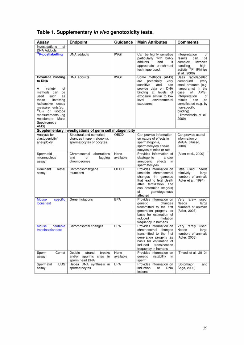
39
Table 1. Supplementary in vivo genotoxicity tests. Assay Endpoint Guidance Main Attributes Comments Investigations of DNA Adducts
32P-postlabelling DNA adducts IWGT Can be highly sensitive
particularly with bulky adducts and if appropriate enrichment technique used.
Interpretation of results can be complex. Involves handling high-activity
32P. (Phillips
et al., 2000) Covalent binding to DNA A variety of methods can be used such as those involving radioactive decay measurements(eg. 14
C-) or isotope measurements (eg Accelerator Mass Spectrometry AMS)
DNA Adducts IWGT Some methods (AMS) are potentially very sensitive and can provide data on DNA binding at levels of exposure similar to low level environmental exposures
Uses radiolabelled compound (very small amounts (e.g. nanograms) in the case of AMS). Interpretation of results can be complicated (e.g. by non-specific binding). (Himmelstein et al., 2009)
Supplementary investigations of germ cell mutagenicity Analysis for clastogenicity/ aneuploidy
Structural and numerical changes in spermatogonia, spermatocytes or oocytes
OECD Can provide information on nature of effects in spermatogonia, spermatocytes and/or oocytes of mice or rats
Can provide useful information on MoGA. (Russo, 2000)
Spermatid micronucleus assay
Chromosomal aberrations and or lagging chromosomes
None available
Provides information of clastogenic and/or aneugenic effects in spermatocytes.
(Allen et al., 2000)
Dominant lethal assay
Chromosomal/gene mutations
OECD Provides information on unstable chromosomal changes in gametes that lead to fetal death after fertilization and can determine stage(s) of gametogenesis affected
Little used. needs relatively large numbers of animals (Adler et al., 1994)
Mouse specific locus test
Gene mutations EPA Provides information on genetic changes transmitted to the first generation progeny as basis for estimation of induced mutation frequency in humans
Very rarely used. Needs large numbers of animals (Adler, 2008)
Mouse heritable translocation test
Chromosomal changes EPA Provides information on chromosomal changes transmitted to the first generation progeny as basis for estimation of induced translocation frequency in humans
Very rarely used. Needs large numbers of animals (Adler, 2008)
Sperm Comet assay
Double strand breaks and/or apurinic sites in sperm head DNA
None available
Provides information on genetic instability in sperm
(Trivedi et al., 2010)
Spermatid UDS assay
Repair DNA synthesis in spermatocytes
EPA Provides information on induction of DNA lesions
(Sotomajor and Sega, 2000)
40
Summary Stage 2 (In Vivo Genotoxicity Testing).
83. The in vivo genotoxicity testing strategy has to be designed on a case-by-
case basis and can be used to address aspects of in vivo mutagenicity, for
example;
1) Investigation of mutagenic end point(s) identified in Stage 1,
2) Investigation of genotoxicity in tumour target tissue(s),
3) Investigation of potential for germ cell genotoxicity,
4) Investigation of in vivo mutagenicity for chemicals which were negative in
Stage 1 but where there is high or moderate and prolonged exposure.
5) Investigation of genotoxicity in site of contact tissues.
The recommended in vivo genotoxicity test(s) include micronucleus assay,
bone marrow cytogenetics, Comet assay in rodents and transgenic rodent
mutation assay. In some instances there may be a need to undertake more
than one in vivo test to perform an initial assessment of in vivo mutagenic
potential (e.g. where endpoints cannot be assessed in one study and there is
a need to investigate multiple end points before reaching conclusions on in
vivo mutagenic potential). Multiple endpoints may be combined in a single
study. If positive results are obtained it is important to consider the evidence
for genotoxic mode of action and check the data for evidence of irrelevant
positive results. Usually negative results obtained in a carefully selected in
vivo test (possibly studying more than one endpoint and tissue) will be
sufficient to address positive results found in vitro. However, a further test(s)
may be needed if some of the genotoxic effects seen in Stage 1 in vitro tests
had not been adequately studied in vivo (e.g. the chemical affects multiple
mutagenic end-points), or other aspects of the genotoxic potential of the
chemical had not been fully resolved (e.g. in the case where an investigation
of heritable effects was required). If equivocal results are obtained, then
supplementary testing may be needed. This may involve repeating some
aspects of the recommended in vitro and/or in vivo genotoxicity tests, or
performing additional investigations (e.g. MoGA investigations, such as DNA
adducts and/or more detailed consideration of heritable effects). The
supplementary in vivo genotoxicity testing strategy should be devised on a
case-by-case basis. There is a need to select the most appropriate assay(s)
41
on a case-by-case basis. All relevant factors such as results from previous
tests, structural alerts and available information on toxicokinetics and
metabolism of the substance, should be considered. In the absence of
appropriate germ cell genotoxicity data, the COM considers it is reasonable to
assume that all somatic cell mutagens have the potential to be germ cell
mutagens.
Possible Future Developments
84. The COM is aware that new assays and toxicogenomic approaches are under
development which might be of value within genotoxicity testing. These
include the detection of gene mutations at the endogenous
phosphatidylinositol glycan complementation group A gene (Pig-A), a reporter
gene in which mutations are currently detected in peripheral red blood cells of
mammals (Bryce et al., 2008a; Dertinger et al., 2011; Miura et al., 2009). This
assay has potential advantage of integration into regulatory toxicity tests
(Dertinger et al., 2010) and it is noted that Pig-A mutations increase with
duration of dosing (Miura et al., 2009). Other potential tests include
investigation of instability in expanded simple tandem repeats in male
gametes and offspring to evaluate heritable mutations (Singer et al., 2006).
The development of new high throughput assays for the assessment of germ
line mutations and the quantification of risk from such data may provide
opportunities to protect future generations from mutated DNA sequences.
Developments within the field of toxicogenomics are also likely to provide new
methods for investigating genotoxic mechanisms and informing on MoGA.
The COM have reviewed data generated in this field several times during
2008 and 2009 up to the drafting of this guidance statement but currently
conclude that the evidence does not support the routine use of toxicogenomic
approaches as an adjunct to genotoxicity testing
(http://www.iacom.org.uk/papers/index.htm). A recent workshop held by the
ILSI-HESI (Health Effects Institute) IVGT (In Vitro Genetic Toxicity Testing)
Project Committee reviewed 16 assays/technologies which were at various
stages of development (defined as emerging to mature). The workshop
highlighted emerging approaches to genotoxicity testing such as Enzyme-
DNA films and DNA adductome studies (Lynch et al., 2011).
42
Annex 1: Sensitivity and Specificity Data Considered by the COM
1. Data for sensitivity (correct identification of rodent carcinogens) and specificity
(correct prediction of non-carcinogens as assessed in rodent carcinogenicity
bioassays) have been obtained from a number of publications. Information is
available for (Q)SAR approaches, screening tests and in vitro genotoxicity
assays .
2. The figures quoted depend on the carcinogenicity data set used (e.g. Gold
Carcinogenicity Potency database (http://potency.berkeley.edu/), the
classification of genotoxicity test results (i.e. positive, negative, equivocal
based on study authors results or subjected to independent peer review) and
whether equivocal and/or technically compromised (inadequate) test results
have been included in the analyses. Sensitivity/specificity data for
genotoxicity tests using a sub-set of genotoxic carcinogens have not been
published, because this would require considerable work to evaluate the
mode of action for carcinogenicity for a large number of chemicals.
Specificity data for identification of chemicals with no in vivo genotoxic activity
(non-genotoxins) have not been published, as there are no published
databases for such chemicals.
3. The sensitivity and specificity data that have been reviewed by the COM are
tabulated in Annex 1, Tables 1-3 (rounded to whole numbers). The list of tests
for each stage is not necessarily exhaustive.
.
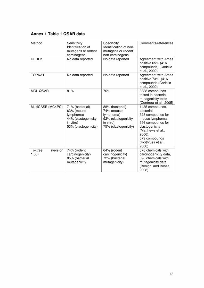
43
Annex 1 Table 1 QSAR data Method Sensitivity
Identification of mutagens or rodent carcinogens
Specificity Identification of non-mutagens or rodent non-carcinogens
Comments/references
DEREK No data reported No data reported Agreement with Ames positive 65% (416 compounds) (Cariello et al., 2002)
TOPKAT No data reported No data reported Agreement with Ames positive 73% (416 compounds (Cariello et al., 2002)
MDL QSAR 81% 76% 3338 compounds tested in bacterial mutagenicity tests (Contrera et al., 2005)
MultiCASE (MC4PC) 71% (bacterial) 63% (mouse lymphoma) 44% (clastogenicity in vitro) 53% (clastogenicity)
88% (bacterial) 74% (mouse lymphoma) 92% (clastogenicity in vitro) 75% (clastogenicity)
1485 compounds, bacterial. 328 compounds for mouse lymphoma. 556 compounds for clastogenicity (Matthews et al., 2006). 679 compounds (Roithfuss et al., 2006)
Toxtree (version 1.50)
74% (rodent carcinogenicity) 85% (bacterial mutagenicity
64% (rodent carcinogenicity) 72% (bacterial mutagenicity)
878 chemicals with carcinogenicity data, 698 chemicals with mutagenicity data (Benigni and Bossa, 2008)
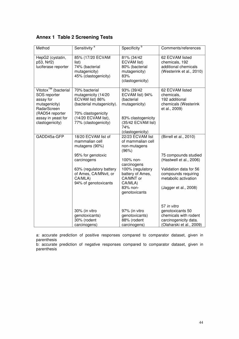
44
Annex 1 Table 2 Screening Tests Method Sensitivity
a Specificity
b
Comments/references
HepG2 (cystatin, p53, Nrf2) luciferase reporter
85% (17/20 ECVAM list) 74% (bacterial mutagenicity) 45% (clastogenicity)
81% (34/42 ECVAM list) 80% (bacterial mutagenicity) 83% (clastogenicity)
62 ECVAM listed chemicals, 192 additional chemicals (Westerink et al., 2010)
VitotoxTM
(bacterial SOS reporter assay for mutagenicity) RadarScreen (RAD54 reporter assay in yeast for clastogenicity)
70% bacterial mutagenicity (14/20 ECVAM list) 86% (bacterial mutagenicity). 70% clastogenicity (14/20 ECVAM list), 77% (clastogenicity)
93% (39/42 ECVAM list) 94% (bacterial mutagenicity) 83% clastogenicity (35/42 ECVAM list) 74% (clastogenicity)
62 ECVAM listed chemicals, 192 additional chemicals (Westerink et al., 2009)
GADD45a-GFP 18/20 ECVAM list of mammalian cell mutagens (90%) 95% for genotoxic carcinogens 63% (regulatory battery of Ames, CA/MNvit, or CA/MLA) 94% of genotoxicants 30% (in vitro genotoxicants) 30% (rodent carcinogens)
22/23 ECVAM list of mammalian cell non-mutagens (96%) 100% non-carcinogens 100% (regulatory battery of Ames, CA/MNT or CA/MLA) 83% non-genotoxicants 97% (in vitro genotoxicants) 88% (rodent carcinogens)
(Birrell et al., 2010) 75 compounds studied (Hastwell et al., 2006) Validation data for 56 compounds requiring metabolic activation (Jagger et al., 2008) 57 in vitro genotoxicants 50 chemicals with rodent carcinogenicity data. (Olaharski et al., 2009)
a: accurate prediction of positive responses compared to comparator dataset, given in parenthesis b: accurate prediction of negative responses compared to comparator dataset, given in parenthesis
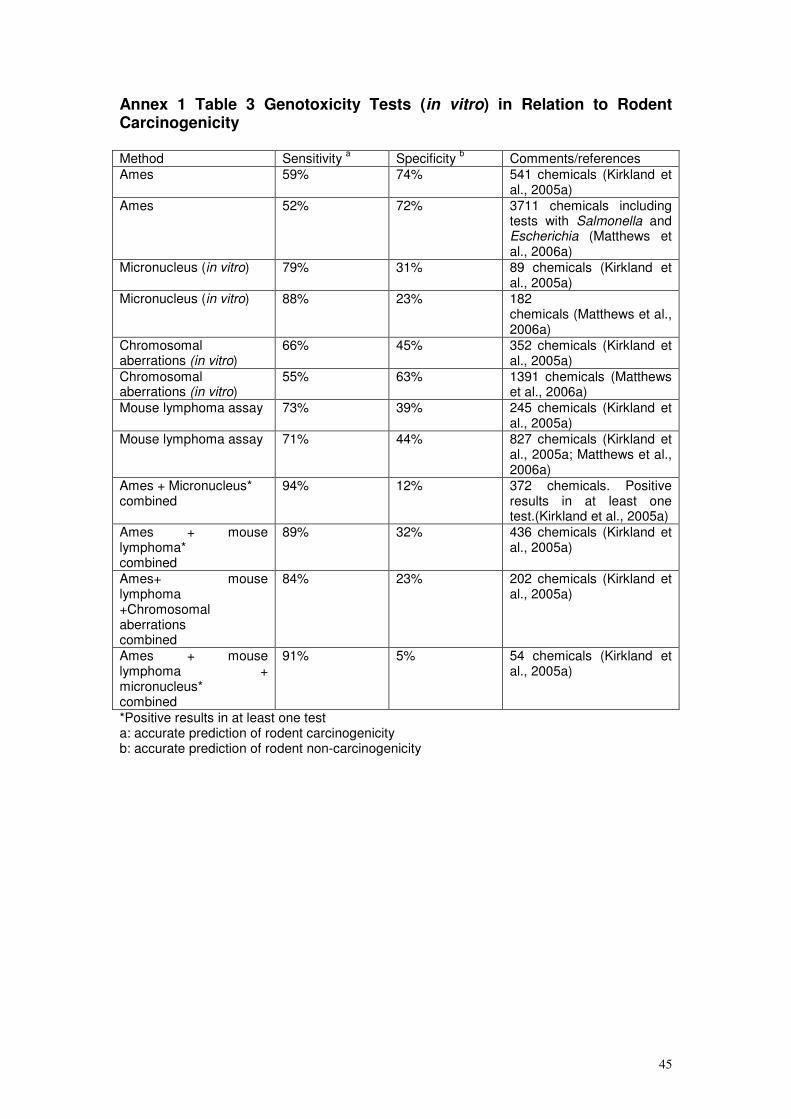
45
Annex 1 Table 3 Genotoxicity Tests (in vitro) in Relation to Rodent Carcinogenicity Method Sensitivity
a Specificity
b Comments/references
Ames 59% 74% 541 chemicals (Kirkland et al., 2005a)
Ames 52% 72% 3711 chemicals including tests with Salmonella and Escherichia (Matthews et al., 2006a)
Micronucleus (in vitro) 79% 31% 89 chemicals (Kirkland et al., 2005a)
Micronucleus (in vitro) 88% 23% 182 chemicals (Matthews et al., 2006a)
Chromosomal aberrations (in vitro)
66% 45% 352 chemicals (Kirkland et al., 2005a)
Chromosomal aberrations (in vitro)
55% 63% 1391 chemicals (Matthews et al., 2006a)
Mouse lymphoma assay 73% 39% 245 chemicals (Kirkland et al., 2005a)
Mouse lymphoma assay 71% 44% 827 chemicals (Kirkland et al., 2005a; Matthews et al., 2006a)
Ames + Micronucleus* combined
94% 12% 372 chemicals. Positive results in at least one test.(Kirkland et al., 2005a)
Ames + mouse lymphoma* combined
89% 32% 436 chemicals (Kirkland et al., 2005a)
Ames+ mouse lymphoma +Chromosomal aberrations combined
84% 23% 202 chemicals (Kirkland et al., 2005a)
Ames + mouse lymphoma + micronucleus* combined
91% 5% 54 chemicals (Kirkland et al., 2005a)
*Positive results in at least one test a: accurate prediction of rodent carcinogenicity b: accurate prediction of rodent non-carcinogenicity
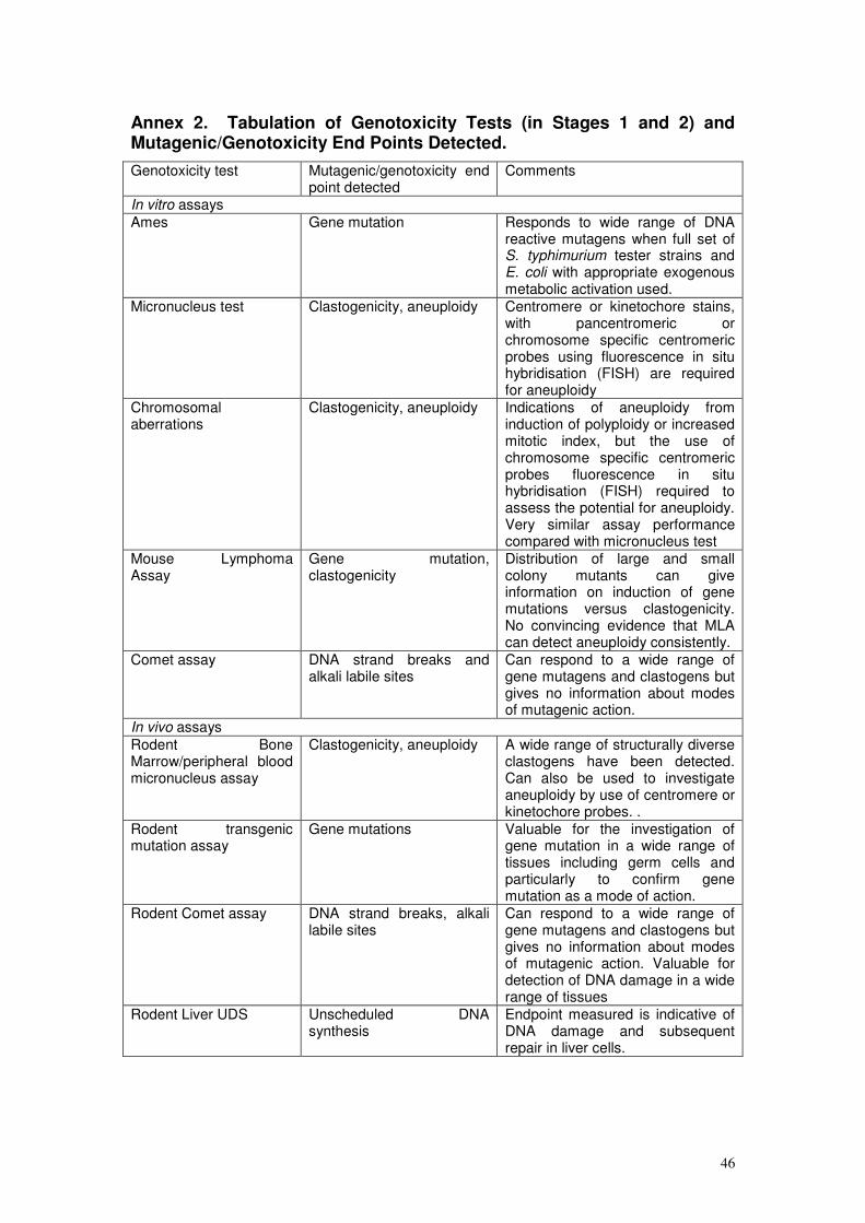
46
Annex 2. Tabulation of Genotoxicity Tests (in Stages 1 and 2) and Mutagenic/Genotoxicity End Points Detected.
Genotoxicity test Mutagenic/genotoxicity end point detected
Comments
In vitro assays
Ames Gene mutation Responds to wide range of DNA reactive mutagens when full set of S. typhimurium tester strains and E. coli with appropriate exogenous metabolic activation used.
Micronucleus test Clastogenicity, aneuploidy Centromere or kinetochore stains, with pancentromeric or chromosome specific centromeric probes using fluorescence in situ hybridisation (FISH) are required for aneuploidy
Chromosomal aberrations
Clastogenicity, aneuploidy Indications of aneuploidy from induction of polyploidy or increased mitotic index, but the use of chromosome specific centromeric probes fluorescence in situ hybridisation (FISH) required to assess the potential for aneuploidy. Very similar assay performance compared with micronucleus test
Mouse Lymphoma Assay
Gene mutation, clastogenicity
Distribution of large and small colony mutants can give information on induction of gene mutations versus clastogenicity. No convincing evidence that MLA can detect aneuploidy consistently.
Comet assay DNA strand breaks and alkali labile sites
Can respond to a wide range of gene mutagens and clastogens but gives no information about modes of mutagenic action.
In vivo assays
Rodent Bone Marrow/peripheral blood micronucleus assay
Clastogenicity, aneuploidy A wide range of structurally diverse clastogens have been detected. Can also be used to investigate aneuploidy by use of centromere or kinetochore probes. .
Rodent transgenic mutation assay
Gene mutations Valuable for the investigation of gene mutation in a wide range of tissues including germ cells and particularly to confirm gene mutation as a mode of action.
Rodent Comet assay DNA strand breaks, alkali labile sites
Can respond to a wide range of gene mutagens and clastogens but gives no information about modes of mutagenic action. Valuable for detection of DNA damage in a wide range of tissues
Rodent Liver UDS Unscheduled DNA synthesis
Endpoint measured is indicative of DNA damage and subsequent repair in liver cells.
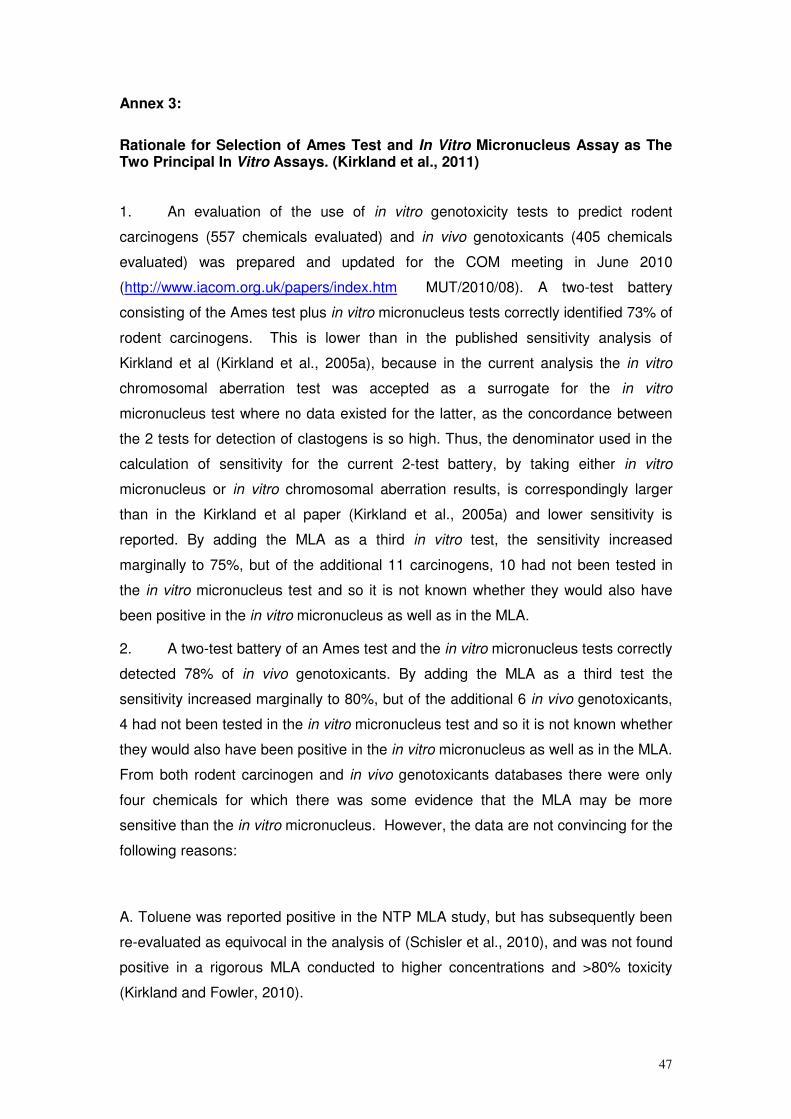
47
Annex 3:
Rationale for Selection of Ames Test and In Vitro Micronucleus Assay as The Two Principal In Vitro Assays. (Kirkland et al., 2011)
1. An evaluation of the use of in vitro genotoxicity tests to predict rodent
carcinogens (557 chemicals evaluated) and in vivo genotoxicants (405 chemicals
evaluated) was prepared and updated for the COM meeting in June 2010
(http://www.iacom.org.uk/papers/index.htm MUT/2010/08). A two-test battery
consisting of the Ames test plus in vitro micronucleus tests correctly identified 73% of
rodent carcinogens. This is lower than in the published sensitivity analysis of
Kirkland et al (Kirkland et al., 2005a), because in the current analysis the in vitro
chromosomal aberration test was accepted as a surrogate for the in vitro
micronucleus test where no data existed for the latter, as the concordance between
the 2 tests for detection of clastogens is so high. Thus, the denominator used in the
calculation of sensitivity for the current 2-test battery, by taking either in vitro
micronucleus or in vitro chromosomal aberration results, is correspondingly larger
than in the Kirkland et al paper (Kirkland et al., 2005a) and lower sensitivity is
reported. By adding the MLA as a third in vitro test, the sensitivity increased
marginally to 75%, but of the additional 11 carcinogens, 10 had not been tested in
the in vitro micronucleus test and so it is not known whether they would also have
been positive in the in vitro micronucleus as well as in the MLA.
2. A two-test battery of an Ames test and the in vitro micronucleus tests correctly
detected 78% of in vivo genotoxicants. By adding the MLA as a third test the
sensitivity increased marginally to 80%, but of the additional 6 in vivo genotoxicants,
4 had not been tested in the in vitro micronucleus test and so it is not known whether
they would also have been positive in the in vitro micronucleus as well as in the MLA.
From both rodent carcinogen and in vivo genotoxicants databases there were only
four chemicals for which there was some evidence that the MLA may be more
sensitive than the in vitro micronucleus. However, the data are not convincing for the
following reasons:
A. Toluene was reported positive in the NTP MLA study, but has subsequently been
re-evaluated as equivocal in the analysis of (Schisler et al., 2010), and was not found
positive in a rigorous MLA conducted to higher concentrations and >80% toxicity
(Kirkland and Fowler, 2010).

48
B. Benzyl acetate was reported positive in the NTP MLA study, and subsequently re-
evaluated as positive by Schisler et al (2010), but an expert panel review (Mitchell et
al, 1997) identified this chemical as "untestable" in the MLA because it reacts with
the plastic of the culture vessels and may thus produce artefacts.
C. Morphine was negative in a non-standard in vitro micronucleus test in which
mouse splenocytes were treated only for 21 hr in the absence of metabolic activation.
It is possible that morphine may induce micronuclei when tested at higher
concentrations over shorter periods in the absence and presence of metabolic
activation in a standard assay.
D. Thiabendazole is an aneugen which, typically, has a very steep dose response. It
has been found positive for induction of micronuclei in vitro in several papers, but is
reported equivocal or negative in other papers, possibly because optimum
concentration spacing, treatment and sampling times were not used.
3. Whilst re-testing of these four chemicals, and of several others for which
neither in vitro micronucleus or chromosomal aberration data exist (Kirkland et al.,
2010), could provide additional reassurance, the Committee concluded that, based
on the large amount of available data, there is no convincing evidence that any
rodent carcinogen or in vivo genotoxicant would fail to be detected by using an in
vitro genotoxicity test battery consisting of Ames test and in vitro micronucleus test.
Summary analyses of sensitivity for the combination of Ames and micronucleus tests
are provided in Annex 1.
4 The revised strategy of two tests (Ames and MNvit) allows for the efficient
identification of all mutagenic end-points but, by reducing the number of mammalian
cell tests from that recommended by COM in 2000, and following improved
methodologies, the risk of misleading positive results is decreased.
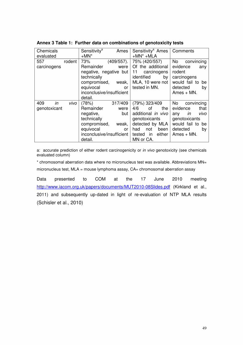
49
Annex 3 Table 1: Further data on combinations of genotoxicity tests
Chemicals evaluated
Sensitivitya Ames +MN*
Sensitivitya Ames +MN* +MLA
Comments
557 rodent carcinogens
73% (409/557). Remainder were negative, negative but technically compromised, weak, equivocal or inconclusive/insufficient detail.
75% (420/557) Of the additional 11 carcinogens identified by MLA, 10 were not tested in MN.
No convincing evidence any rodent carcinogens would fail to be detected by Ames + MN.
409 in vivo genotoxicant
(78%) 317/409 Remainder were negative, but technically compromised, weak, equivocal or inconclusive/insufficient detail.
(79%) 323/409 4/6 of the additional in vivo genotoxicants detected by MLA had not been tested in either MN or CA.
No convincing evidence that any in vivo genotoxicants would fail to be detected by Ames + MN.
a: accurate prediction of either rodent carcinogenicity or in vivo genotoxicity (see chemicals evaluated column)
* chromosomal aberration data where no micronucleus test was available. Abbreviations MN=
micronucleus test, MLA = mouse lymphoma assay, CA= chromosomal aberration assay
Data presented to COM at the 17 June 2010 meeting
http://www.iacom.org.uk/papers/documents/MUT2010-08Slides.pdf (Kirkland et al.,
2011) and subsequently up-dated in light of re-evaluation of NTP MLA results
(Schisler et al., 2010)
50
References Aardema, M., Snyder, R.D., Spicer, C., Divi, K., Morita, T., Mauthe, R., Gibson, D.P., Soelter, S., Curry, P.T., Thybaud, V., et al. (2006). SFTG international collaborative study on in vitro micronucleus test. III. Using CHO cells. Mutation Research 607, 61-87. Aardema, M.J., Albertini, S., Arni, P., Henderson, L.M., Kirsch-Volders, M., Mackay, J.M., Sarrif, D.A., Stringer, D.A., and Taalman, R.D.F. (1998). Aneuploidy: a report of an ECETOC task force Mutation Research 410, 3-79. Ackerman, J., Sharma, R., Hitchcock, J., Hayashi, T., Nagai, Y., Li, S., Lu, S., Miret, J., Tang, K., Spence, F., and Aubrecht, J. (2009). Inter-laboratory evaluation of the bioluminescent Salmonella reverse mutation assay using 10 model chemicals. Mutagenesis 24, 433-438. Adler, I.-D. (2008). Mutagenicity Tests in vivo. In Toxicology and Risk Assessment H. Grein, Snyder, R. , ed. (Wiley and Sons, UK), pp. 371-384. Adler, I.D., Shelby, M.D., Bootman, J., Favor, J., Generoso, W., Pacchierotti, F., Shibuya, T., and Tanaka, N. (1994). Summary report of the Working Group on Mammalian Germ Cell Tests. Mutation Research 312, 313-318. Allen, J.W., Collins, B.W., Lan, A., Afshari, A.J., George, M.H., DeAngelo, A.B., and Fuscoe, J.C. (2000). Erythrocyte and spermatid micronucleus analyses in mice chronically exposed to potassium bromate in drinking water. Environmental and Molecular Mutagenesis 36, 250-252. Ashby, J., and Paton, D. (1993). The influence of chemical structure on the extent and sites of carcinogenesis for 522 rodent carcinogens and 55 different human carcinogen exposures Mutation Research 286, 3-74. Attia, S.M., Badary, O.A., Hamada, S., de Angelis, M.H., and Adler, I.D. (2005). Othovanadate increased the frequency of aneuploid mouse sperm without micronucleus induction in mouse bone marrow erythroctes at the same dose level. Mutation Research 583, 158-167. Aubrecht, J., Osowski, J.J., Persaud, P., Cheung, J.R., Ackerman, J., Lopes, S.H., and Ku, W.W. (2007). Bioluminescent Salmonella reverse mutation assay: a screen for detecting mutagenicity with high throughput attributes. Mutagenesis 22, 335-342. Benfenati, E., Benigni, R., Demarini, D., Helma, C., Kirkland, D., Martin, T., Mazzatorta, P., Ouédraogo-Arras, G., Richard, A., Schilter, B., et al. (2009.). Predictive models for carcinogenicity and mutagenicity: frameworks, state-of-the-art, and perspectives. J Environ Sci Health C Environ Carcinog Ecotoxicol Rev. 27, 57-90. Benigni, R., and Bossa, C. (2008). Structure alerts for carcinogenicity, and the Salmonella assay system: a novel insight through the chemical relational databases technology. Mutation Research 659, 248-261. Benigni, R., Bossa, C., and Worth, A. (2010). Structural analysis and predictive value of the rodent in vivo micronucleus assay. Mutagenesis 25, 335-341. Benigni, R., Netzva, T.I., Benfenati, E., Bossa, C., Franke, R., Helma, C., Hulzebos, E., Marchant, C., Richard, A., Woo, Y.-T., and Yang, C. (2007). The expanding role of predictive toxicology: An update on the (Q)SAR models for mutagens and carcinogens. Journal of Environmental Science and Health Part C 25, 53-97. Birrell, L., Cahill, P., Hughes, C., Tate, M., and Walmsley, R. (2010). AM recommended lists of genotoxic and non-genotoxic chemicals for assessment of new genotoxicity tests. Mutation Research 695, 87-95. Bishop, J.B. (2003). Female specific reproductive toxicities following preconception exposure to xenobiotics. Advances in Experimental Medicine 518, 1-9. Blakey, D., Galloway, S., Kirkland, D., and MacGregor, J.T. (2008). Regulatory aspects of genotoxicity testing: from hazard identification to risk assessment. Mutation Research 657, 84-90. Bowen, D.E., Whitwell, J.H., Lillford, L., Henderson, D., Kidd, D, Mc Garry, S, Pearce, G, , and Beevers, C., Kirkland, DJ; (2011). Evaluation of a multi-endpoint assay in rats, combining the bone-marrow micronucleus test, the Comet assay and the flow-cytometric peripheral blood micronucleus test. Mutation Research 722, 7-19. Brambilla, G., and Martelli, A. (2004). Failure of the standard battery of short-term tests in detecting some rodent and human genotoxic carcinogens. Toxicology 196, 1-19. Brendler-Schwaab, S.Y., Hartmann, A., Pfuhler, S., and Speit, G. (2005). The in vivo comet assay: use and status in genotoxicity testing. Mutagenesis 20, 245-254.
51
Bryce, S., Benis, J., and Dertinger, S. (2008a). In vivo Mutation Assay based on the Endogenous Pig-a Locus. Environmental and Molecular Mutagenesis 49, 256-264. Bryce, S.M., Avlasevich, S.L., Bemis, J.C., Lukamowicz, M., Elhajouji, A., Van Goethem, F., De Boeck, M., Beerens, D., Aerts, H., van Gompel, J., et al. (2008b). Interlaboratory evaluation of a flow cytometric, high content in vitro micronucleus assay. Mutation Research 650, 181-195. Bryce, S.M., Bemis, J.C., Avlasevich, S.L., and Dertinger, S.D. (2007). In vitro micronucleus assay scored by flow cytometry provides a comprehensive evaluation of cytogenetic damage and cytotoxicity. Mutation Research 630, 78-91. Bryce, S.M., Shi, J., Nicolette, J., Diehl, M., Sonders, P., Avlasevich, S.L., Raja, S., Bemis, J.C., and Dertinger, S.D. (2010). High content flow cytometric micronucleus scoring method is applicable to attachment cell lines. Environmental and Molecular Mutagenesis 51, 260-266. Burlinson, B., Tice, R., Speit, G., Agurell, E., Brendler-Schwaab, S., Collins, A., Escobar, P., Honma, M., Kumaravel, T., Nakajima, M., et al. (2007). Fourth International Workgroup on Genotoxicity testing: Results of the in vivo Comet assay workgroup. Mutation Research 627, 31-35. Cariello, N., Wilson, J., Britt, B., Wedd, D., Burlinson, B., and Gombar, V. (2002). Comparison of the computer programs DEREK and TOPKAT to predict bacterial mutagenicity. Mutagenesis 17, 321-329. Cimino, M. (2006). Comparative overview of current international strategies and guidelines for genetic toxicology testing for regulatory purposes. Environmental and Molecular Mutagenesis 47, 362-390. Ciranni, R., Antonetti, M., and Miglore, L. (1995). Vanadium salts induce cytogenetic effects in in vivo treated mice. Mutation Research 343, 53-60. Clare, M., Lorenzon, G., Akhurst, L., Marzin, D., van Delft, J., Montero, R., Botta, A., Bertens, A., Cinelli, S., Thybaud, V., and Lorge, E. (2006). SFTG international collaborative study on in vitro micronucleus test II. Using human lymphocytes. Mutation Research 607, 37-60. Claxton, L., Stewart-Hook, V., and Warren, S. (2001). Methods for the spiral Salmonella mutagenicity assay including specialised applications. . Mutation Research 488, 241-257. Clements, J. (2000). The Mouse Lymphoma Assay. Mutation Research 455, 97-110. Combes, R., Grindon, C., Cronin, M., Roberts, D., and Garrod, J. (2007). Proposed integrated decision-tree testing strategies for mutagenicity and carcinogenicity in relation to the EU REACH legislation. . ATLA 35, 267-287. Contrera, J.F., Mathews, E.J., Kruhlak, N., and Benz, R.D. (2005). In silico screening of chemicals for bacterial mutagenicity using electropological E-state indices and MDL QSAR software. Regulatory Toxicology Pharmacology 43, 313-323. Contrera, J.F., Matthews, E.J., Kruhlak, N.L., and Benz, R.D. (2008). In silico screening of chemicals for genetic toxicity using MDL-QSA, nonparametric discriminant analysis, E-State, connectivity and molecular property descriptors. Toxicology Mechanisms and Methods 18, 207-216. Corvi, R., Albertini, S., Hartung, T., Hoffman, S., Muaurici, D., Pfuhler, S., van Benthem, J., and Vanparys, P. (2008). ECVAM retrospective validation of the in vitro micronucleus test (MNT). Mutagenesis 23, 271-283. CSGMT (1995). The Collaborative Study Group for the Micronucleus Test (CSGMT)(CSGMT/JEMS.MMS, The Mammalian Study Group of the Environmental Mutagen Society of Japan). Protocol recommended by the CSGMT/JEMS. MMS for the short-term mouse peripheral blood micronucleus test. Mutagenesis 10, 153-159. Curren, R.D., Mun, G.C., Gibson, D.P., and Aardema, M.J. (2006). Development of a method for assessing micronucleus induction in a 3D human skin model (EpiDerm
TM). Mutation
Research 607, 192-204. Custer, L., and Sweder, K. (2008). The role of genetic toxicology in drug discovery and optimization. Current Drug Metabolism. 9, 978-985. De Boeck, M., van der Leede, B., Van Goethem, F., De Smedt, A., Steemans, M., A., L., and Vanparys, P. (2005). Flow cytometric analysis of micronucleated reticulocytes: Time- and dose-dependent response of known mutagens in mice, using multiple blood sampling. Environmental Molecular Mutagenesis 46, 30-42. Dean, S., Brooks, T.M., Burlinson, B., Mirsalis, J.C., Myhr, B., Recio, L., and Thybaud, V. (1999). Transgenic mouse mutation assay systems can play an important role in regulatory mutagenicity testing for in vivo for the detection of site-of-contact mutagens. Mutagenesis 14, 141-151.
52
Dearfield, K., Thybaud, V., Cimino, M., Custer, L.L., Czich, A., Harvey, J., Hester, S., Kim, J.H., Kirkland, D., Levy, D.D., et al. (2011). Follow-up actions from positive results of In Vitro genetic toxicology testing. Environmental and Molecular Mutagenesis 52, 177-204. Decordier, I., Papine, A., Plas, G., Roesems, S., Vande Loock, K., Moreno-Palomo, J., Cemeli, E., Anderson, D., Fucic, A., Marcos, R., et al. (2009). Automated image analysis of cytokinesis-blocked micronuclei: an adapted protocol and a validated scoring procedure for biomonitoring. . Mutagenesis 24, 85-93. Dertinger, S., Phonethepswath, S., Franklin, D., Weller, P., Torous, D., Bryce, S., Avlasevich, S., Bemis, J., Hyrien, O., Palis, J., and MacGregor, J. (2010). Integration of mutation and chromosomal damage endpoints into 28-day repeat dose toxicology studies. Toxicological Sciences 115, 401-411. Dertinger, S.D., Bryce, S.M., Phonethepswath, S., and Avlasevich, S.L. (2011). When pigs fly: immunomagnetic separation facilitates rapid determination of Pig-a mutant frequncy by flow cytometric analysis. . Mutation Research. 721, 163-170. DOH (1989). Department of Health. Report on Health and Social Subjects, 35. Guidelines for the testing of chemicals for Mutagenicity. Committee on Mutagenicity of Chemicals in Food, Consumer Products and the Environment. London, HMSO. DOH (2000). Department of Health. Guidance for the testing of chemicals for Mutagenicity. Committee on Mutagenicity of Chemicals in Food, Consumer Products and the Environment. Eastmond, D., Hartwig, A., Anderson, D., Anwar, W.A., Cimino, M.C., Dobrev, I., Douglas, G.R., Nohmi, T., Phillips, D.H., and Vickers, C. (2009). Mutagenicity testing for chemical risk assessment: update of the WHO/IPCS Harmonised Scheme. Mutagenesis 24, 341-349. Eichenlaub-Ritter, U., Adler, I., Carere, A., and Pacchierotti, F. (2007). Gender differences in germ cell mutagenesis and genetic risk. Environmental Research 104, 22-36. Elespuru, R., Agarwal, R., Atrakchi, A., Bigger, C., Heflich, R., Jagannath, D., Levy, D., Moore, M., Ouyang, Y., Robison, T., and Sotomayor, R., Cimino, MC, Dearfield KL. (2009). Current and future application of genetic toxicity assays: the role and value of in vitro mammalian assays. Toxicological Sciences 109, 172-179. Emmert, B., Bunger, J., Keuch, K., Muller, M., Emmert, S., Hallier, E., and Westphal, G. (2006). Mutagenicity of cytochrome P450 2E1 substrates in the Ames test with metabolic competent S. typhimurium strain YG7108pin3ERb5. Toxicology 228, 66-76. Fellows, M., O'Donovan, M., Lorge, E., and Kirkland, D. (2008). Comparison of different methods for an accurate assessment of cytotoxicity in the in vitro micronucleus test II. Practical aspects with toxic agents. Mutation Research 655, 4-21. Fellows, M.D., Boyer, S., and O'Donovan, M.R. (2011). The incidence of positive results in the mouse lymphoma TK assay (MLA) in pharmaceutical screening and their prediction by MultiCase MC4PC. . Mutagenesis 26, 529-532. Fenech, M. (2006). Cytokinesis-block micronucleus assay evolves into a 'cytome' assay for chromosomal instability, mitotic dysfunction and cell death. Mutation Research 600, 58-66. Fenech, M. (2007). Cytokinesis-block micronucleus assay. Nature Protocols 2, 1084-1104. Fischer, S.J., Benson, L.M., Fauq, A., Naylor, S., and Windebank, A.J. (2008 ). Cisplatin and dimethyl sulfoxide react to form an adducted compound with reduced cytotoxicity and neurotoxicity. . NeuroToxicology 29, 444-452. Flamand, N., Marrot, L., Belaidi, J.-P., Bourouf, L., Dourille, E., Feltes, M., and Meunier, J.-R. (2006). Development of genotoxicity test procedures with Episkin® a reconstructed human skin model: Towards new tools for in vitro risk assessment of dermally applied compounds? Mutation Research 606, 39-51. Fluckiger-Isler, S., Baumeister, M., Braun, K., Gervais, V., Hasler-Nguyen, N., Reimann, R., Van Gompel, J., Wunderlich, H.-G., and Engelhardt, G. (2004). Assessment of the performance of the Ames II
TMassay: a collaborative study with 19 coded compounds.
Mutation Research 558, 181-197. Fowler, P. (2009). Industrial Genotoxicity Group, November 2009. Reduction of False/Misleading Positives in in-vitro genetic toxicology testing: Importance of cell selection and toxicity measure. . Fowler, P., Wiiliams, K., Jeffrey , L., Carmichael, P., Ardema, M., Diembeck, W., Fautz, R., Harvey, J., Hewitt, N., Latil, A., et al. (2009a). Reduction of misleading ("false") positive results in mammalian cell genotoxicity assays I. Choice of cell type. Environmental Molecular Mutagenesis 50, 572.
53
Fowler, P., Wiiliams, K., Jeffrey , L., Carmichael, P., Ardema, M., Diembeck, W., Fautz, R., Harvey, J., Hewitt, N., Latil, A., et al. (2009b). Reduction of misleading ("false") positive results in mammalian cell genotoxicity assays II. Importance of accurate toxicity measurement. . Environmental Molecular Mutagenesis 50, 573. Galloway, S. (2000). Cytotoxicity and chromosome aberrations in Vitro: experience in industry and the case for an upper limit on toxicity in the aberration assay. Environmental and Molecular Mutagenesis 35, 191-201. Galloway, S., Lorge, E., Aardema, M.J., Eastmond, D., Fellows, M., Heflich, R., Kirkland, D., Levy, D.D., Lynch, A.M., Marzin, D., et al. (2011). Workshop summary: Top concentration for in vitro mammalian cell genotoxicity assays; and report from working group on toxicity measures and top concentration for in vitro cytogenetics assays (chromosome aberrations and micronucleus). Mutation Research 723, 77-83. Garriott, M.L., Phelps, J.B., and Hoffman, W.P. (2002). A protocol for the in vitro micronucleus test I. Contributions to the development of a protocol suitable for regulatory submissions from an examination of 16 chemicals with different mechanisms of action and different levels of activity. Mutation Research 517, 123-134. Gatehouse, D., Haworth, S., Cebula, T., Kier, L., Matsushima, T., Melcion, C., Nohmi, T., Ohta, T., and Venitt, S. (1994). Recommendations for the performance of bacterial mutation assays. Mutation Research 312, 217-233. Gealy, R., Wright-Bourque, J.L., Kraynak, A.R., McKelvey, T.W., Barnum, J.E., and Storer, R.D. (2007). Validation of a high-throughput in vitro alkaline elution/rat hepatocyte assay for DNA damage. Mutation Research 629, 49-63. Glei, M., Hovhannisyan, G., and Pool-Zobel, B.L. (2009). Use of Comet-FISH in the study of DNA damage and repair: Review. Mutation Research 681, 33-43. Grant, S.G., Zhang, Y.P., Klopman, G., and Rosenkranz, H.S. (2000). Modelling the mouse lymphoma forward mutational assay: the Gene-Tox program database. . Mutation Research 465, 201-229. Hamada, S., Sutou, S., Morita, T., Wakata, A., Asanami, S., Hosoya, S., Ozawa, S., Kondo, K., Nakajima, M., Shimada, H., et al. (2001). Evaluation of the rodent micronucleus assay by a 28-Day treatment protocol: Summary of the 13th collaborative study by the collaborative study group for the micronucleus test (CSGMT)/Environmental Mutagen Society of Japan (JEMS)-Mammalian Mutagenicity Study Group (MMS). Environmental and Molecular Mutagenesis 37, 93-110. Hartmann, A., Agurell, E., Beevers, C., Brendler-Schwaab, S.Y., Burlinson, B., Clay, P., Collins, A.R., Smith, A., Speit, G., Thybaud, V., and Tice, R. (2003). Recommendations for conducting the in vivo alkaline Comet assay. Mutagenesis 18, 45-51. Hartmann, A., Kiskinis, E., Fjallman, A., and Suter, W. (2001). Influence of cytotoxicity and compound precipitation on test results in the alkaline comet assay. . Mutation Research 497, 199-212. Hartmann, A., Schumacher, M., Plappert-Helberg, U., Lowe, P., Suter, W., and Mueller, L. (2004). Use of the alkaline in vivo Comet assay for mechanistic genotoxicity investigations. Mutagenesis 19, 51-59. Hastwell, P., Chai, L., Roberts, K., Webster, T., Harvey, J., Rees, R., and Walmsley, R. (2006). High-specificity and high sensitivity genotoxicicty assessment in a huma cell line: Validation of the GreenScreen HC GADD45a-GFP genotoxicity assay. Mutation Research 607, 160-175. Hayashi, M., Dearfield, K., Kasper, P., Lovell, D., Martus, H., and Thybaud, V. (2010). Compilation and use of genetic toxicity historical control data. Mutation Research (accepted for publication 29 September 2010). Hayashi, M., Dearfield, K., Kasper, P., Lovell, D., Martus, H.J., and Thybaud, V. (2011 ). Compilation and use of genetic toxicity historical control data. . Mutation Research 723, 87-90. Helleday, T., Johansson, F., and Jenssen, D. (2001). The DRAG Test: an assay for detection of genotoxic damage. ATLA 29, 233-241. Himmelstein, M.W., Boogaard, P.J., Cadet, J., Farmer, P.B., Kim, J.H., Martin, E.A., Persaud, R., and Shuker, D.E. (2009). Creating context for the use of DNA adduct data in cancer risk assessment: II. Overview of methods of identification and quantitation of DNA damage. Critical Reviews in Toxicology. 39, 679-694.
54
Hiramoto, K., Nasuhara, A., Michikoshi, K., Kato, T., and Kikugawa, K. (1997). DNA strand-breaking activity and mutagenicity of 2,3-dihydro-3,5-dihydroxy-6-methyl-4-H-pyran-4-one (DDMP), a Maillard reaction product of glucose and glycine. . Mutation Research 395 47-56. Hu, T., Kaluzhny, Y., Mun, G.C., Barnett, B., Karetsky, V., Wilt, N., Klausner, M., Curren, R.D., and Aardema, M.J. (2009). Intralaboratory and interlaboratory evaluation of the EpiDerm
TM 3D human reconstructed skin micronucleus assay. Mutation Research 673, 100-
108. Jagger, C., Tate, M., Cahill, P., Hughes, C., Knight, A.W., Billinton, N., and Walmsley, R.M. (2008). Assessment of the genotoxicity of S9-generated metabolites using the GreenScreen HC GADD45a-GFP assay. Mutagenesis 24, 35-50. Jarabek, A., Pottenger, L., Andrews, L., Casciano, D., Embry, M., Kim, J., Preston, J., Vijayaraj Reddy, M., Schoeny, R., Shuker, D., et al. (2009). Creating context for use of DNA adduct data in cancer risk assessment: I Data organization. Critical Reviews in Toxicology 39, 659-678. Kamber, M., Fluckiger-Isler, S., Engelhardt, G., Jaeckh, R., and WZeiger, E. (2009 ). Comparison of the Ames II and the traditional Ames test responses with respect to mutagenicity, strain specificities, need for metabolism and correlation with rodent carcinogenicity. . Mutagenesis 24, 359-366. Kasper, P., Uno, Y., Mauthe, R., Asano, N., Douglas, G., Matthews, E., Moore, M., Mueller, L., Nakajima, M., Singer, T., and Speit, G. (2007). Follow-up testing of rodent carcinogens not positive in the standard genotoxicity test battery: IWGT workgroup report. Mutation Research 627, 106-116. Kawaguchi, S., Nakamura, T., Yamamoto, A., Honda, G., and Sasaki, Y.F. (2010). Is the comet assay a sensitive procedure for detecting genotoxicity? Journal of Nucleic Acids 2010: , 541050. Kennelly, J.C., Water, R., Ashby, J., Lefevre, P.A., Burlinson, B., Benford, D.J., Dean, S.W., and Mitchell, I.d. (1993). In vivo rat liver UDS assay. In Supplementary Mutagenicity Tests. UKEMS Report. Cambridge University Press pp 52-77. Kirkland, D. (2010). Evaluation of different cytotoxic and cytostatic measures for the in vitro micronucleus test (MNVit). Summary of results of collaborative trial. . Mutation Research 702, 139-147. Kirkland, D., Aardema, M., Henderson, L., and Muller, L. (2005a). Evaluation of the ability of a battery of three in vitro genotoxicity tests to discriminate rodent carcinogens and non-carcinogens 1. Sensitivity, specificity and relative predictivity. Mutation Research 584, 1-256. Kirkland, D., and Fowler, P. (2010). Further analysis of Ames-negative rodent carcinogens that are only genotoxic in mammalian cells at concentrations exceeding 1mM including retest of compounds of concern. . Mutagenesis 25, 539-553. Kirkland, D., Hayashi, M., Jacobson-Kram, D., Kasper, P., MacGregor, J., Muller, L., and Uno, Y. (2007a). Summary of major conclusions from the 4th IWGT, San Francisco, 9-10, 2005. Mutation Research 627, 5-9. Kirkland, D., Pfuhler, S., Tweats, D., Aardema, M., Corvi, R., Darroudi, F., Elhajouji, A., Glatt, H., Hastwell, P., Hayashi, M., et al. (2007b). How to reduce false positive results when undertaking in vitro genotoxicity testing and thus avoid unnecessary follow-up animal tests: Report of an ECVAM Workshop. Mutation Research 628, 31-55. Kirkland, D., Reeve, L., Gatehouse, D., and Vanparys, P. (2011). A core in vitro genotoxicity battery comprising the Ames test plus the in vitro micronucleus test is sufficient to detect rodent carcinogens and in vivo genotoxins. Mutation Research 721, 27-73. Kirkland, D., and Speit, G. (2008). Evaluation of the ability of a battery of three in vitro genotoxicity tests to discriminate rodent carcinogens and non-carcinogens III. Appropriate follow-up testing in vivo. Mutation Research 654, 114-132. Kirkland, D.J., Aardema, M., Banduhn, N., Carmichael, P., Fautz, R., Meunier, J.R., and Pfuhler, S. (2007c). In vitro approaches to develop weight of evidence (WoE) and mode of action (MoA) discussions with positive in vitro genotoxicity results. Mutagenesis 22, 161-175. Kirkland, D.J., Henderson, L., Marzin, D., Muller, L., Parry, J., Speit, G., Tweats, D.J., and Williams, G.M. (2005b). Testing strategies in mutagenicity and genetic toxicology: An appraisal of the guidelines of the European Scientific Committee for Cosmetics and Non-Food Products for the evaluation of hair dyes. Mutation Research 588, 88-105. Kirsch-Volders, M., Sofuni, T., Aardema, M., Albertini, S., Eastmond, D., Fenech, M., Ishidate, M.J., Kirchner, S., Lorge, E., Morita, T., et al. (2003a). Report from the in vitro micronucleus assay workshop group. Mutation Research 540, 153-163.
55
Kirsch-Volders, M., Vanhauwaert, A., DeBoeck, M., and Decordier, I. (2002). Importance of detecting numerical versus structural chromosome aberrations. Mutation Research 504, 137-148. Kirsch-Volders, M., Vanhauwaert, A., Eichenlaub-Ritter, U., and Decordier, I. (2003b). Indirect mechanisms of genotoxicity. Toxicology Letters 140-141, 63-74. Knight, A.W., Little, S., Houck, K., Dix, D., Judson, R., Richard, A., McCarroll, N., Akerman, G., Yang, C., Birrell, L., and Walmsley, R.M. (2009). Evaluation of high-throughput genotoxicity assays used in profiling the US EPA ToxCast
TM chemicals. Regulatory
Toxicology Pharmacology 55, 188-199. Krishna, G., Urda, G., and Theiss, J. (1998). Principles and practices of integrating genotoxicity evaluation into routine toxicology studies: A pharmaceutical industry perspective. Environmental and Molecular Mutagenesis 32, 115-120. Ku, W., Aubrecht, J., Mauthe, R., Schiestl, R., and Fornace Jr, A. (2007a). Genetic toxicity assessment: employing the best science for human safety evaluation Part VII: Why not start with a single test: A transformational alternative to genotoxicity hazard and risk assessment. Toxicological Sciences 99, 20-25. Ku, W.W., Bigger, A., Brambilla, G., Glatt, H., Gocke, E., Guzzie, P., Hakura, A., Honma, M., Martus, H.J., Scott Obach, R., and Roberts, S. (2007b). Strategy for genotoxicity testing - metabolic considerations. Mutation Research 627, 59-77. Laingam, S., Froscio, S.M., and Humpage, A.R. (2008). Flow-cytometric analysis of in vitro micronucleus formation: Comparative studies with WIL2-NS human lymphoblastoid and L5178Y mouse lymphoma cells. Mutation Research 656, 19-26. Lambert, I.B., Singer, T.M., Boucher, S.E., and Douglas, G.R. (2005). Detailed review of transgenic rodent mutation assays. Mutation Research 590, 1-280. Le Hegarat, L., Dumont, J., Josse, R., Huet, S., Lanceleur, R., Mourot, A., Poul, J., Guguen-Guillouzo, C., Guillouzo, A., and Fessard, V. (2010). Assessment of the genotoxic potential of indirect chemical mutagens in HepaRG cells by the comet and the cytokinesis-block micronucleus assays Mutagenesis 25, 555-560. Li, A.P., Gupta, R.S., Heflich, R.H., and J.S., W. (1988). A review and analysis of the Chinese hamster ovary/hypoxanthine guanine phosphoribosyl transferase assay to determine the mutagenicity of chemical agents. A report of phase III of the U.S. Environmental Protection Agency Gene-Tox Program. Mut.Res. 196, 17-36. Long, L., Kirkland, D., Whitwell, J., and Halliwell, B. (2007). Different cytotoxic and clastogenic effects of epigallocatechin gallate in various cell-culture media due to variable rates of oxidation in culture media. Mutation Research 634, 177-183. Lorge, E., Hayashi, M., Albertini, S., and Kirkland, D. (2008). Comparison of different methods for an accurate assessment of cytotoxicity in the in vitro micronucleus test. Mutation Research 655, 1-3. Lorge, E., Thybaud, V., Aardema, M., Oliver, J., Wakata, A., Lorenzon, G., and Marzin, D. (2006). SFTG international collaborative study on the in vitro micronucleus test I. General conditions and overall conclusions of the study. Mutation Research 607, 13-36. Lynch, A.M., Sasaki, J.C., Elespuru, R., Jacobson-Kram, D., Thybaud, V., De Boeck, M., Aardema, M.J., Aubrecht, J., Benz, R.D., Dertinger, S.D., et al. (2011). New and emerging technologies for genetic toxicity testing. Environmental and Molecular Mutagenesis 52, 205-223. Madrigal-Bujaidar, E., Madrigal-Santillan, E.O., Alvarez-Gonzalez, I., Baez, R., and Marquez, P. (2008). Micronuclei Induced by Imipramine and Desipramine in Mice: a subcronic study. Basic & Clinical Pharmacology & Toxicology. 103, 569-573. Maierhofer, C., Jentsch, I., lederer, G.F., C., and Speicher, M.R. (2002). Multicolor FISH in two and three dimensions for clastogenic analysis. Mutagenesis 17, 523-527. Matthews, E.J., Kruhlak, N., Cimino, M., Benz, R.D., and Contrera, J.F. (2006a). An analysis of genetic toxicology, reproductive and developmental toxicity and carcinogenicity data I: Idenitfication of carcinogens using surrogate end points. Regulatory Toxicology and Pharmacology 44, 83-96. Matthews, E.J., Kruhlak, N.L., Cimino, M.C., Benz, R.D., and Contrera, J.F. (2006b). An analysis of genetic toxicology, reproductive and developmental toxicity and carcinogenicity data: II Identification of genotoxicants, reprotoxicants, and carcinogens using in silico methods. Regulatory Toxicology and Pharmacology 44, 97-110.
56
Mekenyan, O., Dimitrov, S., Serafimova, R., Thompson, E., Kotov, S., Dimitrova, N., and Walker, J. (2004). Identification of the structural requirements for mutagenicity by incorporating molecular flexibility and metabolic activation of chemicals I: TA100 model Chemical Research and Toxicology 17, 753-766. Miller, J.E., Vlasakova, K., Glaab, W.E., and Skopek, T.R. (2005 ). A low volume, high-throughput forward mutation assay in Salmonella typhimurium based on fluorouracil resistance. Mutation Research 578, 210-224. Mitchell, I.D., Lambert, T.R., Burden, M., Sunderland, J., Porter, R., and Carlton, J.B. (1995). Is polyploidy an important genotoxic lesion? Mutagenesis 10, 79-83. Miura, D., Dobrovolsky, V.N., Kimoto, T., Kasahara, Y., and Heflich, R.H. (2009). Accumulation and persistence of Pig-A mutant peripheral red blood cells following treatment of rats with a single and split doses of N-ethyl-N-nitrosourea. . Mutation Research 677, 86-92. Moore, M., Honma, M., Clements, J., Bolcsfoldi, G., Burlinson, B., Cifone, M., Clarke, J., Clay, P., Doppalapudi, R., Fellows, M., et al. (2007). Mouse Lymphoma thymidine kinase gene mutation assay: Meeting of the International Workshop on Genotoxicity Testing, San Francisco, 2005, recommendations for 24-h treatment. Mutation Research 627, 36-40. Moore, M., Honma, M., Clements, J., Bolcsfoldi, G., Cifone, M., Delongchamp, R., Fellows, M., Gollapudi, B., Jenkinson, P., Kirby, P., et al. (2003). Mouse Lymphoma Thymidine Kinase Gene Mutation Assay: International Workshop on Genotoxicity Tests Workgroup Report-Plymouth, UK 2002. Mutation Research 540, 127-140. Moore, M.M., Honma, M., Clements, J., Bolcsfoldi, G., B., B., Cifone M, C.J., Delongchamp R, D.R., Fellows M, Gollapudi B, Hou S, Jenkinson P, Lloyd M,, Majeska J, M.B., O'Donovan M, Omori T, Riach C, San R, Stankowski LF Jr, Thakur, and AK, V.G.F., Wakuri S, Yoshimura I. (2006). Mouse lymphoma thymidine kinase gene mutation assay: follow-up meeting of the International Workshop on GenotoxicityTesting--Aberdeen, Scotland, 2003--Assay acceptance criteria, positive controls, and data evaluation. Environmental Molecular Mutagenesis. 47, 1-5. Müller, L., and Kasper, P. (2000). Human biological relevance and the use of threshold-arguments in regulatory genotoxicity assessment: experience with pharmaceuticals. Mutation Research 464, 19-34. Muller, L., Blakely, D., Dearfield, K., Galloway, S., Guzzie, P., Hayashi, M., Kasper, P., Kirkland, D., MacGregor, J., Parry, J., et al. (2003). Strategy for genotoxicity testing and stratification of genotoxicity test results-report on initial activities of the IWGT Expert Group. Mutation Research 540, 177-181. Mun, G.C., Aardema, M., Hu, T., Barnett, B., Kaluzhny, Y., Klausner, M., Karetsky, V., Dahl, E.L., and Curren, R.D. (2009). Further development of the EpiDerm
TM 3D reconstructed
human skin micronucleus (RSMN) assay. Mutation Research 673, 92-99. OECD (1997). Organisation for Economic Co-operation and Development. Ninth addendum to the OECD guidelines for the testing of chemicals. Update of Section 4, Health Effects. Revised guidelines 424, 471, 473, 474, 475, 476, 483, 486, 487 OECD (2004). OECD Series on testing and Assessment. Number 49. The report from the Expert Group on (Quantitative) Structure-Activity Relatiopnships [(Q)SARs] on the principles for the validation of (Q)SARs. (2nd Meeting of the Ad hoc Expert group on QSARs, OECD Headquaters, 20-21 September 2004. JT00176183, ENV/JM/MONO(2004)24, 17-Dec-2005. Ogawa, I., Furukawa, S., Abe, M., Tanaka, Y., and Hayashi, S.U., K. (2009). Multi-endpoint genotoxic assay using L5178Y (Tk
+/--3.7.2c) cells. The Journal of Toxicological Sciences 34,
547-553. Olaharski, A., Albertini, S., Kirchner, S., Platz, S., Uppal, H., Lin, H., and Kolaja, K. (2009). Evaluation of the GreenScreen GADD45a-GFP inidcator assay with non-proprietary and proprietary compounds. Mutation Research 672, 10-16. Oliver, J., Meunier, J.-R., Awogi, T., Elhajouji, A., Ouldelhkim, M.-C., Bichet, N., Thybaud, V., Lorenzon, G., Marzin, D., and Lorge, E. (2006). SFTG international collaborative study on in vitro micronucleus test V. Using L5178Y cells. Mutation Research 607, 125-152. Pacchierotti, F., Adler, I.-D., Eichenlaub-Ritter, U., and Mailher, J.B. (2007). Gender effects on the incidence of aneuploidy in mammalian germ cells. Environ.Res. 104, 46-69. Parry, J., Parry, E., Phrakonkham, P., and Corvi, R. (2010). Analysis of published data for top concnetrations in mammalian cell genotoxicity testing. Mutagenesis 25, 531-538. Parry, J.M. (2006). The use of the in vitro micronucleus assay to detect and assess the aneugenic activity of chemicals. Mutation Research 607, 5-8.
57
Pfuhler, S. (2009). 5th International Workshop on Genotoxicity testing, August 17-19, 2009. Topic 3: In vitro test approaches with better predictivity. ICEM, Florence, August 20-25, 2009. Pfuhler, S., Albertini, S., Fautz, R., Herbold, B., Madle, S., Utesch, D., and Poth, A. (2007). Genetic toxicity assessment: employing the best science for human safety evaluation Part IV: Recomendation of a working group of the Gesellschaft fuer Umwelt-Mutationsforschung (GUM) for a simple and straightforward approach to genotoxicity testing. Toxicological Sciences 97, 237-240. Pfuhler, S., Fellows, M., van Benthem, J., Corvi, R., Curren, R., Dearfield, K., Fowler, P., Frotschl, R., Elhajouji, A., Le Hegarat, L., et al. (2011). In vitro genotoxicity test approaches with better predictivity: Summary of an IWGT workshop. Mutatation Research 723, 101-107. Pfuhler, S., Kirkland, D., Hayashi, M., Vanparys, P., Carmichael, P., Dertinger, S.D., Eastmond, D., Elhajouji, A., Krul, C., Rothfuss, A., et al. (2009). Reduction of use of animals in regulatory genotoxicity testing: Identification and implementation opportunities- Report from an ECVAM workshop. Mutation Research 680, 31-42. Pfuhler, S., Kirst, A., Aardema, M., Banduhn, N., Garsten, G., Daisuke, A., Costabel-Farkas, M., Dufour, E., Fautz, R., Harvey, J., et al. (2010). A tiered approach to the use of alternatives to animal testing for the safety assessment of cosmetics: Genotoxicity. A COLIPA analysis. Regulatory Toxicology and Pharmacology 57, 315-324. Phelps, J.B., Garriott, M.L., and Hoffman, W.P. (2002). A protocol for the in vitro micronucleus test II. Contributions to the validation of a protocol suitable for regulatory submissions from an examination of 10 chemicals with different mechansims of action and different levels of activity. Mutation Research 521, 103-112. Phillips, D.H., Farmer, P.B., Beland, F.A., Nath, R.G., Poirier, M.C., Reddy, M.V., and Turteltaub, K.W. (2000). Methods of DNA adduct determination and their application to testing compounds for genotoxicity. Environmental and Molecular Mutagenesis. 35, 222-233. Pratt, I.S., and Barron, T. (2003). Regulatory recognition of indirect genotoxicity mechanisms in the European Union. Toxicology Letters 140-141, 53-62. Ranaldi, R., Gambuti, G., Eichenlaub-Ritter, U., and Pacchierotti, F. (2008). Triclorfon effects on mouse oocytes following in vivo exposure Mutation Research 651, 125-130. Rapp, A., Bock, C., Dittmar, H., and Greulich, K.-O. (2000). UV-A breakage sensitivty of human chromosomes as measured by COMET-FISH depends on gene density and not on chromosome size. . Journal of Photochemistry and Photobiology B: Biology 56, 109-117. Reifferscheid, G., Arndt, C., and Schmid, C. (2005). Further development of the β-lactamase mutagen assay and evaluation by comparison with Ames fluctuation tests and the umu test. Environmental and Molecular Mutagenesis 46, 126-139. Roithfuss, A., Steger-Hartmann, T., Heinrich, N., and Wichard, J. (2006). Conspirational Prediction of the Chromosome-Damaging Potential of Chemicals. Chem Res Toxicol 19, 1313-1319. Rothfuss, A., Honma, M., Czich, A., Aardema, M., Burlinson, B., Galloway, S., Hamada, S., Kirkland, D., Heflich, R., Howe, J., et al. (2010). Improvement Of In Vitro Genotoxicity Assessment: Combination of Acute Tests and the Link to Standard Toxicity Testing. Mutation Research (accepted for publication). Rothfuss, A., Honma, M, Czich, A, Aardema, MJ, Burlinson, B, Galloway, S, Hamada, S, Kirkland, D, Heflich, RH, Howe, J, Nakajima, M, O'Donovan, M, Plappert-Helbig, U, Priestley, C, Recio, L, Schuler, M, Uno Y, Martus, HJ (2011). Improvement of in vivo genotoxicity assessment: Combination of acute tests and integration into standard toxicity testing. Mutation Research xx, xx. Russo, A. (2000). In vivo cytogenetics in mammalian germ cells. Mutation Research 455, 167-189. Santos, S.J., Singh, N.P., and Natarajan, A.T. (1997). Fluorescence in situ hybridization with comets. . Experimental Cell Research 232, 407-411. Schisler, M., Gollupudi, B., and Moore, M. (2010). Evaluation of the Mouse Lymphoma Mutation Assay (MLA) Data of the U.S. National Toxicology Program (NTP) Using International Workshop on Genotoxicity Tests (IWGT) Criteria. Environmental Molecular Mutagenesis 51, P96. Serafimova, R., Todorov, M., Pavlov, T., Kotov, S., Jacob, E., Aptula, A., and Mekenyan, O. (2007). Identification of the structural requirements for mutagenicity by incorporating molecular flexibility and metabolic activation of chemicals. II. General Ames and mutagenicity model Chemical Research and Toxicology 20, 662-676.
58
Shuey, D.L., Gudi, R., Krsmanovic, L., and Gerson, R.J. (2007 ). Evidence that oxymorphone-induced increases in micronuclei occur secondary to hyperthermia. Toxicological Sciences 95, 369-375. Singer, T., Lambert, I., Williams, A., Douglas, G., and Yauk, C.L. (2006). Detection of induced male germline mutation: Correlations and comparisons between traditional germline mutation assays, transgenic rodent assays and expanded simple tandem repeat instability assays. Mutation Research 598, 164-193. Snyder, R.D., and Smith, A.D. (2005). Computational prediction of genotoxicity: room for improvement. Drug Discovery Today (Biosilico) 10, 1119-1124. Sofuni, T., Honma, M., Hayashi, M., Shimada, H., Tanaka, N., Wakuri, S., Awogi, T., Yamamoto, K.I., Nishi, Y., and Nakadate, M. (1996). Detection of in vitro clastogens and spindle poisons by the mouse lymphoma assay using the microwell method: interim report of an international collaborative study. Mutagenesis 11, 349-355. Sotomajor, R.E., and Sega, G.A. (2000). Unscheduled DNA synthesis assay in mammalian spermatogenic cells: an update. Environmental and Molecular Mutagenesis 36, 255-265. Spanswick, V.J., Hartley, J.M., and Hartley, J.A. (2010). Methods in Molecular Biology, volume 613, Drug-DNA Interaction Protocols, Editied KR Fox, Chapter 17 Measurement of DNA interstrand Crosslinking in Individual Cells Using the Single Cell Gel Electrophoresis (Comet) Assay. 267-282. Speit, G., Vasquez, M., and Hartmann, A. (2009). The comet assay as an indicator test for germ cell genotoxicity. Mutation Research 681, 3-12. . Suzuki, H., Ikeda, N., Kobayashi, K., Terashima, Y., Shimada, Y., Suzuki, T., Hagiwara, T., Hatakeyama, S., Nagaoka, K., Yoshida, J., et al. (2005). Evaluation of liver and peripheral blood micronucleus assays with 9 chemicals using young rats. A study by the Collaborative Study group for the Micronucleus Test (CSGMT)/ Japanese Environmental Mutagenicity Society (JEMS)-Mammalian Mutagenicity Study Group (MMS). Mutation Research 583, 133-145. Thybaud, V., Aardema, M., Clements, J., Dearfield, K., Galloway, S., Hayashi, M., Jacobson-Kram, D., Kirkland, D., MacGregor, J.T., Marzin, D., et al. (2007). Strategy for genotoxicity testing: Hazard identification and risk assessment in relation in vitro testing. Mutation Research 627, 41-58. Thybaud, V., Dean, S., Nohmi, T., de Boer, J., Douglas, G.R., Glickman, B.W., Gorelick, N.J., Heddle, J.A., Heflich, R.H., Lambert, I., et al. (2003). In vivo transgenic mutation assays. Mutation Research 540, 141-151. Torous, D.K., Dertinger, S.D., Hall, N.E., and Tometsko, C.R. (2000). Enumeration of micronucleated reticulocytes in rat peripheral blood: a flow cytometric study. Mutation Research 465, 91-99. Trivedi, P.P., Kushwaha, S., Tripathu, D.N., and Jena, G.B. (2010). Evaluation of male germ cell toxicity in rats: Correlation between sperm head morphology and sperm comet assay. Mutation Research 703, 115-121. Tweats, D., Blakely, D., Heflich, R.H., Jacobs, A., Jacobsen, S.D., Morita, T., Nohmi, T., O'Donovan, M.R., Sasaki, Y., Sofuni, T., and Tice, R. (2007a). Report of the IWGT working group on strategies and interpretation of regulatory in vivo tests I. Increases in micronucleated bone marrow cells in rodents that do not indicate genotoxic hazards. Mutation Research 627, 78-91. Tweats, D.J., Blakely, D., Heflich, R.H., Jacobs, A., Jacobsen, S.D., Morita, T., Nohmi, T., O'Donovan, M.R., Sasaki, Y.F., Sofuni, T., and Tice, R. (2007b). Report of the IWGT working group on strategy/interpretation for regulatory in vivo tests. II. Identification of in vivo-only positive compounds in the bone marrow micronucleus test. Mutation Research 627, 92-105. UKEMS. (1989). Statistical evaluation of mutagenicity test data. UKEMS sub-committee on guidelines for mutagenicity testing. Report . Part III. Editor Kirkland DJ. Published Cambridge University Press. Vasquez, M.Z. (2010). Combining the in vivo comet and micronucleus assays: a practical approach to genotoxicity testing and data interpretation. Mutagenesis 25, 187-199. Vogt, E., Kirsch-Volders, M., Parry, J., and Eichenlaub-Ritter, U. (2008). Spindle formation, chromosome segregation and the spindle checkpoint in mammalian oocytes and susceptibility to meiotic error. Mutation Research. 651, 14-29. Wakata, A., Matsuoka, A., Yamakage, K., Yoshida, J., Kubo, K., Kobayashi, K., Senjyu, N., Itoh, S., Miyajima, H., Hamada, S., et al. (2006). SFTG international collaborative study on in vitro micronucleus test IV. Using CHL cells. Mutation Research 607, 88-124.
59
Wakata, A., Miyamae, Y., Sato, S., Suzuki, T., Morita, T., Asano, N., Awogi, T., Kondo, K., and Hayashi, M. (1998). Evaluation of the Rat Micronucleus Test with Bone Marrow and peripheral Blood: Summary of the 9th Collaborative Study by the CSGMT/JEMS.MMS. Environmental and Molecular Mutagenesis 32, 84-100. Wang, J., Sawyer, J.R., Chen, L., Chen, T., Honma, M., Mei, N., and Moore, M. (2009). The Mouse Lymphoma Assay detects recombination, deletion and aneuploidy. Toxicological Sciences 109, 96-105. Westerink, M.A., Stevenson, J.C.R., Horbach, G.J., Saito, Y., and Schoonen, W.G. (2010). The development of RAD51C, Cystatin A, p53 and Nrf2 luciferase-reported assays in metabolically competent HepG2 cells for the assessment of mechanism-based genotoxicity and oxidative stress in early research phase of drug development. Mutation Research 696, 21-40. Westerink, W.M.A., Stevenson, J.C.R., Lauwers, A., Griffioen, G., Horbach, G.J., and Schoonen, W.G.E.J. (2009). Evaluation of the Vitotox
TM and RadarScreen assays for rapid
assessment of genotoxicity in the early research phase of drug development. Mutation Research 676, 113-130. Witt, I., Plappert, U., de Wall, H., and Hartmann, A. (2007). Genetic toxicity assessment: employing the best science for human safety evaluation part III. The Comet assay as an alternative to in vitro clastogenicity tests for early drug candidate selection. Toxicological Sciences 97, 21-26. Witt, K., Knapton, A., Wehr, C., Hook, G., Mirsalis, J., Shelby, M., and MacGregor, J. (2000). Micronucleated Erythrocyte Frequency in Peripheral Blood of B6C3F1 Mice from Short-Term, prechronic, and Chronic Studies of the NTP Carcinogenesis Bioassay Program. Environmental and Molecular Mutagenesis 36, 163-194. Witt, K.L., Hughes, L.A., Burka, L.T., McFee, A.F., Mathews, J.M., Black, S.L., and Bishop, J.B. (2003). Mouse bone marrow micronucleus test results do not predict the germ cell mutagenicity of N-hydroxymethylacrylamide in the mouse dominant lethal assay. Environmental and Molecular Mutagenesis 41, 111-120. Yin, H., Cukrcam, S., Betzendahl, I., Adler, I.-D., and Eichenlaub-Ritter, U. (1998). Trichlorfon exposure, spindle aberrations and nondisjunction in mammalian oocytes. . Chromosoma 107, 514-522. Zeiger, E., Ashby, J., Bakale, G., Enslein, K., Klopman, G., and Rosenkranz, H.S. (1996). Prediction of Salmonella mutagenicity. . Mutagenesis 11, 471-484.
60
GLOSSARY
Adduct: A chemical grouping which is covalently bound to a large molecule such as
DNA or protein.
Alkylating agents: Chemicals which leave an alkyl group covalently bound to
biologically important molecules such as proteins and nucleic acids (see adduct).
Many alkylating agents are mutagenic, carcinogenic and immunosuppressive.
Ames test: In vitro assay for bacterial gene mutations using strains of Salmonella
typhimurium developed by Ames and his colleagues.
Aneugenic: Inducing aneuploidy (qv).
Aneuploidy: The circumstances in which the total number of chromosomes within a
cell is not an exact multiple of the normal haploid (see 'polyploidy') number.
Chromosomes may be lost or gained during cell division.
Apoptosis: A form of active cell death resulting in fragmentation of the cell into
membrane-bound fragments (apoptotic bodies). These are usually rapidly removed in
vivo by engulfment by phagocytic cells. Apoptosis can occur normally during
development, but is often triggered by toxic stimuli.
Cancer: Synonym for a malignant neoplasm – that is, a tumour (qv) that grows
progressively, invades local tissues and spreads to distant sites (see also tumour).
Carcinogenesis: The origin, causation and development of tumours (qv). The term
applies to benign as well as malignant neoplasms and not just to carcinomas
Carcinogenicity bioassay: Tests carried out in laboratory animals, usually rats and
mice, to determine whether a substance is carcinogenic. The test material is given
throughout life to groups of animals at different dose levels.
Carcinogens: The causal agents which induce tumours. They include external
factors (chemicals, physical agents, viruses) and internal factors such as hormones.
Chemical carcinogens are structurally diverse and include naturally-occurring
substances as well as synthetic compounds. An important distinction can be drawn
between genotoxic (qv) carcinogens which have been shown to react with and
mutate DNA, and non-genotoxic carcinogens which act through other mechanisms.
The activity of genotoxic carcinogens can often be predicted from their chemical
structure - either of the parent compound or of active metabolites (qv). Most chemical
carcinogens exert their effects after prolonged exposure, show a dose-response
61
relationship and tend to act on a limited range of susceptible target tissues.
Carcinogens are sometimes species or sex-specific and the term should be qualified
by the appropriate descriptive adjectives to aid clarity. Several different chemical and
other carcinogens may interact, and constitutional factors (genetic susceptibility,
hormonal status) may also contribute, emphasising the multifactorial nature of the
carcinogenic process.
Chromosomal aberrations: Collective term of particular types of chromosome
damage induced after exposure to exogenous chemical or physical agents which
damage the DNA. (see clastogen).
Chromosome: In simple prokaryotic organisms, such as bacteria and most viruses,
the chromosome consists of a single circular molecule of DNA containing the entire
genetic material of the cell. In eukaryotic cells, the chromosomes are thread-like
structures, composed mainly of DNA and protein, which are present within the nuclei
of every cell. They occur in pairs, the numbers varying from one to more than 100 per
nucleus in different species. Normal somatic cells in humans have 23 pairs of
chromosomes, each consisting of linear sequences of DNA which are known as
genes.
Clastogen: An agent that produces chromosome breaks and other structural
aberrations such as translocations. Clastogens may be viruses or physical agents as
well as chemicals. Clastogenic events play an important part in the development of
some tumours.
Cytogenetic: Concerning chromosomes, their origin, structure and function.
Cytochrome P450 (CYP): An extensive family of haem-containing proteins involved
in enzymic oxidation of a wide range of endogenous and xenobiotic (qv) substances
and their conversion to forms that may be more easily excreted. In some cases the
metabolites produced may be reactive and may have increased toxicity. In other
cases the substances may be natural precursors of hormones (e.g. steroids).
DNA Strand Breakage; A break in double-stranded DNA in which one or both of the
two strands have been cleaved; both strands have not separated from each other.
DNA Strand Break Assay (Comet assay): Alkaline treatment converts certain types
of DNA lesions into strand breaks that can be detected by the alkaline elution
technique or by measuring migration rate through a filter, or by the single gel
electrophoresis or Comet assay in which cells embedded in a thin layer of gel on a
microscope slides are subjected to electric current causing shorter pieces of DNA to
62
migrate out of the nucleus into a Comet tail. The extent of DNA migration is
measured visually under the microscope on stained cells.
Erythrocyte: red blood cell; corpuscle; one of the formed cells in peripheral blood.
Normally, in humans, the mature form is a non-nucleated, yellowish, biconcave disk,
containing haemoglobin and transporting oxygen. Normochromic erythrocyte; one of
normal colour with a normal concentration of haemoglobin. Polychromatic
erythrocyte; one that, on staining, shows shades of blue combined with tinges of
pink indicative of an immature erythrocyte.
Eukaryotes; A class of organisms, which in contrast to prokaryotes (e.g. bacteria),
comprise cells which have a nucleus in which DNA is organised into characteristic
sets of chromosomes. This includes all plants and fungi except the blue-green algae
and all animals.
Fluorescent in situ hybridization (FISH) A technique in which a chemically
modified DNA (or RNA) probe is hybridized with target DNA, usually present as a
chromosome preparation on a microscopic slide. The chemical modification can be
visualized using a fluorescent microscope either directly when the modification
involves use of a fluorescent dye or indirectly with the use of a fluorescently labelled
affinity reagent (e.g. antibody or avidin). Depending upon the type of probe used, this
approach can be used to precisely map genes to a specific region of a chromosome
in a prepared karyotype, enumerate chromosomes, or detect chromosomal deletions,
translocations, or gene amplifications in cancer cells.
Gametogenesis is a process by which diploid or haploid precursor cells undergo cell
division and differentiation to form mature haploid gametes. Depending on the
biological life cycle of the organism, gametogenesis occurs by meiotic division of
diploid gametocytes into various gametes or by mitotic division of haploid
gametogenous cells.
Genome: All the genetic material in the chromosomes of a particular organism; its
size is generally given as its total number of base pairs.
Genotoxic: The ability of a substance to cause DNA damage, either directly or after
metabolic activation (see also carcinogens).
Genotype: The particular genetic pattern seen in the DNA of an individual.
'Genotype' is usually used to refer to the particular pair of alleles that an individual
possesses at a certain location in the genome. Compare this with phenotype.
63
Germ cell: A biological cell that gives rise to the gametes of an organism that
reproduces sexually. The cells undergo mitotic and meiotic cell division in the
gonads followed by cellular differentiation into mature gametes, either oocytes or
sperm.
Heritable translocation test A test that detects heritable structural chromosome
changes (i.e. translocations) in mammalian germ cells as recovered in first-
generation progeny.
Historical negative control data: In the context of the COM guidance on
genotoxicity testing, this term refers to information on the background genotoxicity or
mutagenicity data for a particular assay from a particular laboratory. Historical
control data should be reported as the mean and confidence intervals for the
genotoxicity or mutagenicity indices investigated.
In vitro chromosomal aberration assay: An assay where cultured cell lines or
human lymphocytes are incubated with test substance. At a predetermined time,
cells are arrested in metaphase, harvested and stained, and the metaphase spreads
are then analysed microscopically for the presence of chromosome aberrations.
Kinetochore is the protein structure which is present on chromosomes where the
spindle fibers attach during division to pull the chromosomes apart. The kinetochore
forms in eukaryotes and assembles on the centromere and links the chromosome to
microtubule polymers from the mitotic spindle during mitosis and meiosis. The
kinetochore contains two regions: an inner kinetochore, which is tightly associated
with the centromere DNA; and an outer kinetochore, which interacts with
microtubules.
Kinetochore staining An immunochemical technique used to detect the presence of
centromeric kinetochore proteins in micronuclei and to identify the origin of
micronuclei. In all but a few cases, the presence of kinetochore in a micronucleus
indicates that it was formed by loss of an entire chromosome, whereas a
micronucleus that lacks a kinetochore originated from an acentric chromosome
fragment.
Maximum Tolerated Dose; The highest dose of a substance that can be given
without causing serious weight loss (>10%) or other signs of toxicity.
Metabolic activation: Metabolism of a compound leading to an increase in its
activity, whether beneficial (e.g. activation of a pro-drug) or deleterious (e.g.
activation to a toxic metabolite).
64
Metabolic activation system: A cell-free preparation (e.g. from the livers of rats pre-
treated with an inducing agent added to in vitro tests to mimic the metabolic
activation typical of mammals.
Metabolism: Chemical modification of a compound by enzymes within the body, for
example by reactions such as hydroxylation (see cytochrome P450), epoxidation or
conjugation. Metabolism may result in activation, inactivation, accumulation or
excretion of the compound.
Metabolite: Product formed by metabolism of a compound.
Micronuclei: Isolated or broken chromosome fragments which are not expelled
when the nucleus is lost during cell division, but remain in the body of the cell forming
micronuclei. Centromere positive micronuclei contain DNA and/or protein material
derived from the centromere. The presence of centromere positive micronuclei
following exposure to chemicals in vitro or in vivo can be used to evaluate the
aneugenic (qv) potential of chemicals.
Micronucleus test: See Micronuclei.
Mitogen: A stimulus which provokes cell division in somatic cells.
Mitosis: The type of cell division which occurs in somatic cells when they proliferate.
Each daughter cell has the same complement of chromosomes as the parent cell.
Mode of Genotoxic Action (MoGA): The mode of action of genotoxicant refers to
the underlying events involved in the process whereby the chemical induces
genotoxic effects. In order for a specific mode of action to be supported there needs
to be evidence from robust mechanistic data to establish a biologically plausible
explanation. Mode of genotoxic action should be distinguished from the term
mechanism of action. The latter relates to having sufficient understanding of the
molecular basis of the chemical genotoxicity to establish causality. Thus mechanism
of action is at the other end of a continuum from little or no evidence of mode of
genotoxic action to scientific proof of mechanism of action.
Mouse lymphoma assay: An in vitro assay for gene mutation in mammalian cells
using a mouse lymphoma cell line L5178Y, which is heterozygous for the gene
(carries only one functional gene rather than a pair) for the enzyme thymidine kinase
(TK+/-). Mutation of that single gene is measured by resistance to toxic
trifluorothymidine. Mutant cells produce two forms of colony - large, which represent
mutations within the gene and small, which represent large genetic changes in the
chromosome such as chromosome aberrations. Thus this assay can provide
65
additional information about the type of mutation which has occurred if colony size is
scored.
Mutation: A permanent change in the amount or structure of the genetic material in
an organism or cell, which can result in a change in phenotypic characteristics. The
alteration may involve a single gene, a block of genes, or a whole chromosome.
Mutations involving single genes may be a consequence of effects on single DNA
bases (point mutations) or of large changes, including deletions, within the gene.
Changes involving whole chromosomes may be numerical or structural. A mutation in
the germ cells of sexually reproducing organisms may be transmitted to the offspring,
whereas a mutation that occurs in somatic cells may be transferred only to
descendent daughter cells.
Mutagenic end-points: these comprise of three levels of genetic change, namely
gene mutation, clastogenicity and aneuploidy
No observable effect concentration (NOEC): the highest administered
concentration at which no adverse effect or specific genotoxic effect is seen
Phenotype: The observable physical, biochemical and physiological characteristics
of a cell, tissue, organ or individual, as determined by its genotype and the
environment in which it develops.
Polyploidy: Numerical deviation of the modal number of chromosomes in a cell,
with approximately whole multiples of the haploid number. Endoreduplication is a
morphological form of polypoidy in which chromosome pairs are associated at
metaphase as diplochromosomes.
32P-postlabelling: A sensitive experimental method designed to measure low levels
of DNA adducts induced by chemical treatment.
Prokaryotes: The simplest living organisms namely viruses, bacteria and some blue
green algae. The genetic material in bacteria is arranged into one chromosomal
complex consisting of a single circular molecule of DNA (or RNA in some viruses).
They lack an organised nucleus. Mitosis and meiosis do not occur, although
nucleotide polymerisation replication takes place and division and multiplication
follow.
Recombination: Breakage of DNA structure with balanced or unbalanced rejoining
of DNA
S9: metabolic activation system comprising of the post-mitochondrial supernatant
(S9) from the homogenised livers of rats treated with P450 dependent drug-
66
metabolizing enzyme inducers such as Arochlor 1254 or phenobarbitone/β-
naphthoflavone. S9 is combined with a mix of co-factors which optimize the activity
of the mixed function oxidases and form a NADPH generating system which has the
capacity to metabolise chemicals in vitro.
Sensitivity: In the context of the COM guidance on a strategy for genotoxicity
testing, the correct identification of rodent carcinogens or in vivo genotoxins using
genotoxicity (mutagenicity) assays based on a defined set of carcinogenicity data
(e.g. Gold Carcinogenicity Potency database)
Screening test; High-Throughput procedures designed to provide rapid information
on toxicological end points for a large number of compounds
Specificity: In the context of the COM guidance on a strategy for genotoxicity
testing, the correct prediction of non-carcinogens as assessed in rodent
carcinogenicity bioassays using genotoxicity (mutagenicity) assays based on a
defined set of carcinogenicity data (e.g. Gold Carcinogenicity Potency database).
Specific locus test: A technique used to detect recessive induced mutations in
diploid organisms; a strain that carries several known recessive mutants in a
homozygous condition is crossed with a non mutant strain that has been treated to
induce mutations in its germ cells; induced recessive mutations allelic with those of
the test strain will be expressed in the progeny.
Spindle apparatus: In cell biology, the spindle apparatus is the structure that
separates the chromosomes into the daughter cells during cell division. It is part of
the cytoskeleton in eukaryotic cells. It is also referred to as the mitotic spindle during
mitosis and the meiotic spindle during meiosis.
Structure Activity Relationships: the relationship between chemical structure and
genotoxic effect based on predictions using computerised models (also Quantitative
Structure Activity Relationships)
Test substance: A chemical element and its compounds in the natural state or
obtained by any manufacturing process, including any additive necessary to preserve
its stability and any impurity deriving from the process used, but excluding any
solvent which may be separated without affecting the stability of the substance or
changing its composition.
Threshold: Dose or exposure concentration below which an effect is not expected.
Topoisomerases: Enzymes which catalyze and guide the unknotting of DNA by
creating transient breaks in the DNA using a conserved tyrosine as the catalytic
67
residue. In so-called circular DNA, in which double helical DNA is bent around and
joined in a circle, the two strands are topologically linked, or knotted. Topoisomerase
I solves the problem caused by tension generated by winding/unwinding of DNA. It
wraps around DNA and makes a cut permitting the helix to spin. Once DNA is
relaxed, topoisomerase reconnects broken strands
Toxicogenomics: A new scientific subdiscipline that combines the emerging
technologies of genomics and bioinformatics to identify and characterise
mechanisms of action of known and suspected toxicants. Currently, the premier
toxicogenomic tools are the DNA microarray and the DNA chip, which are used for
the simultaneous monitoring of expression levels of hundreds to thousands of genes.
Toxicokinetics: The description of the fate of chemicals in the body, including a
mathematical account of their absorption, distribution, metabolism and excretion.
(see pharmacokinetics)
Transgenic: Genetically modified to contain genetic material from another species
(see also genetically modified organism).
Transgenic rodent gene mutation models: Animals which have extra (exogenous)
fragments of DNA incorporated into their genomes. This includes transgenic mice
containing reporter genes to assess in-vivo mutagenicity in recoverable bacterial
gene (lacZ or lac I). DNA can be isolated from a wide range of tissues following
exposure to a test substance and the genes assessed for induced mutations.
Translation: In molecular biology, the process during which the information in mRNA
molecules is used to construct proteins.
Tumour (Synonym - neoplasm): A mass of abnormal, disorganised cells, arising
from pre-existing tissue, which are characterised by excessive and uncoordinated
proliferation and by abnormal differentiation. Benign tumours show a close
morphological resemblance to their tissue of origin; grow in a slow expansile fashion;
and form circumscribed and (usually) encapsulated masses. They may stop growing
and they may regress. Benign tumours do not infiltrate through local tissues and they
do not metastasise. They are rarely fatal. Malignant tumours (synonym - cancer)
resemble their parent tissues less closely and are composed of increasingly
abnormal cells in terms of their form and function. Well differentiated examples still
retain recognisable features of their tissue of origin but these characteristics are
progressively lost in moderately and poorly differentiated malignancies:
undifferentiated or anaplastic tumours are composed of cells which resemble no
known normal tissue. Most malignant tumours grow rapidly, spread progressively
68
through adjacent tissues and metastasise to distant sites. Tumours are
conventionally classified according to the anatomical site of the primary tumour and
its microscopical appearance, rather than by cause.
Unscheduled DNA Synthesis (UDS): DNA synthesis that occurs at some stage in
the cell cycle other than the S period (the normal or 'scheduled' DNA synthesis
period), in response to DNA damage. It is usually associated with DNA repair.
Weight of Evidence A quantitative ranking of evidence, or the qualitative appraisal
of many different forms of evidence (e.g toxicological or genotoxicity data) to arrive at
a conclusion regarding potential hazard (such as mutagenicity).
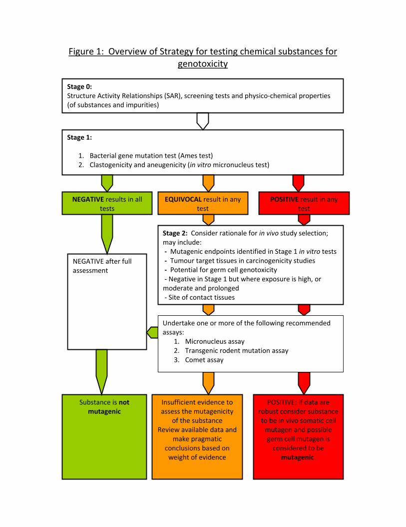
Figure 1: Overview of Strategy for testing chemical substances for genotoxicity
NEGATIVE after full assessment
Stage 1:
1. Bacterial gene mutation test (Ames test) 2. Clastogenicity and aneugenicity (in vitro micronucleus test)
NEGATIVE results in all tests
POSITIVE result in any test
EQUIVOCAL result in any test
Stage 0: Structure Activity Relationships (SAR), screening tests and physico‐chemical properties (of substances and impurities)
Stage 2: Consider rationale for in vivo study selection; may include: ‐ Mutagenic endpoints identified in Stage 1 in vitro tests ‐ Tumour target tissues in carcinogenicity studies ‐ Potential for germ cell genotoxicity ‐ Negative in Stage 1 but where exposure is high, or moderate and prolonged ‐ Site of contact tissues
Substance is not mutagenic
Insufficient evidence to assess the mutagenicity
of the substance Review available data and
make pragmatic conclusions based on weight of evidence
POSITIVE: if data are robust consider substance to be in vivo somatic cell mutagen and possible germ cell mutagen is considered to be
mutagenic.
Undertake one or more of the following recommended assays:
1. Micronucleus assay 2. Transgenic rodent mutation assay 3. Comet assay
Figure 2: Screening (Stage 0) and in vitro tests (Stage 1)
Stage 1 Core Tests: 1. Bacterial gene mutation (Ames test) 2. Clastogenicity and aneugenicity (in vitro micronucleus test)
NEGATIVE results in all Stage 1 tests
POSITIVE result in any Stage 1 test
EQUIVOCAL result in any Stage 1 test
Consider: • Reproducibility • Historical control data • Mode of Genotoxic Action (MoGA)
• Results of stage 0 • Additional in vitro tests (e.g. Mammalian cell mutation, mammalian cytogenetics, Comet assay, or human reconstituted skin)
Consider: • Mode of Genotoxic Action (MoGA)
• Results of stage 0 • Misleading positive results (e.g. bacterial specific metabolism, or excessive mammalian cell cytotoxicity)
Substance is not mutagenic
Insufficient evidence to assess the mutagenicity
of the substance
Substance should be considered to be an in
vitro mutagen.
Proceed to Stage 2 only where in vivo testing is permitted*
Stage 0: Structure Activity Relationships (SAR), screening tests, and physico‐chemical properties (substance/impurities)
Consider other factors that indicate additional evaluation is required: • Structural alerts, • Results of other tests (e.g. rodent tumours)
If high, or moderate and prolonged exposure, consider proceeding to Stage 2 only where in vivo testing is permitted* * In situations where in vivo testing is prohibited,
further in vitro testing should be considered
Figure 3: Testing for in vivo mutagenic potential (Stage 2)
Stage 2: Rationale for in vivo study selection may include:
• Mutagenic endpoints identified in Stage 1 in vitro tests • Tumour target tissues in carcinogenicity studies • Potential for germ cell genotoxicity • Negative Stage 1 but where exposure is high, or moderate and prolonged • Site of contact tissues
Devise and justify initial testing strategy which may incorporate one of the following recommended assays:
1. Micronucleus assay 2. Transgenic rodent mutation assay 3. Comet Assay
Insufficient evidence to assess the mutagenicity
of the substance Review available data and make pragmatic conclusions based on weight of evidence
Substance is not mutagenic in vivo
NEGATIVE results in all appropriate Stage 2 tests
POSITIVE result in any Stage 2 test
EQUIVOCAL result in any Stage 2 test
Consider: • Reproducibility • Historical control data • Mode of Genotoxic Action (MoGA) • Results of stage 0, 1 & 2 tests. • Toxicokinetic and metabolic information.
• If further in vitro or in vivo testing is warranted. Select appropriate tests on a case‐by‐case basis: (e.g. DNA adducts)
Consider: • Historical control data • Mode of Genotoxic Action (MoGA)
• Results of other Stage 0, 1 & 2 tests
• Misleading positive results
Substance should be considered to be an in
vivo somatic cell mutagen and possible germ cell mutagen.