Embed Size (px)
Citation preview
Citation: Mezzadri J, Silva K, Moraes GF and Manfron APT. Guided Periodontal Surgery: Digital Workflow for Correction of a Gingival Smile. J Dent App. 2021; 7(1): 463-466.
J Dent App - Volume 7 Issue 1 - 2021ISSN : 2381-9049 | www.austinpublishinggroup.com Manfron et al. © All rights are reserved
Journal of Dental ApplicationsOpen Access
Abstract
To promote an effective and predictable treatment planning in cases of gingival smile correction, a periodontal surgical guided from virtual planning could be used. The guide was designed using planning software, based on the patient’s facial aesthetic analysis, photos and intraoral scanning. As a result, the surgical procedure by digital planning provided a more predictable, personalized and safe treatment and outcome to the patient.
The present case report describes a gingival smile correction, using digital planning combined with high-power diode laser for periodontal surgery. After 3 months of preservation, there was less exposure of the gingiva in the smile and a high level of patient aesthetic satisfaction. Considering the importance of the correct treatment planning, it may be concluded that use of surgical guided made from a digital planning provided a predictable, personalized and safe treatment to the patient.
Keywords: Computer aided design; Gingiva; Periodontics; Smile; Surgery
IntroductionGingivoplasty is a minimally invasive surgery, in which a part of
the gingival tissue is removed for therapeutic or aesthetic purposes, resulting in a harmonious smile to the patient [1-3]. More recently, the use of digital planning provides something more precise and dynamic, with more predictable results [3,4]. It is based on diagnostic methods such as photos, intraoral and facial scanning and Cone Beam Computed Tomography (CBCT) scans that provide assessment of the ideal relationship between hard and soft tissues for surgical planning of gingival smile correction, using surgical guides [3-6].
Moreover, the use of high-power diode laser instead traditional scalpel in soft tissue surgery, allows greater transaoperative control with reduction of bleeding, as well as possibility of performing the procedure in a surgical field reduced of microorganisms due to laser antimicrobial activity [7].
Therefore, the purpose of this study was to report a clinical case, in which digital planning was used for a gingival smile correction, using surgical guide combined with diode laser for periodontal surgery.
Case Presentation A female, 16-year-old patient was referred to clinical service
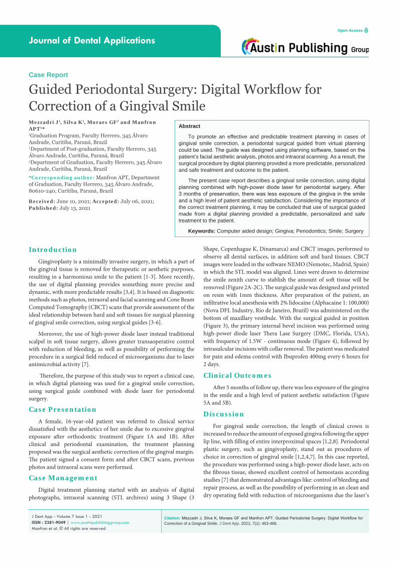
dissatisfied with the aesthetics of her smile due to excessive gingival exposure after orthodontic treatment (Figure 1A and 1B). After clinical and periodontal examination, the treatment planning proposed was the surgical aesthetic correction of the gingival margin. The patient signed a consent form and after CBCT scans, previous photos and intraoral scans were performed.
Case ManagementDigital treatment planning started with an analysis of digital
photographs, intraoral scanning (STL archives) using 3 Shape (3
Case Report
Guided Periodontal Surgery: Digital Workflow for Correction of a Gingival SmileMezzadri J1, Silva K1, Moraes GF2 and Manfron APT3*1Graduation Program, Faculty Herrero, 345 Álvaro Andrade, Curitiba, Paraná, Brazil2Department of Post-graduation, Faculty Herrero, 345 Álvaro Andrade, Curitiba, Paraná, Brazil3Department of Graduation, Faculty Herrero, 345 Álvaro Andrade, Curitiba, Paraná, Brazil
*Corresponding author: Manfron APT, Department of Graduation, Faculty Herrero, 345 Álvaro Andrade, 80610-240, Curitiba, Paraná, Brazil
Received: June 10, 2021; Accepted: July 06, 2021; Published: July 13, 2021
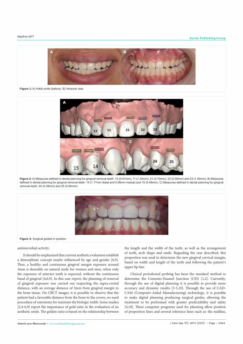
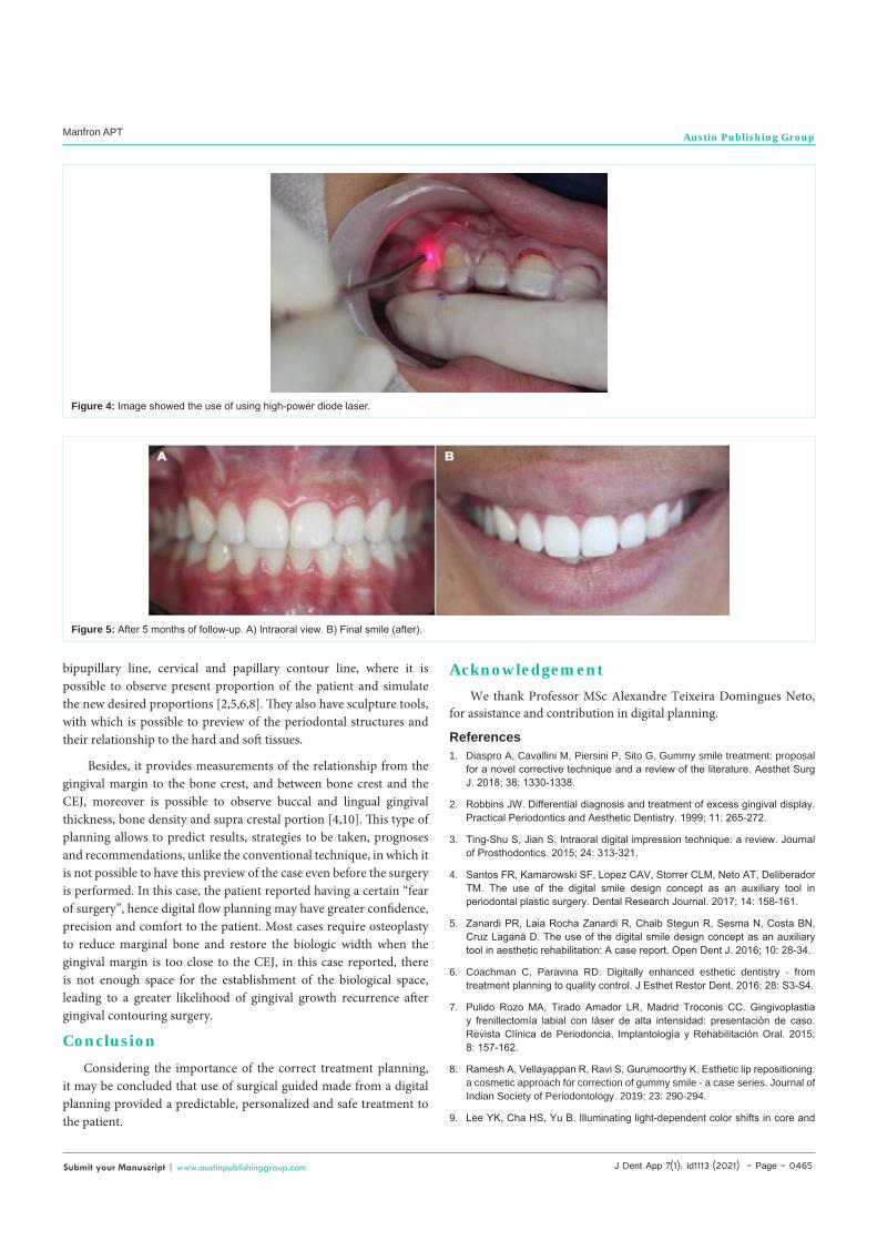
Shape, Copenhague K, Dinamarca) and CBCT images, performed to observe all dental surfaces, in addition soft and hard tissues. CBCT images were loaded in the software NEMO (Nemotec, Madrid, Spain) in which the STL model was aligned. Lines were drawn to determine the smile zenith curve to stablish the amount of soft tissue will be removed (Figure 2A-2C). The surgical guide was designed and printed on resin with 1mm thickness. After preparation of the patient, an infiltrative local anesthesia with 2% lidocaine (Alphacaine 1: 100,000) (Nova DFL Industry, Rio de Janeiro, Brazil) was administered on the bottom of maxillary vestibule. With the surgical guided in position (Figure 3), the primary internal bevel incision was performed using high-power diode laser Thera Lase Surgery (DMC, Florida, USA), with frequency of 1.5W - continuous mode (Figure 4), followed by intrasulcular incisions with collar removal. The patient was medicated for pain and edema control with Ibuprofen 400mg every 6 hours for 2 days.
Clinical OutcomesAfter 5 months of follow up, there was less exposure of the gingiva
in the smile and a high level of patient aesthetic satisfaction (Figure 5A and 5B).
DiscussionFor gingival smile correction, the length of clinical crown is
increased to reduce the amount of exposed gingiva following the upper lip line, with filling of entire interproximal spaces [1,2,8]. Periodontal plastic surgery, such as gingivoplasty, stand out as procedures of choice in correction of gingival smile [1,2,4,7]. In this case reported, the procedure was performed using a high-power diode laser, acts on the fibrous tissue, showed excellent control of hemostasis according studies [7] that demonstrated advantages like: control of bleeding and repair process, as well as the possibility of performing in an clean and dry operating field with reduction of microorganisms due the laser’s
J Dent App 7(1): id1113 (2021) - Page - 0464
Manfron APT Austin Publishing Group
Submit your Manuscript | www.austinpublishinggroup.com
antimicrobial activity.
It should be emphasized that current aesthetic evaluation establish a dimorphism concept maybe influenced by age and gender [6,9]. Thus, a healthy and continuous gingival margin exposure around 3mm is desirable on natural smile for women and men, when only the exposure of anterior teeth is expected, without the continuous band of gingival [4,6,9]. In this case report, the planning of removal of gingival exposure was carried out respecting the supra-crestal distance, with an average distance of 3mm from gingival margin to the bone tissue. On CBCT images, it is possible to observe that the patient had a favorable distance from the bone to the crown, no need procedure of osteotomy for maintain the biologic width. Some studies [2,4-6,9] report the importance of gold ratio in the evaluation of an aesthetic smile. The golden ratio is based on the relationship between
the length and the width of the teeth, as well as the arrangement of teeth, arch shape and smile. Regarding the case described, this proportion was used to determine the new gingival cervical margin, based on width and length of the teeth and following the patient’s upper lip line.
Clinical periodontal probing has been the standard method to determine the Cemento-Enamel Junction (CEJ) [1,2]. Currently, through the use of digital planning it is possible to provide more accuracy and dynamic results [3-5,10]. Through the use of CAD-CAM (Computer-Aided Manufacturing) technology, it is possible to make digital planning producing surgical guides, allowing the treatment to be performed with greater predictability and safety [4,10]. These computer programs used for planning allow position of proportion lines and several reference lines such as: the midline,
Figure 1: A) Initial smile (before). B) Intraoral view.
Figure 2: A) Measures defined in dental planning for gingival removal teeth: 12 (0.61mm), 11 (1.33mm), 21 (0.70mm), 22 (0.58mm) and 23 (1.05mm). B) Measures defined in dental planning for gingival removal teeth: 14 (1.17mm distal and 0.48mm mesial) and 15 (0.48mm). C) Measures defined in dental planning for gingival removal teeth: 24 (0.38mm) and 25 (0.64mm).
Figure 3: Surgical guided in position.
J Dent App 7(1): id1113 (2021) - Page - 0465
Manfron APT Austin Publishing Group
Submit your Manuscript | www.austinpublishinggroup.com
bipupillary line, cervical and papillary contour line, where it is possible to observe present proportion of the patient and simulate the new desired proportions [2,5,6,8]. They also have sculpture tools, with which is possible to preview of the periodontal structures and their relationship to the hard and soft tissues.
Besides, it provides measurements of the relationship from the gingival margin to the bone crest, and between bone crest and the CEJ, moreover is possible to observe buccal and lingual gingival thickness, bone density and supra crestal portion [4,10]. This type of planning allows to predict results, strategies to be taken, prognoses and recommendations, unlike the conventional technique, in which it is not possible to have this preview of the case even before the surgery is performed. In this case, the patient reported having a certain “fear of surgery”, hence digital flow planning may have greater confidence, precision and comfort to the patient. Most cases require osteoplasty to reduce marginal bone and restore the biologic width when the gingival margin is too close to the CEJ, in this case reported, there is not enough space for the establishment of the biological space, leading to a greater likelihood of gingival growth recurrence after gingival contouring surgery.
Conclusion
Considering the importance of the correct treatment planning, it may be concluded that use of surgical guided made from a digital planning provided a predictable, personalized and safe treatment to the patient.
Figure 4: Image showed the use of using high-power diode laser.
Figure 5: After 5 months of follow-up. A) Intraoral view. B) Final smile (after).
AcknowledgementWe thank Professor MSc Alexandre Teixeira Domingues Neto,
for assistance and contribution in digital planning.
References1. Diaspro A, Cavallini M, Piersini P, Sito G. Gummy smile treatment: proposal
for a novel corrective technique and a review of the literature. Aesthet Surg J. 2018; 38: 1330-1338.
2. Robbins JW. Differential diagnosis and treatment of excess gingival display. Practical Periodontics and Aesthetic Dentistry. 1999; 11: 265-272.
3. Ting-Shu S, Jian S. Intraoral digital impression technique: a review. Journal of Prosthodontics. 2015; 24: 313-321.
4. Santos FR, Kamarowski SF, Lopez CAV, Storrer CLM, Neto AT, Deliberador TM. The use of the digital smile design concept as an auxiliary tool in periodontal plastic surgery. Dental Research Journal. 2017; 14: 158-161.
5. Zanardi PR, Laia Rocha Zanardi R, Chaib Stegun R, Sesma N, Costa BN, Cruz Laganá D. The use of the digital smile design concept as an auxiliary tool in aesthetic rehabilitation: A case report. Open Dent J. 2016; 10: 28-34.
6. Coachman C, Paravina RD. Digitally enhanced esthetic dentistry - from treatment planning to quality control. J Esthet Restor Dent. 2016; 28: S3-S4.
7. Pulido Rozo MA, Tirado Amador LR, Madrid Troconis CC. Gingivoplastia y frenillectomía labial con láser de alta intensidad: presentación de caso. Revista Clínica de Periodoncia, Implantología y Rehabilitación Oral. 2015; 8: 157-162.
8. Ramesh A, Vellayappan R, Ravi S, Gurumoorthy K. Esthetic lip repositioning: a cosmetic approach for correction of gummy smile - a case series. Journal of Indian Society of Periodontology. 2019; 23: 290-294.
9. Lee YK, Cha HS, Yu B. Illuminating light-dependent color shifts in core and

J Dent App 7(1): id1113 (2021) - Page - 0466
Manfron APT Austin Publishing Group
Submit your Manuscript | www.austinpublishinggroup.com
veneer layers of dental all-ceramics. Journal of Biomedical Optics. 2014; 19: 95002.
10. Liu X, Yu J, Zhou J, Tan J. A digitally guided dual technique for both gingival
and bone resection during crown lengthening surgery. J Prosthet Dent. 2018; 119: 345-349.