Embed Size (px)
Citation preview
1
GUIDELINES FOR PET/CT IMAGING OF NEUROENDOCRINE
NEOPLASMS WITH 68Ga-DOTA-SOMATOSTATIN ANALOGUES AND
18F-DOPA.
Murat Fani BOZKURT, Irene VIRGOLINI, Sona BALOGOVA, Mohsen BEHESHTI,
Domenico RUBELLO, Clemens DECRISTOFORO, Valentina AMBROSINI,Andreas
KJAER, Roberto DELGADO BOLTON, Jolanta KUNIKOWSKA, Wim OYEN, Arturo
CHITI, Francesco GIAMMARILE, Christian BOY, Bernd KRAUSE, Anders SUNDIN,
Stefano FANTI
Abstract
Neuroendocrine neoplasms are a heterogenous group of tumours, for which nuclear medicine
plays an important role in the diagnostic work-up, follow-up and somatostatin receptor
targeted therapies.These guidelines are aimed to assist nuclear medicine physicians in
recommending, performing, interpreting and reporting the results of somatostatin receptor
(SSTR) PET/CT imaging using 68Ga-DOTA-somatostatin analogues (SSA), and 18F-DOPA
for various neuroendocrine neoplasms (NENs). The previous procedural guideline by EANM
regarding the use of PET/CT tumour imaging with 68Ga-DOTA-SSAs has been revised and
updated with the relevant and recent literature in the field with contribution of distinguished
experts.
Keywords: Neuroendocrine tumour; neurendocrine neoplasm; carcinoid; PET/CT; 68Ga-
DOTATATE; 68Ga-DOTATOC; 68Ga-DOTANOC; 18F-DOPA; 18F-FDG; thyroid medullary
cancer; pheochromocytoma; paraganglioma; foregut-NET; midgut-NET; hindgut-NET;
hyperinsulism in infants
2
Background information and definitions
Neuroendocrine neoplasms (NENs) are a heterogenous group of neoplasms that originate
from cells of neuroendocrine origin in many different organs but most frequently in the
gastrointestinal tract and the lungs. Less common locations include thymus and other organs
with endocrine function such as adrenal medulla, pituitary and thyroid.
NENs can be classified into three histopathological grades according to the World Health
organisation (WHO) 2010 classification. This classification depends on the proliferation
marker Ki-67 index and the mitotic index of the tumour (Table 2). According to a
histopathological grading system, NENs can also be classified into four types. Type 1 refers
to NENs with low Ki-67 and mitotic index, which are benign well-differentiated NENs. Type
2 refers to the well-differentiated NENs with an uncertain behaviour. Type 3 NENs are well-
differentiated neuroendocrine carcinomas (NECs) with mostly low-grade malignant features.
Type 4 refers to poorly-differentiated high-grade NECs with aggressive behaviour.
Additionally, there is a tumour type consisting of adenocarcinoma with neurendocrine
differentiation, which is called “MANEC” (mixed adenoneuroendocrine carcinomas).
The majority of NENs express somatostatin receptors (SSTR), which can be used as targets
for radionuclide imaging and therapy. Somatostatin is a small cyclic neuropeptide that is
found in neurons and endocrine cells and has a high density in the brain, peripheral neurons,
endocrine pancreas and gastrointestinal tract [1-10]. Since naturally occuring somatostatin is
very unstable, synthetic more stable analogues have been developed [1, 11].
Scintigraphy with radiolabeled somatostatin analogues (SSA), first with123I and followed by
111In and 99mTc, has been an important part of the imaging work up of patients with SSTR-
positive NENs with a detection rate between 50 and 100% in different studies [1-11]. These
varying detection rates between studies may at least partly be explained by the use of different
SSA-preparations and varying acquisition techniques such as planar imaging only, combined
planar imaging and SPECT or SPECT/CT. SSTR scintigraphy presents some limitations that
may decrease the diagnostic efficacy. This is mostly due to high physiological uptake such as
the liver as well as the lack of detection of smaller lesions because of the suboptimal physical
characteristics of the radiopharmaceuticals and the relatively low spatial resolution of gamma
cameras [12-13].
3
The transition to SSTR imaging by PET/CT with 68Ga-DOTA-SSA, has brought a new vision
with regard to spatial resolution, tumour-to-normal tissue contrast and patient comfort due to
earlier and shorter acquisition times compared to SSTR planar and SPECT imaging . The
most commonly used SSA-preparations as radiopeptides for PET/CT imaging are [68Ga-
DOTA0 -Tyr3]octreotide (68Ga-DOTA-TOC, 68Ga-edotreotide), [68Ga-DOTA0 -1NaI3
]octreotide (68Ga-DOTA-NOC) and [68Ga-DOTA0 -Tyr3]octreotate (68Ga-DOTA-TATE) [14-
16]. All these radiopeptides bind to the SSTR subtype 2, which is predominantly expressed in
NENs but each has different affinity profiles for other SSTR subtypes [17] (Table 1). 68Ga-
DOTA-NOC along with the highest affinity to SSTR2 shows a high affinity to SSTR3 and 5,
68Ga-DOTA-TOC binds also to SSTR subtype 5 (with lower affinity compared to DOTA-
NOC) while 68Ga-DOTA-TATE predominantly binds to SSTR subtype 2. Finally, 68Ga-
DOTA-lanreotide binds to SSTR subtype 2 and 5 [18-20]. Recently, also 64Cu-DOTA-TATE
has been used for PET/CT imaging in NEN patients and was found superior to SPECT based
somatostatin receptor imaging [19].
These above mentioned radiopeptides function as SSTR agonists. Recently some SSTR
antagonists have, however, been introduced. Limited data on SSTR antagonist radiopeptides
excist, especially for PET applications, which might more efficiently localise NENs due to
binding to a higher number of receptor sites (practically all SSTR subtypes) and with more
stable binding [21].
SSTRs are not only targets for radionuclide imaging of NENs but also for peptide receptor
radionuclide therapy (PRRT) for which the beta-emitters 177Lu and 90Y are currently used,
generally conjugated to DOTA-TOC and DOTA-TATE. Confirmation of sufficient tumour
receptor expression, traditionally with SSTR planar and SPECT imaging but increasingly with
PET/CT, is needed to select patients eligible for PRRT. The treatment decision is also based
on some factors such as the tumour load and location of the disease. For pre-therapy
diagnostic 68Ga-DOTA-SSA PET/CT imaging the appropriate radiopeptide should preferably
be chosen i.e. 68Ga-DOTA-TATE before PRRT with 177Lu-/90Y-DOTA-TATE and 68Ga-
DOTA-TOC when PRRT with 177Lu-/90Y- DOTA-TOC is considered [22]. Due to the short
half-life of 68Ga (68 minutes), 68Ga-DOTA-SSA cannot be used for dosimetry, which is
instead usually derived from scintigraphy during PPRT with 177Lu-DOTA-labelled peptides.
4
Along with SSTR, NENs can be imaged by using other molecular and metabolic targets,
based on their neuroendocrine functional features. Most widely studied for this application
has been 3,4-dihydroxy-6-[18F]fluoro-l-phenylalanine (18F-DOPA). Higher accuracy for
PET/CT with 18F-DOPA has been shown for imaging of well differentiated NENs as
compared to conventional radiological and planar and/or SPECT imaging [23]. Currently, the
main clinical indication for NEN imaging with 18F-DOPA is tumours with low/variable SSTR
expression, such as neuroectodermal tumours [23]. Furthermore, since several types of
malignant and non malignant lesions may show variable expression of SSTR, 18F-DOPA, as a
tracer of catecholamine metabolic pathway, may be helpful in the characterisation of
medullary thyroid cancer (MTC), jejuno-ileal (midgut) NEN, pheochromocytoma,
neuroblastoma or paraganglioma in patients suspected of harbouring synchrone/metachrone
metastatic malignancy (e.g. breast cancer). Although 18F-DOPA has no theranostic role in
assessing the patient´s suitability for treatment with SSA, it may assist to identify lesions with
low or absent SSTR expression, consequently predicting poor or no response to SSA therapy .
Similarly, in therapy monitoring and surveillance, 18F-DOPA may be helpful in identifying
new lesions to define disease progression. Major drawbacks with the use of 18F-DOPA are
limited availability in several European countries and costly synthesis.
As a problem solving tool when 68Ga-DOTA-SSA- and 18F-DOPA-PET/CT does not suffice,
PET/CT with the serotonine precursor 11C-5-hydroxy-tryptophan (5-HTP) has been shown
suitable as a general tracer for NEN imaging and is currently available in two European
centres [24,25].
Furthermore, radiolabelled peptide analogues targeting the cholecystokinin-2 receptor have
been developed for NEN imaging. Initial clinical studies with 99mTc and 111In labelled gastrin
analogues show very promising results in patients with MTC [26,27] as well as in patients
with other NENs [28]. Recently, a new radiolabelled gastrin analogue with very promising
characteristics for clinical translation, in terms of high metabolic stability, prolonged tumour
uptake and low kidney retention, has been developed and will be studied in patients with
metastatic MTC in the near future [29], however, evidence regarding possible PET-
applications are still lacking.
PET/CT imaging with 2-[18F]fluoro-2-deoxy-D-glucose (18F-FDG) plays a role not only for
lesion detection of G2 and G3 NENs, but additionally provides important prognostic
information (28). While most of the low grade highly differentiated NENs show high uptake
5
of 68Ga-DOTA-SSA and 18F-DOPA, poorly differentiated NENs show preferential uptake of
18F-FDG indicating a more aggressive behaviour and worse prognosis [30].
Tumours that may be visualised with68Ga-DOTA-SSA PET/CT include:
Tumours with high expression of somatostatin receptors [31-38)
Gastro-entero-pancreatic tumours (GEP) functioning and non-functioning (e.g.:
gastrinoma, insulinoma, glucagonoma, VIPoma, etc.)
Broncho-pulmonary NENs
Sympatho-adrenal system tumours (e.g. paraganglioma)
Meningioma
Tumours with low or varying expression of receptors [39-40]
Breast carcinoma
Melanoma
Lymphoma
Prostate carcinoma
Non-small cell lung cancer
Head and neck cancer
Sarcoma
Renal cell carcinoma
Differentiated thyroid carcinoma
Astrocytoma
6
Tumours with neuroendocrine/neuroectodermal features that may be visualised with
18F-DOPA PET/CT include [33, 44-47]:
Jejuno-ileal(midgut) NENs
Pheochromocytoma
Paraganglioma
Neuroblastoma
Medullary Thyroid Cancer
Other Tumours/tumour-like conditions with high 18F-DOPA uptake include:
Brain tumors
Beta cell hyperplasia (especially for the indication of congenital hyperinsulinemic
hypoglycemia).
Tumours with neuroendocrine features that show high18F-FDG uptake:
Neuroendocrine carcinomas (NECs)
Neuroendocrine neoplasms (NENs) with high histologic grade or G2 NENs with
suspect aggressive behaviour.
Medullary thyroid cancer (MTC)
Mixed adenoneuroendocrine cancers (MANEC)
Synchrone/metachrone non-NEN malignancies
7
Clinical indications for PET/CT imaging of NENs with 68Ga-DOTA-SSA
Clinical indications for 68Ga-DOTA-peptide PET/CT imaging of NENs are primary tumour
localisation and characterisation, tumour staging, detection of recurrent disease and restaging,
as well as selecting patients eligible for PRRT. The results of these different imaging
applications have been reported in the literature as follows:
Diagnosis and staging: Localise primary tumours and detect sites of metastasis
(staging) [34-41, 48-49]
Re-staging: Follow-up of NEN patients to detect residual, recurrent or progressive
disease (detection of recurrent disease and restaging) [34-41, 48-51]
Management decisions: Select patients with metastatic disease for PRRT [34-41, 50].
Monitor the response to therapy (surgery, radiotherapy, chemotherapy or PRRT) [52].
Detection of the primary occult NEN when there is biochemocal evidence and/or
symptoms of NEN disease with no evidence of a primary tumour on conventional
radiological imaging.
Detection of the primary tumour in patients with metastasis from an unknown primary
NEN on conventional radiological imaging [48].
Characterisation of a broncho-pulmonary mass as a NEN when other diagnostic
modalities were inconclusive.
Detection, characterisation and restaging in case of biochemical NEN recurrence.
The sensitivity of 68Ga-DOTA-SSA PET/CT is likely to vary among tumour types and
patients, and even between lesions in the same patient, depending on their SSTR density. Due
to the short half life of 68Ga, conjugated peptides cannot be used for dosimetry, which is
usually derived from 177Lu-DOTA-labelled peptides. The sensitivity of 68Ga-DOTA-SSA
PET/CT may theoretically be reduced also in patients receiving therapeutic doses of
somatostatin analogues such as octreotide, but this issue still needs to be clarified [53]. On
empirical grounds, prior to PET with 68Ga-DOTA-SSA, it has been recommended to
discontinue therapy with SSA (when possible and not contraindicated) to avoid possible
8
SSTR blockade [54]. However, there are literature reports of improved tumour/non-tumour
ratio, following pre-treatment with SSA as a consequence of non-saturability of SSTR
expressed by malignant cells in contrast to SSTR expressed in normal tissues [55,56].
If discontinuation of SSA treatment is undertaken, it has been suggested that the time interval
between interruption of therapy and 68Ga-DOTA-SSA PET/CT depends on the type of SSA
used: 1 day is suggested for short-lived SSA and 3–4 weeks for long-acting SSA preparations.
However, this issue is still not definitely clarified and many centres do not require SSA
withdrawal before PET/CT imaging. Some centers suggest that the best option is to perform
the PET/CT study just prior to the scheduled monthly dose of long-acting octreotide [54].
However, taken together there is no clear evidence that discontinuation of somatostatin
analogues prior to PET imaging with 68Ga-DOTA-SSA is necessary.
68Ga-DOTA-SSA PET/CT imaging in non-NENs
Less frequently 68Ga-DOTA-SSA PET/CT can be used in non-NEN imaging, particularly
when PRRT is considered. Except for the determination of SSTR status 68Ga-DOTA-SSA
PET/CT cannot be considered as the first-choice functional modality in the management of
patients with non-NENs.
Indications for 18F-DOPA PET/CT for NENs:
PET/CT with 18F-DOPA targets tumours with enhanced intracellular transport and
decarboxylation of the amino acid DOPA and is for NEN imaging approved in several EU
countries for the following indications:
Diagnosis
Diagnosis and localisation of glomus tumours in patients with a gene mutation of the
succinate dehydrogenase D variant
Localisation of pheochromocytoma and paraganglioma
Diagnosis and localisation of insulinomas in the case of hyperinsulinism in infants and
children
9
Staging
Phaeochromocytoma and paraganglioma
Well differentiated NENs of the digestive tract
Detection in case of reasonable suspicion of recurrences or residual disease
Phaeochromocytoma and paraganglioma
Medullary thyroid cancer with elevated serum levels of calcitonin
Well differentiated NENs of the digestive tract
Other endocrine digestive tumours when SSTR imaging is negative
Indications for 18F-FDG PET/CT imaging of NENs:
Localisation of NECs and high-grade poorly-differentiated NETs with aggressive
behaviour
Prognosis
Localisation of synchrone/metachrone non-NEN malignancy
The proposed PET/CT imaging strategies with the use of the abovementioned currently
available radiopharmaceuticals and based on the different NEN types are tabulated in Table
3.
Precautions for PET/CT imaging
10
Pregnancy (suspected or confirmed): In the case of a diagnostic procedure in a patient
who is or may be pregnant, a clinical decision is necessary to consider the benefits
against the possible harm of carrying out the procedure.
Breastfeeding: If radiopharmaceutical administration is considered necessary,
breastfeeding should be interrupted and can be restarted when 7 physical half-lives for
the radionuclide in radiopharmaceutical has elapsed when the level of radiation in the
milk will not result in a radiation dose to the child greater than 1 mSv.
The ionising radiation from 68Ga-DOTA-SSA must be carefully evaluated in subjects
under 18 years of age. However, the dosimetry of 68Ga- SSA is more favorable than
that of 111ln-pentetreotide.
It has been recommended to temporarily withdraw SSA therapy (when possible) to avoid
possible SSTR blockade (see patient preparation). In some patients the withdrawal of therapy
might not be tolerated [54-56] .
Pre-examination procedures for 68Ga-SSA PET/CT Imaging
1) Patient preparation
• The physician or the technologist should give the patient a detailed information about the
procedure.
• It has been advocated by some authors to withdraw “cold” octreotide therapy (when
possible and not contraindicated) to avoid possible SSTR blockade. The time interval between
interruption of therapy and 68Ga-DOTA-SSA PET/CT depends on the type of SSA used: one
day is suggested for short-lived SSAs and at least 4-7 weeks for long-acting SSA preparations
[54-56].
• There is no need for fasting before the procedure.
2) Pre-injection
All information useful for optimal interpretation of the study should be considered by the
nuclear medicine physician:
11
• relevant history of suspected or known primary tumour
• absence or presence of functional symptoms
• laboratory test results (hormones and tumour markers)
• results of other imaging modalities (CT, MRI, US, X-ray)
• results of recent biopsy, (including tumour grading and ki-67), surgery, chemotherapy,
radiotherapy or radionuclide therapy
• history of recent SSA therapy and of PRRT
3) Administration of 68Ga-DOTA-SSA (DOTA-TOC, DOTA-NOC, DOTA-TATE)
• The radiopharmaceutical should be administered as a short bolus injection using an
indwelling catheter to avoid extravasation and immediately followed by injection of about 50
mL of physiological saline.
• The administered amount of activity should be determined by taking into account the
Directive 97/43/EURATOM. It is expected that Diagnostic Reference Levels (DRL) for the
radiopharmaceuticals will not to be exceeded for standard procedures when good and normal
practice regarding diagnostic and technical performance is applied. It should be noted that in
each country Nuclear Medicine physicians should respect the DRLs and the rules stated by
the local regulations. Activities higher than the DRLs must be justified. For the
aforementioned reasons, the following activity for 68Ga-DOTA-TOC, 68Ga-DOTA-NOC,
68Ga-DOTA-TATE should be considered only as a general indication, based on literature data
and current experience.
• The activity administered ranges from 100 to 200 MBq, depending on the PET scanner
technical characteristics and the patient´s body weight. As with other PET-tracers the injected
activity should be calculated according to the patients body-weight. The recommended
activity is 2 MBq/kg body-weight and can be reduced in fat patients because fat accumulate
very little 68Ga-DOTA-SSA. In adult patients, as a rule at least 100 MBq of 68Ga-DOTA-SSA
needs to be administered to obtain good image quality. The experience in paediatric patients is
very limited. When the use of 68Ga-DOTA-SSA is considered necessary in a child 2 MBq/kg
12
is similarly recommended in order to reduce the administered activity according to the
recommendations of the EANM Paediatric Task Group [57].
Definitive dosimetric data for 68Ga-DOTA-TOC, DOTA-NOC and DOTA-TATE are
available in the literature and the mean effective dose is 0.023, 0.025, and 0.026 mSv/MBq,
respectively, in several dosimetric studies [58-60]. The organ receiving the largest radiation
dose is the spleen, followed by kidneys and bladder.
The amount of peptide in the injected 68Ga-DOTA-SSA preparation (DOTA-TOC, DOTA-
NOC, DOTA-TATE) should be below 50 µg (in discussion in PharmEur); this amount is not
expected to have any clinically significant pharmacological effect. The radiopharmaceutical
should not be injected into intravenous lines together with solutions for parenteral nutrition.
4) Post-injection
Patients should void before scanning. Elimination of the extra fluid intake will help to flush
out any radioactivity (either 68Ga labelled DOTA-SSA or 68Ga transferrin) from the
circulation and by glomerular filtration. This will reduce the background activity as well as
the radiation dose to kidneys and bladder.
Pre-examination procedures for 18F-DOPA PET Imaging
1) Patient preparation
• The physician or the technologist should give the patient detailed information about
the procedure
• Oral premedication with carbidopa (L-alpha-hydrazino-alpha-methyl-b-3,4-
dihydroxyphenyl propionic acid), an inhibitor of the aromatic aminoacid decarboxylase
enzyme, is controversial. The posology of carbidopa usually ranges between 100 and 200 mg
(or 2 mg/kg of body weight) [61,62]. Timmers et al. [63] reported that, compared with
baseline 18F-DOPA PET, carbidopa pre-treatment resulted in the detection of 3 additional
lesions in 3 of 11 patients with pheochromocytoma or extra-adrenal paraganglioma. In
contrast, in one infant of the Ribeiro’s series the diffuse uptake of 18F-DOPA in the pancreas
completely disappeared under carbidopa treatment while the kidney activity was still present:
13
the patient had histologically proven diffuse abnormal pancreatic cells scattered in the whole
pancreas [64]. Similar findings have been reported by Kauhanen et al. in 2 of 3 adults with
insulinoma [65]. These findings do not favour the use of carbidopa in patients with pancreatic
tumours since pancreatic physiological uptake disappears, and tumour uptake could also
disappear along with this. Carbidopa effect on 18F-DOPA uptake in insulinomas is not fully
elucidated. No final consensus has been reached about the usefulness of carbidopa in patients
with insulinoma-related hyperinsulinemic hypoglycaemia [66,67]. Currently, no preparation
of carbidopa (without levodopa) is commercially available.
• Fasting: On an empirical basis, to avoid interaction with amino acids from food, 18F-
DOPA should be administered to patients fasting for a minimum of 4 hours without limiting
water intake.
. Medication Withdrawal: No special interactions have been reported and no therapeutic
discontinuation of is needed.
. Posology and time of acquisition: According to extensive literature data, the
recommended activity of 18F-DOPA for adults is 2 to 4 MBq/kg body-weight administered by
slow intravenous injection over approximately one minute [68, 69].
The use of 18F-DOPA in children and adolescents has to be considered carefully, based upon
clinical needs and assessing the risk/benefit ratio in this patient group. The activities to be
administered to children and adolescents may be calculated according to the recommendations
of the European Association of Nuclear Medicine (EANM) paediatric dosage card
(http://www.eanm.org/publications/dosage_calculator.php?navId=285 ).
To detect foci in the liver, intestine or pancreas area, early “static” images can be acquired
starting 5 minutes after injection, or a “dynamic” acquisition starting right after the injection
during 10 minutes. Whole-body images are usually acquired 60 minutes after injection.
Physiological biodistribution of 68Ga-DOTA-SSAs
68Ga-DOTA-SSAs are rapidly cleared from the blood. Arterial activity elimination is bi-
exponential and no radioactive metabolites are detected within 4 h in serum and urine.
Maximal tumour activity accumulation is reached 70±20 min post injection. Kidney uptake
averages <50% of that of the spleen. Excretion is almost entirely through the kidneys [13].

14
SSTRs are expressed by many neuroendocrine and non-neuroendocrine cells of the body, and
many normal organs show 68Ga-DOTA-SSA accumulation including the liver, spleen,
pituitary, thyroid, kidneys, adrenal glands, salivary glands, stomach wall and bowel. The
pancreas shows variable uptake of 68Ga-DOTA-SSAs and although all five SSTR subtypes
are present, the SSTR receptor subtype 2 is preferably found and is located in the islets of
Langerhans. A well known potential pitfall is a variable but sometimes very high focal 68Ga-
DOTA-SSA accumulation in the uncinate process of the pancreas that may mimic a
pancreatic NEN [70]. Also, the prostate gland and the glandular tissue in the breast may show
diffuse low-grade physiological 68Ga-DOTA- SSA uptake.
The biodistribution of the various 68Ga-DOTA-SSA preparations may vary depending on the
receptor coverage of the peptide. According to literature data, 68Ga-DOTA-TATE, which has
mainly affinity to SSTR subtype 2, shows more intense physiological uptake in the pituitary
and salivary glands [60], than 68Ga-DOTANOC, binding to the SSTR subtypes 2, 3 and 5.
Although the SSTR subtype binding profile of 68Ga-DOTANOC is wider than that of 68Ga-
DOTATATE, the NEN uptake of 68Ga-DOTA-TATE was in a PET/CT imaging study shown
more intense and with higher lesion-to-background ratio compared with 68Ga-DOTA-NOC
[71]. By contrast, PET/CT imaging with 68Ga-DOTA-NOC in another study detected
significantly more lesions than using 68Ga-DOTA-TATE in patients with
gastroenteropancreatic-NENs [72]. However, there is still a debate in the literature whether
the uptake pattern differences due to the dissimilar SSTR subtype profiles of the various 68Ga-
DOTA-SSA significantly affects the tumour imaging yield. In direct comparisons of 68Ga-
DOTA-SSA preparations in the same patients, small imaging differenses have been found,
usully for merely singular or a small number of lesions. Thus, to date there are no studies in
larger patient groups that convincingly shows that the diagnostic performance of 68Ga-DOTA-
TOC, 68Ga-DOTA-TATE and 68Ga-DOTA-NOC are dissimilar.
Physiological Biodistribution of 18F-DOPA
The biodistribution of 18F-DOPA was investigated in a cohort of 107 patients and showed
physiological uptake in the basal ganglia, liver, adrenal glands which was very variable [68,
73]. In the pancreas the accumulation was most prominent in the uncinate process and less
intense in the body and tail. Further, very intense and variable 18F-DOPA uptake was seen in
the excretory organs such as the gallbladder and biliary tract. The tracer was excreted through
15
the kidneys, in which the highest activity was found together that in the urine. Bowel uptake
was an unusual finding and when present, was low and diffuse. Low uptakes were also found
in the myocardium, muscles and in some cases a very faint uptake in the mammary glands,
the oral cavity, the esophagus. In the literature it is reported that children can present 18F-
DOPA uptake in the growth plates [74].
Effect of carbidopa premedication on the biodistribution of 18F-DOPA
Oral premedication with carbidopa, to block the aromatic amino acid decarboxylase enzyme,
is controversial and is for NEN visualisation of less common use than for neurologic imaging
purposes. The pre-administration of carbidopa increases the 18F-DOPA uptake in the basal
ganglia, lungs, myocardium and liver and decreases the pancreatic uptake [63]. Physiological
excretion into the biliary tract and the urinary system is unaffected by carbidopa pre-
medication. Similar effects of carbidopa premedication are reported in children [74].
Preparation of 68Ga-DOTA-SSAs:
Currently different types of 68Ge/68Ga-generators are being used, all of them providing 68Ga in
strongly acidic hydrochloric acid solutions (0.05-0.6 M HCl) [65]. For radiolabelling of
DOTA-SSAs, different techniques are being employed, usually using semi- or fully
automated systems. These are either based on prepurification and concentration of the
generator eluate using an anion-exchange or cation-exchange technique. Alternatively, a
fraction of the generator eluate may be used directly for radiolabelling. The procedure is
performed using a suitable buffer at elevated temperature followed by purification of the
preparation solution on a C-18 cartridge and appropriate aseptic formulation. Either method
employed must ensure that the level of germanium-68 (68Ge) in the final preparation is lower
than 0.001% of the total 68Ga radioactivity. Quality parameters to be tested may vary
dependent on the technique applied and are currently defined within a monograph of the
European Pharmacopeia for 68GaDOTA-TOC (Gallium- (68Ga) Edotreotide Injection, No.
2482). Quality control protocols may include tests for radionuclidic purity, radiochemical
purity (HPLC, TLC), chemical purity (buffer, solvents) as well as sterility and endotoxin
testing using validated methods. Generally, quality control should be performed according to
the governing monograph or national regulations; whichever is applicable. A review on
production technologies and quality aspects can be found in previous publications [75,76].
Recently, generators with a marketing authorisation have become available and radiolabelling
16
kits are in the pipeline. This will simplify kit-based preparation of 68Ga-DOTA-SSAs,
potentially reducing the requirements for purification, GMP compliance and quality control.
PET/CT scanner quality control
A strict quality control programme should be routinely performed according to the rules of
each country, as stated in the Council Directives 97/43/ EURATOM.
Image acquisition of 68Ga-DOTA conjugate peptides
Data acquisition is performed by means of a dedicated PET/CT scanner in 3D mode. The
timing for image acquisition ranges between 45 and 90 minutes after injection and varies on
the basis of the 68Ga-DOTA-SSA used. There are no unequivocal data in the literature in this
respect, but according to the experience of various centres the best results are achieved with
image acquisition at 45 to 60 minutes after injection for 68Ga-DOTA-TATE and 60 to 90
minutes for 68Ga-DOTA-TOC and 68Ga-DOTA-NOC. The acquisition is performed as a
whole body scan from head to mid thighs. Image reconstruction should be performed by an
iterative reconstruction algorithm using the system’s implementation and settings.
Reconstructions may be performed with or without time of flight information, depending on
the systems capabilities. When possible it is, however, recommended to acquire and
reconstruct data with time of flight information. Reconstructions should be performed
including all regular corrections, such as normalisation, (CT based) attenuation correction,
dead time, decay correction and, preferably, model based scatter correction.
Image interpretation for PET/CT imaging with 68Ga-DOTA-SSAs
Normal biodistribution and abnormal accumulations should be evaluated by a nuclear
medicine physician. Tracer accumulations other that physiological or areas of accumulations
higher than that of the background activity can be considered as pathological. Clearly
demarkated findings with higher tracer uptake as compared to that of the liver are classified as
definitely positive for enhanced SSTR expression and thus indicative for a SSTR expressing
neoplasm. Linear, non-focal intestinal uptake with moderate intensity is considered
physiological. Pancreas may show variable physiological tracer uptake and a well known
pitfall is physiological uptake in the uncinate process [70].
Interpretation criteria
17
To evaluate 68Ga-DOTA-SSA PET/CT studies, the following issues should be taken into
consideration:
• clinical question raised in the request for 68Ga-DOTA-SSA PET/CT imaging
• clinical patient history, recent biochemical test results
• comprehension of the physiological tracer distribution
• anatomical localisation of the 68Ga-DOTA-SSA uptake with corresponding separate and
fused CT images; correlation with other imaging modalities (CT, MRI) is strongly
recommended
• intensity grading of the 68Ga-DOTA-SSA uptake that can also be expressed semi-
quantatively (SUVmax)
• 68Ga-DOTA-SSAs may show variable uptake in different tumour types, with respect to
tumour histology, SSTR expression and density, anatomical site and size of the lesion(s)
• causes of false negative results
• causes of false positive results
Image interpretation for 18F-DOPA PET/CT
18F-DOPA visualises a very specific metabolic process and presents non-specific
accumulation only corresponding to its excretory pathways. In other normal tissues, 18F-
DOPA has minimal uptake and therefore provides high lesion-to-background contrast.
Patients who are referred for 18F-DOPA PET/CT generally have a clinical suspicion of
disease, based on their clinical records and/or biochemical results and/or previous imaging
findings. Thus, it is helpful to be a priori aware of this information, to be aware of the
physiological tracer uptake patterns, and to have knowledge of the 18F-DOPA PET/CT
apparence of the different tumour types, their patterns of spread and of the anatomical sites
for recurrent disese.
Of special notice is the large variability in 18F-DOPA uptake in the pancreas, especially in the
uncinate process that, in some cases, can minic a pancreatic NEN. Similarly, highly variable
accumulation in the adrenal glands must be taken into consideration to avoid misinterpretation
18
of a normal adrenal as a pheochromocytoma. A high adrenal 18F-DOPA uptake, when
homogeneous and symmetrical, and not associated with a morphological finding of a tumour
on CT/MRI, should be considered as physiological. Because the liver accumulation of 18F-
DOPA is relatively constant this can serve as a normal tissue reference and be helpful for
semiquantitative measurements of the lesion-to-liver ratio.
In the 18F-DOPA PET/CT image reading, any focal uptake besides areas of physiologic tracer
distribution can be considered as pathological. When paragangliomas/pheochromocytomas
are suspected, either a non-physiological extraadrenal focal uptake, or asymmetrical adrenal
uptake together with a concordant enlarged gland, or an adrenal uptake higher than that of the
liver together with a concordant enlarged gland on CT/MRI, can be considered as
pathological.
Reporting of the Scans
For both 68Ga-DOTA-SSA and 18F-DOPA PET/CT imaging, the nuclear medicine physician
should record: the clinical question, a concise patient’s clinical history, type and date of
examination, administered activity and route of administration, CT parameters and dosimetry,
relevant medications (patient preparation, previous therapy with cold somatostatin analogues,
Carbidopa premedication, withdrawal period, chemotherapy, etc.), laboratory and other
imaging studies results.
The report should describe:
1. the procedure (the type of 68Ga-DOTA-SSA and its administered activity, the administered
activity of 18F-DOPA, acquisition time, duration of imaging, the area imaged)
2. the findings (site and size of the lesion(s), uptake intensity, SUVmax etc.)
3. comparative data - the findings should be related to previous PET/CT scans performed with
the same tracer or to 18FDG PET/CT, if performed, or to results of other imaging modalities
such as CT/MRI when appropriate
4. interpretation: a clear diagnosis should be made if possible, accompanied - when
appropriate - by a description of the study limitations (potential causes of false negative or
false positive results). Additional diagnostic examinations or an adequate follow-up should be
suggested, when required.
19
Potential Pitfalls in 68Ga-DOTA-SSA PET/CT Imaging
• A usually intense but sometimes variable tracer accumulation is seen in the spleen and
accessory spleen(s) if present, in kidneys, adrenals and pituitary. After spleenectomy,
accumulation in accessory spleen(s) may be misinterpreted. Benign adrenocortical adenomas
may also show 68Ga-DOTA-SSA uptake. The liver uptake is generally lower than that of the
spleen. The thyroid and salivary glands are (mostly) faintly visible. A variable and sometimes
very high tracer uptake in the uncinate process of the pancreas should not be misinterpreted.
Particularly in patients referred for 68Ga-DOTA-SSA PET/CT because of a supsected
pancreatic NEN, the finding of a high tracer uptake in the uncinate process should be
thouroughly correlated to radiological imaging (CT/MRI).
• Contamination with urine of clothes and/or skin may cause false positive images.
• SSA therapy or endogenous production of somatostatin by a tumour may interfere with
tumour detection by reducing or enhancing tumour detectability.
• Variable tumour differentiation and heterogeneous SSTR expression may influence the
affinity for 68Ga-DOTA-SSAs and thereby the diagnostic accuracy
• False negative findings may be due to tumour dedifferentiation or small lesion size
• False positive findings can be encountered in the presence of activated lymphocytes that
can express SSTR at sites of inflammation/infection.
• Syncronus tumours other than NENs may express SSTR and can be detected
Potential Pitfalls in 18F-DOPA PET/CT Imaging
An intense focal accumulation in the gallbladder and, in some patients, also in the common
bile duct may mimic an intestinal tumour or a hepatic metastasis [77]. Knowledge of the
normal tracer biodistribution and its physiological excretion together with the use of
correlative radiological imaging (CT/MRI) should help the reader to avoid misinterpretation
in this respect.
20
The urinary excretion of the tracer can also be the cause of several pitfalls. The intense uptake
of the tracer in the kidneys can mask a pathologic uptake in the tail of the pancreas. Moreover,
the kidney activity can hide a pathologic uptake in the adrenals, especially in patients with
dilatation of the urinary collective system (superior caliceal groups and kidney pelvis).
Activity in the ureters, especially if focal as opposed to the usually segmental pattern, can
mimic retroperitoneal uptake and should be correlated to radiological imaging (CT/MRI). In
order to minimise possible image interpretation problems from urinary bladder activity, the
patients are always ask to void immediately before start of the PET/CT examination. If
needed, the 110 minutes half life of 18F offers the possibility to acquire late 18F-DOPA images
after diuretic administration or after ambulation and hydration to help discriminating between
pathological and physiological image findings.
The physiologically intense and very variable 18F-DOPA uptake in the pancreas can lead to
pitfalls. A physiological uptake in the uncinate process can be interpreted as a pancreatic NEN
or confused with a retroperitoneal lymph node metastasis (false positive). Alternatively, a
pancreatic NEN with the same uptake intensity as that of the pancreas may be overlooked
(false negative). Moreover, physiological pancreatic uptake may potentially interfere with
visualisation of the left adrenal. When 18F-DOPA is performed because of suspected adrenal
pathology, premedication with carbidopa can in these cases prevents masking of an adrenal
lesion by blocking the pancreatic uptake and also increases the uptake in the lesions.
The utility of 18F-DOPA PET/CT in adult patients with hyperinsulinemic hypoglycemia can
be cumbersome since there is few difference between pathologic or non pathologic areas of
the pancreas (which show a very variable physiologic uptake of the tracer. Moreover
premedication with carbidopa could lead to another possible methodological pitfall when
considering patients with hyperinsulinemic hypoglycemia since carbidopa (a peripheral
AADC inhibitor) decreases the whole pancreatic uptake decreasing also the lesion to
background ratio [78]. Disappearance of 18F-DOPA focal pancreatic hot spots has been
reported after premedication with carbidopa in patients with hyperinsulinemic hypoglycemia
[64,74,78].
Pitfalls related to pathology

21
Possible sources of false-negative 18F-DOPA PET/CT results can be related to small lesion
size and tumor de-differentiation. Genetic factors may also affect the 18F-DOPA uptake in
paraganglioma. On one hand, succinate dehydrogenase B-subunit (SDHB) gene mutations
may result in extra-adrenal paragangliomas for which 18F-DOPA PET shows a lower
sensitivity than for non-SDHB-related lesions [63]. On the other hand, the fact that only
neuroendocrine cells are able to take up and decarboxylate 18F-DOPA and store the resulting
amine, leads to few false-positive 18F-DOPA PET findings.
Koopmans et al. studied prospectively 53 patients with GEP-NENs and recorded 100%
patient based sensitivity, 95% region based sensitivity and 96% lesion-based sensitivity,
which were better than CT, SRS and CT & SRS combined and without false positive results
[79].
In a meta-analysis on PET/CT imaging in recurrent MTC, 18F-DOPA have shown better
results in terms of sensitivity and specificity but with a complementary role for 18F-FDG in
more agressive tumours, thus reflecting the two different metabolic pathways of these tracers.
It was reported that false positive 18F-DOPA findings were uncommon, and that false negative
results could be mainly related to small lesions or to de-differentiation. The diagnostic
performance of 18F-DOPA in recurrent MTC improved in patients with higher serum
calcitonin levels [80, 81].
Technical pitfalls
PET/CT represents a major technologic advance, consisting of two complementary modalities
which provide both functional and anatomic information and whose combined strength tends
to overcome their respective weaknesses. With combined PET/CT, the superimposition of the
precise structural detail provided by CT allows an accurate anatomical localisation of the PET
findings and a correlation of the PET findings with the morphological findings.
With CT based attenuation correction there is a potential risk of overestimating the true
activity concentration in very high attenuating materials such as metallic implants [82]. In
these instances, comparison with the non-attenuated PET images is usually helpful to differ
attenuation correction induced from true tracer accumulations.
Another possible pitfall can be caused by misregistration between the PET and CT image
volumes. Superimposition of a focal radiotracer uptake on the wrong anatomic position in the
22
CT can be caused by breathing, patient motion, bowel motility, etc and can lead to
misinterpretation.
23
References:
1) Bombardieri E, Maccauro M, De Deckere E, Savelli G, Chiti A. Nuclear medicine
imaging of neuroendocrine tumours. Ann Oncol. 2001;12 Suppl 2:S51-61.
2) Olsen JO, Pozderac RV, Hinkle G, Hill T, O'Dorisio TM, Schirmer WJ, Ellison
EC, O'Dorisio MS. Somatostatin receptor imaging of neuroendocrine tumors with
indium-111 pentetreotide (Octreoscan). Semin Nucl Med. 1995 Jul;25(3):251-61.
3) Briganti V, Sestini R, Orlando C, et al. Imaging of somatostatin receptors by
indium-111-pentetreotide correlates with quantitative determination of somatostatin
receptor type 2 gene expression in neuroblastoma tumors. Clinical Cancer Res 1997;
3: 2385-2391.
4) Chiti A, Briganti V, Fanti S, Monetti N, Masi R, Bombardieri E. Results and
potential of somatostatin receptor imaging in gastroenteropancreatic tract tumours. Q
J Nucl Med. 2000 Mar;44(1):42-9.
5) Chiti A, Fanti S, Savelli G, Romeo A, Bellanova B, Rodari M, van Graafeiland
BJ, Monetti N, Bombardieri E. Comparison of somatostatin receptor imaging,
computed tomography and ultrasound in the clinical management of neuroendocrine
gastro-entero-pancreatic tumours. Eur J Nucl Med. 1998 Oct;25(10):1396-403.
6) Krenning EP, Kwekkeboom DJ, Bakker WH, Breeman WA, Kooij PP, Oei HY,
van Hagen M, Postema PT, de Jong M, Reubi JC, et al. Somatostatin receptor
scintigraphy with [111In-DTPA-D-Phe1]- and [123I-Tyr3]-octreotide: the Rotterdam
experience with more than 1000 patients. Eur J Nucl Med. 1993 Aug;20(8):716-31.
7) Seregni E, Chiti A, Bombardieri E. Radionuclide imaging of neuroendocrine
24
tumours: biological basis and diagnostic results. Eur J Nucl Med 1998; 25: 639-658.
8) Jamar F, Fiasse R, Leners N, Pauwels S. Somatostatin receptor imaging with
indium-111-pentetreotide in gastroenteropancreatic neuroendocrine tumors: safety,
efficacy and impact on patient management. J Nucl Med 1995; 36: 542-549.
9) Lebtahi R, Cadiot G, Sarda L, Daou D, Faraggi M, Petegnief Y, Mignon M, le
Guludec D. Clinical impact of somatostatin receptor scintigraphy in the management
of patients with neuroendocrine gastroenteropancreatic tumors. J Nucl Med. 1997
Jun;38(6):853-8.
10) Kowalski J, Henze M, Schuhmacher J, Maecke HR, Hofmann M, Haberkorn U.
Evaluation of positron emission tomography imaging using [68Ga]-DOTA-D-Phe1
Tyr3-octreotide in comparison to [111In]-DTPAOC SPECT. First results in patients
with neuroendocrine tumors. Mol Imaging Biol 2003; 5: 42-48.
11) Reubi JC, Waser B. Concomitant expression of several peptide receptors in
neuroendocrine tumors: molecular basis for in vivo multireceptor tumour targeting Eur J
Nucl Med Mol Imaging. 2003 May;30(5):781-93.
12) Buchmann I, Henze M, Engelbrecht S, Eisenhut M, Runz A, Schäfer M, Schilling T,
Haufe S, Herrmann T, Haberkorn U. Comparison of 68Ga-DOTATOC PET and 111In-
DTPAOC (Octreoscan) SPECT in patients with neuroendocrine tumours. Eur J Nucl Med
Mol Imaging. 2007 Oct;34(10):1617-26.
13) Hofmann M, Maecke H, Börner R, Weckesser E, Schöffski P, Oei L, Schumacher J,
Henze M, Heppeler A, Meyer J, Knapp H. Biokinetics and imaging with the somatostatin
receptor PET radioligand (68)Ga-DOTATOC: preliminary data. Eur J Nucl Med. 2001
Dec;28(12):1751-7.
14) Wild D, Schmitt JS, Ginj M, Mäcke HR, Bernard BF, Krenning E, De Jong M, Wenger S,
Reubi JC. DOTA-NOC, a high-affinity ligand of somatostatin receptor subtypes 2, 3 and 5
25
for labelling with various radiometals. Eur J Nucl Med Mol Imaging. 2003
Oct;30(10):1338-47.
15) Campana D, Ambrosini V, Pezzilli R, Fanti S, Maria A, Labate M, Santini D, Ceccarelli
C, Nori F, Franchi R, Corinaldesi R, Tomassetti P. Standardized Uptake Values of 68Ga-
DOTANOC PET: A Promising Prognostic Tool in Neuroendocrine Tumors. J Nucl Med.
2010 Feb 11.
16) Gabriel M, Decristoforo C, Kendler D, Dobrozemsky G, Heute D, Uprimny C, Kovacs P,
Von Guggenberg E, Bale R, Virgolini IJ. 68Ga-DOTA-Tyr3-octreotide PET in
neuroendocrine tumors: comparison with somatostatin receptor scintigraphy and CT. J
Nucl Med. 2007 Apr;48(4):508-18.
17) Reubi JC. Peptide receptors as molecular targets for cancer diagnosis and therapy Endocr
Rev. 2003 Aug;24(4):389-427.
26
18) Putzer D., Kroiss A., Waitz D., Gabriel M., Traub-Weidinger T., Uprimny C., von
Guggenberg E., Decristoforo C., Warwitz B., Widmann G., Virgolini Irene J.:
Somatostatin receptor PET in neuroendocrine tumours: (68)Ga-DOTA (0),Tyr (3)-
octreotide versus (68)Ga-DOTA (0)-lanreotide.European Journal of Nuclear Medicine and
Molecular Imaging. 2013 Feb;40(3):364-372.
19) Pfeifer A, Knigge U, Mortensen J, Oturai P, Berthelsen AK, Loft A, Binderup T,
Rasmussen P, Elema D, Klausen TL, Holm S, von Benzon E, Højgaard L, Kjaer A.
Clinical PET of neuroendocrine tumors using 64Cu-DOTATATE: first-in-humans study.J
Nucl Med. 2012 Aug;53(8):1207-15. doi: 10.2967/jnumed.111.101469. Epub 2012 Jul 10
20) Binderup T, Knigge U, Mellon Mogensen A, Palnaes Hansen C, Kjaer A. Quantitative
gene expression of somatostatin receptors and noradrenaline transporter underlying
scintigraphic results in patients with neuroendocrine
tumors.Neuroendocrinology. 2008;87(4):223-32. doi: 10.1159/000113128. Epub 2008 Jan
14.
21) Wang X, Fani M, Schulz S, Rivier J, Reubi JC, Maecke HR. Comprehensive evaluation of
a somatostatin-based radiolabelled antagonist for diagnostic imaging and radionuclide
therapy.Eur J Nucl Med Mol Imaging. 2012 Dec;39(12):1876-85
22) Virgolini I; Innsbruck Team.Peptide receptor radionuclide therapy (PRRT): clinical
significance of re-treatment? Eur J Nucl Med Mol Imaging. 2015 Dec;42(13):1949-54.
doi: 10.1007/s00259-015-3153-z.
23) Kauhanen A, Seppanen M, Ovaska J, et al. The clinical value of [18F]fluoro-
dihydroxyphenylalanine positron emission tomography in primary diagnosis, staging, and
restaging of neuroendocrine tumors. Endocr Relat Cancer 2009; 16:255–265.
24) Orlefors H, Sundin A, Garske U, et al. Whole-body (11)C-5-hydroxytryptophan positron
emission tomography as a universal imaging technique for neuroendocrine tumors:
comparison with somatostatin receptor scintigraphy and computed tomography.J Clin
Endocrinol Metab. 2005 Jun;90(6):3392-400.
25) Koopmans KP1, Neels OC, Kema IP et al. Improved staging of patients with carcinoid
and islet cell tumors with 18F-dihydroxy-phenyl-alanine and 11C-5-hydroxy-tryptophan
positron emission tomography. J Clin Oncol. 2008 Mar 20;26(9):1489-95. doi:
10.1200/JCO.2007.15.1126.
26) Fröberg AC1, de Jong M, Nock BA, Breeman WA, Erion JL, Maina T, Verdijsseldonck
M, de Herder WW, van der Lugt A, Kooij PP, Krenning EP. Comparison of three
27
radiolabelled peptide analogues for CCK-2 receptor scintigraphy in medullary thyroid
carcinoma.Eur J Nucl Med Mol Imaging. 2009 Aug;36(8):1265-72. doi: 10.1007/s00259-
009-1098-9. Epub 2009 Mar 6.
27) Gotthardt M, Béhé MP, Beuter D, Battmann A, Bauhofer A, Schurrat T, Schipper M,
Pollum H, Oyen WJ, Behr TM. Improved tumour detection by gastrin receptor
scintigraphy in patients with metastasised medullary thyroid carcinoma.Eur J Nucl Med
Mol Imaging. 2006 Nov;33(11):1273-9. Epub 2006 Jul 11.
28) Gotthardt M, Béhé MP, Grass J, Bauhofer A, Rinke A, Schipper ML, Kalinowski M,
Arnold R, Oyen WJ, Behr TM.Added value of gastrin receptor scintigraphy in comparison
to somatostatin receptor scintigraphy in patients with carcinoids and other neuroendocrine
tumours.Endocr Relat Cancer. 2006 Dec;13(4):1203-11.
29) Pawlak D, Rangger C, Kolenc Peitl P, Garnuszek P, Maurin M, Ihli L, Kroselj M, Maina
T, Maecke H, Erba P, Kremser L, Hubalewska-Dydejczyk A, Mikołajczak R, Decristoforo
C. From preclinical development to clinical application: Kit formulation for radiolabelling
the minigastrin analogue CP04 with In-111 for a first-in-human clinical trial. Eur J Pharm
Sci. 2016 Mar 31;85:1-9. doi: 10.1016/j.ejps.2016.01.023. Epub 2016 Jan 27.
30) Binderup T, Knigge U, Loft A, Federspiel B, Kjaer A. 18F-fluorodeoxyglucose positron
emission tomography predicts survival of patients with neuroendocrine tumors. Clin
Cancer Res. 2010 Feb 1;16(3):978-85. doi: 10.1158/1078-0432.CCR-09-1759. Epub 2010
Jan 26.
31) Nilica B, Waitz D, Stevanovic V, Uprimny C, Kendler D, Buxbaum S, Warwitz B,
Gerardo L, Henninger B, Virgolini I, Rodrigues M.Direct comparison of 68Ga-DOTA-
TOC and 18F-FDG PET/CT in the follow-up of patients with neuroendocrine tumour
treated with the first full peptide receptor radionuclide therapy cycle. Eur J Nucl Med Mol
Imaging. 2016 Feb 27. [Epub ahead of print]PMID 26922350
32) Gabriel M, Decristoforo C, Kendler D, Dobrozemsky G, Heute D, Uprimny C, Kovacs P,
Von Guggenberg E, Bale R, Virgolini IJ. 68Ga-DOTA-Tyr3-octreotide PET in
neuroendocrine tumors: comparison with somatostatin receptor scintigraphy and CT. J
Nucl Med. 2007 Apr;48(4):508-18.
33) Ambrosini V, Marzola MC, Rubello D, Fanti S. (68)Ga-somatostatin analogues PET and
(18)F-DOPA PET in medullary thyroid carcinoma. Eur J Nucl Med Mol Imaging. 2010
Jan;37(1):46-8.

28
34) Conry BG, Papathanasiou ND, Prakash V, Kayani I, Caplin M, Mahmood S, Bomanji JB.
Comparison of (68)Ga-DOTATATE and (18)F-fluorodeoxyglucose PET/CT in the
detection of recurrent medullary thyroid carcinoma. Eur J Nucl Med Mol Imaging. 2010
Jan;37(1):49-57.
35) Kayani I, Bomanji JB, Groves A, Conway G, Gacinovic S, Win T, Dickson J, Caplin M,
Ell PJ. Functional imaging of neuroendocrine tumors with combined PET/CT using 68Ga-
DOTATATE (DOTA-DPhe1,Tyr3-octreotate) and 18F-FDG. Cancer. 2008
Jun;112(11):2447-55.
36) Ambrosini V, Tomassetti P, Castellucci P, Campana D, Montini G, Rubello D, Nanni C,
Rizzello A, Franchi R, Fanti S. Comparison between 68Ga-DOTA-NOC and 18F-DOPA
PET for the detection of gastro-entero-pancreatic and lung neuroendocrine tumours. Eur J
Nucl Med Mol Imaging. 2008 Aug;35(8):1431-8.
37) Fanti S, Ambrosini V, Tomassetti P, Castellucci P, Montini G, Allegri V, Grassetto G,
Rubello D, Nanni C, Franchi R. Evaluation of unusual neuroendocrine tumours by means
of 68Ga-DOTA-NOC PET. Biomed Pharmacother. 2008 Dec;62(10):667-71.
38) Kayani I, Conry BG, Groves AM, Win T, Dickson J, Caplin M, Bomanji JB. A
comparison of 68Ga-DOTATATE and 18F-FDG PET/CT in pulmonary neuroendocrine
tumors. J Nucl Med. 2009 Dec;50(12):1927-32.
39) Ambrosini V, Castellucci P, Rubello D, Nanni C, Musto A, Allegri V, Montini GC,
Mattioli S, Grassetto G, Al-Nahhas A, Franchi R, Fanti S. 68Ga-DOTA-NOC: a new PET
tracer for evaluating patients with bronchial carcinoid. Nucl Med Commun 2009
Apr;30(4):281-6.
40) Schartinger V.H., Dudás J., Decristoforo C., Url C., Schnabl J., Göbel G., Virgolini Irene
J., Riechelmann H., Rasse M., Waitz D., Putzer D.: 68Ga-DOTA0-Tyr3-octreotide positron
emission tomography in head and neck squamous cell carcinoma. European Journal of
Nuclear Medicine and Molecular Imaging. 2013 Sep;40(9):1365-1372.
29
41) Traub-Weidinger T, Putzer D, von Guggenberg E, Dobrozemsky G, Nilica B, Kendler D,
Bale R, Virgolini Irene J: Multiparametric PET imaging in thyroid malignancy
characterizing tumour heterogeneity: somatostatin receptors and glucose metabolism.Eur J
Nucl Med Mol Imaging. 2015 Dec;42(13):1995-2001.
42) Putzer, D., Waitz, D., Donnemiller, E., Virgolini Irene (2009): Multimodal neuroimaging
in diagnosis and follow-up. Memo, Springer Verlag Volume 2, Supplement 3, Pages 20-
23.
43) Klutmann S, Bohuslavizki KH, Brenner W, Behnke A, Tietje N, Kröger S, Hugo HH,
Mehdorn HM, Clausen M, Henze E. Somatostatin receptor scintigraphy in postsurgical
follow-up examinations of meningioma. J Nucl Med. 1998 Nov;39(11):1913-7.
44) Henze M, Dimitrakopoulou-Strauss A, Milker-Zabel S, Schuhmacher J, Strauss LG, Doll
J, Mäcke HR, Eisenhut M, Debus J, Haberkorn U. Characterization of 68Ga-DOTA-D-
Phe1-Tyr3-octreotide kinetics in patients with meningiomas. J Nucl Med. 2005
May;46(5):763-9.
45) Nanni C, Fantini L, Nicolini S, Fanti S. Non FDG PET. Clin Radiol 2010; 65:536–548.
46) Chen W. Clinical applications of PET in brain tumors J Nucl Med. 2007 Sep;48(9):1468-
81.
47) Kauhanen S, Seppänen M, Minn H, et al. Fluorine-18-L-dihydroxyphenylalanine (18F-
DOPA) positron emission tomography as a tool to localize an insulinoma or beta-cell
hyperplasia in adult patients. J Clin Endocrinol Metab. 2007 Apr;92(4):1237-44. 30.
48) Prasad V, Ambrosini V, Hommann M, Hoersch D, Fanti S, Baum RP. Detection of
unknown primary neuroendocrine tumours (CUP-NET) using (68)Ga-DOTA-NOC
receptor PET/CT. Eur J Nucl Med Mol Imaging. 2010 Jan;37(1):67-77.
49) Putzer D, Gabriel M, Henninger B, Kendler D, Uprimny C, Dobrozemsky G, Decristoforo
C, Bale RJ, Jaschke W, Virgolini IJ. Bone metastases in patients with neuroendocrine
tumor: 68Ga-DOTA-Tyr3-octreotide PET in comparison to CT and bone scintigraphy. J
Nucl Med. 2009 Aug;50(8):1214-21.
50) Ambrosini V, Nanni C, Zompatori M, Campana D, Tomassetti P, Castellucci P, Allegri V,
Rubello D, Montini G, Franchi R, Fanti S. (68)Ga-DOTA-NOC PET/CT in comparison
with CT for the detection of bone metastasis in patients with neuroendocrine tumours. Eur
J Nucl Med Mol Imaging. 2010 Jan 27.
30
51) Ugur O, Kothari PJ, Finn RD, Zanzonico P, Ruan S, Guenther I, Maecke HR, Larson SM.
Ga-66 labeled somatostatin analogue DOTA-DPhe1-Tyr3-octreotide as a potential agent
for positron emission tomography imaging and receptor mediated internal radiotherapy of
somatostatin receptor positive tumors. Nucl Med Biol. 2002; 29:147-57.
52) Gabriel M, Oberauer A, Dobrozemsky G, Decristoforo C, Putzer D, Kendler D, Uprimny
C, Kovacs P, Bale R, Virgolini IJ. 68Ga-DOTA-Tyr3-octreotide PET for assessing
response to somatostatin-receptor-mediated radionuclide therapy. J Nucl Med.
2009;50:1427-34.
53) Öksüz MÖ, Winter L, Pfannenberg C, Reischl G, Müssig K, Bares R, Dittmann H.Peptide
receptor radionuclide therapy of neuroendocrine tumors with(90)Y-DOTATOC: is
treatment response predictable by pre-therapeutic uptake of(68)Ga-DOTATOC? Diagn
Interv Imaging. 2014 Mar;95(3):289-300.
54) Virgolini I, Ambrosini V, Bomanji JB, Baum RP, Fanti S, Gabriel M, Papathanasiou ND,
Pepe G, Oyen W, De Cristoforo C, Chiti A.Eur J Nucl Med Mol
Imaging. 2010 Oct;37(10):2004-10. doi: 10.1007/s00259-010-1512-3.
55) Velikyan I, Sundin A, Eriksson B, Lundqvist H, Sörensen J, Bergström M, Långström B.
In vivo binding of [68Ga]-DOTATOC to somatostatin receptors in neuroendocrine
tumours-impact of peptide mass. Nucl Med Biol. 2010;37(3):265-75.
56) Ezziddin S, Lohmar J, Yong-Hing CJ, Sabet A, Ahmadzadehfar H, Kukuk G, Biersack
HJ, Guhlke S, Reichmann K. Does the pretherapeutic tumor SUV in 68Ga DOTATOC
PET predict the absorbed dose of 177Lu octreotate? Clin Nucl Med. 2012;37(6):e141-7
57) Machado JS, Beykan S, Herrmann K, Lassmann M. Recommended administered
activities for (68)Ga-labelled peptides in paediatric nuclear medicine. Eur JNucl Med Mol
Imaging. 2016 Jan 9. [Epub ahead of print] PubMed PMID: 26746241.
31
58) Hartmann H, Zöphel K, Freudenberg R, Oehme L, Andreeff M, Wunderlich G, Eisenhofer
G, Kotzerke J. Radiation exposure of patients during 68Ga-DOTATOC PET/CT
examinations. Nuklearmedizin. 2009;48(5):201-7. doi: 10.3413/nukmed-0214. Epub 2009
Jul 28.
59) Pettinato C, Sarnelli A, Di Donna M, Civollani S, Nanni C, Montini G, Di Pierro D,
Ferrari M, Marengo M, Bergamini C . 68Ga-DOTANOC: biodistribution and dosimetry in
patients affected by neuroendocrine tumors..Eur J Nucl Med Mol Imaging. 2008
Jan;35(1):72-9. Epub 2007 Sep 14.
60) Walker RC, Smith GT, Liu E, Moore B, Clanton J, Stabin M.Measured human dosimetry
of 68Ga-DOTATATE.J Nucl Med. 2013 Jun;54(6):855-60. doi:
10.2967/jnumed.112.114165. Epub 2013 Mar 20.
61) K. P., J. W. de Groot, J. T. Plukker, E. G. de Vries, I. P. Kema, W. J. Sluiter, P. L. Jager,
and T. P. Links. 2008. '18F-dihydroxyphenylalanine PET in patients with biochemical
evidence of medullary thyroid cancer: relation to tumor differentiation', J Nucl Med, 49:
524-31.
62) Eriksson, B., H. Orlefors, K. Oberg, A. Sundin, M. Bergstrom, and B. Langstrom. 2005.
'Developments in PET for the detection of endocrine tumours', Best practice & research.
Clinical endocrinology & metabolism, 19: 311-24.
63) Timmers, H. J., M. Hadi, J. A. Carrasquillo, C. C. Chen, L. Martiniova, M. Whatley, A.
Ling, G. Eisenhofer, K. T. Adams, and K. Pacak. 2007. 'The effects of carbidopa on
uptake of 6-18F-Fluoro-L-DOPA in PET of pheochromocytoma and extraadrenal
abdominal paraganglioma', J Nucl Med, 48: 1599-606.
64) Ribeiro MJ, De Lonlay P, Delzescaux T, et al. Characterization of hyperinsulinism in
infancy assessed with PET and 18F-fluoro-L-DOPA. J Nucl Med 2005 46:560–566.
65) Kauhanen, S., M. Seppanen, and P. Nuutila. 2008. 'Premedication with carbidopa masks
positive finding of insulinoma and beta-cell hyperplasia in [(18)F]-dihydroxy-phenyl-
alanine positron emission tomography', J Clin Oncol, 26: 5307-8; author reply 08-9.
66) Imperiale, A., T. Bahougne, B. Goichot, P. Bachellier, D. Taieb, and I. J. Namer. 2015.
'Dynamic 18F-FDOPA PET Findings After Carbidopa Premedication in 2 Adult Patients
With Insulinoma-Related Hyperinsulinemic Hypoglycemia', Clin Nucl Med, 40: 682-4.
32
67) Imperiale, A., F. Sebag, M. Vix, F. Castinetti, L. Kessler, F. Moreau, P. Bachellier, B.
Guillet, I. J. Namer, O. Mundler, and D. Taieb. 2015. '18F-FDOPA PET/CT imaging of
insulinoma revisited', Eur J Nucl Med Mol Imaging, 42: 409-18.
68) Koopmans KP, Brouwers AH, De Hooge MN, et al. Carcinoid crisis after injection of 6-
18F-fluorodihydroxyphenylalanine in a patient with metastatic carcinoid. J Nucl Med.
2005;46:1240–1243
69) Nataf, V., M. Balard, V. de Beco, K. Kerrou, F. Gutman, D. Grahek, F. Montravers, and J.
N. Talbot. 2006. 'Safety of 18F-DOPA injection for PET of carcinoid tumor', J Nucl Med,
47: 1732; author reply 32.
70) Virgolini I, Gabriel M, Kroiss A, von Guggenberg E , Prommegger R, Warwitz B, Nilica
B, Lic IGR, Rodrigues M,
Uprimny C. Current Knowledge on the Sensitivity of the
68Ga-Somatostatin Receptor Positron Emission Tomography and the SUVmax Reference
Range for Management of Pancreatic Neuroendocrine Tumours. Eur J Nucl Med Mol
Imaging, in print)
71) Kabasakal L, Demirci E, Ocak M, Decristoforo C, Araman A, Ozsoy Y, Uslu I, Kanmaz
B.Comparison of ⁶⁸Ga-DOTATATE and ⁶⁸Ga-DOTANOC PET/CT imaging in the same
patient group with neuroendocrine tumours.Eur J Nucl Med Mol Imaging. 2012
Aug;39(8):1271-7. doi: 10.1007/s00259-012-2123-y. Epub 2012 Apr 20.
72) Wild D, Bomanji JB, Benkert P, Maecke H, Ell PJ, Reubi JC, Caplin ME. Comparison of
68Ga-DOTANOC and 68Ga-DOTATATE PET/CT within patients with
gastroenteropancreatic neuroendocrine tumors. J Nucl Med. 2013 Mar;54(3):364-72. doi:
10.2967/jnumed.112.111724. Epub 2013 Jan 7.
73) Grassetto G, Massaro A, Cittadin S, et al . Kinetic of 18F-DOPA in basal ganglia of non-
human primate. Eur J Nucl Med Mol Imaging 2009; (Suppl 2):145–539
74) Lopci E, D’Ambrosio D, Nanni C et al. Feasibility of carbidopa premedication in
pediatric patients :A pilot study. Cancer Biother Radiopharm. 2012 Dec., 27(10):729-33.
75) Decristoforo C1, Pickett RD, Verbruggen A. Feasibility and availability of ⁶⁸Ga-labelled
peptides. Eur J Nucl Med Mol Imaging. 2012 Feb;39 Suppl 1:S31-40. doi:
10.1007/s00259-011-1988-5.
76) Velikyan I. 68Ga-Based radiopharmaceuticals: production and application relationship.
Molecules. 2015 Jul 16;20(7):12913-43. doi: 10.3390/molecules200712913.
33
77) Balan KK.Visualization of the gall bladder on F-18 FDOPA PET imaging: a potential
pitfall. Clin Nucl Med. 2005 Jan;30(1):23-4.
78) Tessonnier L, Sebag F, Ghander C, et al. Limited value of 18F-F-DOPA PET to localize
pancreatic insulin-secreting tumors in adults with hyperinsulinemic hypoglycemia. J Clin
Endocrinol Metab. 2010 Jan;95(1):303-7.
79) Koopmans KP, de Vries EG, Kema IP,et al Staging of carcinoid tumours with 18F-DOPA
PET: a prospective, diagnostic accuracy study. Lancet Oncol. 2006 Sep;7(9):728-34.
80) Treglia G, Rufini V, Salvatori M, Giordano A, Giovanella L. PET Imaging in Recurrent
Medullary Thyroid Carcinoma. Int J Mol Imaging. 2012;2012:324686. doi:
10.1155/2012/324686. Epub 2012 Jul 18.
81) Treglia G, Cocciolillo F, de Waure C, et al. Diagnostic performance of 18F-
dihydroxyphenylalanine positron emission tomography in patients with paraganglioma: a
meta-analysis. Eur J Nucl Med Mol Imaging. 2012 Jul;39(7):1144-53.
82) Blake MA, Singh A, Setty BN, et al. Pearls and pitfalls in interpretation of abdominal and
pelvic PET-CT Radiographics. 2006 Sep-Oct;26(5):1335-53.
34
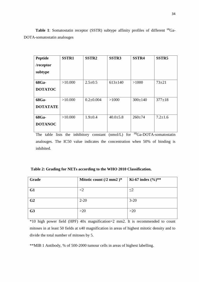
Table 1: Somatostatin receptor (SSTR) subtype affinity profiles of different 68Ga-
DOTA-somatostatin analouges
Peptide
/receptor
subtype
SSTR1 SSTR2 SSTR3 SSTR4 SSTR5
68Ga-
DOTATOC
>10.000 2.5±0.5 613±140 >1000 73±21
68Ga-
DOTATATE
>10.000 0.2±0.004 >1000 300±140 377±18
68Ga-
DOTANOC
>10.000 1.9±0.4 40.0±5.8 260±74 7.2±1.6
The table lists the inhibitory constant (nmol/L) for 68Ga-DOTA-somatostatin
analouges. The IC50 value indicates the concentration when 50% of binding is
inhibited.
Table 2: Grading for NETs according to the WHO 2010 Classification.
Grade Mitotic count (/2 mm2 )* Ki-67 index (%)**
G1 <2 ≤2
G2 2-20 3-20
G3 >20 >20
*10 high power field (HPF) 40x magnification=2 mm2. It is recommended to count
mitoses in at least 50 fields at x40 magnification in areas of highest mitotic density and to
divide the total number of mitoses by 5.
**MIB 1 Antibody, % of 500-2000 tumour cells in areas of highest labelling.
35
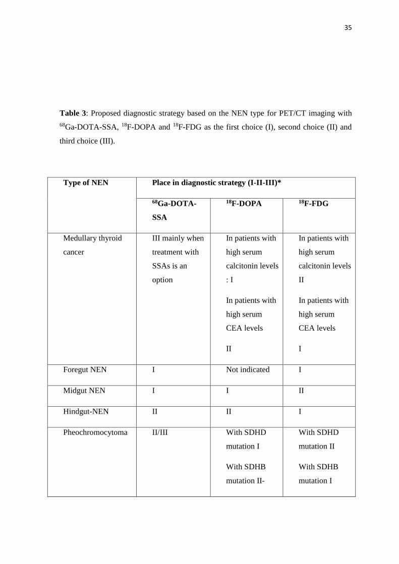
Table 3: Proposed diagnostic strategy based on the NEN type for PET/CT imaging with
68Ga-DOTA-SSA, 18F-DOPA and 18F-FDG as the first choice (I), second choice (II) and
third choice (III).
Type of NEN Place in diagnostic strategy (I-II-III)*
68Ga-DOTA-
SSA
18F-DOPA 18F-FDG
Medullary thyroid
cancer
III mainly when
treatment with
SSAs is an
option
In patients with
high serum
calcitonin levels
: I
In patients with
high serum
CEA levels
II
In patients with
high serum
calcitonin levels
II
In patients with
high serum
CEA levels
I
Foregut NEN I Not indicated I
Midgut NEN I I II
Hindgut-NEN II II I
Pheochromocytoma II/III With SDHD
mutation I
With SDHB
mutation II-
With SDHD
mutation II
With SDHB
mutation I
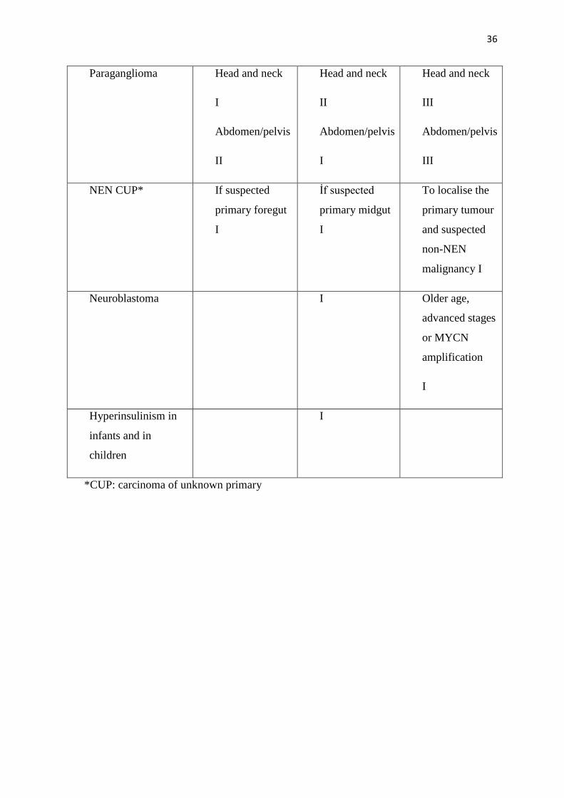
36
Paraganglioma Head and neck
I
Abdomen/pelvis
II
Head and neck
II
Abdomen/pelvis
I
Head and neck
III
Abdomen/pelvis
III
NEN CUP* If suspected
primary foregut
I
İf suspected
primary midgut
I
To localise the
primary tumour
and suspected
non-NEN
malignancy I
Neuroblastoma I Older age,
advanced stages
or MYCN
amplification
I
Hyperinsulinism in
infants and in
children
I
*CUP: carcinoma of unknown primary