Embed Size (px)
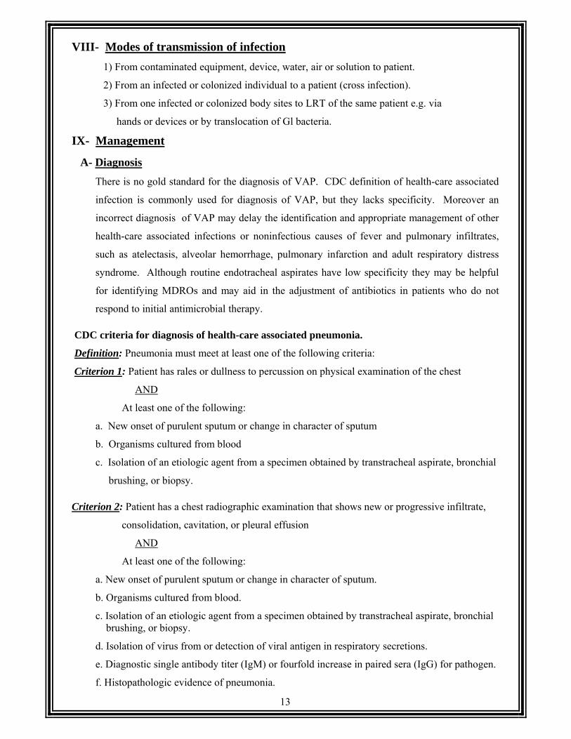
Citation preview

1
State of Kuwait Ministry of Health Infection Control Directorate
Guidelines for Prevention of
Ventilator Associated Pneumonia (VAP)
May, 2005

2
CONTENTS
Page I. Introduction 1
II. Definition 1
III. Aim 2
IV. Epidemiology 2
V. Microbial Aetiology 3
VI. Risk Factors 6
VII. Pathogenesis 8
VIII. Modes of Transmission
11
IX. Management: 11
X. Recommendations for Prevention of VAP 17
XI. Appendices 29
XII. References 40

3
I- Introduction
Pneumonia is the second most common health-care associated infection 2nd to urine tract infection,
accounting for 13 - 18% of all health-care associated infections. Critically ill patients who require
mechanical ventilation are especially vulnerable to develop ventilator-associated pneumonia (VAP).
The overall health-care associated infection rate of critically ill patients in the ICU approaches
40 - 60%. Respiratory infections account for 30 - 60% of all such infections. Incidence of VAP is
estimated to range from 10 - 25%.
Negative outcomes associated with VAP include increased mortality and morbidity and prolonged
hospital stay. In some studies, results showed that critically ill patients in whom VAP developed
were twice as likely to die as were those who did not acquire pneumonia. The risk of pneumonia
increased from 6.5% in those ventilated for 10 days, to 28% in those ventilated for 30 days.
Patients with VAP stay an average of 4.3 days longer in the ICU and have an absolute risk increase
of 5.8% for mortality.
Because of its reported frequency, associated high fatality rate, and attendant cost, health-care
associated pneumonia is a major infection control problem.
In the current policy, epidemiology, risk factors, pathogenesis, and management of VAP are
described. Based on a review of literature of evidence-based clinical practice, relevant randomized,
controlled trials, systematic reviews of mechanically ventilated adults, and CDC guidelines several
recommendations for preventing VAP are presented.
II- Definition
• Ventilator Associated pneumonia (VAP) is defined as Nosocomial pneumonia in a patient on
mechanical ventilatory support (by endotracheal tube or tracheostomy) for > 48 hrs.
• Ventilator-associated pneumonia that occurs within 48 -72 hrs after tracheal intubation is
usually termed early-onset pneumonia; it often results from aspiration, which complicates the
intubation process.
● Ventilator-associated pneumonia that occurs after this period is considered late-onset
pneumonia.

4
III- Aim The aim of the policy is to provide basic information regarding etiology, epidemiology, risk factors
and preventive measures to reduce the incidence of VAP.
IV- Epidemiology
• Pneumonia comprises 60% of all deaths due to health-care associated infections.
• Pneumonia could prolong hospitalization by 4-9 days. Conservative estimates of the direct cost of excess hospital stay due to pneumonia is $1.2 billion a year for USA.
• VAP is the 2nd most common health-care associated infection worldwide as well as in
Kuwait.
• In ICU, 86% of health-care associated pneumonias are associated with mechanical ventilation (MV).
• VAP is more frequent in patient with Acute Respiratory Distress Syndrome (ARDS) (55%)
than in other mechanically ventilated patient (28%) because the longer MV period the more risk of developing VAP.
Incidence
International:
• 12 infections per 1000 patient days.
• 20 infections per 1000 ventilator days.
In Kuwait: {Report of Kuwait National health-care associated Infection Surveillance - Directorate of Infection Control for the years 1999 – 2003}
• Main ICU average - 29.4 infections per 1000 ventilator days.
• NICU average - 8.2 infections per 1000 ventilator days.
• PICU average - 1.4 infections per 1000 ventilator days.

5
V- Microbial aetiology of VAP according to USA Figures
In adult ICU:
Organism Percentage
- Gram negative bacilli 30 - 60 %
- Staph aureus 10 - 30 %
- Streptococcus pneumoniae 5 - 20 %
- Haemophilus influenzae 2 - 5 %
- Legionella spp 0 - 20 %
- Polymicrobial 20 - 40 %
- Unknown 40 - 60 %
In pediatric ICU:
Organism Percentage
- Pseudomonas aeruginosa 21.8 %
- Staph aureus 16.9 %
- Haemophilus influenzae 10.2 %
- Enterobacter spp 9.3 %
- Klebsiella pneumoniae 5.3 %
- Serratia marcescens 3.6 %
- Escherichia coli 3.6 %
- Streptococcus pneumoniae 3.4 %
Source: Adapted from health-care associated Infections in PICU in USA – Michael J. Richards et. al. and National health-care associated Infections Surveillance System

6
Microbial aetiology of VAP according to Kuwait Figures
In Adult ICU:
ORGANISMS NUMBER OF INFECTIONS MRSA 43
Candida Albicans 28
S. aureus 20
S. epidermidis 8
Serratia spp. 11
Xanthomonas 4
Pseudomonas aeruginosa 69
Candida spp 20
Strep. Pneumoniae 4
E. coli 11
Acinetobacter 50
Alcaligenes 1
Haemophilus spp. 4
Enterobacter cloae 15
Klebsiella pneumonia 32
Enterococci 6
Diph. Spp. 1
GNB 1
Stenotrophomotophili 10
Branhgtarhelu 1
CNS 3
Strep. pyogene 1
Providensia 1
No pathogens isolated 1
Culture not done 3
Morganella morgani 1
Proteus spp. 2
Strept. fecalis 1
Halmia alver 1
Total 353

7
In Pediatric ICU: (2003)
ORGANISMS NUMBER OF INFECTIONS Enterococcus faecalis 1
Pseudomonas aeruginosa 13
Acinetobacter 3
S. aureus 3
Stenotrophomonas maltaphilia 6
Klebsiella pneumonia 8
Candida spp 2
Strept. Pneumoniae 2
MRSA 1
Enterobacter 2
Serratia spp. 2
Culture not done 1
E. coli 1
Spingomonas Pacimeoble 1
Total 46
In Neonatal ICU: ORGANISMS NUMBER OF INFECTIONS Culture not done 10
CNS 2
Pseudomonas aeruginosa 2
Enterococcus faecalis 1
Stenotrophomonas maltaphilia 1
Candida Albican 1
No pathogen isolated 1
E. coli 1
Clinically diagnosed 1
Total 20

8
VI- Risk Factors:
Can be grouped into the following general categories: (Fig. 1)
(1) Host factors:
• Age > 65 yrs.
• Underlying illness including:
- Chronic pulmonary diseases.
- Immunosupression.
- Depressed conciousness.
(2) Factors that enhance colonization of the oropharynx or stomach by microorganisms including:
• Administration of antimicrobials.
• Admission to ICU.
• Underlying chronic lung diseases.
• Coma.
• Administration of antacids for prevention of stress bleeding in critically ill patients.
(3) Conditions favouring aspiration reflex like:
• Endotracheal intubation
• Insertion of nasogastric tube.
• Supine position.
• Depressed level of consciousness [Glasgow Coma Scale (GCS) < 9.]
• Dysphagia from neurological or esophageal disorders.
(4) Prolonged use of mechanical ventilation.
Patients receiving continuous mechanically assisted ventilation have 6-21 times increased
risk for VAP. The risk was attributed partially to carriage of oropharyngeal organisms upon
passage through endotracheal tube into the trachea during intubation and depressed host
defenses secondary to the patient’s severe underlying illness.
(5) Lack of anatomical barriers:
leakage around the endotracheal cuff.
Impaired cough and mucocillary clearance.
Injury of the epithelial layer.
(6) Cross contamination via hands of health care workers (HCW) through:
• tracheal suctioning
• manipulation of the ventilator circuit, or endotracheal tubes

9
(7) Contamination of devices used on the respiratory tract which may be potential reservoirs and
vehicles for infectious microorganisms:
• Devices used on the respiratory tract for respiratory therapy:
Endotracheal tube (Emergency intubation versus elective)
Nebulizer
Bronchoscopy and Spirometer
Anaesthesia devices
Vaccinations. Reduce stay ICU surveillance Reduce exposure and duration of suppression Limit use of antimicrobials
Perform respiratory therapy Stress bleeding prophylaxis
Perform respiratory therapy Patient education Use aseptic technique.
Encourage early ambulation Limit sedation Use pain control medication
Patient monitoring
Proper patient position Staff education and management of Proper disinfection, sterilization secretions/condensate/ Restrict infected staff, visitors circuit tubing
Maintain cooling towers, water supplies, air flow
Figure 1. Intrinsic and extrinsic host factors contributing to the chain of health-care associated
pneumonia and ventilator-associated pneumonia caused by bacterial pathogens. (Adapted from
Craven DE et al. Semin Respir Crit Care Med. 1997.)
Risk Factors.
Intrinsic
Extremes of age
Underlying conditions Immune suppression
Chronic lung disease
Life style factors: Smoking , malnutrition
Acute condition: Coma , Trauma,
Mechanical Ventilation
Extrinsic
ICU stay
Exposure to antimicrobials/ other agents
Invasive procedures/
surgery (head, neck ,
chest, abdomen)
Exposure to
contaminated equipment, devices, hands, individuals, air, water, solutions
Pneumonia

10
VII- Pathogenesis of VAP The pathogenesis of VAP is related to the number and virulence of organisms entering the lower
airway and the response of the host’s mechanical, humoral, and cellular defenses to the invasion
(See Figure 2).
The development of pneumonia requires the pathogen to reach the alveoli and the host defenses to be
overwhelmed. The endogenous sources of microorganisms are nasal carriers, sinusitis, mouth,
oropharynx, gastric, or tracheal colonization, and hematogenous spread. The exogenous sources of
microorganisms are biofilm of the tracheal tube, ventilator circuits, nebulizers, and humidifiers.
Health care workers may also play role in this setting.
Figure 2. Risk factors for colonization of the aero-digestive tract and trachea before the
development of tracheobronchitis and health-care associated / ventilator- associated pneumonia.
(Adapted from Craven DE et al. Curr Opin Crit Care. 2002)
Antimicrobials/Medications
Surgery/ invasive devices
Contaminated hands, gloves, devices, water, solutions
Aero-digestive colonization.
Aspiration
Tracheal colonization: Inoculum and virulence
Inoculaton/ Inhalation
Mechanical, cellular, humoral Lung defenses overwhelmed
Tracheobronchitis
Bacteremia Pneumonia Translocation from GI
Host Factors

11
Different microorganism can be found depending on the onset time of pneumonia and the local
pattern variation encountered between different institutions and countries. (Early versus late) See
Table 1.
Table 1. Bacteria Associated With Early-and Late-Onset Ventilator – Associated Pneumonia.
Bacterial pathogen Crude frequency (%) Onset
Streptococcus pneumoniae: Pencillin-resistant Multidrug-resistant
10 - 20 Early
Haemophilus influenzae 5 - 15 Early Staphylococcus aureus Methicillin-resistant Methicillin-sensitive
20 -30 Early/ Late
Gram –ve bacilli Pseudomonas aeruginosa Acinetobacter species Klebsiella species Enterobacter species Serratia species
30 - 60 Late
Legionella pneumophila 0 - 15 Late
Local trauma and inflammation caused by an endotracheal tube and possible leakage of
contaminated secretions around the cuff and into the upper trachea increases lower airway
colonization and the risk of tracheobronchitis and VAP (Figure 3)
In addition , microorganism aggregated in biofilm on the surface of the endotracheal tube may have
pathogenetic relevance for colonization of the trachea and possible embolization to the lung
following maneuvers such as endotracheal suctioning and bronchoscopy. Host factors, the types of
bacteria colonizing the pharynx, and the recent use of antibiotics may also alter pharyngeal
colonization and adherence of bacteria.

12
The stomach is also a potential reservoir for Multi-Drug Resistant Organisms(MDROs) bacteria that
may cause retrograde colonization of the oropharynx and increase the risk of VAP, particularly in
patients with increased gastric volume or alkaline pH. The use of antacids or histamine type 2 (H2) blockers has been associated with an increased risk of
VAP compared with use of sucralfate, but patients receiving sucralfate have had a higher rate of
clinically significant GI bleeding. Furthermore, patients in a supine body position, those who have a nasogastric or orogastric tube,
and those who are receiving enteral feeding are at much greater risk for VAP than are patients who
are maintained in the semi-upright position.
Figure 3. Schematic representation of an intubated patient with collection of subglottic secretions above the endotracheal tube cuff. Note that the placement of an orogastric tube rather than a nasogastric tube reduces the risk sinusitis and ventilator-associated pneumonia. (Adapted from Craven DE, Steger KA. Infect Control Hosp Epidemiol. 1997.)
Endotracheal tube
Orogastric tube
Pooled Secretions
Trachea
Esophagus
CuffStomach
Endotracheal tube
Orogastric tube (to suction)
13
VIII- Modes of transmission of infection 1) From contaminated equipment, device, water, air or solution to patient.
2) From an infected or colonized individual to a patient (cross infection).
3) From one infected or colonized body sites to LRT of the same patient e.g. via
hands or devices or by translocation of Gl bacteria.
IX- Management
A- Diagnosis
There is no gold standard for the diagnosis of VAP. CDC definition of health-care associated
infection is commonly used for diagnosis of VAP, but they lacks specificity. Moreover an
incorrect diagnosis of VAP may delay the identification and appropriate management of other
health-care associated infections or noninfectious causes of fever and pulmonary infiltrates,
such as atelectasis, alveolar hemorrhage, pulmonary infarction and adult respiratory distress
syndrome. Although routine endotracheal aspirates have low specificity they may be helpful
for identifying MDROs and may aid in the adjustment of antibiotics in patients who do not
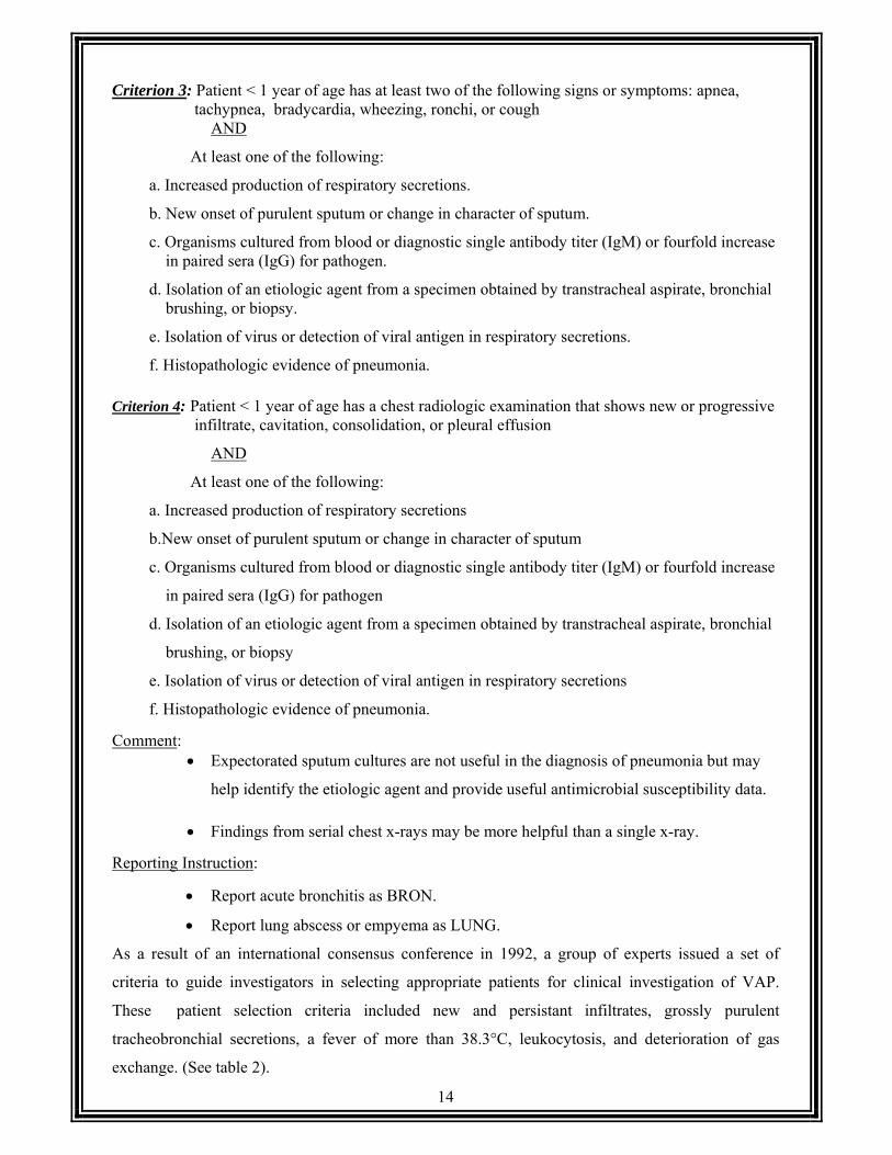
respond to initial antimicrobial therapy. CDC criteria for diagnosis of health-care associated pneumonia.
Definition: Pneumonia must meet at least one of the following criteria:
Criterion 1: Patient has rales or dullness to percussion on physical examination of the chest
AND
At least one of the following:
a. New onset of purulent sputum or change in character of sputum
b. Organisms cultured from blood
c. Isolation of an etiologic agent from a specimen obtained by transtracheal aspirate, bronchial
brushing, or biopsy.
Criterion 2: Patient has a chest radiographic examination that shows new or progressive infiltrate,
consolidation, cavitation, or pleural effusion
AND
At least one of the following:
a. New onset of purulent sputum or change in character of sputum.
b. Organisms cultured from blood.
c. Isolation of an etiologic agent from a specimen obtained by transtracheal aspirate, bronchial brushing, or biopsy.
d. Isolation of virus from or detection of viral antigen in respiratory secretions.
e. Diagnostic single antibody titer (IgM) or fourfold increase in paired sera (IgG) for pathogen.
f. Histopathologic evidence of pneumonia.
14
Criterion 3: Patient < 1 year of age has at least two of the following signs or symptoms: apnea, tachypnea, bradycardia, wheezing, ronchi, or cough
AND
At least one of the following:
a. Increased production of respiratory secretions.
b. New onset of purulent sputum or change in character of sputum.
c. Organisms cultured from blood or diagnostic single antibody titer (IgM) or fourfold increase in paired sera (IgG) for pathogen.
d. Isolation of an etiologic agent from a specimen obtained by transtracheal aspirate, bronchial brushing, or biopsy.
e. Isolation of virus or detection of viral antigen in respiratory secretions.
f. Histopathologic evidence of pneumonia.
Criterion 4: Patient < 1 year of age has a chest radiologic examination that shows new or progressive infiltrate, cavitation, consolidation, or pleural effusion
AND
At least one of the following:
a. Increased production of respiratory secretions
b.New onset of purulent sputum or change in character of sputum
c. Organisms cultured from blood or diagnostic single antibody titer (IgM) or fourfold increase
in paired sera (IgG) for pathogen
d. Isolation of an etiologic agent from a specimen obtained by transtracheal aspirate, bronchial
brushing, or biopsy
e. Isolation of virus or detection of viral antigen in respiratory secretions
f. Histopathologic evidence of pneumonia. Comment:
• Expectorated sputum cultures are not useful in the diagnosis of pneumonia but may
help identify the etiologic agent and provide useful antimicrobial susceptibility data.
• Findings from serial chest x-rays may be more helpful than a single x-ray. Reporting Instruction:
• Report acute bronchitis as BRON.
• Report lung abscess or empyema as LUNG.
As a result of an international consensus conference in 1992, a group of experts issued a set of
criteria to guide investigators in selecting appropriate patients for clinical investigation of VAP.
These patient selection criteria included new and persistant infiltrates, grossly purulent
tracheobronchial secretions, a fever of more than 38.3°C, leukocytosis, and deterioration of gas
exchange. (See table 2).

15
Table 2. Consenses Diagnostic Criteria for VAP Definitions Recommendations Definite pneumonia The patient meets the clinical criteria for suspicion of VAP of new (progressive)
or persistent infiltrate and purulent tracheal secretions and demonstrates one of the following:
1. Radiographic evidence, preferably CT evidence, of pulmonary abscess and
positive needle aspirate culture from the abscess. 2. Pathogenic evidence of pneumonia on histologic examination of lung tissue
obtained by open-lung biopsy or at postmortem examination immediately after death (and within 3 days of the bronchoscopic procedure when used to, confirm the diagnosis of VAP made by bronchoscopy) that demonstrates abscess formation or an area of consolidation with intense polymorphonuclear leukocyte accumulation plus a positive quantitative culture of lung parenchyma (>104 microorganisms per gram of lung tissue).
Probable Pneumonia In the absence of any of the above criteria for pneumonia, the patients meets the clinical criteria for suspicion of VAP and demonstrates one of the following:
1. The presence of positive quantitative culture of a sample of secretions from the lower respiratory tract obtained by a technique that minimizes contamination with upper respiratory tract flora (PSB, BAL, protected BAL).
2. The presence of positive blood culture unrelated to another source and obtained
within 48h before and after respiratory sampling. The microorganism(s) recovered should be identical to the organism recovered from a culture of lower respiratory tract secretions.
3. The presence of positive pleural fluid culture in the absence of previous pleural
instrumentation. The microorganism(s) recovered should be identical to the organism recovered from a culture of lower respiratory tract secretions.
4. Pathogenic evidence of pneumonia on histologic examination of lung tissue
obtained by open-lung biopsy or at postmortem examination immediately after death (and within 3 days of the bronchoscopic procedure when used to confirm the diagnosis of VAP made by bronchoscopy) that demonstrates abscess formation or an area of consoldation with intense polymorphonuclear leukocyte accumulation plus a negative quantitative culture of lung parenchyma (<104 microorganisms per gram of lung tissue).
Definite absence of In the absence of any of the above criteria for pneumonia, the absence of Pneumonia pneumonia is definitive if one of the following criteria is met:
1. Postmortem exam within 3 days of the suspicion of pneumonia showing no histologic sign of lung infection.
2. Definite alternative etiology with no bacterial growth on the reliable respiratory
specimen. 3. Cytologic identification of a process other than pneumonia (e.g. lung cancer)
without significant bacterial growth on the reliable respiratory specimen. Probable absence Lack of significant growth on the reliable respiratory specimen with one of Of pneumonia the following:
1. Resolution without antibiotic therapy of 1 of the following – fever, radiologic infiltrate, or radiologic infiltrate and a definitive diagnosis.
2. Persistent fever and radiologic infiltrate, with a definite alternative diagnosis established.

16
Advantages and limitations of current diagnostic methods of VAP are summarized in Table 3. Table 3. Current Methods for the Diagnosis of Ventilator-Associated Pneumonia (VAP):
Advantages and Disadvantages:
Method of Diagnosis Advantages Disadvantages
Clinical • Easy to perform. • Gram stain may be helpful.
• Poor specificity, especially in patients with chest radiograph abnormalities.
Non-quantitative cultures
• Non invasive. • Inexpensive . • Gram stain may be helpful for
initial antibiotic treatment and interpretation of culture results.
• May increase antibiotic use or alter outcome compared with quantitative microbiology.
Quantitative endotracheal aspirate analysis
• Simple and easy to perform. • Gram stain may be helpful. • Less expensive than
bronchoscopy and non-bronchoscopy with PSB or BAL, but less specific.
• Good negative predictive value with Diagnostic threshold 105 CFU/ml.
• Less specific. • If used alone for diagnosis,
it can lead to unnecessary antibiotic therapy or overtreatment with broad spectrum antibiotics.
Non-bronchoscopic (blind) BAL/ PSB
• Simpler and less expensive than bronchoscopy with PSB or BAL .
• Correlates with bronchoscopies BAL.
• Costs of quantitative cultures.
• Special skills required. • Limited data for
comparison.
Bronchoscopic BAL/PSB
• Can observe sampling site. • Specificity > 95%. • Greater sensitivity for ≥ 103 CFU/mL for PSB and ≥ 104 CFU/mL for BAL. • Cytocentrifugation may be
helpful for early identificaion of cause.
• May decrease antibiotic use and development of resistance.
• May be associated with better patient outcomes.
• Useful in immuno-compromised and nonresponding patients.
• Antibiotic use in last 24 hours may decrease sensitivity .
• False negative results in early cases.
• Need quantitative bacteriology and meticulous and prompt processing of specimens.
• Costly, not widely available.
• Complication rate greater for BAL than PSB.
BAL : Bronchoalveolar Lavage; PSB: Protected Specimen Brush; CFU: Colony-forming unit.

17
Several principles for the management of VAP are beginning to emerge (Figure 4):
- Early, appropriate initial antibiotic coverage for all suspected pathogens and de-escalation or
streamlining of antibiotic therapy based on clinical response and microbiologic data appear
to improve patient outcomes.
- The use of a specific antibiotic regimen should also be based on the local epidemiology of
MDROs in the ICU.
- Stopping antibiotic therapy in patients who do not meet the criteria for a diagnosis of VAP is
important. Negative lower respiratory tract cultures in a patient who has not changed
antibiotics in the past 72 hrs can be used to stop antibiotic therapy.
- Emperic therapy should include agents from different antibiotic class than what the
patient has received recently.
- The optimal duration of therapy for patients with VAP is unknown, but studies are in
progress that may shorten the duration of treatment.
- Shorter courses of antibiotics have been used in early-onset disease where there is prompt
response to initial therapy.
- Longer courses of antibiotics may be needed for patients with Legionella infection or for
patients with complications of VAP, such as empyema, lung abscess, or additional sites of
infection.
- Initial antibiotic regimens for early-onset or late-onset, mild to severe ventilator-
associated pneumonia will be based upon local antibiotic policy.
PMNs : Polymorphonuclear Leucocytes, ETA: Endotracheal Aspirate; QEA: Quantitative Endotracheal Aspirate; BAL: Bronchoalveolar Lavage; PSB: Protected Specimen Brush; ID: Infectious Diseases. Figure 4. Management algorithm for ventilator-associated pneumonia (VAP)
Clinical suspicion of VAP
Sputum presentYes No
Assess Gram statin for PMNs and bacteria
Culture: ETA , QEA, BAL, PSB
Obtain ETA or QEA Blind BAL/ PSB
Bronchoscope BAL / PSB
Assess risk factors Initiate appropriate antibiotic therapy.
Clinical Improvement? Yes No
Review microbiologic results Streamline antibiotic therapy
Assess microbiologic results, antibiotics, and diagnosis, obtain ID/ pulmonary consultation

18
Recommendations for prevention of ventilator associated pneumonia (VAP)
Categorization of Recommendations
Based on a review of the literature, several recommendations for preventing VAP are presented. The
CDC has promoted some of these suggestions; others are based on results in studies that have been
published since the 1994 CDC guidelines were published. Each recommendation is categorized on
the basis of existing scientific evidence, theoretical rationale, applicability, and potential economic
impact. In addition, a new category accommodates recommendations that are made on the basis of
existing national or state health regulations. The following categorization scheme is applied in this
guideline:
Category IA. Strongly recommended for implementation and strongly supported by well-designed experimental, clinical, or epidemiological studies.
Category IB. Strongly recommended for implementation and supported by certain clinical or epidemiological studies and by strong theoretical rationale.
Category IC. Required for implementation, as mandated by federal or state regulation or standard.
Category II. Suggested for implementation and supported by suggestive clinical or epidemiological studies or by strong theoretical rationale.
No recommendation; unresolved issue. Practices for which insufficient evidence or no consensus exists about efficacy.
I. STAFF EDUCATION AND INFECTION SURVEILLANCE
A. Staff Education
Educate healthcare workers about the epidemiology of, and infection-control procedures for,
preventing VAP. Also involve the workers in the implementation of interventions to prevent VAP by
using performance-improvement tools and techniques (IA).
B. Surveillance
1. Conduct surveillance for bacterial pneumonia in ICU patients who are at high risk for VAP (e.g.,
patients with mechanically assisted ventilation or selected postoperative patients) to determine
trends and help to identify outbreaks and other potential infection-control problems. Include data
on the causative microorganisms and their antimicrobial susceptibility patterns. Express data as

19
rates (e.g., number of infected patients or infections per 100 ICU days or per 1,000 ventilator
days) to facilitate intra hospital comparisons and trend determination. (IB).
2. In the absence of specific clinical, epidemiological, or infection-control objectives, do not
routinely perform surveillance cultures of patients or of equipments or devices used for
respiratory therapy, pulmonary-function testing, or delivery of inhalation anesthesia (II)
II. INTERRUPTION OF TRANSMISSION OF MICROORGANISMS
A. Sterilization or Disinfection, and Maintenance of Equipment and Devices
Proper cleaning and sterilization or disinfection of reusable equipment is an important
component of a program to reduce infection associated with respiratory therapy and
anesthesia equipment. Many devices used on the respiratory tract have been categorized as
semicritical in the Spaulding classification system of appropriate sterilization or disinfection
of medical devices because they come into direct or indirect contact with mucus membranes
but do not ordinarily penetrate body surfaces and the associated infection risk following the
use of these devices in patients is less than that associated with devices that penetrate
normally sterile tissues.
1. General Measures
a) Thoroughly clean all equipment and devices to be sterilized or disinfected (IA)
b) Sterilize (by autoclaving) for semicritical equipment or devices. If it is neither possible or
cost effective to sterilize these devices by steam autoclave or ethylene oxide, they can be
subjected to high level disinfection by using utensil disinfector machine at 94°C for 4
minutes or by using liquid chemical disinfectant. Follow disinfection with appropriate
rinsing, drying, and packaging, taking care not to contaminate the disinfected items in the
process (IA)
c) Use sterile water for rinsing reusable semicritical respiratory equipment and devices when
rinsing is needed after they have been chemically disinfected. Data are insufficient regarding
safety of using tap water for rinsing (followed by drying) reusable semicritical respiratory
devices after their disinfection or between their uses on the same patient. If this is not
feasible, rinse the device with filtered water (i.e., water that has been through a 0.2µ filter) or
tap water, and then rinse with isopropyl alcohol and dry with forced air or in a drying cabinet
(IB)
d) Do not reprocess an equipment or device that is manufactured for single use only.
20
2. Mechanical ventilators
The internal machinery of mechanical ventilators is not considered an important source of bacterial
contamination of inhaled gas. Thus, routine sterilization or disinfection of the internal
machinery is considered unnecessary. (II)
3. Breathing circuits, humidifiers, and heat-and-moisture exchangers (HMEs)
• Breathing circuits with humidifiers (See appendix 1)
a) Do not routinely change more frequently than 48 hr, including ventilator tubing and exhalation
valve, and the attached humidifier that is in use on an individual patient. Change the circuit when
it is visibly soiled or mechanically malfunctioning. (IA)
b) Breathing-circuit--tubing condensate
- Periodically drain and discard any condensate that collects in the tubing of a mechanical
ventilator, taking precautions not to allow condensate to drain toward the patient (IB)
- Wear gloves to perform the previous procedure and/or when handling the fluid (IB)
- Decontaminate hands with antiseptic hand wash and water (if hands are visibly soiled) or with an
alcohol-based hand rub after performing the procedure or handling the fluid (IA)
c) Humidifier fluids
- Use sterile water to fill bubbling humidifiers. (II)
- No recommendation can be made for the preferential use of a closed or continuous-feed
humidification system (Unresolved issue). In Kuwait a closed system is recommended for
use as humidification system.
• Ventilator breathing circuits with HMEs
a) Changing HME
- Change an HME that is in use on a patient when it malfunctions mechanically or becomes
visibly soiled or when its water content increase causes resistance on the ventilator (II).
- Do not routinely change more frequently than every 48 hours an HME that is in use on a
patient (II)
b) Do not change routinely (in the absence of gross contamination or malfunction) the
breathing circuit attached to an HME while it is in use on a patient (II)

21
4. Oxygen humidifiers
Change the humidifier tubing (including any nasal prongs or mask) that is in use on one patient
when it malfunctions or becomes visibly contaminated. (II).
5. Small-volume medication nebulizers: in-line and hand-held nebulizers
a) Between treatments on the same patient clean with liquid detergent and wipe with alcohol 70%,
and dry small-volume in-line or hand-held medication nebulizers. (IB)
b) Use only sterile fluid for nebulization, and dispense the fluid into the nebulizer aseptically. (IA)
c) Whenever possible, use aerosolized medications in single-dose vials. If multidose medication
vials are used, follow manufacturers' instructions for handling, storing, and dispensing the
medications. (IB)
6. Mist tents
a) Between uses on different patients, replace mist tents and their nebulizers, reservoirs, and tubings
with those that have been subjected to sterilization or high-level disinfection. (II)
b) No recommendation can be made about the frequency of routinely changing mist-tent nebulizers,
reservoirs, and tubings while in use on one patient. (Unresolved issue).
c) Subject mist-tent nebulizers, reservoirs, and tubings that are used on the same patient to daily
low-level disinfection (e.g., with 2% acetic acid) followed by air-drying (II) or according to
manufacturer’s instructions.
7. Other devices used in association with respiratory therapy
a) Respirometer and ventilator thermometer: between their uses on different patients, sterilize or
subject to high-level disinfection portable respirometers and ventilator thermometers (IB)
b) Resuscitation bags
- Between their uses on different patients, sterilize or subject to high-level disinfection reusable
hand-powered resuscitation bags (IB). (See appendix 2)
- Our recommendation in Kuwait is to change the hydrophopic filter whenever visibly
soiled or when used for a known infected case.
8. Anesthesia machines and breathing systems or patient circuits
a) Do not routinely sterilize or disinfect the internal machinery of anesthesia equipment. (IB)
b) Clean and then sterilize or subject to high-level liquid chemical disinfection if available
reusable components of the breathing system or patient circuit (ie. the y-piece; face mask;

22
reservoir bag and humidifiers) and use disposable tracheal tubes, and inspiratory and
expiratory breathing tubing, between uses on different patients, in accordance with the device
manufacturers' instructions for their reprocessing (IB). (See appendix 3)
c) Follow published guidelines or manufacturers' instructions about in-use maintenance,
cleaning, and disinfection or sterilization of other components or attachments of the
breathing system or patient circuit of anesthesia equipment (IB)
d) No recommendation can be made for placing a bacterial filter in the breathing system or
patient circuit of anesthesia equipment (Unresolved issue)
9. Pulmonary-function testing equipment
a) Do not routinely sterilize or disinfect the internal machinery of pulmonary-function testing
machines between uses on different patients (II)
b) Change the mouthpiece of a peak flow meter or the mouthpiece and filter of a spirometer
between uses on different patients (II)
10. Room-air "humidifiers" and faucet aerators
a) Do not use large-volume room-air humidifiers that create aerosols (e.g., by venturi principle,
ultrasound, or spinning disk, and thus actually are nebulizers) unless they can be sterilized or
subjected to high-level disinfection at least daily and filled only with sterile water (II)
b) Faucet aerators
- No recommendation can be made about the removal of faucet aerators from areas for
immunocompetent patients (Unresolved issue).
- If Legionella spp. are detected in the water of a transplant unit and until Legionella spp. are
no longer detected by culture, remove faucet aerators in the unit (II)
B. Prevention of Person-to-Person Transmission of Bacteria
1. Standard Precautions
a) Hand hygiene
Meticulous hand washing is the first step in reducing VAP. Decontaminate hands by washing them
with either antiseptic hand wash and water (if hands are visibly dirty or contaminated with
proteinaous material or are soiled with blood or body fluids) or by using an alcohol-based antiseptic
agent (e.g., hand rub) if hands are not visibly soiled. Decontaminate hands as described previously

23
after contact with mucous membranes, respiratory secretions, or objects contaminated with
respiratory secretions, whether or not gloves are worn and before and after contact with a patient
who has an endotracheal or tracheostomy tube in place, and before and after contact with any
respiratory device that is used on the patient, whether or not gloves are worn (IA)
b) Gloving
- Wear gloves when suctioning patients orally or through the endotracheal tube. Gloves are also
needed when handling respiratory secretions or objects contaminated with respiratory secretions of
any patient. Gloves are also recommended when closed-suction devices are used. (IB)
- Change gloves and decontaminate hands as described previously between contacts with different
patients; after handling respiratory secretions or objects contaminated with secretions from one
patient and before contact with another patient, object, or environmental surface; and between
contacts with a contaminated body site and the respiratory tract of, or respiratory device on, the same
patient (IA)
Although these recommendations seem simple, many times staff are observed leaving a patient’s
room with gloves on, and proceed to enter data on the patient’s record, answer the phone, and
perform other duties.
c) Gowning
When soiling with respiratory secretions from a patient is anticipated, wear a gown and change it
after soiling occurs and before providing care to another patient (IB)
2. Care of patients with tracheostomy
a) Perform tracheostomy under aseptic conditions (II).
b) When changing a tracheostomy tube, wear a gown, use aseptic technique, and replace the tube with one that has undergone sterilization or high-level disinfection (IB)
c) No recommendation can be made for the daily application of topical antimicrobial agent(s) at the tracheostomy site (Unresolved issue).
However, in Kuwait it is not recommended to make daily topical application of antimicrobial agent(s) at the tracheostomy site.
3. Suctioning of respiratory tract secretions (See also Section IV-B-1-e)
Endotracheal Suctioning The current standard of care is to suction patients only when need is
determined by auscultation of adventitious lung sounds or other assessments. The rationale for this
standard is to reduce trauma to the airways. However, some patients have minimal secretions and
24
may not have to be suctioned for several hours. Because stagnant mucus and lack of a cough reflex
are risk factors for the development of VAP, suctioning and interventions to facilitate effective
coughing may be needed periodically.
Maintaining aseptic technique when endotracheal suctioning is needed to reduce contamination of
the oropharyngeal cavity. When suction catheters are used, it is important to rinse the secretions, to
remove mucus from the suction catheter and to reduce the likelihood of bacterial growth.
a) No recommendation can be made for the preferential use of either the multiuse closed-system
suction catheter or the single-use open-system suction catheter for prevention of pneumonia
(Unresolved issue)
b) No recommendation can be made about wearing sterile rather than clean gloves when
performing endotracheal suctioning (Unresolved issue).
c) No recommendation can be made about the frequency of routinely changing the in-line
suction catheter of a closed-suction system in use on one patient (Unresolved issue)
d) If the open-system suction is employed, use a sterile, single-use catheter (II).
e) Use only sterile fluid to remove secretions from the suction catheter if the catheter is to be
used for re-entry into the patient's lower respiratory tract (II).
In Kuwait it is recommended to rinse the tube connected to the vacuum bottle catheter (in-line catheter) with sterile saline after each use. Galipot used for rinsing in-line catheter should be discarded after each use.
IV. MODIFYING RISK FACTORS FOR INFECTION
A. Increasing Host Defense Against Infection: Administration of immune modulators
1. Pneumococcal vaccination (Consider Vaccination whenever indicated)
a- Administer the pneumococcal polysaccharide vaccine to persons aged > 65 years.
b- Persons aged 5 - 64 years
▪ Who have chronic cardiovascular disease, chronic pulmonary disease (e.g., chronic
obstructive pulmonary disease [COPD] or emphysema, but not asthma), diabetes mellitus,
alcoholism, chronic liver disease (e.g., cirrhosis), or cerebrospinal fluid (CSF) leaks.
▪ Who have functional or anatomic asplenia.
25
▪ Who are living in special environments or social settings; immunocompromised persons
aged >5 years with HIV infection, leukemia, lymphoma, Hodgkin's disease, multiple
myeloma, generalized malignancy, chronic renal failure, nephrotic syndrome, or other
conditions associated with immunosuppression (e.g., receipient of Homeopetic stem cell
transplantation- HSCT, solid-organ transplant, or immunosuppressive chemotherapy,
including long-term systemic corticosteroids) and persons in long-term care facilities (IA)
2. No recommendation can be made for the routine administration of preparations of
granulocyte-colony stimulating factor (GCSF) or intravenous gamma globulin for
prophylaxis against VAP (Unresolved issue)
3. No recommendation can be made for the routine enteral administration of glutamine for
prevention of VAP (Unresolved issue)
B. Precaution for Prevention of Aspiration
As soon as the clinical indications for their use are resolved, remove devices such as endotracheal,
tracheostomy, and/or enteral (i.e., oro- or nasogastric or jejunal) tubes from patients (IB)
Because aspiration of oropharyngeal secretions is a primary route for acquiring VAP, strategies to
reduce aspiration of secretions are recommended.
1. Prevention of aspiration associated with endotracheal intubations
a) Use of noninvasive ventilation (NIV) to reduce the need for and duration of endotracheal intubation:
- When feasible and not medically contraindicated, use noninvasive positive-pressure ventilation
(NIV) delivered continuously by face or nose mask, instead of performing endotracheal intubations
in patients who are in respiratory failure and are not needing immediate intubations (e.g., those who
are in hypercapneic respiratory failure secondary to acute exacerbation of COPD or cardiogenic
pulmonary edema) (II)
- When feasible and not medically contraindicated, use NIV as part of the weaning process (from
mechanically assisted ventilation) to shorten the period of endotracheal intubation (II)
b) Early Extubation: Because the likelihood of pneumonia increases with prolonged
ventilation period; separation from mechanical ventilation as soon as possible must be a

26
priority. Accidental extubation and subsequent reintubation increases the risk for VAP.
Therefore, strategies to prevent unplanned extubation are warranted.
c) As much as possible, avoid repeated endotracheal intubations in patients who have received
mechanically assisted ventilation. (II)
d) Unless contraindicated by the patient's condition, perform orotracheal rather than
nasotracheal intubations on patients. (IB)
e) If feasible, use an endotracheal tube with a dorsal lumen above the endotracheal cuff to allow
drainage (by continuous or frequent intermittent suctioning) of tracheal secretions that
accumulate in the patient's subglottic area. (II)
f) Endotracheal cuff: Before deflating the cuff of an endotracheal tube in preparation for tube
removal, or before moving the tube, ensure that secretions are cleared from above the tube
cuff (II). The incidence of VAP increases when the endotracheal cuff pressure is less than 20
cm water pressure. It is important to check cuff pressures routinely and to maintain at least
20 cm of pressure.
g) Repositioning Endotracheal Tubes: Oral endotracheal tubes are repositioned and retaped
according to hospital protocol, usually once a day. The rationale for repositioning tubes is to
prevent breakdown of the oral mucosa and mouth. There is no guidance in reported research
regarding frequency of repositioning tubes. Because pooled secretions above the
endotracheal cuff are associated with VAP, it is important that thorough oral suctioning be
performed before repositioning tubes. Optimal frequency of repositioning tubes should be
established.
h) Aspiration of Subglottic Secretions: Studies evaluated a special dual-lumen endotracheal
tube that has a port through which subglottic secretions are continually aspirated. Results in a
randomized trial of this new tube versus conventional endotracheal tubes showed a
significant decrease in VAP in the patients with continuous subglottic aspiration and a delay
in onset of VAP. Failure of the continuous aspiration device was cited as a risk factor for
VAP. This is a fairly new treatment that appears to be beneficial, and ongoing evaluation is
recommended. Alternative methods for aspiration may be tried for example, thoroughly
suctioning the oropharynx of patients every 1 to 2 hours in an attempt to decrease the amount
of pooled secretions around the endotracheal cuff.
2. Prevention of aspiration associated with enteral feeding

27
a) Bed elevation: In the absence of medical contraindication(s), elevation of the head of bed to
an angle of 30 - 45 degrees is effective in reducing the risk of aspiration in high risk patients
(e.g., a person receiving mechanically assisted ventilation and/or who has an enteral tube in
place). (II)
b) Routinely verify appropriate placement of the feeding tube. (IB)
c) No recommendation can be made for the preferential use of small-bore tubes for enteral feeding. (Unresolved issue)
d) No recommendation can be made for preferentially administering enteral feedings continuously or intermittently. (Unresolved issue)
e) No recommendation can be made for preferentially placing the feeding tubes, (e.g., jejunal tubes) distal to the pylorus. (Unresolved issue)
f) Gastric Residuals Patients receiving tube feeding must be closely monitored for aspiration.
Checking residual volumes is one method of assessment. High residual gastric volumes may
occur when gastric emptying is impaired, increasing the likelihood of regurgitation and
aspiration. No standard for checking residuals has been established. Based on review of the
literature and on clinical experience, residuals should be checked every 2 hours when
feedings are initiated. Once tube feedings are progressing without difficulty, residuals can be
checked every 4 to 6 hours.
g) Gastric Versus Intestinal Feeding: Where to place feeding tubes in the stomach or the
small intestine is another issue related to aspiration. In addition, it is believed that critically
ill patients better tolerate early enteral feeding into the small intestine. However, one
researcher found that aspiration was associated with both feeding methods. Although further
investigation is needed, rationale for feeding into the small intestine supports its use to
reduce the risk of VAP. (Unresolved issue)
h) Evaluating Swallowing: Oral feeding is initiated in patients with tracheostomies and in some
patients who are nasally intubated. It is important to avoid oral feedings in patients with artificial
airways until a dysphagia evaluation and a rehabilitation of swallowing are completed, since
subclinical aspiration is common in this group.
3. Prevention or modulation of oropharyngeal colonization
The first step in preventing VAP is to reduce colonization by pathogens of the oropharynx (Basic
nursing care principles are essential).
28
a) Oral Hygiene Although considered a standard nursing intervention, oral hygiene is often
neglected when caring for intubated patients. Many times, it is performed by quickly
swabbing the mouth. Oral care involves brushing the patient’s teeth, use of solutions and
mouthwash to cleanse the mouth, and thorough suctioning of oral secretions. Systematic oral
assessment is recommended in intubated patients.
b) Nasal Hygiene Meticulous nasal care and cleansing of the nasopharynx may reduce bacterial
colonization. As in oral care, nasal care is often neglected as a part of routine hygiene. Most
patients have a nasogastric or nasoenteric tube in place, and the endotracheal tube may be
placed nasally. The tubes remain taped for prolonged periods, and secretions accumulate and
crust in the nares. Protocols for routinely cleansing the nose and suctioning nasopharyngeal
secretions should be implemented and evaluated.
c) Oropharyngeal cleaning and decontamination with an antiseptic agent: develop and
implement a comprehensive oral-hygiene program (that might include the use of an antiseptic
agent) for patients in acute-care settings or residents in long-term care facilities that are at
high risk for VAP. (II)
d) Turning and Positioning: Stagnant mucus in the lower airways is a medium for bacterial
growth, should pathogens reach the lower airways. Routine turning and positioning assists in
mobilization of secretions.
e) Chlorhexidine oral rinse: Use an oral chlorhexidine gluconate (0.12%) rinse during the
perioperative period on adult patients who undergo cardiac surgery. (II)
f) Oral decontamination with topical antimicrobial agents: No recommendation can be made
for the routine use of topical antimicrobial agents for oral decontamination to prevent VAP.
(Unresolved issue)
4. Prevention of gastric colonization
Stress Ulcer Prophylaxis Each patient should be individually evaluated for the need for
medications to prevent stress ulcers.
a) No recommendation can be made for the preferential use of sucralfate, H2-antagonists,
and/or antacids for stress-bleeding prophylaxis in patients receiving mechanically assisted
ventilation. (Unresolved issue)

29
b) No recommendation can be made for the routine selective decontamination of the digestive
tract (SDD) of all critically ill, mechanically ventilated, or ICU patients. (Unresolved issue)
c) No recommendation can be made for routinely acidifying gastric feeding. (Unresolved issue)

30
Appendix – 1
Recommended patient circuit configurations

31
Appendix – 2
Cleaning, disinfection and sterilization of Ambubag Mark III resuscitator
Parts exposed to expiratory gases
Parts not exposed to patient expiratory gases
Note: Do not disassemble parts further than shown

32
Appendix – 2
Applicable methods for cleaning, disinfection and sterilization of Ambubag Mark
III resuscitator
● Applicable
○ not applicable
Methods Cleaning Disinfecting-sterilising Washing Disinfecting Auto-
claving (a), (b), (c) and (d) refer to the figure above
Resuscitator parts
Man
ual w
ashi
ng
Was
hing
Mac
hine
(WM
)
W.M
. hea
t dis
infe
ctin
g
Boi
ling
Che
mic
al
121º
C
134º
C
Eac
h Pa
tient
Patient valve (a)
●
●
●
●
●
●
●
Reg
ular
ly
Bag (b) ● ● ● ● ● ● ● Oxygen reservoir attachment (c) ● ○ ○ ○ ● ○ ○ Gas filter adapter (d) ● ● ● ● ● ● ● Extension tube ● ● ● ● ● ● ● Oxygen supply tube ● ● ● ● ● ● ●
.

33
Appendix – 3a (Cleaning, disinfection and sterilization of parts of for anesthesia machine in general)
Anesthetic Apparatus Sterilization / Disinfection
Airways Machine wash, then sterilize with ethylene oxide. If it does not withstand high temperatures, otherwise autoclave.
Endotracheal & Endobronchial tubes Disposable
Endotracheal tube Metal or polypropylene
Machine wash and autoclaved.
Corrugated tubing and valves Machine wash then sterilize.
Face masks Machine wash then sterilize.
Forceps and Introducers Metal, machine wash and autoclave.
Humidifiers
Disposable humidifiers are preferable. If not available autoclave if possible the water reservoir should be emptied, cleaned and autoclaved, daily and between patients use. Refill with sterile water immediately prior to use.
Mouth gags, Tooth guards, & Props Machine wash and autoclave after use.
Laryngoscope blades Machine wash then sterilize.
Suction ends Disposable.
Suction tubing Machine wash and autoclave after use. Change daily if not disposable.
Suction bottles
Machine wash rinse with distilled water, then autoclave. Add aqueous Hibitane 1:200 to measure up a distance of 6cm immediately prior to use, change the suction bottle every 8 hrs. If not in use keep it dry and change daily.

34
Appendix – 3b
Cleaning, disinfection, and sterilization of ventilator parts and surfaces for “ 840
ventilator system.”
Part Procedure Comments
Ventilator exterior (including touch screen and flex arm) GUI (Graphic user interface)
Wipe clean with a damp cloth and mild soap solution. Then wipe with Isopropyl alcohol ( 70% solution) Vacuum vents at the back of the GUI (Graphic user interface) to remove dust.
- Do not allow liquid or sprays to penetrate the ventilator or cable connections. - Do not attempt to sterilize the ventilator by exposing to ethylene oxide (ETO) gas. - Do not use pressurized air to clean or dry the ventilator, including the GUI vents.
Caution: • To avoid damaging filter materials used on the back of
the GUI, do not use hydrogen peroxide to clean the GUI.

35
Cleaning, disinfection, and sterilization of ventilator parts and surfaces for “ 840
ventilator system.” [ Continued]
Part Procedure Comments
Patient circuit tubing Disassemble and clean, then autoclave, pasteurize, or chemically disinfect. Single patient use: Discard.
If submerged in liquid, use preassurized air to blow moisture from inside the tubing before use. Inspect for nicks and cuts, and replace if damaged. Run SST to check for leaks when a new circuit is installed.
Caution:
• Steam sterilization is a viable sterilizing method for 840 Ventilator patient circuits supplied by Nellcor Puritan Bennett, but it may shorten the tubing’s life span. Discoloration (yellowing) and decreased tubing flexibility are expected side effects of steam sterilizing this tubing. These effective cumulative and irreversible.
In-line water traps Disassemble and clean, then
autoclave, pasteurize, or chemically disinfect.
Inspect for cracks and replace if damaged.
Coupling and connectors Autoclave, pasteurizing or chemically disinfect.
If submerged in liquid, use preassurized air to blow moisture form inside the part before use. Inspect for nicks and cuts, and replace if damaged.
Collector vial Reusable: clean, then autoclave or chemically disinfect. Single-patient use : Discard.
Inspect for cracks and replace if damaged.

36
Cleaning, disinfection, and sterilization of ventilator parts and surfaces for “ 840
ventilator system.” [ Continued]
Part Procedure Comments
Expiratory and inspiratory bacteria filters
Reusable: Autoclave. Before discarding, disinfect or sterilize according to your institution’s protocol. Single patient use:Discard.
Effective sterilization of Nellcor Puritan Bennett inspiratory and expiratory filters occurs by steam autoclaving at 1320C (2700F) for 20 minutes for gravity displacement cycles. Do not chemically disinfect or expose to ETO gas. Check filter resistance before reuse. Follow manufacturer’s recommendations for reusability.
Compressor inlet filter Every 250 hours or as necessary: wash in mild soap solution, rinse, and air dry.
Replace filter element if torn or damaged.
Drain bag, tubing, and clamp Discard bag when filled to capacity or at circuit change. Clean and autoclave reusable bubing. Wipe clamp clean with alcohol or pasteurize.
Do not autoclave clamp. Replace clamp if visibly damaged.
Air inlet filter bowl Wash exterior with mild soap solution if needed.
Avoid exposure to aromatic solvents, especially ketones. Replace if cracks or crazing are visible.
Other accessories Follow manufacturer’s instructions.

37
Appendix – 3c (i)
Cleaning , disinfection and sterilization of ventilator parts and surfaces for “ 7200ae ventilator system.”
Part Recommended Action Cautions
Ventilator exterior, front panel, and con-sole cover eg. housing, basket, tray, gas supply hoses , and power cord
Wipe clean with a damp cloth and mild detergent.
Do not use liquid bactericide. Do not allow moisture to sit between keyboard panel and console cover.
All other outside surfaces, including flex arm
Wipe clean with alcohol or bactericide.
Do not allow liquid to penetrate the ventilator or keyboard display pane. Do not attempt to sterilize the ventilator by exposing to ETO gas.
Gas supply water traps
Wash in mild solution of soap and water.
Do not steam-autoclave, chemically disinfect, or expose to ETO gas.
Accessory equipment surfaces.
Wipe clean with a damp cloth and mild detergent .
Consult appropriate operator’s manual for details.
• Patient circuit tubing • In-line water
traps • Nebulizer • Collector vial
• Use disposable items if feas-ible or disassemble and cle-an in the utensil disinfector machine connecting the tub-ing circuit to a special rack for flushing then steam auto-clave or chemical disinfect or exposure to ETO gas.
• Change patient circuit tubing every 4 days and between different patients.
• If submerged in liquid during cleaning andsterilizing, blow moisture from inside tubing with pressurized air before using. Inspect for nicks and cuts.
• Check for cracks. • Ensure that nebulizer jet passages are
cleaned with the jet cleaning rod provided with the nebulizer.
Coupling and connectors
Steam – autoclave or chemically disinfect.
If submerged in liquid during cleaning and sterilizing, blow moisture from inside tubing with pressurized air before using. Inspect for nicks and cuts.
Bacterial filters Discard disposable or single -patient use filters. Wipe the exterior surface of the reus-able filters with damp cloth and mild detergent and change when visibly soiled or showing resistance and between patients. The num-ber of autoclavable cycles depend on manufacturer recommendations
Do not chemically disinfect or expose to ETO gas. Check resistance of filter before reusing.
Exhalation flow sensor and internal exhalation valve.
Do not clean. Do not attempt to remove the flow sensor and valve. Do not flush them with liquids or preassurized air. To clean the exhalation flow circuit, remove and clean the exhalation bacterial filter, collector vial, and tee. No further cleaning is required.

38
Appendix – 3c (ii)
Disassembly of the Patient Service Circuit of 7200 ae ventilator system
Disconnect tubing from humifier
Disconnect tubing from collector vial
Disconnect tubing from in-line water traps
Disconnect and remove temp-erature sensex
Disconnect and remove tubing from patient wye

39
Appendix – 3d
Cleaning , disinfection and sterilization of ventilator parts and surfaces for “Galilio
ventilator system.”
Part (Material) How to decontaminate Remarks
Ventilator exterior Wipe with an appro-priate bactericidal agent after each patient use. Eg. alcohol
Do not clean the ventilator interior. This can damage internal parts.
Breathing tubes (silicone rubber)
Steam autoclave, chemically disinfect, or ETO sterilize.
Roll tubes into large coils. Do not twist, kink, or cross tubes when sterilizing them. The tubing lumen should not have vapor or moisture before wrapping for autoclaving. Avoid exposing silicone rubber breathing tubes to grease, oil, silicone-based lubricants, organic solvents (benzene, ether, ketone, and chlorinated hydrocarbons), acids, concentrated alkaline cleaning products, and phenols and derivatives.
Flow Sensor Refer to the accompanying instructions
Inspiratory filter, reusable autoclavable
Steam autoclave Inspect the filter media for cracks or foreign matter; replace if necessary. Replace after 20 autoclave cycle. Do not chemically disinfect or expose to ETO gas.
Expiratory valve
membrane
(silicone rubber)
Steam autoclave, chemically disinfect, or ETO sterilize.
Inspect the membrane for damage; replace if necessary. Replace after 30 autoclave cycles.

40
Cleaning , disinfection and sterilization of ventilator parts and surfaces for “Galilio
ventilator system.” (Continued)
Part (Material) How to decontaminate Remarks
Nebulizer jar, reusable (polysulfone)
Steam autoclave or chemically disinfect.
Expiratory valve cover (polysulfone) Y-Piece, water traps, adapters, connectors (polysulfone) temperature probe housing (polysulfone and silicone rubber)
Steam autoclave, chemically disinfect, or ETO sterilize.
Solutions such as Medizyme, Pyroneg, Control 3, Solution 2, and Cidex have been tested according to the manufacturer’s guidelines. Other brand names with similar active ingredients may also be suitable. Do not autoclave if medications containing chlorinated or aromatic hydrocarbons are used.
Humidifier and chamber, temperature probe, and other accessories.
Follow the manufacturer’s guidelines.
Small – bore tubing for Paux measurement.
Discard every 48 hours or when changing breathing circuit.

41
References 1. Chastre J, Fagon JY. Ventilator-Associated Pneumonia. Am J Respir Crit Care Med 2002;
165:867-903.
2. Prod’hom G, Leuenberger P, Koerfer J, et al. Nosocomial pneumonia in mechanically ventilated
patients receiving antacid, ranitidine, or sucralfate as prophylaxis for stress ulcer. A randomized
controlled trial. Ann Intern Med. 1994; 120:653-662.
3. Pugin J Auckenthaler R, Mili N, et al. Diagnosis of ventilator –associated pneumonia by
bacteriologic analysis of bronchoscopic and non-bronchoscopic “blind” bronchoalveolar lavage
fluid. Am Rev Respir Dis. 1991; 143:1121-1129
4. Squier C, Yu VL , Stout JE. Waterborne nosocomial infections, Curr Infect Dis Rep. 2000;2:490-
496.
5. Ibrahim EH, Ward S, Sherman G, et al. Experience with a clinical guideline for the treatment of
ventilator-associated pneumonia. Crit Care Med. 2002;29:1109-1115.
6. Craven DE, Steger KA. Nosocomial pneumonia in mechanically ventilated adult patients:
epidemiology and prevention in 1996. Semin Respir
7. Inglis TJ, Millar MR, Jones JG, Robinson DA. Tracheal tube biofilm as a source of bacterial
colonization of the lung. J Clin Microbiol. 1989:27:2014-2018.
8. Niederman MS. Gram-negative colonization of the respiratory tract: pathogenesis and clinical
consequences. Semin Respir Infect. 1990;5: 173-184.
9. Bonten MJ, Gaillard CA, de Leeuw PW, Stobberingh EE. Role of colonization of the upper
intestinal tract in the pathogenesis of ventilator-associated pneumonia. Clin Infect Dis.
1997;24:309-319.
10. Niederman MS, Craven DE. Devising strategies for preventing nosocomial pneumonia-should we
ignore the stomach? Clin Infect Dis. 1997;24:320-323.
11. Drakulovic MB, Torres A, Bauer TT, et al. Supine body position as a risk factor for nosocomial
pneumonia in mechanically ventilated patients: a randomized trial. Lancet. 1999;354:1851-1858.
12. Tryba M. Risk of acute stress bleeding and nosocomial pneumonia in ventilated intensive care unit
patients: sucralfate versus antacids. Am J Med. 1987; 83:117-124.
13. Tryba M. Role of acid suppressants in intensive care medicine. Best Pract Res Clin Gastroenterol.
2001; 15:447-461.
14. Cook D, Guyatt G, Marshall J, et al. A comparison of sucralfate and ranitidine for the prevention of
upper gastrointestinal bleeding in patients requiring mechanical ventilation. Canadian Critical Care
Trials Group. N Engl J Med. 1998; 338:791-797.

42
15. Torres A, Serra-Batlles J, Ros E, et al. Pulmonary aspiration of gastric contents in patients receiving
mechanical ventilation: the effect of body position . Ann Intern Med. 1992; 116:540-543.
16. Niederman MS, Torres A, Summer W. Invasive diagnostic testing is not needed routinely to
manage suspected ventilator-associated pneumonia. Am J Respir Crit Care Med. 1994;150:565-
569.
17. Singh N, Rogers P, Atwood CW, et al. Short-course empiric antibiotic therapy for patients with
pulmonary infiltrates in the intensive care unit. A proposed solution for indiscriminate antibiotic
prescription. Am J Respir Crit Care Med. 2000;162:505-511.
18. Ioanas M, Ferrer R, Angrill J, et al. Microbial Investigation In Ventilator-Associated Pneumonia.
Eur Respir J. 2002;17:791-801.
19. Fagon JY, Chastr J, Wolff M, et al. Invasive and noninvasive strategies for management of
suspected ventilator-associated pneumonia. A randomized trial. Ann Intern Med. 2000; 132:621-
630.
20. Sanchez-Nieto JM, Torres A, Garcia-Cordoba F, et al. Impact of invasive and noninvasive
quantitative culture sampling on outcome of ventilator- associated pneumonia: a pilot stude. Am J
Respir Crit Care Med. 1998;157:371-376.
21. Wermert D, Marquette CH, copin MC, et al. Influence of pulmonary bacteriology and histology on
the yield of diagnostic procedures in ventilator-acquired pneumonia. Am J Respir Crit Care Med.
1998;158:139-147.
22. Fagon JY, Chastre J, Hance AJ, et al. Nosocomial pneumonia in ventilated patients: a cohort study
evaluating attributable mortality and hospital stay. Am J Med. 1993;94:281-288.
23. De Lassence A, Joly-Guillou ML, Martin-Lefevre L, et al. Accuracy of delayed cultures of plugged
telescoping catheter samples for diagnosting bacterial pneumonia. Crit Care Med. 2001; 29:1311-
1317.
24. Rello J, Gallego M, Mariscal D, et al. The value of routine microbial investigation in ventilator-
associated pneumonia. Am J Respir Crit Care Med. 1997; 156: 196-200.
25. Iregui M, Ward S, Sherman G, et al. Clinical importance of delays in initiation of appropriate
antibiotic treatment for ventilator-associated pneumonia. Chest. 2002;122:262-268.
26. Hospital-acquired pneumonia in adults: diagnosis, assessment of severity, initial antimicrobial
therapy, and preventive strategies. A consensus statement, American Thoracic Society, November
1995, Am J Respir Crit Care Med. 1996; 153:1711-1725.
27. Luna CM, Vujacich P, Niederman MS, et al. Impact of BAL data in the therapy and outcome of
ventilator-associated pneumonia. Chest. 1997;111:676-685.
43
28. Tablan OC, Anderson LJ, Arden NH, et al. Guideline for prevention of nosocomial pneumonia. The
Hospital Infection Control Practices Advisory Committee, Centers for Disease Control and
Prevention. Infect Control Hosp Epidemiol. 1994;15:587-627.
29. Antonelli M, Conti G, Rocco M, et al. A comparison of noninvasive positive-pressure ventilation
and conventional mechanical ventilation in patients with acute respiratory failure. N Engl J Med.
1998;339:429-435.
30. Craven DE, D Rosa FG, Thornton D. Nosocomial pneumonia: entering concepts in diagnosis,
management, and prophylaxis. Curr Opin Crit Care. 2002; 8:421-429.
31. Craven DE, Steger KA. Hospital-acquired pneumonia: perspectives for the healtcare epidemiologist.
Infect Control Hosp Epidemiol.1997;18: 783-795.
32. Craven DE, Steger KA, Fleming CA. Preventive hospital-acquired pneumonia: current concepts and
strategies. Semin Respir Crit Care Med. 1997;18:185-199.
33. Bauer TT, Ferrer R, Angrill J, Schultze-Werninghaus G, Torres A. Ventilator-associated
pneumonia: incidence, risk factors, and microbiology. Seminars in Respiratory infections. 2000;
15(4): 272-9.
34. Alcon A, Fabregas N, Torres A. Hospital-acquired pneumonia: etiologic considerations. Infectious
Diseases Clinics of North America 2003; 17(4): 679-95.


![Pneumonia (Ventilator-associated [VAP] and non-ventilator](https://img.pdfslide.net/doc/110x75/61c3dfa934191a172140c0d5/pneumonia-ventilator-associated-vap-and-non-ventilator-.jpg)