Embed Size (px)
DESCRIPTION
Guidelines for the Management of Community-Acquired Pneumonia in ChildrenChildren Pneumonia
Citation preview

Guidelines for the Management of Community-Acquired Pneumonia in Children
PING-ING LEE1, CHENG-HSUN CHIU2, PO-YEN CHEN3, CHIN-YUN LEE1, TZOU-YIEN LIN2 AND
Taiwan Pediatric Working Group for “Guideline on the management of CAP in children” 4
I. ETIOLOGY
A. The prevalent pathogen is different in different agegroups.
B. Atypical pneumonia1. Viral infections prevail in children younger than
5 years of age, especially respiratory syncytialvirus. Other viral etiologies include influenzavirus, parainfluenza virus, adenovirus, humanmetapneumovirus, rhinovirus, and cytomegalovirus.
2. Mycoplasma pneumoniae infections prevail inchildren older than 2-5 years of age. M. pneumoniaeand Chlamydophila pneumoniae are responsiblefor 40-50% of atypical pneumonia in children of thisage in Taiwan. Legionella pneumophila infectionis relatively uncommon in children. Chlamydiatrachomatis infection may occur in children youngerthan 6 months of age.
C. Pyogenic bacterial pneumonia1. Streptococcus pneumoniae is the single most
common cause of pyogenic bacterial pneumoniain children beyond the first few weeks of life.Haemophilus influenzae type b, Staphylococcusaureus are also possible offending bacteriapathogens in children younger than 5 years.
2. S. aureus is one common pathogen in pneumoniaassociated with chest trauma or influenza virusinfection.
D. Mycobacterium tuberculosis infection is still prevalentin Taiwan and should be put into the list of differentialdiagnoses for community-acquired pneumonia (CAP)in children.
E. Mixed infection is not uncommon in children withCAP.
Department of Pediatrics, National Taiwan University Hospital and National Taiwan University College of Medicine1,Taipei, Taiwan; Division of Infectious Diseases, Department of Pediatrics, Chang Gung Children’s Hospital, ChangGung Memorial Hospital, Chang Gung University, College of Medicine2, Taiwan; Section of Pediatric Infectious Diseases,Department of Pediatrics, Taichung Veterans General Hospital3, Taichung; Taiwan Pediatric Working Group for “Guidelineon the management of CAP in children” 4 : Chin-Yun Lee, Ping-Ing Lee, Frank Leigh Lu, Li-Min Huang, Wu-Shi-un Hsieh, Ren-Bin Tang, Wen-Jue Soong, Chih-Chien Wang, Fu-Yuan Huang, Rey-In Lien, Cheng-Hsun Chiu, Yhu-Chering Huang, Kin-Sun Wong, Po-yen Chen, Ching-Chuan Liu, Kao-Pin Hwang, Yung-Zen Lin.Received: June 7, 2007. Revised: July 7, 2007. Accepted: August 7, 2007.Address reprint requests to: Dr. Tzou-Yien LIN, 5-7 Fu-Hsin Street, Kweishan333, Taoyuan, Taiwan.E-mail: [email protected]
167
The causes of community-acquired pneumonia (CAP)in children as reported in the medical literature must beinterpreted with caution, largely because many methodsfor assignment of etiology are inadequate. Pyogenicbacteria present the most difficult challenge, becausethe normal upper respiratory tract flora frequently containspotential pathogens and sputum collection may be difficultin young children. The presence of bacteremia confirmsthe cause, but blood culture is positive in less than onetenth of children with bacterial pneumonia.1
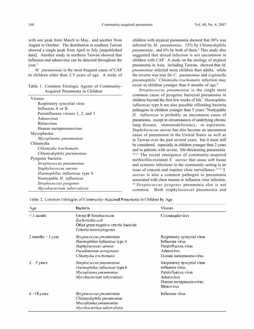
Epidemiologic information frequently is useful inguiding the search for the cause of pneumonia. Certainviruses, particularly respiratory syncytial virus (RSV),rhinoviruses, and influenza virus, as well as Mycoplasmapneumoniae, are strongly seasonal in temperate areas.However, as being located in subtropical region, theseasonal tendency of these pathogens in Taiwan is notas obvious as that in temperate areas.2,3 In other instances,the pattern of family illness can hint at the cause,especially the highly contagious influenza virus. Table1 lists the etiological agents for pneumonia in children,and Table 2 shows the distribution of these agents inchildren by age.
Respiratory viruses are the most common cause ofCAP in children younger than 5 years old. RSV is mostprevalent in children younger than 1-2 years of age.4
Adenovirus has been reported to be associated with severediseases in Taiwan.5 In temperate areas, RSV andinfluenza virus infections occur in winter epidemics, andparainfluenza viruses and rhinoviruses are more commonin autumn and spring. Infections due to adenovirusesoccur throughout the year. However, a recent study inTaiwan showed that monthly distribution of RSVinfections in Northern Taiwan showed a bimodal pattern
Review Article
Community-acquired pneumonia Vol. 48, No. 4, 2007168
children with atypical pneumonia showed that 26% wasinfected by M. pneumoniae, 15% by Chlamydophilapneumoniae, and 6% by both of them.6 This study alsosuggested that mixed infection is not uncommon inchildren with CAP. A study on the etiology of atypicalpneumonia in Asia, including Taiwan, showed that M.pneumoniae infected more children than adults, whilethe reverse was true for C. pneumoniae and Legionellapneumophila.7 Chlamydia trachomatis infection mayoccur in children younger than 6 months of age.8
Streptococcus pneumoniae is the single mostcommon cause of pyogenic bacterial pneumonia inchildren beyond the first few weeks of life.7 Haemophilusinfluenzae type b are also possible offending bacteriapathogens in children younger than 5 years.9 NontypableH. influenzae is probably an uncommon cause ofpneumonia, except in circumstances of underlying chroniclung disease, immunodeficiency, or aspiration.Staphylococcus aureus has also become an uncommoncause of pneumonia in the United States as well asin Taiwan over the past several years, but it must stillbe considered, especially in children younger than 2 yearsand in patients with severe, life-threatening pneumonia.10,11 The recent emergence of community-acquiredmethicillin-resistant S. aureus that cause soft tissueand systemic infections in the community setting is anissue of concern and requires close surveillance.11-13 S.aureus is also a common pathogen in pneumoniaassociated with chest trauma or influenza virus infection.14 Streptococcus pyogenes pneumonia also is notcommon. Both staphylococcal pneumonia and
Table 1. Common Etiologic Agents of Community-Acquired Pneumonia in Children
VirusesRespiratory syncytial virusInfluenza A or BParainfluenza viruses 1, 2, and 3AdenovirusRhinovirusHuman metapneumovirus
MycoplasmaMycoplasma pneumoniae
ChlamydiaChlamydia trachomatisChlamydophila pneumoniae
Pyogenic bacteriaStreptococcus pneumoniaeStaphylococcus aureusHaemophilus influenzae type bNontypable H. influenzaeStreptococcus pyogenesMycobacterium tuberculosis
with one peak from March to May, and another fromAugust to October. The distribution in southern Taiwanshowed a single peak from April to July [unpublisheddata]. Another study in northern Taiwan showed thatinfluenza and adenovirus can be detected throughout theyear.3
M. pneumoniae is the most frequent cause of CAPin children older than 2-5 years of age. A study of
P.I. LEE, C.H. CHIU, P.Y. CHEN, et al. 169Acta Paediatr Tw
streptococcal pneumonia are rapidly progressive andsevere, frequently leading to hypoxemia and effusionwithin hours. Mycobacterium tuberculosis that is stillprevalent in Taiwan should be put into the list ofdifferential diagnoses for CAP in children.15
II. CLINICAL MANIFESTATIONS
A. Features of pyogenic bacteria pneumonia1. Sudden deteriorated respiratory condition after an
apparently mild respiratory infection.2. Severely debilitated with poor activity when the
body temperature is normal.3. Tachypnea (respiratory rate > 60/min for infants
< 11 months, > 40/min for children between 1 yearand 4 years, and > 30/min for children older than5 years).
4. Oxygen saturation 92%, cyanosis.5. Septic signs, such as consciousness disturbance,
bleeding tendency, and hypotension.6. Signs of respiratory distress, including nasal flaring,
grunting, and chest wall retraction.7. Lung consolidation, cavity formation.
B. Features of atypical pneumonia:1. Children retain normal activity without features of
pyogenic bacteria pneumonia.2. Conjunctivitis, otitis media, skin rash, and
wheezing may be more common.
There have been many studies conducted in an effortto differentiate bacterial etiologies of pneumonia fromviral infections based on clinical manifestations. Ingeneral, none of the symptoms or signs can be consideredspecific. The onset of pyogenic bacterial pneumonia maybe abrupt and may follow days of mild viral respiratoryillness. The patient is ill, sometimes toxic appearing.Tachypnea, respiratory distress, hypoxemia, and lungconsolidation or cavity formation are predictive of severeor pyogenic bacteria pneumonia16-18 A study fromdeveloping world showed that oxygen desaturation wasassociated with a greater risk of death, and tachypneais closely related to hypoxemia.19 Several scoring systemshave been proposed to predict the severity and mortalityof CAP However, none of them has been modifiedfor children, and none has been examined in pediatricpatients.19,20
In contrast to pyogenic bacterial pneumonia, childrenwith atypical pneumonia, including those caused byM. pneumoniae, C. pneumoniae, L. pneumophilaand viruses, usually appear healthy without apparentrespiratory distress. Presence of arthralgia and erythemamultiforme may suggest M. pneumoniae infection. Ascompared with pyogenic bacterial pneumonia, some
studies suggest that children with atypical pneumoniamay have a higher incidence of conjunctivitis,21 otitismedia,21 and wheezing.22,23 However, some featuresthought to be specific for viral illness were not observedmore frequently in children with atypical pneumonia,including rhinorrhea, illness in family members, andmyalgia.22,23
III. DIAGNOSIS
A. Acute phase reactants cannot reliably differentiatebetween pyogenic bacterial pneumonia and atypicalpneumonia in children.
B. Image studies:1. Chest radiography
a. Chest radiography should be considered inchildren with an unexplained fever after excludingthe possibility of common infectious diseases,and in those with a prolonged fever.
b. Chest X-ray findings can hardly differentiateamong different etiologies. Bulging interlobarfissures and cavitations are suggestive of pyogenicbacteria infection.
2. Chest ultrasonography is useful to evaluate thepresence of consolidation and pleural effusion, andis helpful to guide thoracocentesis or chest tubing.
3. Computerized tomography of the chest may providedetails of pneumonia, and is indicated beforesurgical interventions.
C. Microbiological investigations:1. Sputum:
a. Gram stain, and acid-fast stain if necessary,should be done before the initiation of antibiotics.
b. The result of sputum culture may not representthe true etiology of pneumonia. However,with a qualitative count of gram stain(polymorphonuclear cells > 25/high-power fieldand epithelium < 10/ high-power field, with orwithout phagocytosis of polymorphonuclear cells),it does provide some help to adjust theantimicrobial agent during the disease course.
c. For patients with suspected M. tuberculosisinfection, acid-fast stain and mycobacteria cultureof the sputum should be done for at least 3 times.For children whose sputum is not available,gastric lavage for mycobacterial examinationshould be done in the early morning before mealsfor 3 consecutive days.
d. Direct fluorescent antigen test is available forL. pneumophila.
2. Nasopharyngeal or oropharyngeal swab: Thespecimens may be sent for virus culture and viralantigen detection that are more useful for young

Community-acquired pneumonia Vol. 48, No. 4, 2007170
children.3. Blood culture should be performed in all children
with suspected pyogenic bacterial pneumonia.4. A high titer of cold agglutinin may suggest
mycoplasma pneumonia. However, its specificityis low.
5. A 4-fold rise of specific serum IgG titer or a singlepositive IgM response indicates acute infection.
6. Urinary antigen tests are available for L.pneumophila serogroup I and S. pneumoniae.Although the pneumococcal antigen test is lessspecific in children, it has a good negativepredictive value for the diagnosis of S. pneumoniaepneumonia.
7. Tuberculin skin test should be performed when M.tuberculosis infection is suspected.
C. Invasive procedures:1. The pleural fluid from thoracocentesis should be
tested for:a. White count and differentials, protein, sugar,
lactate dehydrogenase and pH value.b. Gram stain and acid-fast stain.c. Antigen test for S. pneumoniae and H.
influenzae type b may be helpful.d. Culture for bacteria and, if suspected, virus and
M. tuberculosis.2. Bronchoalveolar lavage and lung biopsy may be
considered in some difficult cases.
White cell count, neutrophil count, percentage ofimmature neutrophil, erythrocyte sedimentation rate, andC-reactive protein (CPR) may reflect the severity ofinfections, and are therefore believed to be able todifferentiate between pyogenic and nonpyogenic infections.However, serum CRP was not useful to distinguishbetween pneumococcal, chlamydial, or viral etiologyin children with pneumonia in a prospective study.24
Following an acute-phase stimulus, CRP values peakat approximately 48 hours.25 Timing of CRP test shouldbe considered in interpretation. Acute phase reactantsmay only be useful to monitor the treatment response,and to distinguish between fever and hyperthermia thatis not caused by an inflammatory response.
There has been some debates about the optimal timingfor chest X-ray examination in children with respiratorysymptoms. One study of 522 children aged 2 to 59 monthsthat were randomly allocated to have a chest radiographor not showed that there was a marginal improvementin time to recovery which was not clinically significant.It was concluded that routine use of chest radiographyis not beneficial in ambulatory children aged over 2 monthswith acute lower respiratory-tract infection.26 Anotherstudy of 278 children aged 5 years or less suggested thatchest radiography should be considered a routine
diagnostic test in children with a temperature of 39 or greater and white count of 20,000/mm3 or greater
without an alternative major source of infection. In thatstudy, pneumonia was found in 32 of 79 (40%) of thosewith findings suggestive of pneumonia and in 38 of 146(26%) of those without clinical evidence of pneumonia.27 Although chest radiography should not be performedroutinely in children with mild uncomplicated acute lowerrespiratory tract infection,18 it may be indicated in selectivepatients, including an unexplained fever after excludingthe possibility of common infectious diseases, and aprolonged fever with or without respiratory manifestations.
Chest X-ray findings can hardly differentiate amongdifferent etiologies, especially for interstitial infiltrationsand pneumonic patches. Lung consolidation and pleuraleffusion may be observed in pyogenic bacterial pneumoniaand pneumonia caused by M. pneumoniae, C.pneumoniae, and L. pneumophila.6 Bulging interlobarfissures and cavitations are suggestive of pyogenic bacteriainfection.28,29
Chest ultrasonography is simple, ready to use, andnot associated with radiation. It is useful to evaluatethe presence of consolidation and pleural effusion, andis helpful to guide thoracocentesis or chest tubing, andfor follow-up. Therefore, it may be considered whenchest X-ray shows the presence of consolidation or pleuraleffusion.
Computerized tomography (CT) of the chest mayprovide details of pneumonia, including the extent ofconsolidation, cavitation, lung abscess, and empyema.It is indicated before surgical interventions, such as video-assisted thoracoscopic surgery (VATS), and decorticationof empyema. It may also be useful to evaluate complicatedpneumonia with poor clinical response. High-resolutionCT has been suggested to be more sensitive than chestradiograph to detect pulmonary infiltrates.30 However,it should not be used routinely.
For the majority of patients treated as outpatients,a specific microbiological diagnosis may not be necessary.The investigations are important for patients admittedto hospital with pneumonia. Sputum gram staining,and acid-fast staining if necessary, should be performedbefore the initiation of antibiotics. The sputum may behard to be obtained in children and the result of sputumculture may not represent the true etiology of pneumonia.However, with a quantitative count of gram stain, itdoes provide some help to adjust the antimicrobial agentlater on.
In Taiwan, it is recommended that at least 3 sputumspecimens should be sent for acid-fast stain andmycobacterial culture in patients with suspected M.tuberculosis infection. Because the sputum cannot beobtained in many children, gastric lavage in the earlymorning before meals for 3 consecutive days is also
P.I. LEE, C.H. CHIU, P.Y. CHEN, et al. 171Acta Paediatr Tw
a proper specimen for mycobacteria study.31
Direct fluorescent antigen test is a reliable test forL. pneumophila. It may be performed in selectedpatients. Sputum obtained from bronchoscope has bettersensitivity.
Because viral etiologies are more prevalent in youngerchildren with CAP, nasopharyngeal or oropharyngealswab may be sent for virus culture and viral antigendetection, including RSV, influenza virus A and B,parainfluenza virus, and adenovirus. However, laboratoryquality should be certificated for these tests.
Blood culture should be performed in all childrenwith suspected bacterial pneumonia before receivingantibiotics. However, the isolation rate is no more than10-20%.32 In older children, 2 blood cultures may beattempted to increase the diagnostic sensitivity.32
Cold agglutinin is a nonspecific antibody responsein M. pneumoniae infection. It is a sensitive test, butits specificity is low and an elevated titer may also beseen in other causes of CAP. Several serological testsare available for the diagnosis of M. pneumoniae,Chlamydophila pneumoniae, Chlamydia trachomatis,L. pneumophila, and common respiratory viruses. A4-fold rise of IgG titer or a single positive IgM responseindicates acute infection. A single high titer of IgG isnot diagnostic.
Legionella uninary antigen test identifies only L.pneumophila serogroup I, which is claimed to be themost common type causing clinical illness. A study inTaiwan showed that urine antigen test can detect only17.3% of 237 patients with L. pneumophila infection.33 A negative test does no exclude the diagnosis.
Pneumococcal urinary antigen test is an acceptabletest to augment diagnostic methods for S. pneumoniaeinfection. The sensitivity ranged between 50% and 80%,and the specificity is about 90% in adults.34,35 Studiesinvolving children have documented the lack of specificity.36,37 Although many authors suggest that a low specificityof the test may be attributed to that the test may givea false-positive result in children with colonization, itis more appropriate to say that the test may also be positivein S. pneumoniae infections other than pneumonia,such as otitis media.38 The test has a high sensitivityand a good negative predictive value for the diagnosisof S. pneumoniae pneumonia in children.
Bacille Calmette-Guérin (BCG) is routinely givento children in Taiwan. Although the vaccination mayinterfere with the interpretation of tuberculin reaction,studies in Taiwan showed than BCG vaccination did notappear to limit the usefulness of tuberculin skin test asa tool for diagnosing tuberculosis.39 The tuberculinreactivity toward BCG is usually lost 5-10 yr aftervaccination.40
Significant pleural effusion should be aspirated for
etiological diagnosis, especially when the effusion is> 10 mm in thickness on the lateral decubitus view orchest ultrasonography.32,41 Gram stain and acid-fast stainshould be routinely done. White count and differentials,protein, sugar, lactate dehydrogenase and pH value arehelpful to differentiate among transudate, anduncomplicated or complicated parapneumonic pleuraleffusions.41 The sensitivity of culture to define theoffending bacteria is usually limited but can be improvedby antigen detection.42,43 Culture for M. tuberculosisand viruses, though being less frequently seen, shouldbe done in suspected cases.
Invasive procedures, including bronchoalveolar lavageand lung biopsy, should not be routinely done. Analysisof sputum from bronchoalveolar lavage may have a bettercorrelation with pneumonia. However, it is technicallydifficult in young children and can only be consideredin some difficult cases.
IV. GENERAL MANAGEMENT
A. Decision for hospitalization1. Children with the following conditions that may
be suggestive of a grave illness are not recommendedto be cared at home:a. Features of severe bacterial pneumonia (see II-
A).b. Signs of dehydration.c. Neonates and children with immunodeficiency.d. Caretakers not able to provide appropriate
observation or supervision.2. If the clinical condition is aggravated, or is not
improving after 48 hours on treatment at home, thechild should be reviewed by a pediatric specialist.
B. Children who have hypoxemia or respiratory distressshould receive oxygen therapy.
C. Intravenous fluids, if necessary, may be given at80% maintenance level with monitoring of serumelectrolytes.
Children with CAP may be cared at home, especiallyfor those caused by atypical pathogens. As listed in II-A, several clinical manifestations are predictive for asevere bacteria pneumonia that should be cared inhospitals. Oral intake usually decreases in childrenwith pneumonia. If there are obvious signs of dehydration,the child should also be hospitalized. CAP in neonatesand children with immunodeficiency are prone to beingmore severe. Therefore, they should be treated moreaggressively in the hospital.
Hypoxemia and respiratory distress are important riskfactors of a severe disease.44 Oxygen therapy given bynasal cannula, head box, face mask, or oxygen tent
Community-acquired pneumonia Vol. 48, No. 4, 2007172
should be given to children with hypoxemia, especiallyfor those with oxygen saturation 92%.18 If bloodoxygenation is not improved after oxygen therapy, patientshould be cared for at an intensive care unit with positive-pressure respiratory support, such as intubation and useof ventilator.
V. ANTIBIOTIC THERAPY
A. Principle1. Empiric use of antibiotics should take into
consideration the age and the disease severity ofthe patients. Appropriate antibiotics should be givenas soon as possible after registration for hospitalizedpatients.
2. Parenteral antibiotics should be given to childrenwith severe pneumonia.
3. If fever or some grave clinical manifestations persistbeyond 48 hours after treatment, the treatment planshould be re-evaluated and a follow-up chest imagestudy should be considered.
4. Oral switch of antibiotics: If the clinical conditionimproves rapidly with all of the followingcharacteristics suggestive of a stabilized illness,intravenous antibiotics may be considered to beswitched to oral ones.a. Absence of septic signs, empyema, necrotizing
pneumonia and lung abscess.b. Stabilized vital signs for at least 48 hours,
including body temperature, heart rate,
respiratory rate, and blood pressure.c. No growth on blood culture.d. May be fed orally.
5. Duration of antibiotic treatmenta. Mycoplasma pneumonia and chlamydia
pneumonia may be treated by appropriate oralantibiotics for 10 days. If azithromycin is used,the treatment should be continued for only 3-5 days.
b. Legionnaires' disease: For immunocompetentchildren, azithromycin may be used for 5-10 days,and other macrolides and fluoroquinolones maybe used for 10-14 days. For immunocompromisedchildren, macrolides plus fluoroquinolones orrifampin may be used for 14-21 days.
c. Antibiotics should be given according to thetreatment response, and are usually used for atleast 7-10 days.
d. Duration of antibiotic therapy may need to beprolonged in complicated infections, such asthose complicated by bacteremia or meningitis,Pseudomonas aeruginosa infection, empyema,necrotizing pneumonia, and lung abscess.
B. Choice of antibiotics when the pathogen is known:Current antibiotic-resistance rate of some importantrespiratory pathogens in Taiwan include 70% ofpenicillin-nonsusceptible S. pneumoniae (minimuminhibitory concentration 0.12 g/mL), about 60%of -lactamase-producing H. influenzae, and 50-70% of community-acquired methicillin-resistant S.aureus.
P.I. LEE, C.H. CHIU, P.Y. CHEN, et al. 173Acta Paediatr Tw
D. Choice of antibiotics under special circumstances:1. Broad-spectrum and potent antibiotics should be
used in children with sepsis, meningitis, orcomplications that may endanger the life.
2. For children with disorders such as bronchiectasis,chronic lung disease, and severe neuromusculardisorders and have a history of recurrent pneumonia,repetitive use of antibiotics, or prolonged use ofsteroids, enteric gram-negative bacteria, includingP. aeruginosa, are more likely to be the offendingpathogen. Empiric therapy may includeantipseudomonal -lactams with or withoutaminoglycosides.
3. When S. aureus infection is a concern, such aschest trauma or influenza-associated pneumonia,antibiotics effective against methicillin-resistant S.aureus may be added to the empiric therapy.
E. Recommended dosage of empirical antibiotics (forchildren older than 1 month of age):1. Penicillin: 300,000-400,000 units/kg/day, q4-6h.2. Ampicillin: 150-200 mg/kg/day, q6h.3. Amoxicillin: 80-90 mg/kg/day, po tid.4. Oxacillin: 100-300 mg/kg/day, q4-6h.
5. Ampicillin/sulbactam: 150-200 ampicillin mg/kg/day, q6-8h.
6. Amoxicillin/clavulanate: 150-200 amoxicillin mg/kg/day, iv q6-8h; 80-90 amoxicillin mg/kg/day,po bid-tid.
7. Cefazolin: 50-100 mg/kg/daym, iv q8-6hv.8. Cefuroxime: 100-200 mg/kg/day, iv q6-8h; 20-
30 mg/kg/day, po bid, may double the dose forsevere infection.
9. Ticarcillin/clavulanate: 200-300 ticarcillin mg/kg/day, q6-8h.
10. Piperacillin/tazobactam: 200-300 piperacillin mg/kg/day, q6-8h.
11. Cefotaxime: 150-200 mg/kg/day, q6h.12. Ceftriaxone: 100 mg/kg/day, q12h - qd.13. Ceftazidime: 100-150 mg/kg/day, q6-8h.14. Cefepime: 100-150 mg/kg/day, q8-12h.15. Imipenem: 60-100 mg/kg/day, q6h.16. Meropenem: 60-100 mg/kg/day, q6-8h.17. Erytrhomycin: 40 mg/kg/day, q6h.18. Clarithromycin: 15 mg/kg/day, q12h.19. Azithromycin: 10-12 mg/kg/day, qd.20. Tetracycline: 25-50 mg/kg/day, po bid-qid.

Community-acquired pneumonia Vol. 48, No. 4, 2007174
21. Minocycline: 4 mg/kg loading, then 2 mg/kg poq12h.
22. Doxycycline: 4.4 mg/kg/day loading, then 2.2-4.4 mg/kg po qd.
23. Vancomycin: 20-60 mg/kg/day, q6-8h.24. Teicoplanin: 10 mg/kg q12h loading doses for 3
doses, then 10-20 mg/kg, qd.25. Linezolid: 20-30 mg/kg/day, q8-12h.26. Rifampin: 10-15 mg/kg/day, qd.27. Gentamicin: 6-7.5 mg/kg/day, bid-qd.28. Tobramycin: 6-7.5 mg/kg/day, bid-qd.29. Netilmicin: 5.5-8.0 mg/kg/day, bid-qd.30. Amikacin: 15-25 mg/kg/day, bid-qd.31. Ciprofloxacin: 20-40 mg/kg/day, q12h.
Age and disease severity are the two most importantfactors in deciding whether antibiotics should be usedor which antibiotics should be chosen. For example,viral infections are more prevalent in young children,4 and it is recommended that young children presentingwith mild symptoms of lower respiratory tract infectionneed not be treated with antibiotics.18 Studies showedthat initiation of antibiotics within 4 to 8 hours after arrivalat hospital correlated strongly with the outcome.45,46
Appropriate antibiotics should be given as soon as possibleafter registration for hospitalized patients.
Orally administered antibiotics are safe and effectivefor children with community-acquired pneumonia thatis not associated with clinical manifestations suggestiveof a grave illness. Parenteral antibiotics can ensure arapidly rising high serum concentration and should begiven to children with clinical manifestations suggestiveof a severe pneumonia and to those who cannot be fedorally.
Some viral pneumonia may have a prolonged fever,and some bacterial pneumonia may have a persistent feverafter using appropriate antibiotics, especially for thosewith consolidation and pleural effusion.6 However,clinical conditions of bacteria pneumonia that areresponsive to antibiotics usually improved within 48 hoursafter treatment with defervescence.6 If fever or somegrave clinical manifestations persist beyond 48 hoursafter treatment, the treatment plan should be re-evaluatedand a follow-up chest image study should be considered.
There have been limited data published regardingintravenous-to-oral sequential antibiotic therapy in Taiwan.47 Some randomized studies suggested that intravenous-to-oral switch of antibiotics may be feasible for someclinically stable and antibiotic-responsive CAP.32 Sucha practice may be able to reduce the cost of treatmentand the length of stay in the hospital. We recommendthat oral switch of antibiotics may be applied to CAPin children without evidence of sepsis, empyema,necrotizing pneumonia, and lung abscess when the
vital signs have been stabilized for at least 48 hoursand when the patient can be fed orally. Generally, theantibiotic switch can take place after 2-4 days ofintravenous therapy.32
There is no appropriate randomized study to definethe optimal duration of antibiotic therapy for CAP. Mostrecommendations are conjectural, and many physiciansrecommend treatment for 1-2 weeks.32 The recommendeddurations of treatment in this guideline are based on theexperiences of experts and some statements in textbooks.Seven to 10 days of treatment is usually enough with2 exceptions. One is Legionnaires' disease that maybe more severe than other causes of atypical pneumonia,especially when it occurs in immunocompromised children.The recommended duration is longer. The other iscomplicated pneumonia that may require a longer durationof treatment, including those complicated by bacteremiaor meningitis, Pseudomonas aeruginosa infection,empyema, necrotizing pneumonia, and lung abscess.Community-acquired P. aeruginosa sepsis with orwithout pneumonia is most frequently seen in infants.48 One study showed that 8-day therapy for P. aeruginosapneumonia led to relapse more commonly than did 15-day therapy.49
When the pathogen is known, the antibiotic shouldbe chosen according to the antibiotic susceptibility pattern.Antibiotic resistance among pneumococci is increasingand the incidence of severe pneumococcal pneumoniais apparently increasing in recent years.50 Being the sameas our previous version of guideline,51 a penicillinminimum inhibitory concentration (MIC) of < 1 g/mLwas defined as penicillin susceptible, MIC 4 g/mLas penicillin resistant, and an intermediate MIC aspenicillin intermediate. Recently, penicillin MIC'sof about 70% of S. pneumoniae strains in Taiwanare 0.12 g/mL, while only less than 5% is penicillin-resistant (MIC 4 g/mL).52 Therefore, most penicillin-nonsusceptible S. pneumoniae infection can be treatedby a high dose of penicillin and its analogue. On theother hand, erythromycin resistance in S. pneumoniaehas remained high (94%) in Taiwan in recent years.52
Likewise, trimethoprim-sulfamethoxazole resistance rateis also high (65%).53
Recent studies in Taiwan showed that 56% of H.influenzae isolates produce -lactamase, as did nearlyall Moraxella catarrhalis isolates (95.7%). Only1.7% of H. influenzae were -lactamase negative andamoxicillin resistant.53 Antibiotics used for these gram-negative bacteria should be stable to -lactamase. Theresistance rate to trimethoprim-sulfamethoxazole is 52%for H. influenzae.53
Having being a predominant pathogen in nosocomialinfections in Taiwan for many years, methicillin-resistanceS. aureus is now becoming more and more common

P.I. LEE, C.H. CHIU, P.Y. CHEN, et al. 175Acta Paediatr Tw
in community-acquired infections. Recent datademonstrated that 50-70% of community strains of S.aureus obtained from pediatric patients is resistant tomethicillin.12,13 Vancomycin and other agents activeagainst methicillin-resistance S. aureus may be consideredin selected cases, especially for those with chest traumaand influenza.54
M. pneumoniae, C. pneumoniae and C. trachomatisare rarely resistant to erythromycin and other macrolidesthat should be the drug of choice for these infections.Different macrolides have similar therapeutic efficacy.6 Tetracyclines may be used as an alternative only whenthe child is older than 8 years to avoid their potentialdetrimental effects on bone and teeth. Althoughfluoroquinolones may be used as the first-line drug fortreating L. pneumophila infection,32,55 new macrolidesare a more preferred agent in children. Rifampin orfluoroquinolones may be added in severe infections.
When the pathogen is unknown, either before theculture result is available or due to a negative result ofmicrobiological test, antibiotics may be given empiricallybased on the knowledge of predominant pathogens ineach age groups.
For neonates younger than 1 month, Escherichiacoli, group B streptococcus and other bacteria arecommon pathogens. They may be treated empiricallyby ampicillin + aminoglycosides, or by ampicillin +cefotaxime or ceftriaxone when meningitis is a concern.Common bacterial pathogens of CAP in children between2 months and 5 years of age include S. pneumoniaeand H. influenzae type b. -lactams stable to -lactamasemay be used empirically. S. pneumoniae becomes thesingle most important etiology of CAP in children beyond6 years of age. Penicillin may be used as empirical therapyfor clinically stable patients.
M. pneumoniae and C. pneumoniae infections arenot infrequent after 2 years of age.6,7 For children olderthan 2 years with suspected atypical pneumonia,macrolides are the antibiotic of choice. However, suchinfections are not totally absent in children younger than2 years,6 and C. trachomatis is a possible etiology ofCAP in infants.8 Macrolide antibiotics may be used inspecial circumstances.
The choice of antibiotics should also take into accountthe severity of illness and comorbidities. Severalguidelines for management of CAP in adults stratifypatients into groups based on site of therapy (i.e.outpatient, inpatient, or intensive care unit),comorbidities (including cardiopulmonary disease,diabetes mellitus, renal failure, malignancy), and riskfactors for infection with drug-resistant bacteria.32,55,56
To avoid too many stratifications, the working groupchoose to stratify pediatric patients by the age only.However, some points deserve further attention.
A few retrospective studies suggested that dualtherapy with -lactams and a macrolide may reducemortality associated with bacteremic pneumococcuspneumonia.57,58 Two possible explanations are theimmunonodulating effect of macrolides and a concomitantinfection by atypical pathogens that may be susceptibleto macrolides. A well-designed prospective study isneeded to prove such an observation, and the data inpediatric patients are still lacking. However, addinga macrolide for children with suspected pyogenicpneumonia may be warranted since mixed infections arenot uncommon in children with CAP.
With life-threatening complications, such as sepsisand meningitis, CAP in children may be treatedempirically with broad-spectrum and potent antibiotics,such as vancomycin plus a third-generation cephalosporinor other antibiotics effective against commonly seen gram-negative bacteria.
Enteric gram-negative bacteria, such as P. aeruginosa,may pose some impact on selection of an appropriateantibiotic for treatment of CAP. Several risk factors havebeen recognized in adult patients.56 Patients who residein a nursing home, or have underlying cardiopulmonarydisease or multiple comorbidities, or have received recentantimicrobial therapy are more likely to be infected byenteric organisms. Risk factors for P. aeruginosainfection include structural lung disease (e.g.bronchiectasis), steroid therapy, recent use of broad-spectrum antibiotic, and malnutrition. Although similardata are lacking for children, the working group makessimilar recommendations. When children with disorderssuch as bronchiectasis, chronic lung disease, and severeneuromuscular disorders have a history of recurrentpneumonia, repetitive use of antibiotics, or prolongeduse of steroids, they tended to be infected by gram-negative bacteria, including P. aeruginosa. Empirictherapy may include antipseudomonal -lactams(including ceftazidime, piperacillin, ticarcillin/clavulanate,piperacillin/tazobactam, cefepime, imipenem, andmeropenem) with or without aminoglycosides.
Because some antibiotic-resistant bacteria, especiallypenicillin-nonsusceptible S. pneumoniae, are highlyprevalent in Taiwan, the dosage of some antibiotics shouldbe modified. As mentioned before, less than 5% ofpenicillin-nonsusceptible S. pneumoniae is truly resistantto penicillin with a MIC 4 g/mL in Taiwan,52
increasing the dose of penicillin, ampicillin, amoxicillin,ampicillin/sulbactam, amoxicillin/clavulanate, cefuroxime,and cefotaxime may be an effective way to treat infectionscaused by penicillin-intermediate S. pneumoniae. Therecommended dosage of various antibiotics in presentguideline is for empirical use of antibiotics. The dosageof antibiotics may be adjusted when the pathogen andits antibiotic susceptibility pattern are known. For

Community-acquired pneumonia Vol. 48, No. 4, 2007176
example, when dealing with penicillin-susceptible S.pneumoniae infection, the dose of penicillin may belowered down.
As suggested by recent pharmacokinetic andpharmacodynamic studies, some antibiotics are time-dependent for their therapeutic effect, including -lactamsand macrolides. Frequent dosing may improve theirperformance. On the other hand, Some antibiotics areconcentration-dependent, including aminoglycosides andfluoroquinolones. Such antibiotics should be given witha longer dosing interval to maximize their therapeuticeffect.59 In the era of increasing resistance, an dosingschedule with optimized pharmacokinetic andpharmacodynamic features can not only bring about abetter treatment response, but also prevent the emergenceof resistant bacteria. The recommended dosage in thisguideline was set up according to this principle.
VI. POST-TREATMENT EVALUATION ANDMANAGEMENT OF COMPLICATION
A. If the fever persists, the clinical condition is notimproved, or aggravating signs appeared aftertreatment, the following conditions should beconsidered.1. Inadequate dose of antibiotics.2. Antimicrobials not effective for offending pathogen,
such as antibiotic-resistant bacteria, tuberculosis.3. Viral infection or mixed infection.4. Extrapulmonary focus of infection.5. Complication of pneumonia, such as lung abscess,
empyema.6. Drug fever.
B. Complication of pneumonia:1. Pleural effusion, empyema.2. Necrotizing pneumonia, lung abscess.3. Acute respiratory distress syndrome.4. Others, such as bronchopleural fistula.
C. Management of pleural effusion and empyema:1. Diagnosis: lateral decubitus chest radiography or
chest ultrasonography. The latter is preferred.2. Pleural tapping: Examinations of pleural fluid should
include white count and differentials, pH value,glucose, protein, gram stain, acid-fast stain,bacterial culture, mycobacteria culture. Bacteriaantigen detection may also be considered.
3. With one of the following conditions, drainageof pleural fluid should be required:a. Pus-like effusion.b. Positive finding of gram stain or bacterial culture
of pleural fluid.c. Large amount of fibrinous substances or septations
in pleural cavity.
d. Massive pleural effusion associated withrespiratory distress.
e. pH of pleural fluid < 7.2.4. Draining procedure
a. Simple chest tube drainage: not recommended.b. Chest tube drainage with firbrinolytic agents: Use
streptokinase 2,500 U/mL or urokinae 1,000 U/mL with a dose of 3-4 mL/kg that does not exceed100 mL. The agent may be given once per daywith retention of the agent for 2-4 hours eachtime. The therapy may be instituted for 2-3 daysor until there is a significant improvement of chestimages. Tissue plasminogen activator may beused with a dose of 2-5 gm in 50-250 mL saline.
c. Video-assisted thoracoscopic surgery (VATS):Computerized tomography of the chest shouldbe performed previous to this procedure todelineate the extent of pleural effusion. EarlyVATS that is performed within 4 days after thediagnosis is more effective than late VATS.
d. If the above mentioned draining procedures failto improve the condition, such as persistent highfever and severe respiratory distress, open surgeryfor drainage may be considered.
D. Management of necrotizing pneumonia and lungabscess: Chest ultrasonography or computerizedtomography should be performed. If the clinicalcondition does not improve after appropriateantimicrobial therapy and drainage, open surgery maybe considered.
If the fever persists, the clinical condition is notimproved or even aggravated after treatment, severalpossibilities should be considered.60 Because penicillin-nonsusceptible S. pneumoniae is highly prevalent inTaiwan,50,52 an adequate dose of antibiotics as mentionedin present guideline is a prerequisite to ensure treatmentsuccess. For young infants and children with risk factors,antibiotics with a broader antibacterial spectrum may benecessary to be effective against potential pathogens,including methicillin-resistant S. aureus10,11 and entericgram-negative bacteria.56 As mentioned previously, M.tuberculosis infection is prevalent in Taiwan and shouldbe regarded as a possible etiology in CAP unresponsiveto empiric antibiotic therapy.15
Viral infection and extrapulmonary focus of infectionare also possible causes for an unresponsive CAP. Astudy in Taiwan showed that children with a severepneumonic change (consolidation or pleural effusion)or extrapulmonary manifestations (e.g. encephalitis,hepatitis) tended to have a prolonged fever afterappropriate macrolide treatment in children with eitherM. pneumoniae or C. pneumoniae infection.6 Adequatedrainage of lung abscess and empyema may be necessary
P.I. LEE, C.H. CHIU, P.Y. CHEN, et al. 177Acta Paediatr Tw
for some complicated pneumonia.Drug fever is easily overlooked because affected
patients may have an extremely high temperature anda high CRP value. Sulfonamides and -lactams arecommon causes of drug fever. However, it should beassumed that any drug can cause drug fever, includingnonantibiotics.60 Fever may developed a few days afterusing an antibiotic.61 A relatively good activity withre-emergence of fever after defervescence for some daysafter treatment is characteristic for drug fever. Otherhelpful clues to drug fever are skin rashes, relativebradycardia, neutropenia, eosinophilia, atypicallymphocytosis, elevations of the serum transaminases.61,62 The diagnosis of drug fever may be confirmed byobserving a subsidence of fever after withholding theoffending medication. Drug fever usually subsides within72 hours after the sensitizing drug is discontinued if arash is not present.61
Parapneumonic pleural effusion is not uncommonin children with bacterial pneumonia and is a commoncause of prolonged fever after treatment. A study inTaiwan showed that 56% of pneumococcus pneumoniain children is complicated.50 It has been recommendedthat for all adult patients with acute bacterial pneumonia,the presence of a parapneumonic effusion should beconsidered.63 The effusion may be delineated by lateraldecubitus chest radiography or by chest ultrasonography.Chest ultrasonography is preferred because it is moreaccurate relative to lateral decubitus chest radiographyfor the diagnosis of small pleural effusions.64
Aspirated pleural fluid should be sent for necessarytests. Some studies showed that commercially availablepneumococcal antigen test that was designed forcerebrospinal fluid samples may also be useful for pleuralfluid.65,66 Although available data are limited, the workinggroup suggests that such a bacteria antigen test may beused for pleural fluid when S. pneumoniae infectionis one of the possible pathogen.
It is a common agreement that a frankly purulenteffusion or an effusion containing bacteria as evidencedby either culture or gram stain should be drained to hastenthe recovery and to avoid complications. Dilemma occurswhen the effusion does not appear purulent. A meta-analysis suggested that a low pH < 7.21-7.29 was themost accurate predictor of the need for drainage.67 Thecut-off point for pH is controversial. The presentrecommendation adopts a pH of 7.2 as a cut-off, similarto that recommended by the American College of ChestPhysicians.63
There are several drainage procedures. A recentreview showed that the pooled mortality was higher forthe no drainage (6.6%), therapeutic thoracentesis (10.3%), and tube thoracostomy (8.8%) than for the
fibrinolytic (4.3%), VATS (4.8%), and surgery (1.9%).The pooled proportion of patients needing a secondintervention was also higher for the no drainage,therapeutic thoracentesis, and tube thoracostomymanagement approaches.63
Adding streptokinase, urokinase or tissue plasminogenactivator into the chest cavity may facilitate the drainageby causing lysis of fibrins and septations. Fibrinolyticagents are resolved in normal saline that is instituted intoa properly positioned chest tube. The drainage is heldfor several hours for firbrinolytic agents to take effect.Several studies in Taiwan have shown that intrapleuralfibrinolytic treatment is safe and effective in children,and it can obviate the need for surgery.68,69 There is nota consensus on the dosage of firbrinolytic agents.However, the working group suggests one dosageschedule according to the experience in Taiwan.
Most studies agree that debridement of the pleuralspace by VATS is effective for the management of pleuralempyema, including studies in children. Data alsosuggested that the main prognostic factor for thoracoscopictreatment of pleural empyema is the interval betweendiagnosis and surgery.70,71 A 4-day limit, correspondingto the natural process of empyema organization, maysignificantly affect the efficacy of VATS.70,72 Therefore,VATS should be attempted within 4 days after diagnosiswhen necessary. Open surgery is another option for thetreatment of pleural empyema in children.
Necrotizing pneumonia and lung abscess are notuncommon in children with CAP. Diagnosis may beconfirmed by ultrasonography or computerizedtomography. Prolonged antibiotic therapy may be required.Infrequently, open surgery may be needed in complicatedcases refractory to medical therapy.
VII. PREVENTION
A. General principles: reduce the risk of exposure torespiratory pathogens by droplet precautions.
B. Immunization:1. Bacille Calmette-Guérin vaccine: routine for all
neonates and 7-year-old children who has a negativetuberculin reaction.
2. Influenza vaccine:a. Routine for children aged 6-23 months.b. Recommended for children older than 23 months
with high-risk conditions.3. Pneumococcal vaccine: 23-valent pneumococcal
polysaccharide vaccine (PPV23) for children olderthan 2 years and 7-valent pneumococcal conjugatevaccine (PCV7) for children older than 2 months.Recommended schedule for those having notreceived pneumococcal vaccines:

Community-acquired pneumonia Vol. 48, No. 4, 2007178
C. Preventive therapy:1. Tuberculosis: isoniazid 10 mg/kg/day (maximum
300 mg/day) for 9 months recommended for children12 years with evidence of latent tuberculosis
infection and a history of close contact with patientswith infectious tuberculosis.
2. Haemophilus influenzae type b infection: rifampin20 mg/kg (maximum 600 mg) daily for 4 days forall household contacts when at least 1 contact isyounger than 4 years of age.
Pathogens responsible for CAP are transmitted bydroplet, while most bacterial pneumonia may becomplications of some preceding virus infection.73
Preventive measures for CAP include droplet precautionsfor hospitalized children, strict hand hygiene procedures,and that infected children should be excluded from schooland day care facilities until they are no longer consideredcontagious.
According to the guidelines for the diagnosis andtreatment of tuberculosis in Taiwan, one dose of BCGshould be given to all neonates with a body weight
2,500 gm. Tuberculin skin test is done at school entry(7 years of age) for children whose BCG scar is 2 mmin diameter. One dose of BCG should be given to thosewho have a negative tuberculin reaction.
Currently, influenza vaccine is a routine for childrenaged between 6 and 23 months in Taiwan. The vaccineis also recommended for children older than 23 monthsof age with risk factors, including chronic pulmonarydiseases (e.g. bronchopulmonary dysplasia, cysticfibrosis, bronchiolitis obliterans, laryngotracheomalasia,asthma), hemodynamically significant cardiac disease,immunosuppresive disorders or therapy, humanimmunodeficiency virus infection, hemoglobinopathies,disorders requiring long term salicylate therapy (e.g.rheumatoid arthritis, Kawasaki disease), chronic renaldysfunction, chronic metabolic disease (including diabetesmellitus), and any condition that can compromiserespiratory function or handling of respiratory tractsecretions or that can increase the risk of aspiration.74
Two pneumococcal vaccines are available in Taiwan,
including a 23-valent polysaccharide pneumococcalvaccine (PPV23) for use in children aged over 2 year,and a 7-valent pneumococcal conjugate vaccine (PCV7)for children between 2 months and 9 years of age. ThePCV7 is recommended for routine vaccination at 2, 4,6, and 12-18 months of age. Catch-up vaccination isalso recommended for children up to 23 months of agewith fewer doses of PCV7. The cost-effectiveness ofpneumococcal vaccines in healthy children between 24and 59 months of age remain to be studied. Pneumococcalvaccines may be given to all children older than 24months of age with risk factors, includinghemoglobinopathies, congenital or acquired immunedeficiency, human immunodeficiency virus infection,chronic pulmonary disease, chronic renal disorders,diabetes mellitus, anatomical abnormalities associatedwith higher rates or severity, cerebrospinal leaks,hemodynamically significant heart disease, and chronicpulmonary disease.75
Recent revision of the guidelines for the diagnosisand treatment of tuberculosis in Taiwan recommends thatisoniazid chemoprophylaxis may be given to children
12 years with evidence of latent tuberculosis infectionby the tuberculin reaction and a history of close contactwith patients with infectious tuberculosis. The risk ofinvasive Haemophilus influenzae type b disease isincreased among household contacts who are less than4 years of age. Rifampin 20 mg/kg (maximum 600 mg)daily for 4 days is recommended for all householdcontacts in such occasions regardless of the age ofhousehold contacts and the Haemophilus influenzae typeb vaccination history.
REFERENCES
1. Isaacs D. Problems in determining the etiology of com-munity-acquired childhood pneumonia. Pediatr Infect DisJ 1989; 8:143-8.
2. Huang YC, Lin TY, Chang LY, Wong KS, Ning SC. Ep-idemiology of respiratory syncytial virus infection amongpaediatric inpatients in northern Taiwan. Eur J Pediatr2001; 160:581-2.
3. Lin TY, Huang YC, Ning HC, Tsao KC. Surveillance ofrespiratory viral infections among pediatric outpatients in
P.I. LEE, C.H. CHIU, P.Y. CHEN, et al. 179Acta Paediatr Tw
northern Taiwan. J Clin Virol 2004; 30:81-5.4. Chen CJ, Jeng MJ, Yuan HC, Wu KG, Soong WJ,
Hwang B. Epidemiology of respiratory syncytial virus inchildren with respiratory tract infection. Acta PaediatrTaiwan 2005; 46:72-6.
5. Chuang YY, Chiu CH, Wong KS, et al. Severe aden-ovirus infection in children. J Microbiol Immunol Infect2003; 36:37-40.
6. Lee PI, Wu MH, Huang LM, Chen JM, Lee CY. An open,randomized, comparative study of clarithromycin and eryth-romycin in the treatment of children with community-ac-quired pneumonia. J Microbiol Immunol Infect 2007 (inpress).
7. Ngeow YF, Suwanjutha S, Chantarojanasriri T, et al. AnAsian study on the prevalence of atypical respiratory patho-gens in community-acquired pneumonia. Intern J Infect Dis2005; 9:144-53.
8. Chiang YC, Shyur SD, Huang LH, et al. Chlamydia tra-chomatis pneumonia: experience in a medical center. ActaPaediatr Taiwan 2005; 46:284-8.
9. Wang CH, Lin TY. Invasive Haemophilus influenzae dis-eases and purulent meningitis in Taiwan. J Formos MedAssoc 1996; 95:599-604.
10. Schultz KD, Fan LL, Pinsky J, et al. The changing faceof pleural empyemas in children: epidemiology andmanagement. Pediatrics 2004; 113:1735-40.
11. Alfaro C, Fergie J, Purcell K. Emergence of communi-ty-acquired methicillin-resistant Staphylococcus aureus incomplicated parapneumonic effusions. Pediatr Infect DisJ 2005; 24:274-76.
12. Fang YH, Hsueh PR, Hu JJ, et al. Community-acquiredmethicillin-resistant Staphylococcus aureus in children innorthern Taiwan. J Microbiol Immunol Infect 2004; 37:29-34.
13. Chen CJ, Huang YC, Chiu CH, Su LH, Lin TY. Clini-cal features and genotyping analysis of community-acquiredmethicillin-resistant Staphylococcus aureus infections inTaiwanese children. Pediatr Infect Dis J 2005; 24:40-5.
14. Bhat N, Wright JG, Broder KR, et al. Influenza-associ-ated deaths among children in the United States, 2003-2004. N Engl J Med 2005; 353:2559-67.
15. Lin YS, Huang YC, Chang LY, Lin TY, Wong KS. Clinicalcharacteristics of tuberculosis in children in the north ofTaiwan. J Microbiol Immunol Infect 2005; 38:41-6.
16. Palafox M, Guiscafre H, Reyes H, et al. Diagnostic valueof tachypnea in pneumonia defined radiologically. Arch DisChild 2000; 82:41-5.
17. Pereira JC, Escuder MM. The importance of clinical symp-toms and signs in the diagnosis of community-acquiredpneumonia. J Trop Pediatr 1998; 44:18-24.
18. British Thoracic Society of Standards of Care Committee.BTS guidelines for the management of community acquiredpneumonia in childhood. Thorax 2002; 57:1-24.
19. Aujesky D, Auble TE, Yealy DM, et al. Prospective com-parison of three validated prediction rules for prognosis incommunity-acquired pneumonia. Am J Med 2005; 118:384-92.
20. Capelastegui A, Espana PP, Quintana JM, et al. Vali-dation of a predictive rule for the management of commu-nity-acquired pneumonia. Eur Respir J 2006; 27:151-7.
21. Ramsey BW, Marcuse EK, Foy HM, et al. Use of bac-terial antigen detection in the diagnosis of pediatric low-er respiratory tract infections. Pediatrics 1986; 78:1-9.
22. Turner RB, Lande AE, Chase P, Hilton N, Weinberg D.Pneumonia in pediatric outpatients: cause and clinicalmanifestations. J Pediatr 1987; 111:194-200.
23. Forgie IM, O’Neill KP, Lloyd-Evans N, et al. Etiology ofacute lower respiratory tract infections in Gambian children.
I. Acute lower respiratory tract infections in infants present-ing at the hospital. Pediatr Infect Dis J 1991; 10:33-41.
24. Heiskanen-Kosma T, Korppi M. Serum C-reactive proteincannot differentiate bacterial and viral aetiology of commu-nity-acquired pneumonia in children in primary healthcaresettings. Scand J Infect Dis 2000; 32:399-402.
25. Vigushin DM, Pepys MB, Hawkins PN. Metabolic andscintigraphic studies of radioiodinated human C-reactiveprotein in health and disease. J Clin Invest 1993; 91:1351-7.
26. Swingler GH, Hussey GD, Zwarenstein M. Randomisedcontrolled trial of clinical outcome after chest radiographin ambulatory actue lower-respiratory infection in children.Lancet 1998; 351:404-8.
27. Bachur R, Perry H, Harper MB. Occult penumonias:empir ic chest radiographs in febri le chi ldren withleukocytosis. Ann Emerg Med 1999; 33:166-73.
28. Korvick JA, Hackett AK, Yu VL, Muder RR. Klebsiellapneumonia in the modern era: clinicoradiographiccorrelations. South Med J 1991; 84:200-4.
29. Francis JB, Francis PB. Bulging (sagging) fissure sign inHemophilus influenzae lobar pneumonia. S Med J 1978;71:1452-3.
30. Syrjala H, Broas M, Suramo I, et al. High-resolution com-puterized tomography for the diagnosis of community-ac-quired pneumonia. Clin Infect Dis 1998; 27:358-63.
31. Voss LM. Management of tuberculosis in children. J Pae-diatr Child Health 2000; 36:530-6.
32. Mandell LA, Marrie TJ, Grossman RF, et al. Canadianguidelines for the initial management of community-ac-quired pneumonia: an evidence-based update by theCanadian Infectious Diseases Society and the CanadianThoracic Society. Clin Infect Dis 2000; 31:383-421.
33. Su HP, Tseng LR, Chou CY, Chung TC, Pan TM. Le-gionella pneumophila infection in the Taiwan area. J In-fect Chemother 2005; 11:244-9.
34. Domínguez J, Galí N, Blanco S, et al. Detection of Strep-tococcus pneumoniae antigen by a rapid immunochromato-graphic assay in urine samples. Chest 2001; 119:243-9.
35. Murdoch DR, Laing RT, Mills GD, et al. Evaluation ofa rapid immunochromatographic test for detection of Strep-tococcus pneumoniae antigen in urine samples from adultswith community-acquired pneumonia. J Clin Microbiol 2001;39:3495-8.
36. Navarro D, Garcia-Maset L, Gimeno C, Escribano A,Garcia-de-Lomas J, Spanish Pneumococcal InfectionStudy Network. Performance of the Binax NOW Strepto-coccus pneumoniae urinary antigen assay for diagnosis ofpneumonia in children with underlying pulmonary diseas-es in the absence of acute pneumococcal infection. J ClinMicrobiol 2004; 42:4853-5.
37. Dowell SF, Garman RL, Liu G, Levine OS, Yang YH.Evaluation of Binax NOW, an assay for the detection ofpneumococcal antigen in urine samples, performed amongpediatric patients. Clin Infect Dis 2001; 32:824-5.
38. Ramsey BW, Marcuse EK, Foy HM, et al. Use of bac-terial antigen detection in the diagnosis of pediatric low-er respiratory tract infections. Pediatrics 1986; 78:1-9.
39. Bowerman RJ. Tuberculin skin testing in BCG-vaccinat-ed populations of adults and children at high risk for tuber-culosis in Taiwan. Int J Tuberc Lung Dis 2004; 8:1228-33.
40. Munoz FM, Starke JR. Tuberculosis (Mycobacteriumtuberculosis). In: Behrman RE, Kliegman RM, JensonHB, eds. Nelson Textbook of Pediatrics. 17th ed.Philadelphia: Churchill Saunders, 2004:958-72.
41. Light RW, Girard WM, Jenkinson SG, et al. Parapneu-monic effusions. Am J Med 1980; 69:507-12.
42. Requejo HI, Guerra ML, Dos Santos M, et al. Immun-

Community-acquired pneumonia Vol. 48, No. 4, 2007180
odiagnoses of community-acquired pneumonia in childhood.J Trop Paediatr 1997; 43:208-12.
43. Scheinmann P. Berche P. Ferroni A, et al. Microbiologicaldiagnosis of empyema in children: comparative evaluationsby culture, polymerase chain reaction, and pneumococ-cal antigen detection in pleural fluids. Clin Infect Dis 2006;42:1135-40.
44. Smyth A, Carty H, Hart CA. Clinical predictors of hy-poxaemia in children with pneumonia. Ann Trop Paediatr1998; 18:31-40.
45. Meehan TP, Fine MJ, Krumholz HM, et al. Quality ofcare, process and outcomes in elderly patients withpneumonia. J Am Med Assoc 1997; 278:2080-4
46. Houck PM, Bratzler DW, Nsa W, Ma A, Barlett JG. Tim-ing of antibiotic administration and outcomes for Medicarepatients hospitalized with pneumonia. Arch Intern Med2004; 164:637-44.
47. Lee PI, Lee CY, Chen JM, Huang LM, Hwang KC, LeeMJ. Efficacy and safety of oral cefixime therapy in com-mon infectious diseases in children. Acta Paediatr Taiwan1991; 32:145-150.
48. Huang YC, Lin TY, Wang CH. Community-acquiredPseudomonas aeruginosa sepsis in previously healthyinfants and children: analysis of forty-three episodes.Pediatr Infect Dis J 2002; 21:1049-52.
49. Chastre J, Wolff M, Fagon JY, et al. Comparison of 8vs 15 days of antibotic therapy for ventilator-associatedpneumonia in adults: a randomized trial. J Am Med Assoc2003; 290:2588-98.
50. Hsieh YC, Hsueh PR, Lu CY, Lee PI, Lee CY, HuangLM. Clinical manifestations and molecular epidemiologyof necrotizing pneumonia and empyema caused by Strep-tococcus pneumoniae in children in Taiwan. Clin Infect Dis2004; 38:830-5.
51. Anonymous. Guidelines on antimicrobial therapy of pneu-monia in Taiwan. The Infectious Diseases Society of theRepublic of China. J Microbiol Immunol Infect 1999; 32:292-4.
52. Hsueh PR. Decreasing rates of resistance to penicillin, butnot erythromycin, Streptococcus pneumniae after introduc-tion of a policy to restrict antibiotic usage in Taiwan. ClinMircobiol Infect 2005; 11:925-7.
53. Hsueh PR, Liu YC, Shyr JM, et al. Multicenter surveil-lance of antimicrobial resistance of Streptococcuspneumoniae, Haemophilus influenzae, and Moraxellacatarrhalis in Taiwan during the 1998-1999 respiratoryseason. Antimicrob Agents Chemother 2000; 44:1342-5.
54. Hageman JC, Uyeki TM, Francis JS, et al. Severe com-munity-acquired pneumonia due to Staphylococcus aureus,2003-04 influenza season. Emerg Infect Dis 2006; 12:984-9.
55. Mandell LA, Barlett JG, Dowell SF, File TM, Musher DM,Whitney C. Update of practice guidelines for the manage-ment of community-acquired pneumonia in immunocom-petent adults. Clin Infect Dis 2003; 37:1405-33.
56. American Thoracic Society. Guidelines for the manage-ment of adults with community-acquired pneumonia:diagnosis, assessment of severity, antimicrobial therapy,and prevention. Am J Respir Crit Care Med 2001; 163:1730-54.
57. Mufson MA, Stanek RJ. Bacteremic pneumococcal pneu-monia in one American city: a 20-year longitudinal study,1978-1997. Am J Med 1999; 107(Suppl 1):S34-S43.
58. Martinez JA, Horcajada JP, Almeld M, et al. Addition ofa macrolide to a -lactam based empirical antibiotic reg-imen is associated with lower in-hospital mortality for pa-tients with bacteremic pneumococcal pneumonia. ClinInfect Dis 2003; 36:396-8.
59. Craig WA. Re-evaluating current antibiotic therapy. RespirMed 2001; 95(Suppl A):S12-S19.
60. Schlossberg D. Clinical approach to antibiotic failure. MedClin N Am 2006; 90:1265-77.
61. Cunha BA. Antibiotic side effects. Med Clin N Am 2001;85:149-85.
62. Oizumi K. Onuma K. Watanabe A. Motomiya M. Clini-cal study of drug fever induced by parenteral administra-tion of antibiotics. Tohoku J Exp Med 1989; 159:45-56.
63. Colice GL, Curtis A, Deslauriers J, et al. Medical andsurgical treatment of parapneumonic effusions: an evi-dence-based guideline. Chest 2000; 118:1158-71.
64. Kocijancic I, Vidmar K, Ivanovi-Herceg Z. Chest sonog-raphy versus lateral decubitus radiography in the diagnosisof small pleural effusions. J Clin Ultrasound 2003; 31:69-74.
65. Requejo HI, Guerra ML, Dos Santos M, Cocozza AM.Immunodiagnoses of community-acquired pneumonia inchildhood. J Trop Pediatr 1997; 43:208-12.
66. Le Monnier A, Carbonnelle E, Zahar JR, et al. Micro-biological diagnosis of empyema in children: comparativeevaluations by culture, polymerase chain reaction, andpneumococcal antigen detection in pleural fluids. Clin InfectDis 2006; 42:1135-40.
67. Heffner JE, Brown LK, Barbieri C, DeLeo JM. Pleuralfluid chemical analysis in parapneumonic effusions. Ameta-analysis. Am J Resipir Crit Care Med 1995; 151:1700-8.
68. Yao CT, Wu JM, Liu CC, Wu MH, Chuang HY, WangJN. Treatment of complicated parapneumonic pleural ef-fusion with intrapleural streptokinase in children. Chest2004; 125:566-71.
69. Chen JP, Lue KH, Liu SC, Cheng SL, Sheu JN. Intra-pleural urokinase treatment in children with complicatedparapneumonic effusion. Acta Paediatr Taiwan 2006; 47:61-6.
70. Kalfa N, Allal H, Lopez M, et al. Thoracoscopy in pe-diatric pleural empyema: a prospective study of prognosticfactors. J Pediatr Surg 2006; 41:1732-7.
71. Waller DA, Rengarajan A, Nicholson FHG, Rajesh PB.Delayed referral reduces the success of video-assistedthoracoscopic debridement for post-pneumonic empyema.Respir Med 2001; 95:836-40.
72. Kalfa N, Allal H, Montes-Tapia F, et al. Ideal timing ofthoracoscopic decortication and drainage for empyema inchildren. Surg Endosc 2004; 18:472-7.
73. Madhi SA, Klugman KP, the Vaccine Trialist Group. Arole for Streptococcus pneumoniae in virus-associatedpneumonia. Nat Med 2004; 10:811-3.
74. Smith NM, Bresee JS, Shay DK, et al. Prevention andControl of Influenza. Recommendation of the AdvisoryCommittee on Immunization Practices. Morb Mortal WklyRep 2006; 55(RR10):1-42.
75. Overturf GD. American Academy of Pediatrics. Committeeon Infectious Diseases. Technical report: prevention ofpneumococcal infections, including the use of pneumococ-cal conjugate and polysaccharide vaccines and antibioticprophylaxis. Pediatrics 2000; 106(2 Pt 1):367-76.


![Community acquired pneumonia [cap] in children](https://img.pdfslide.net/doc/110x75/5454e4c4af795946778b8712/community-acquired-pneumonia-cap-in-children.jpg)

![Antibiotics for Community-Acquired Pneumonia in Children [2010]](https://img.pdfslide.net/doc/110x75/577cdcd81a28ab9e78ab8e9b/antibiotics-for-community-acquired-pneumonia-in-children-2010.jpg)