Embed Size (px)
Citation preview
GUIDELINES
ON THE DIAGNOSIS AND MANAGEMENT
OF
STABLE ANGINA
Revised: September 2012
2
CONTENTS PAGES INTRODUCTION 1. What is Angina? 3 2. Why Is Angina Important 4 3. The Need for Guidelines 5 4. Principles Underpinning Guidelines 5 STAGE 1 CASE MANAGEMENT AND REFERRAL PATHWAYS 1. Patients with Known Stable Angina 8 2. Patients with Worsening Known Stable Angina 9 3. Unstable Angina 9 4. New Onset Chest Pains – Suspected angina 9 5. Communications 10 STAGE 2 DIAGNOSIS 1. Clinical Assessment 11 2. Baseline Tests 12 3. Pre-test Probability of CAD 13 4. Diagnostic Investigation based on Pre-Test Probability of CAD. 15 STAGE 3 RISK STRATIFICATION 19 STAGE 4 DRUG TREATMENT 1. Drug Treatment of Acute Episode 21 2. Prophylactic Drug Treatment of Symptoms 21 3. Drug Treatment to improve Prognosis in Stable Angina 26 STAGE 5 CORONARY ANGIOGRAPHY 29 STAGE 6 REVASCULARISATION 1. Selection of Patients for Revascularisation 31 2. Selection of Method of Revascularisation 32 3. Specific Patient and Lesion Subsets 33 STAGE 7 LIFESTYLE AND RISK FACTOR MODIFICATION 1. Cardiac Rehabilitation 34 2. Refractory Angina 39
3
INTRODUCTION
1. What Is Angina? These guidelines update the existing CMCN Stable Angina Guidelines (November 2007) and take account of:- • NICE Clinical Guideline 95 "Chest Pain of Recent Onset" (March 2011) • NICE Clinical Guideline 126 "Management of Stable Angina" (July 2011) • QIPP Chest Pain pathways – “Stable Angina Initial Assessment in Primary Care
and New Onset of Chest Pain” (20.10.11); “Stable Angina guidelines and treatment” (17.4.12)
Angina pectoris (angina) is a clinical syndrome characterised by discomfort in the chest, jaw, shoulder, back or arms, brought on by exercise or emotion and relieved by rest or nitroglycerin. Conventionally, the term angina is reserved for cases in which the discomfort is due to myocardial ischaemia resulting from atheromatous coronary artery disease (CAD). Less commonly anginal-type chest pain sounding similar, or even identical, to true angina can arise in the absence of CAD due to:
dynamic coronary artery problems (e.g. coronary spasm, cardiac syndrome X, endothelial dysfunction),
non-coronary cardiac problems (e.g. aortic stenosis, cardiomyopathy, vasculitis etc),
non-cardiac causes mimicking angina (e.g. oesophageal, musculo-skeletal or psychogenic problems).
Myocardial ischaemia, which underlies true angina, results from an imbalance between the supply of and demand for myocardial oxygen. Myocardial oxygen supply is essentially the coronary flow and is itself dependent on the luminal cross-sectional area of the coronary artery and coronary arterial tone, both adversely affected by atherosclerotic plaque. Myocardial oxygen demand is determined by heart rate, myocardial contractility (force of contraction) and wall stress, all of which increase with exercise and emotion. Imbalance, caused by demand exceeding supply, initiates a sequential ischaemic cascade of metabolic abnormalities, perfusion mismatch, contractile dysfunction, ECG changes and then angina. The pain of angina is mediated by the release of adenosine, from ischaemic myocardium, that stimulates A1 receptors on cardiac nerve endings. The stable angina threshold frequently varies from day to day or even during the same day. This symptom variability, including the occurrence of rest pain, results from
4
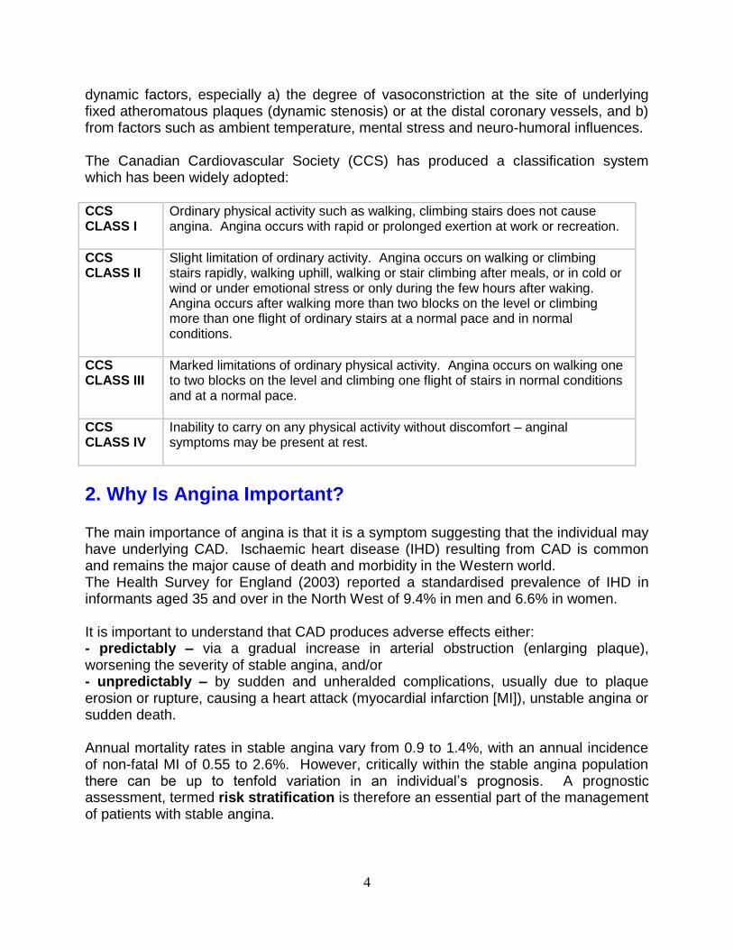
dynamic factors, especially a) the degree of vasoconstriction at the site of underlying fixed atheromatous plaques (dynamic stenosis) or at the distal coronary vessels, and b) from factors such as ambient temperature, mental stress and neuro-humoral influences. The Canadian Cardiovascular Society (CCS) has produced a classification system which has been widely adopted: CCS CLASS I
Ordinary physical activity such as walking, climbing stairs does not cause angina. Angina occurs with rapid or prolonged exertion at work or recreation.
CCS CLASS II
Slight limitation of ordinary activity. Angina occurs on walking or climbing stairs rapidly, walking uphill, walking or stair climbing after meals, or in cold or wind or under emotional stress or only during the few hours after waking. Angina occurs after walking more than two blocks on the level or climbing more than one flight of ordinary stairs at a normal pace and in normal conditions.
CCS CLASS III
Marked limitations of ordinary physical activity. Angina occurs on walking one to two blocks on the level and climbing one flight of stairs in normal conditions and at a normal pace.
CCS CLASS IV
Inability to carry on any physical activity without discomfort – anginal symptoms may be present at rest.
2. Why Is Angina Important? The main importance of angina is that it is a symptom suggesting that the individual may have underlying CAD. Ischaemic heart disease (IHD) resulting from CAD is common and remains the major cause of death and morbidity in the Western world. The Health Survey for England (2003) reported a standardised prevalence of IHD in informants aged 35 and over in the North West of 9.4% in men and 6.6% in women. It is important to understand that CAD produces adverse effects either: - predictably – via a gradual increase in arterial obstruction (enlarging plaque), worsening the severity of stable angina, and/or - unpredictably – by sudden and unheralded complications, usually due to plaque erosion or rupture, causing a heart attack (myocardial infarction [MI]), unstable angina or sudden death.
Annual mortality rates in stable angina vary from 0.9 to 1.4%, with an annual incidence of non-fatal MI of 0.55 to 2.6%. However, critically within the stable angina population there can be up to tenfold variation in an individual’s prognosis. A prognostic assessment, termed risk stratification is therefore an essential part of the management of patients with stable angina.
5
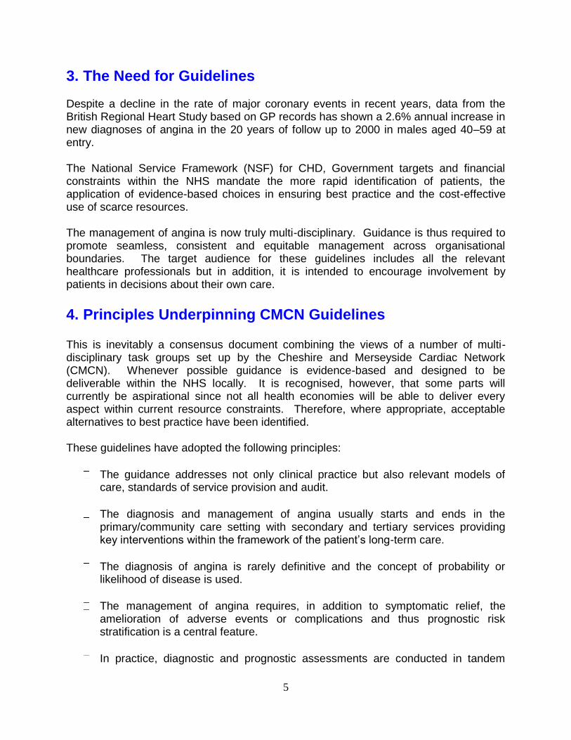
3. The Need for Guidelines Despite a decline in the rate of major coronary events in recent years, data from the British Regional Heart Study based on GP records has shown a 2.6% annual increase in new diagnoses of angina in the 20 years of follow up to 2000 in males aged 40–59 at entry. The National Service Framework (NSF) for CHD, Government targets and financial constraints within the NHS mandate the more rapid identification of patients, the application of evidence-based choices in ensuring best practice and the cost-effective use of scarce resources. The management of angina is now truly multi-disciplinary. Guidance is thus required to promote seamless, consistent and equitable management across organisational boundaries. The target audience for these guidelines includes all the relevant healthcare professionals but in addition, it is intended to encourage involvement by patients in decisions about their own care.
4. Principles Underpinning CMCN Guidelines This is inevitably a consensus document combining the views of a number of multi-disciplinary task groups set up by the Cheshire and Merseyside Cardiac Network (CMCN). Whenever possible guidance is evidence-based and designed to be deliverable within the NHS locally. It is recognised, however, that some parts will currently be aspirational since not all health economies will be able to deliver every aspect within current resource constraints. Therefore, where appropriate, acceptable alternatives to best practice have been identified. These guidelines have adopted the following principles:
The guidance addresses not only clinical practice but also relevant models of care, standards of service provision and audit.
The diagnosis and management of angina usually starts and ends in the primary/community care setting with secondary and tertiary services providing key interventions within the framework of the patient’s long-term care.
The diagnosis of angina is rarely definitive and the concept of probability or likelihood of disease is used.
The management of angina requires, in addition to symptomatic relief, the amelioration of adverse events or complications and thus prognostic risk stratification is a central feature.
In practice, diagnostic and prognostic assessments are conducted in tandem
6
rather than sequentially as the same clinical and investigational tools are used for both. However, for clarity of understanding and presentation, these linked assessments are described separately.
Modern medical management now includes a number of effective and locally well-developed treatment modalities in addition to drug therapy, including cardiac rehabilitation and refractory angina management. This guidance attempts to define their place in patient management.
Invasive and interventional approaches carry risks as well as benefits and are not infinitely available. Guidance is given on appropriate indications, treatment choices and patient prioritisation to allow best possible use of local resources compatible with good standards of care.
Successful implementation of these guidelines will require investment in the on-going education of a large constituency of relevant healthcare professionals.
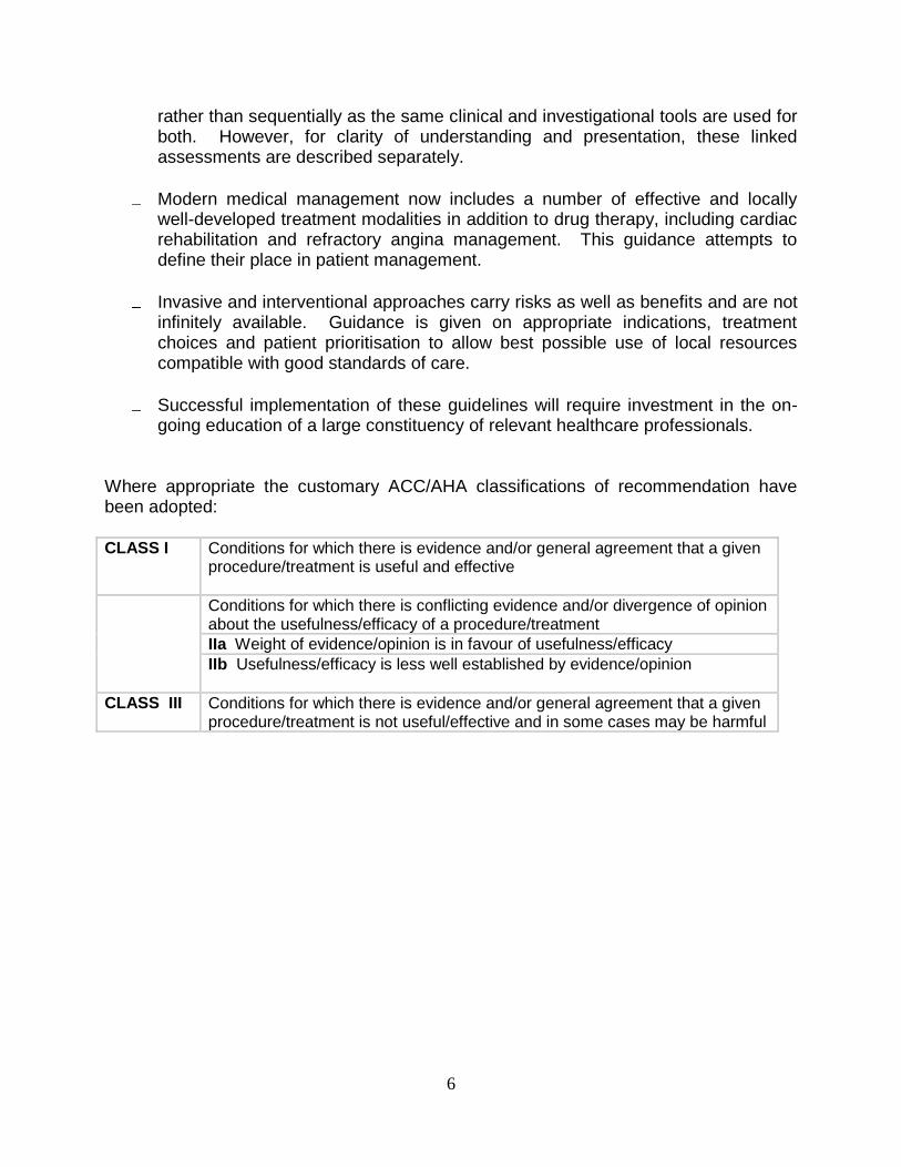
Where appropriate the customary ACC/AHA classifications of recommendation have been adopted: CLASS I Conditions for which there is evidence and/or general agreement that a given
procedure/treatment is useful and effective
Conditions for which there is conflicting evidence and/or divergence of opinion about the usefulness/efficacy of a procedure/treatment
IIa Weight of evidence/opinion is in favour of usefulness/efficacy
IIb Usefulness/efficacy is less well established by evidence/opinion
CLASS III Conditions for which there is evidence and/or general agreement that a given procedure/treatment is not useful/effective and in some cases may be harmful
7
STAGE 1 CASE MANAGEMENT and REFERRAL PATHWAYS
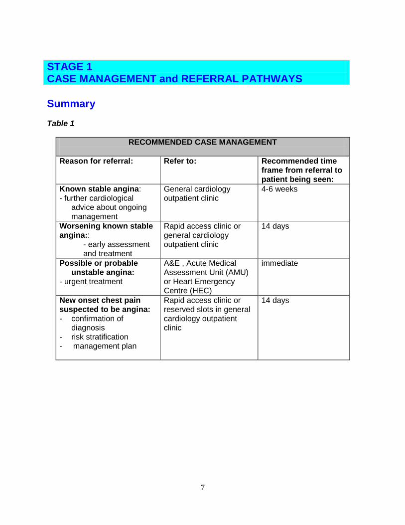
Summary Table 1
RECOMMENDED CASE MANAGEMENT
Reason for referral: Refer to: Recommended time frame from referral to patient being seen:
Known stable angina: - further cardiological
advice about ongoing management
General cardiology outpatient clinic
4-6 weeks
Worsening known stable angina::
- early assessment and treatment
Rapid access clinic or general cardiology outpatient clinic
14 days
Possible or probable unstable angina:
- urgent treatment
A&E , Acute Medical Assessment Unit (AMU) or Heart Emergency Centre (HEC)
immediate
New onset chest pain suspected to be angina: - confirmation of
diagnosis - risk stratification - management plan
Rapid access clinic or reserved slots in general cardiology outpatient clinic
14 days
8
STAGE 1 CASE MANAGEMENT and REFERRAL PATHWAYS
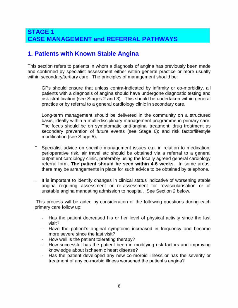
1. Patients with Known Stable Angina This section refers to patients in whom a diagnosis of angina has previously been made and confirmed by specialist assessment either within general practice or more usually within secondary/tertiary care. The principles of management should be:
GPs should ensure that unless contra-indicated by infirmity or co-morbidity, all patients with a diagnosis of angina should have undergone diagnostic testing and risk stratification (see Stages 2 and 3). This should be undertaken within general practice or by referral to a general cardiology clinic in secondary care.
Long-term management should be delivered in the community on a structured basis, ideally within a multi-disciplinary management programme in primary care. The focus should be on symptomatic anti-anginal treatment; drug treatment as secondary prevention of future events (see Stage 6); and risk factor/lifestyle modification (see Stage 5).
Specialist advice on specific management issues e.g. in relation to medication, perioperative risk, air travel etc should be obtained via a referral to a general outpatient cardiology clinic, preferably using the locally agreed general cardiology referral form. The patient should be seen within 4-6 weeks. In some areas, there may be arrangements in place for such advice to be obtained by telephone.
It is important to identify changes in clinical status indicative of worsening stable angina requiring assessment or re-assessment for revascularisation or of unstable angina mandating admission to hospital. See Section 2 below.
This process will be aided by consideration of the following questions during each primary care follow up:
- Has the patient decreased his or her level of physical activity since the last
visit? - Have the patient’s anginal symptoms increased in frequency and become
more severe since the last visit? - How well is the patient tolerating therapy? - How successful has the patient been in modifying risk factors and improving
knowledge about ischaemic heart disease? - Has the patient developed any new co-morbid illness or has the severity or
treatment of any co-morbid illness worsened the patient’s angina?
9
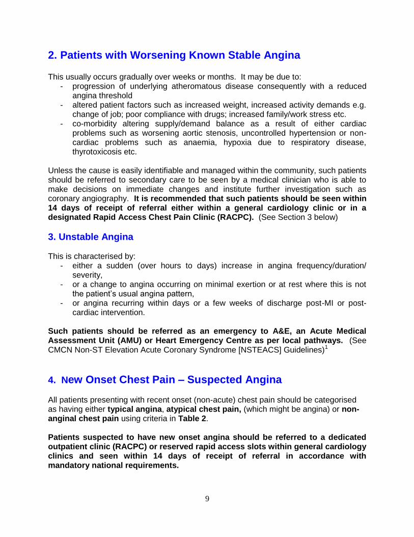
2. Patients with Worsening Known Stable Angina This usually occurs gradually over weeks or months. It may be due to:
- progression of underlying atheromatous disease consequently with a reduced angina threshold
- altered patient factors such as increased weight, increased activity demands e.g. change of job; poor compliance with drugs; increased family/work stress etc.
- co-morbidity altering supply/demand balance as a result of either cardiac problems such as worsening aortic stenosis, uncontrolled hypertension or non-cardiac problems such as anaemia, hypoxia due to respiratory disease, thyrotoxicosis etc.
Unless the cause is easily identifiable and managed within the community, such patients should be referred to secondary care to be seen by a medical clinician who is able to make decisions on immediate changes and institute further investigation such as coronary angiography. It is recommended that such patients should be seen within 14 days of receipt of referral either within a general cardiology clinic or in a designated Rapid Access Chest Pain Clinic (RACPC). (See Section 3 below)
3. Unstable Angina This is characterised by:
- either a sudden (over hours to days) increase in angina frequency/duration/ severity,
- or a change to angina occurring on minimal exertion or at rest where this is not the patient’s usual angina pattern,
- or angina recurring within days or a few weeks of discharge post-MI or post-cardiac intervention.
Such patients should be referred as an emergency to A&E, an Acute Medical Assessment Unit (AMU) or Heart Emergency Centre as per local pathways. (See CMCN Non-ST Elevation Acute Coronary Syndrome [NSTEACS] Guidelines)1
4. New Onset Chest Pain – Suspected Angina All patients presenting with recent onset (non-acute) chest pain should be categorised as having either typical angina, atypical chest pain, (which might be angina) or non-anginal chest pain using criteria in Table 2. Patients suspected to have new onset angina should be referred to a dedicated outpatient clinic (RACPC) or reserved rapid access slots within general cardiology clinics and seen within 14 days of receipt of referral in accordance with mandatory national requirements.
10
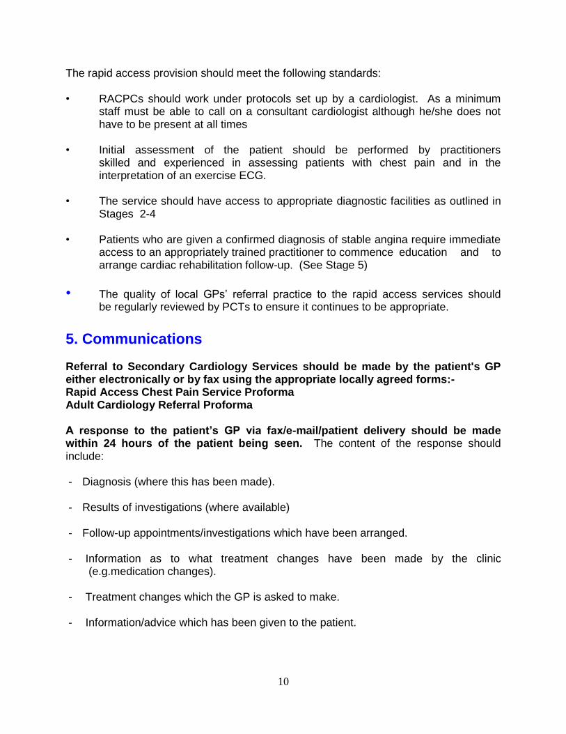
The rapid access provision should meet the following standards: • RACPCs should work under protocols set up by a cardiologist. As a minimum
staff must be able to call on a consultant cardiologist although he/she does not have to be present at all times
• Initial assessment of the patient should be performed by practitioners
skilled and experienced in assessing patients with chest pain and in the interpretation of an exercise ECG.
• The service should have access to appropriate diagnostic facilities as outlined in
Stages 2-4 • Patients who are given a confirmed diagnosis of stable angina require immediate
access to an appropriately trained practitioner to commence education and to arrange cardiac rehabilitation follow-up. (See Stage 5)
• The quality of local GPs’ referral practice to the rapid access services should be regularly reviewed by PCTs to ensure it continues to be appropriate.
5. Communications Referral to Secondary Cardiology Services should be made by the patient's GP either electronically or by fax using the appropriate locally agreed forms:- Rapid Access Chest Pain Service Proforma Adult Cardiology Referral Proforma A response to the patient’s GP via fax/e-mail/patient delivery should be made within 24 hours of the patient being seen. The content of the response should include: - Diagnosis (where this has been made). - Results of investigations (where available) - Follow-up appointments/investigations which have been arranged. - Information as to what treatment changes have been made by the clinic
(e.g.medication changes). - Treatment changes which the GP is asked to make. - Information/advice which has been given to the patient.
11
STAGE 2
DIAGNOSIS
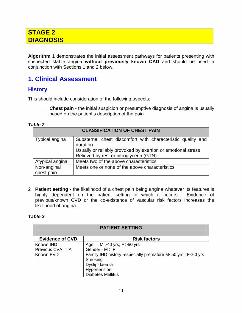
Algorithm 1 demonstrates the initial assessment pathways for patients presenting with suspected stable angina without previously known CAD and should be used in conjunction with Sections 1 and 2 below.
1. Clinical Assessment
History
This should include consideration of the following aspects:
Chest pain - the initial suspicion or presumptive diagnosis of angina is usually based on the patient’s description of the pain.
Table 2
CLASSIFICATION OF CHEST PAIN
Typical angina Substernal chest discomfort with characteristic quality and duration Usually or reliably provoked by exertion or emotional stress Relieved by rest or nitroglycerin (GTN)
Atypical angina Meets two of the above characteristics
Non-anginal chest pain
Meets one or none of the above characteristics
2 Patient setting - the likelihood of a chest pain being angina whatever its features is
highly dependent on the patient setting in which it occurs. Evidence of previous/known CVD or the co-existence of vascular risk factors increases the likelihood of angina.
Table 3
PATIENT SETTING
Evidence of CVD Risk factors Known IHD Previous CVA, TIA Known PVD
Age- M >40 yrs; F >50 yrs Gender - M > F Family IHD history -especially premature M<50 yrs ; F<60 yrs Smoking Dyslipidaemia Hypertension Diabetes Mellitus
12
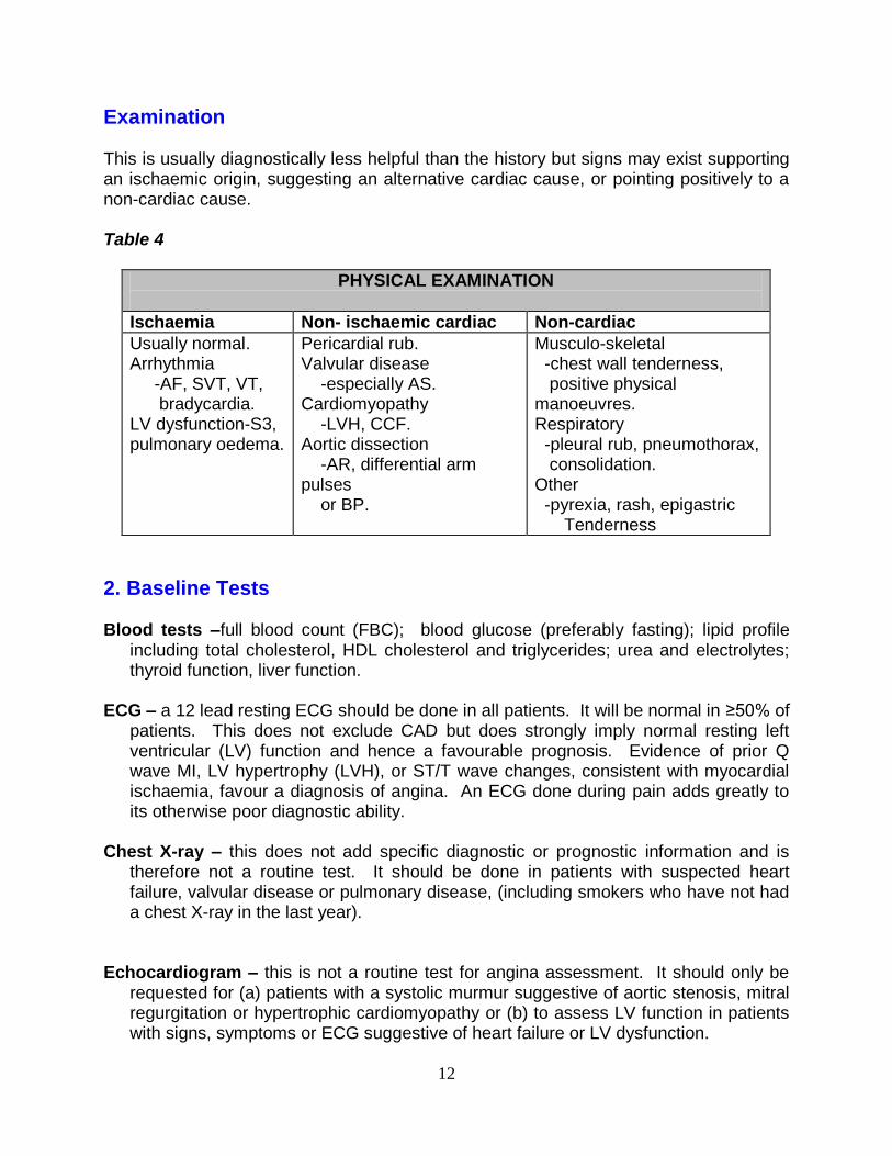
Examination This is usually diagnostically less helpful than the history but signs may exist supporting an ischaemic origin, suggesting an alternative cardiac cause, or pointing positively to a non-cardiac cause. Table 4
PHYSICAL EXAMINATION
Ischaemia Non- ischaemic cardiac Non-cardiac
Usually normal. Arrhythmia -AF, SVT, VT, bradycardia. LV dysfunction-S3, pulmonary oedema.
Pericardial rub. Valvular disease -especially AS. Cardiomyopathy -LVH, CCF. Aortic dissection -AR, differential arm pulses or BP.
Musculo-skeletal -chest wall tenderness, positive physical manoeuvres. Respiratory -pleural rub, pneumothorax, consolidation. Other -pyrexia, rash, epigastric Tenderness
2. Baseline Tests Blood tests –full blood count (FBC); blood glucose (preferably fasting); lipid profile
including total cholesterol, HDL cholesterol and triglycerides; urea and electrolytes; thyroid function, liver function.
ECG – a 12 lead resting ECG should be done in all patients. It will be normal in ≥50% of
patients. This does not exclude CAD but does strongly imply normal resting left ventricular (LV) function and hence a favourable prognosis. Evidence of prior Q wave MI, LV hypertrophy (LVH), or ST/T wave changes, consistent with myocardial ischaemia, favour a diagnosis of angina. An ECG done during pain adds greatly to its otherwise poor diagnostic ability.
Chest X-ray – this does not add specific diagnostic or prognostic information and is
therefore not a routine test. It should be done in patients with suspected heart failure, valvular disease or pulmonary disease, (including smokers who have not had a chest X-ray in the last year).
Echocardiogram – this is not a routine test for angina assessment. It should only be
requested for (a) patients with a systolic murmur suggestive of aortic stenosis, mitral regurgitation or hypertrophic cardiomyopathy or (b) to assess LV function in patients with signs, symptoms or ECG suggestive of heart failure or LV dysfunction.
13
Table 5
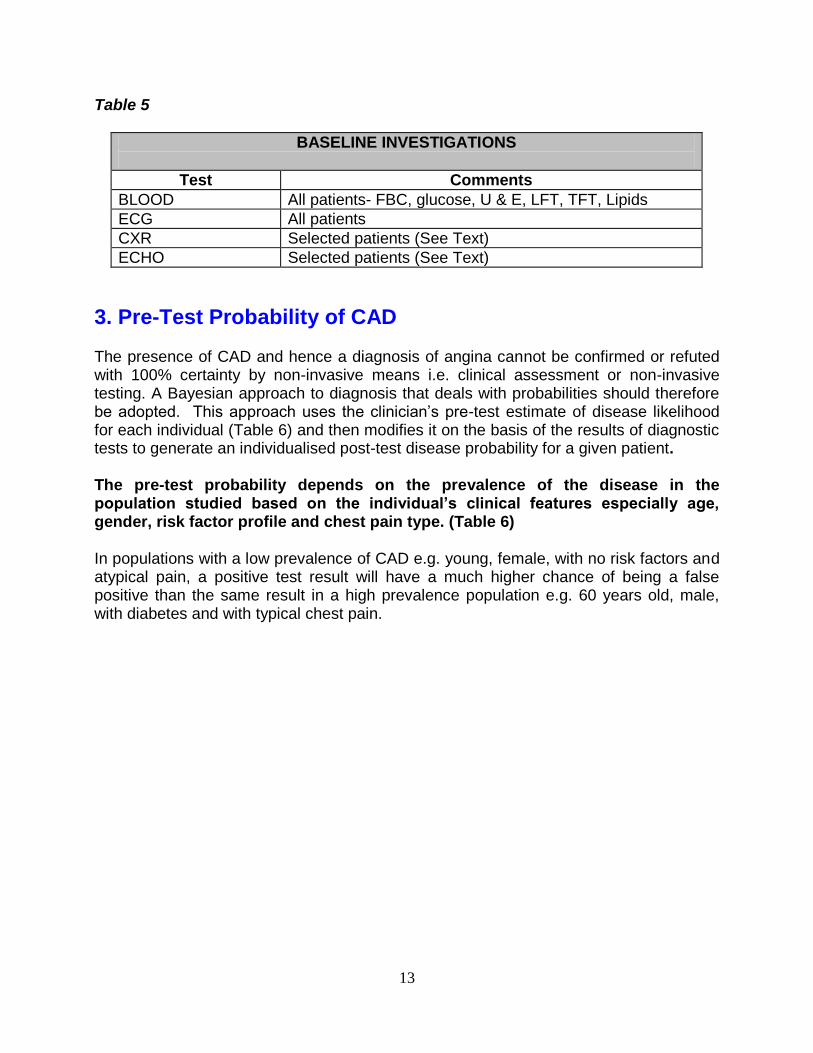
BASELINE INVESTIGATIONS
Test Comments
BLOOD All patients- FBC, glucose, U & E, LFT, TFT, Lipids
ECG All patients
CXR Selected patients (See Text)
ECHO Selected patients (See Text)
3. Pre-Test Probability of CAD The presence of CAD and hence a diagnosis of angina cannot be confirmed or refuted with 100% certainty by non-invasive means i.e. clinical assessment or non-invasive testing. A Bayesian approach to diagnosis that deals with probabilities should therefore be adopted. This approach uses the clinician’s pre-test estimate of disease likelihood for each individual (Table 6) and then modifies it on the basis of the results of diagnostic tests to generate an individualised post-test disease probability for a given patient. The pre-test probability depends on the prevalence of the disease in the population studied based on the individual’s clinical features especially age, gender, risk factor profile and chest pain type. (Table 6) In populations with a low prevalence of CAD e.g. young, female, with no risk factors and atypical pain, a positive test result will have a much higher chance of being a false positive than the same result in a high prevalence population e.g. 60 years old, male, with diabetes and with typical chest pain.
14
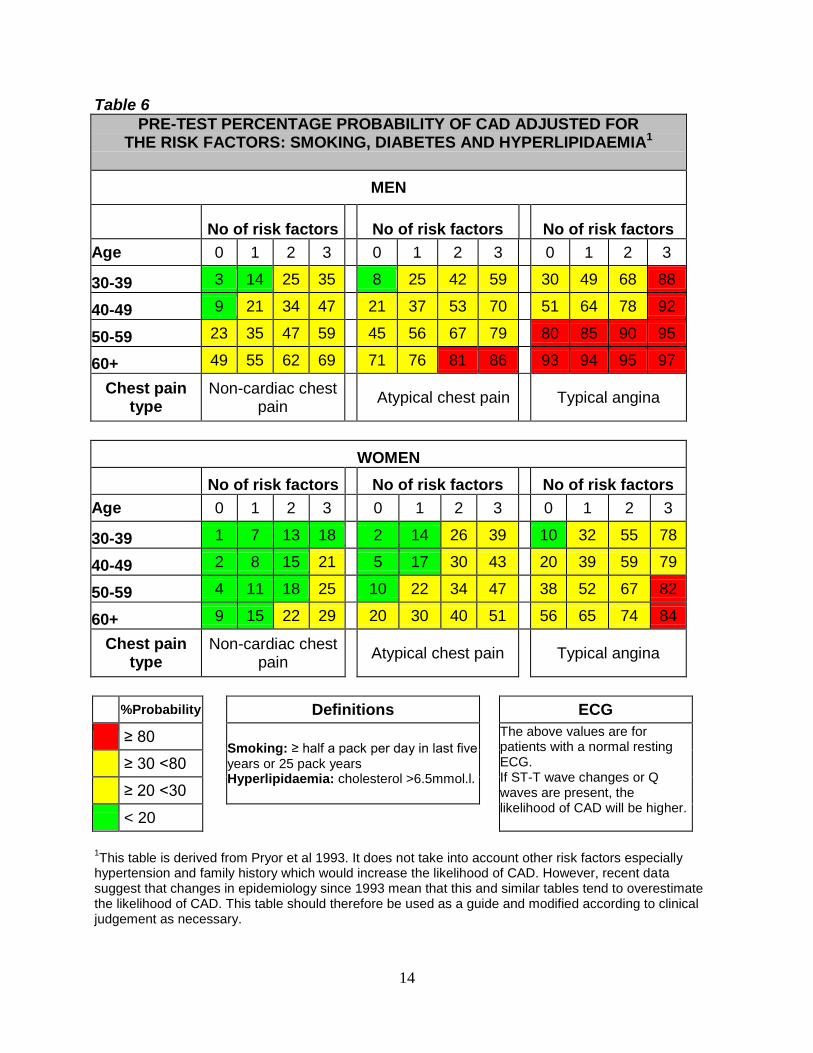
Table 6
PRE-TEST PERCENTAGE PROBABILITY OF CAD ADJUSTED FOR THE RISK FACTORS: SMOKING, DIABETES AND HYPERLIPIDAEMIA1
MEN
No of risk factors
No of risk factors
No of risk factors
Age 0 1 2 3 0 1 2 3 0 1 2 3
30-39 3 14 25 35 8 25 42 59 30 49 68 88
40-49 9 21 34 47 21 37 53 70 51 64 78 92
50-59 23 35 47 59 45 56 67 79 80 85 90 95
60+ 49 55 62 69 71 76 81 86 93 94 95 97
Chest pain type
Non-cardiac chest pain
Atypical chest pain Typical angina
WOMEN
No of risk factors No of risk factors No of risk factors
Age 0 1 2 3 0 1 2 3 0 1 2 3
30-39 1 7 13 18 2 14 26 39 10 32 55 78
40-49 2 8 15 21 5 17 30 43 20 39 59 79
50-59 4 11 18 25 10 22 34 47 38 52 67 82
60+ 9 15 22 29 20 30 40 51 56 65 74 84
Chest pain type
Non-cardiac chest pain
Atypical chest pain
Typical angina
%Probability Definitions ECG
≥ 80 Smoking: ≥ half a pack per day in last five years or 25 pack years Hyperlipidaemia: cholesterol >6.5mmol.l.
The above values are for patients with a normal resting ECG. If ST-T wave changes or Q waves are present, the likelihood of CAD will be higher.
≥ 30 <80
≥ 20 <30
< 20
1This table is derived from Pryor et al 1993. It does not take into account other risk factors especially
hypertension and family history which would increase the likelihood of CAD. However, recent data suggest that changes in epidemiology since 1993 mean that this and similar tables tend to overestimate the likelihood of CAD. This table should therefore be used as a guide and modified according to clinical judgement as necessary.
15
4. Diagnostic Investigation Based on Pre-Test Probability of CAD Once a pre-test probability has been calculated for an individual patient from Table 6, this figure should be used as the basis for deciding on appropriate diagnostic testing as outlined in algorithm 2. This can be summarised according to pre-test probability as:- <10% - angina/CAD very unlikely so no routine diagnostic test necessary 10-29% - CT calcium score +/- CT coronary angiography 30-60% - Functional test for myocardial ischaemia i.e. myocardial perfusion
imaging (MPI) stress echo or occasional stress MRI depending on local availability
>60-90% - Left heart catheterisation for diagnosis and prognosis >90% - No diagnostic test required; left heart catheterisation for prognosis if
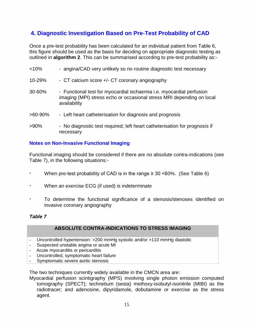
necessary Notes on Non-Invasive Functional Imaging Functional imaging should be considered if there are no absolute contra-indications (see Table 7), in the following situations:-
∙ When pre-test probability of CAD is in the range ≥ 30 <60%. (See Table 6)
∙ When an exercise ECG (if used) is indeterminate
∙ To determine the functional significance of a stenosis/stenoses identified on
invasive coronary angiography Table 7
ABSOLUTE CONTRA-INDICATIONS TO STRESS IMAGING
- Uncontrolled hypertension: >200 mmHg systolic and/or >110 mmHg diastolic - Suspected unstable angina or acute MI - Acute myocarditis or pericarditis - Uncontrolled, symptomatic heart failure - Symptomatic severe aortic stenosis
The two techniques currently widely available in the CMCN area are: Myocardial perfusion scintigraphy (MPS) involving single photon emission computed
tomography (SPECT); technetium (sesta) methoxy-isobutyl-isonitrile (MIBI) as the radiotracer; and adenosine, dipyridamole, dobutamine or exercise as the stress agent.
16
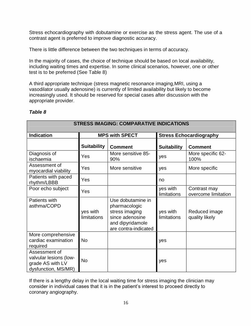
Stress echocardiography with dobutamine or exercise as the stress agent. The use of a contrast agent is preferred to improve diagnostic accuracy. There is little difference between the two techniques in terms of accuracy. In the majority of cases, the choice of technique should be based on local availability, including waiting times and expertise. In some clinical scenarios, however, one or other test is to be preferred (See Table 8) A third appropriate technique (stress magnetic resonance imaging,MRI, using a vasodilator usually adenosine) is currently of limited availability but likely to become increasingly used. It should be reserved for special cases after discussion with the appropriate provider. Table 8
STRESS IMAGING: COMPARATIVE INDICATIONS
Indication MPS with SPECT Stress Echocardiography
Suitability
Comment
Suitability
Comment
Diagnosis of ischaemia
Yes More sensitive 85-90%
yes More specific 62-100%
Assessment of myocardial viability
Yes More sensitive yes More specific
Patients with paced rhythm/LBBB
Yes no
Poor echo subject Yes
yes with limitations
Contrast may overcome limitation
Patients with asthma/COPD
yes with limitations
Use dobutamine in pharmacologic stress imaging since adenosine and dipyridamole are contra-indicated
yes with limitations
Reduced image quality likely
More comprehensive cardiac examination required
No yes
Assessment of valvular lesions (low-grade AS with LV dysfunction, MS/MR)
No yes
If there is a lengthy delay in the local waiting time for stress imaging the clinician may consider in individual cases that it is in the patient’s interest to proceed directly to coronary angiography.
17
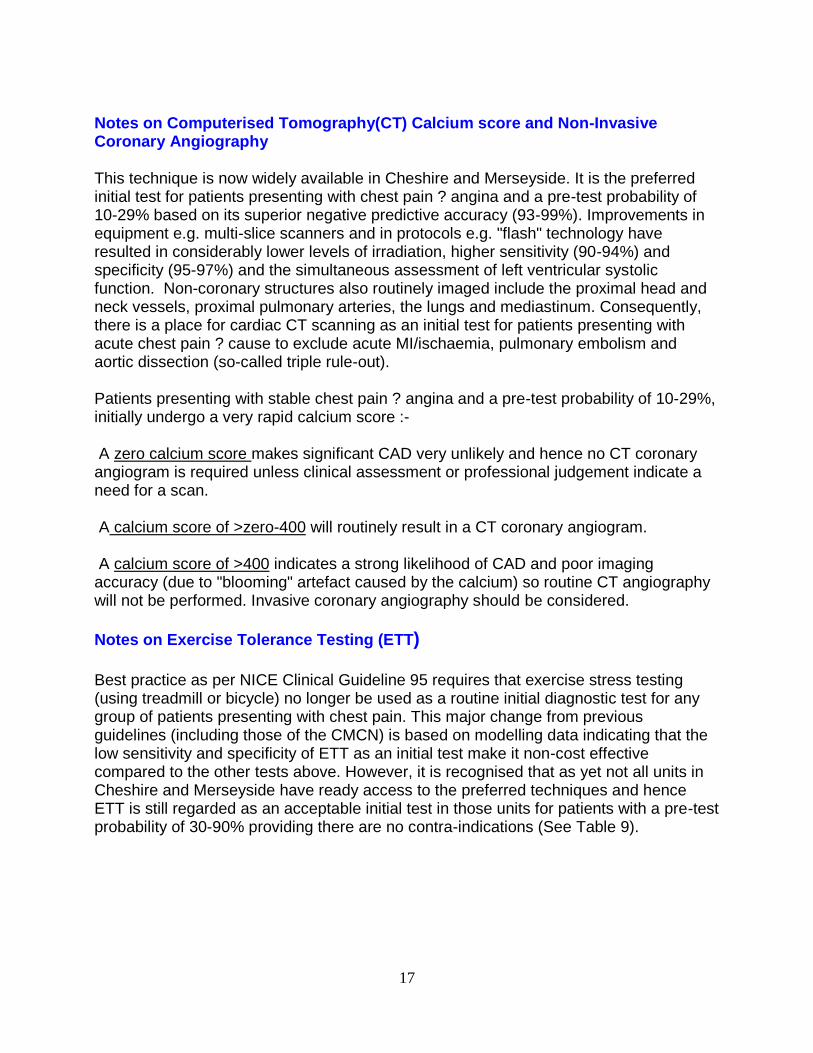
Notes on Computerised Tomography(CT) Calcium score and Non-Invasive Coronary Angiography This technique is now widely available in Cheshire and Merseyside. It is the preferred initial test for patients presenting with chest pain ? angina and a pre-test probability of 10-29% based on its superior negative predictive accuracy (93-99%). Improvements in equipment e.g. multi-slice scanners and in protocols e.g. "flash" technology have resulted in considerably lower levels of irradiation, higher sensitivity (90-94%) and specificity (95-97%) and the simultaneous assessment of left ventricular systolic function. Non-coronary structures also routinely imaged include the proximal head and neck vessels, proximal pulmonary arteries, the lungs and mediastinum. Consequently, there is a place for cardiac CT scanning as an initial test for patients presenting with acute chest pain ? cause to exclude acute MI/ischaemia, pulmonary embolism and aortic dissection (so-called triple rule-out). Patients presenting with stable chest pain ? angina and a pre-test probability of 10-29%, initially undergo a very rapid calcium score :- A zero calcium score makes significant CAD very unlikely and hence no CT coronary angiogram is required unless clinical assessment or professional judgement indicate a need for a scan. A calcium score of >zero-400 will routinely result in a CT coronary angiogram. A calcium score of >400 indicates a strong likelihood of CAD and poor imaging accuracy (due to "blooming" artefact caused by the calcium) so routine CT angiography will not be performed. Invasive coronary angiography should be considered.
Notes on Exercise Tolerance Testing (ETT) Best practice as per NICE Clinical Guideline 95 requires that exercise stress testing (using treadmill or bicycle) no longer be used as a routine initial diagnostic test for any group of patients presenting with chest pain. This major change from previous guidelines (including those of the CMCN) is based on modelling data indicating that the low sensitivity and specificity of ETT as an initial test make it non-cost effective compared to the other tests above. However, it is recognised that as yet not all units in Cheshire and Merseyside have ready access to the preferred techniques and hence ETT is still regarded as an acceptable initial test in those units for patients with a pre-test probability of 30-90% providing there are no contra-indications (See Table 9).

18
Table 9
CONTRA-INDICATIONS TO DIAGNOSTIC EXERCISE ECG TESTING
Absolute contra-indications - Uncontrolled hypertension:>200 mmHg systolic and/or >110 mmHg diastolic - LBBB on ECG - Pre-excitation pattern i.e. delta waves - Paced rhythm - Uncontrolled arrhythmia - Suspected unstable angina - More than 2 mm resting ST depression, particularly if the patient is on digoxin - Acute myocarditis or pericarditis - Uncontrolled, symptomatic heart failure - Symptomatic severe aortic stenosis
- Inability to perform exercise ECG due to co-morbidity or disability
Relative contra-indications – caution required - Suspected significant outflow tract obstruction due to moderate aortic stenosis or
hypertrophic obstructive cardiomyopathy - Other significant valvular disorder e.g. mitral stenosis or aortic regurgitation - ACS: MI/high risk unstable angina ≤ 3 weeks; left main stem stenosis
- High degree atrioventricular block
Useful Probability Range
Outside the range ≥ 30% to <90%, the test has little diagnostic accuracy producing excessive false positives in very low prevalence populations and failing to add to the pre-test probability in very high prevalence populations. Within this range, its accuracy is reasonable. (sensitivity 68% specificity 77%) for the detection CAD using a diagnostic threshold of 1mm horizontal or down-sloping ST depression.
.
19
STAGE 3
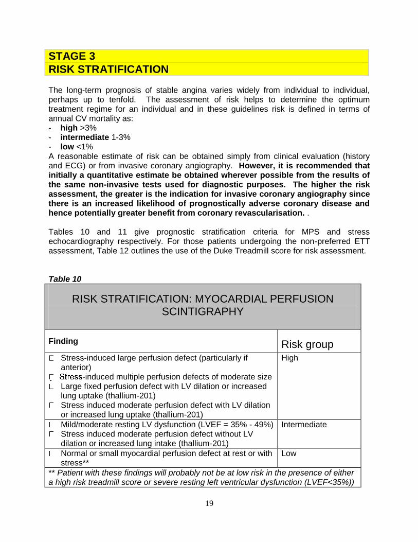
RISK STRATIFICATION The long-term prognosis of stable angina varies widely from individual to individual, perhaps up to tenfold. The assessment of risk helps to determine the optimum treatment regime for an individual and in these guidelines risk is defined in terms of annual CV mortality as: - high >3% - intermediate 1-3% - low <1% A reasonable estimate of risk can be obtained simply from clinical evaluation (history and ECG) or from invasive coronary angiography. However, it is recommended that initially a quantitative estimate be obtained wherever possible from the results of the same non-invasive tests used for diagnostic purposes. The higher the risk assessment, the greater is the indication for invasive coronary angiography since there is an increased likelihood of prognostically adverse coronary disease and hence potentially greater benefit from coronary revascularisation. . Tables 10 and 11 give prognostic stratification criteria for MPS and stress echocardiography respectively. For those patients undergoing the non-preferred ETT assessment, Table 12 outlines the use of the Duke Treadmill score for risk assessment. Table 10
RISK STRATIFICATION: MYOCARDIAL PERFUSION SCINTIGRAPHY
Finding Risk group Stress-induced large perfusion defect (particularly if
anterior) -induced multiple perfusion defects of moderate size
Large fixed perfusion defect with LV dilation or increased lung uptake (thallium-201)
Stress induced moderate perfusion defect with LV dilation or increased lung uptake (thallium-201)
High
Mild/moderate resting LV dysfunction (LVEF = 35% - 49%) Stress induced moderate perfusion defect without LV
dilation or increased lung intake (thallium-201)
Intermediate
Normal or small myocardial perfusion defect at rest or with stress**
Low
** Patient with these findings will probably not be at low risk in the presence of either a high risk treadmill score or severe resting left ventricular dysfunction (LVEF<35%))
20
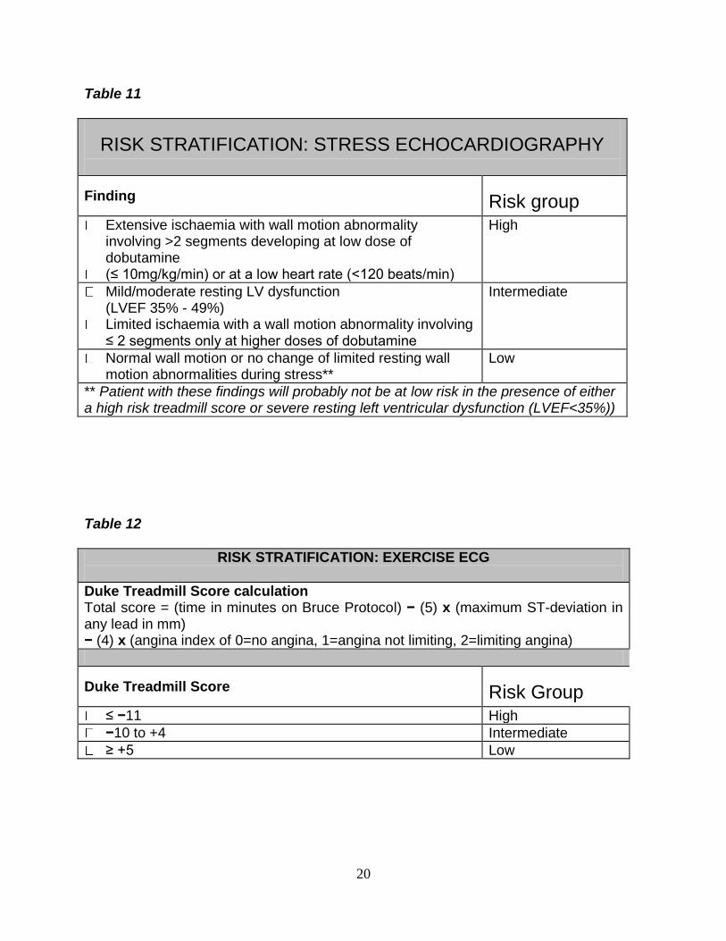
Table 11
RISK STRATIFICATION: STRESS ECHOCARDIOGRAPHY
Finding Risk group Extensive ischaemia with wall motion abnormality
involving >2 segments developing at low dose of dobutamine
(≤ 10mg/kg/min) or at a low heart rate (<120 beats/min)
High
Mild/moderate resting LV dysfunction (LVEF 35% - 49%)
Limited ischaemia with a wall motion abnormality involving ≤ 2 segments only at higher doses of dobutamine
Intermediate
Normal wall motion or no change of limited resting wall motion abnormalities during stress**
Low
** Patient with these findings will probably not be at low risk in the presence of either a high risk treadmill score or severe resting left ventricular dysfunction (LVEF<35%))
Table 12
RISK STRATIFICATION: EXERCISE ECG
Duke Treadmill Score calculation Total score = (time in minutes on Bruce Protocol) − (5) x (maximum ST-deviation in any lead in mm) − (4) x (angina index of 0=no angina, 1=angina not limiting, 2=limiting angina)
Duke Treadmill Score Risk Group ≤ −11 High
−10 to +4 Intermediate
≥ +5 Low
21
STAGE 4
DRUG TREATMENT Drug therapy is an important component of angina treatment (see algorithm 3) involving the treatment of an acute attack of angina; drug prophylaxis to eliminate or reduce the frequency of anginal chest pain;and drugs given to improve prognosis. These 3 therapeutic areas are summarised in algorithm 3 and discussed in greater detail in this section. Key details about individual drugs will be given in the following sections but full information on dosages, formulations, contraindications, use in pregnancy and during lactation and side effects should be sought from the British National Formulary (BNF) and summaries of product characteristics (SMPCs).
Drug Treatment of Acute Episode Glyceryl trinitrate (GTN) is the drug of choice. It reduces pre-load and after-load and induces coronary vasodilatation. It is effective quickly (usually < five minutes) and lasts 20-30 minutes. It can be repeated as necessary but recurrent chest pain over a short period should raise the question of worsening angina or acute coronary syndrome. It can be taken in different formulations, tablets, spray and buccal tablets, which should be tailored to suit the patient and context. Attention should be paid to the likelihood of headache on first use when GTN in tablet form is particularly appropriate because the patient can be advised to remove it before the headache becomes severe.
Prophylactic Drug Treatment of Symptoms Six classes of drug are widely accepted as effective for symptomatic prophylaxis in reducing the frequency and severity of anginal chest pain and/or breathlessness where this is an angina-equivalent due to ischaemia. . There is no universally acknowledged strategy for the optimal cost effective use of these drugs but the following reflects national consensus (NICE Clinical Guideline 126). However, individual patients react very differently in terms of benefit and efficacy and so in practice choice is often determined by patient response.
22
Recommended Strategy This is outlined in Algorithm 3. In summary:-
Commence with monotherapy using a first-line agent (beta blocker or rate-limiting calcium blocker) in small dosage.
If symptoms continue after 2-4 weeks, add the other first line line agent to optimise symptom control, assessed subjectively by clinician and objectively by exercise ECG if necessary.
Titrate dose upwards until the maximally-tolerated doses are reached.
There is little objective evidence of added benefit from a third or fourth drug although it is acceptable to try with the proviso that they be stopped if the patient does not respond
Consider the potential adverse effects on blood pressure (BP), heart rate (HR) and left ventricular function (LVF).
The failure of a maximally tolerated drug treatment to control symptoms adequately constitutes an indication for consideration of revascularisation even in the absence of other indications.
Anti-anginal agents, especially beta blockers, when no longer required, should be tailed off rather than stopped abruptly unless they are causing significant side effects.
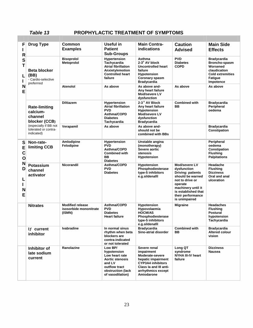
Table 13 summarises the agents which are described in detail in the sections that follow.
23
Table 13 PROPHYLACTIC TREATMENT OF SYMPTOMS
F I R S T L I N E
Drug Type Common Examples
Useful in Patient Sub-Groups
Main Contra-indications
Caution Advised
Main Side Effects
Beta blocker (BB) - Cardio-selective preferrred
Bisoprolol Metoprolol
Hypertension Tachycardia Atrial fibrillation Anxiety/emotion Controlled heart failure
Asthma 2-3
o AV block
Uncontrolled heart failure Hypotension Coronary spasm Bradycardia
PVD Diabetes COPD
Bradycardia Broncho-spasm Worsened claudication Cold extremities Fatigue Impotence
Atenolol As above As above and- Any heart failure Mod/severe LV dysfunction
As above As above
Rate-limiting calcium-channel blocker (CCB)
(especially if BB not tolerated or contra-indicated)
Diltiazem Hypertension Atrial fibrillation PVD Asthma/COPD Diabetes Tachycardia
2-3 o
AV Block Any heart failure Hypotension Mod/severe LV dysfunction Bradycardia
Combined with BB
Bradycardia Peripheral oedema
Verapamil As above As above and- should not be combined with BBs
Bradycardia Constipation
S E C O N D L I N E
Non-rate-limiting CCB
Amlodipine Felodipine
Hypertension PVD Asthma/COPD Combined with BB Diabetes
Unstable angina (monotherapy) Severe aortic stenosis Hypotension
Peripheral oedema Constipation Flushing Palpitations
Potassium channel activator
Nicorandil Asthma/COPD PVD Diabetes
Hypotension Phosphodiesterase type-5 inhibitors e.g.sildenafil
Mod/severe LV dysfunction Driving: patients should be warned not to drive or operate machinery until it is established that their performance is unimpaired
Headache Flushing Dizziness Oral and anal ulceration
Nitrates Modified release isosorbide mononitrate (ISMN)
Asthma/COPD PVD Diabetes Heart failure
Hypotension Hypovolaemia HOCM/AS Phosphodiesterase type-5 inhibitors e.g.sildenafil
Migraine Headaches Flushing Postural hypotension Tachycardia
Iƒ current
inhibitor
Ivabradine
In normal sinus rhythm when beta blockers are contra-indicated or not tolerated
Bradycardia Sino-atrial disorder
Combined with BB
Bradycardia Altered colour vision
Inhibitor of late sodium current
Ranolazine
Low BP/ hypotension Low heart rate Aortic stenosis and LV outflow tract obstruction (lack of vasodilation)
Severe renal impairment Moderate-severe hepatic impairment CYP3A4 inhibitors Class Ia and III anti-arrhythmics except Amiodarone
Long QT syndrome NYHA III-IV heart failure
Dizziness Nausea

24
First Line Agents Beta Blockers (BBs) BBs are the recommended first choice for exercise or emotion induced angina. Whilst no more effective in reducing symptoms than other agents, extrapolation from successful post-MI trials suggests they may also reduce mortality in patients with stable angina although this has not been proved in a placebo controlled trial. See also section 3 ‘Drug Treatment to Improve Prognosis in Stable Angina: Anti-anginal Drugs’. BBs work by blockade of the β1 adrenoceptor, so reducing myocardial oxygen demand (especially on exercise) by slowing the heart rate, lowering the blood pressure and reducing myocardial contractility. Perfusion of ischaemic areas may be improved by prolonging diastole and by ‘reverse coronary steal’ due to increased vascular resistance in non-ischaemic areas. Cardio-selective BBs are recommended because they preferentially block β1 adrenoceptors in the heart and blood vessels rather than elsewhere especially in the bronchi. In contrast to standard BBs, some newer agents such as carvedilol and nebivolol, have an additional arteriolar vasodilating action. Since this reduces the likelihood of cold peripheries and worsened claudication such drugs may be useful where these are concerns. All BBs are effective in angina but Table 13 will help guide the choice of agent for an individual patient. Rate Limiting Calcium-channel Blockers (CCBs)
CCBs are now regarded as alternative first line agents to BBs. They work by interfering with the inward displacement of calcium ions through the slow (L-type) channels of active membranes. The target tissues are therefore the myocardium (reduced contractility), cardiac conducting system (reduced heart rate) and vascular smooth muscle (peripheral vasodilation). However, CCBs are a heterogeneous group of compounds with important differences in pharmacological action due largely to variable effects on the target tissues. CCBs are generally divided into two subgroups with similar but not identical properties. The rate-limiting CCB group consists of diltiazem and verapamil. These principally reduce myocardial oxygen demand through their major effects on the conducting system (bradycardia, impaired AV conduction) and the myocardium (negative inotropic response) reducing contractility. They also have a moderate effect on vascular smooth muscle causing some coronary and peripheral vasodilation.
25
They are recommended as first-line agents when BBs are contra-indicated or not tolerated, especially when used as monotherapy. They are also safe and effective as second-line agents in combination with other anti-anginal classes but special precautions are necessary with BBs see Table 13.
Second-Line Agents All the following have proven anti-anginal efficacy. They should be used as second-line i.e. in addition to a first line drug if the other first line agent is ineffective/not tolerated or third line i.e. in addition to both first line drugs as a therapeutic trial to be stopped if ineffective. Non-Rate Limiting CCBs
The larger non-rate limiting group includes all other CCB agents, the vast majority of which are analogues of the prototype dihydropyridine drug, nifedipine. These agents have a major effect on the vascular smooth muscle, causing marked coronary and especially peripheral vasodilation and lesser effects on the myocardium and conducting system. These are therefore second-line agents and ideally combined with a BB because the latter will block the reflex tachycardia often associated with vasodilation. Potassium-Channel Activators
Nicorandil is the only available drug in this group. It has both arterial and venous dilating properties due to its potassium channel activation and an associated nitrate activity. It is licensed for angina treatment and prophylaxis as monotherapy (second- line agent). It may also be useful in combination with other drug classes though it is not licenced for this indication (Off-licence use requires additional patient discussion) Nitrates
Nitrates are veno- and vasodilatory. Their principal benefit in angina derives from reduced preload due to venodilation but also from coronary vasodilation. Three main agents are available: GTN, isosorbide mononitrate (ISMN) and isosorbide dinitrate (ISDN), which is converted to its active metabolite ISMN. GTN, either sub-lingually or preferably by the buccal route, is valuable for situational prophylaxis prior to exertion. (For use in the relief of an acute episode, see above.) For long-term prophylaxis, ISMN is preferred to ISDN because it avoids the variability of conversion to ISMN. It is also preferred to GTN (buccal, patch etc) because of its ease of use and patient acceptability. The problems with nitrates relate to headaches (which can be severe but tend to reduce with continued use) and reduced efficacy (nitrate tolerance) on continuous usage with
26
long-lasting or transdermal preparations. Nitrate tolerance can be avoided by appropriate dosing techniques, which allow 8-12 hour nitrate low or nitrate-free periods. This can be achieved by limiting use of ISMN to a once-daily modified release preparation or to two asymmetrically timed conventional formulations or by removing the patch at night. The choice between different preparations of ISMN will depend on price and patient preference but in general, a single daily dose of a modified release preparation is preferred. Iƒ Current Inhibitors
Ivabradine is the only currently available drug in this group. It inhibits the cardiac
pacemaker Iƒ current in the sino-atrial node selectively and specifically resulting in
reduced heart rate but not reduced BP. By reducing myocardial oxygen demand, it is as symptomatically effective as atenolol but has no BB related side effects. It is licensed as an alternative agent in patients who cannot tolerate a BB. It is restricted to use in sinus rhythm and has no value in atrial fibrillation. Late Sodium Current Blocker Ranolazine is the only available drug in this recently introduced class. It is recommended as add-on therapy for the symptomatic treatment of patients with stable angina inadequately controlled on (or intolerant of) first-line anti-anginal drugs. Although its exact mode of action is unknown, it appears to exert its anti-anginal effects via the novel action of inhibiting the late sodium current in cardiac cells so reducing intra-cellular sodium accumulation and hence decreasing intra-cellular calcium overload. The main advantage claimed is that it is haemodynamically independent with its anti-anginal effect being achieved without significant alteration of heart rate, blood pressure or vasodilation. It should be avoided in severe renal and hepatic impairment and should not be used concomitantly with other CYP3A4 inhibitors e.g. ketoconazole, erythromycin, diltiazem, simvastatin or Class Ia or III anti-arrhythmia apart from amiodarone.
Drug Treatment to Improve Prognosis in Stable Angina An essential component of stable angina management is the optimal control of risk factors for progression of atherosclerosis and for the occurrence of acute cardiac adverse events. This involves optimal control of hypertension, dyslipidaemia and diabetes as well as smoking cessation using both non-pharmacological methods and drug therapy. A detailed account of these aspects is outside the scope of these guidelines but this section summarises recommendations on drug therapy for secondary prophylaxis whilst Stage 5 addresses aspects of lifestyle modification and non-pharmacological risk factor management.
27
Anti-Platelet Therapy Low dose aspirin is the drug of choice and is the cornerstone of pharmacological
prevention of arterial thrombosis. It irreversibly inhibits cyclo-oxygenase-1 (COX-1) in platelets and this prevents thromboxane production. Optimal anti-thrombotic dosage is 75mg since gastro-intestinal side effects, especially bleeding, increase as the dose increases.
Clopidogrel is a thienopyridine, which non-competitively blocks the platelet adenosine
diphosphate (ADP) receptor, producing anti-thrombotic effects similar to aspirin. It is not recommended as first-line anti-platelet medication except in patients with contra-indications to low dose aspirin. The combination of clopidogrel with aspirin is not recommended in the management of stable angina unlike its recommended use in management post-NSTEACS or post-PCI. (See CMCN Guidelines on the Use of Clopidogrel in the management of CAD.)
Dipyridamole and warfarin are not recommended for anti-thrombotic prophylaxis in
stable angina. As part of treatment for other conditions and under specialist supervision, they may be prescribed with aspirin. (Off licence.)
Lipid-Lowering Therapy Statins reduce the risk of fatal and non-fatal vascular events in secondary prophylaxis,
including stable angina. Reference should be made to the CMCN guidelines Statins in the Secondary Prevention of CVD.
Fibrates, nicotinic acid (NIACIN), ezetemibe and resins may be needed, alone or in
combination with statins, to control severe dyslipidaemia especially when low HDL and/or high triglycerides predominate or remain after cholesterol reduction. However, there is no strong evidence to support their routine use and they are not recommended as standard therapy in stable angina.
Anti-Anginal Drugs BBs without ISA in addition to reducing anginal symptoms have also been shown to
reduce mortality by 24% during long-term secondary prophylaxis post-MI. Some BBs (metoprolol, bisoprolol, carvedilol) also effectively reduce cardiac events in patients with heart failure, commonly in the setting of CAD.
Calcium antagonists are not recommended for use on prognostic grounds in
uncomplicated angina though rate-limiting CCBs may be used post-MI, in the absence of heart failure, as alternatives when BBs are not tolerated.
Nitrates have no beneficial effects on prognosis in stable angina and are therefore not
recommended for secondary prophylaxis.
28
Potassium channel openers. Nicorandil was shown to have some cardio-protective
properties in the IONA trial, reducing major coronary events (predominantly the soft endpoint of recurrent hospital admission for chest pain), in stable angina as an add-on to conventional therapy. The size and importance of this effect remains a subject of debate.
Other Drugs ACE Inhibitors are well established for the treatment of hypertension, heart failure and
diabetes with microalbuminuria. Three trials (HOPE, EUROPA and PEACE), have studied their effects in secondary prophylaxis in stable CAD or in patients at very high risk of developing it (diabetes with risk factor). Cost efficacy has been shown for the high- and medium-risk cohorts but not proven in the low- risk. Therefore, routine use of ACE inhibitors (ramipril or perindopril in relevant trial doses) is only recommended for stable angina patients with co-existing hypertension, diabetes, heart failure, asymptomatic LV dysfunction or post-MI.
Angiotensin receptor blocking drugs (ARBs) are appropriate treatment for
hypertension, heart failure or diabetic renal dysfunction in angina patients only when ACE inhibitors are indicated but not tolerated. They are not recommended as first-line agents especially in non-diabetics with preserved LV function.
Hormone Replacement Therapy (HRT). Although epidemiological evidence supported
substantial cardiovascular benefits of HRT in post-menopausal women, subsequent prospective trials have shown no benefit or potential harm. Routine use of HRT is thus not recommended on cardiovascular grounds and current users should discontinue if possible, or taper doses to the minimum required for non-cardiovascular purposes.
COX-2 Inhibitors and NSAIDS. COX-2 inhibitors should be avoided in stable angina
because, unlike aspirin (a COX-1 inhibitor), they reduce the formation of prostacyclin which has beneficial vaso-dilatory and platelet inhibiting effects, so predisposing to elevated BP, accelerated atherogenesis, stroke and thrombosis or plaque rupture.
Standard NSAIDS are non-selective, reversible COX inhibitors. Their effects on platelet function and thrombosis are thus unpredictable. Paracetamol should be the preferred analgesic but if NSAIDs are needed they should be used in the lowest dose for shortest time and wherever possible in combination with low-dose aspirin to assure effective platelet inhibition.
29
STAGE 5
CORONARY ANGIOGRAPHY Coronary angiography holds a fundamental position in the investigation of patients with stable angina. It provides reliable anatomical information to identify the presence or absence of coronary lumen stenosis; defines therapeutic options including the suitability of medical treatment or myocardial revascularisation and determines prognosis by defining the extent and severity of coronary artery stenosis. This allows classification into one-two-three vessel disease or left main stem CAD. However, it is important to recognise its limitations which include the following: - It does not diagnose coronary atheroma since vessel wall disease may be present when the lumen is normal. - It does not give information on myocardial ischaemia since it does not assess
the functional importance of any anatomical stenosis. - It is insensitive in detection of a thrombus. - It is ineffective in determining which plaques have characteristics likely to lead to
acute coronary events. Plaques resulting in unstable angina and MI commonly produce less than 50% stenosis before the acute event and will therefore be angiographically “silent”.
Complications and Consent In the vast majority of cases, coronary angiography for stable angina should be a day case procedure. It can be carried out with adequate quality and safety in either a tertiary or DGH setting. However, it is an invasive investigation and as such has inherent risks and complications. The composite rate of death, MI or stroke associated with routine diagnostic catheterisation in patients is of the order of 0.1% to 0.2%. The composite rate of major complications is about 1%. Where possible the complication rate of the relevant hospital should be known and quoted. In making the decision to proceed to invasive coronary angiography, it is important to take into account the patient’s willingness to accept the risks of the procedure and his/her willingness to proceed to any therapeutic intervention that might arise. Obviously, it is important to consider the patient’s suitability on the basis of comorbidity and frailty.
Recommendations for Angiography As shown in algorithm 3, coronary angiography is principally indicated for :- - symptoms i.e. where optimal drug therapy fails to adequately control angina or is
not tolerated by the patient - for prognosis i.e. where clinical or non-invasive risk stratification indicates a high
risk of future death or acute cardiac events - or where there is an over-riding patient preference for coronary angiography often
to obtain greater diagnostic clarity for occupational or insurance purposes.
30
Audit Each institution should make adequate provision for quality assurance and audit of its catheter laboratory procedures. Quality assurance requires that angiography be carried out by practitioners competent in the procedure or by trainees under adequate supervision and that operators carry out sufficient cases per year to maintain competence. In addition, CMCN has established a data set for coronary angiography which will aid quality assurance, allow bench marking and underpin audit.
STAGE 6
REVASCULARISATION Following coronary angiography and assessment of left ventricular function (either by left ventricular angiography or non-invasively), patients may be considered for coronary revascularisation. There are two well-established approaches to revascularisation for the treatment of chronic stable angina caused by coronary atherosclerosis: surgical revascularisation (CABG) and percutaneous coronary intervention (PCI). As in the case of pharmacological therapy, the potential objectives of revascularisation are two-fold: firstly to improve survival or survival free of infarction and secondly to diminish or eradicate symptoms. The individual risk of the patient as well as symptomatic status must be a major factor in the decision-making process.
Currently both methods of revascularisation are facing rapid development. Advances in CABG techniques include the greater use of arterial conduits, the introduction of minimally-invasive techniques and the increase in use of off-pump surgery. PCI has seen a rapid development in the movement away from simple balloon angioplasty towards the insertion of metal stents and particularly drug–eluting stents (DES), which are metal stents coated with anti-proliferative or anti-mitotic agents, designed to reduce the rate of in-stent re-stenosis. Current and future guidelines on the practice of PCI and CABG will have a direct bearing on the management of stable angina.
31
1. Selection of Patients for Revascularisation Therapy In general, patients who have indications for coronary angiography and in whom catheterisation reveals severe coronary artery stenosis are potential candidates for myocardial revascularisation. The principal indications for revascularisation are:
To improve prognosis – this relates to effects on mortality or morbidity especially MI. This indication is relevant where coronary angiography has shown anatomy associated with a high risk of adverse events which can be ameliorated by CABG and includes significant stenosis of the left mainstem (LM), significant proximal three vessel disease, and significant two-vessel disease including high grade stenosis of the proximal left anterior descending artery (LAD) especially in the presence of impaired LV systolic function. In general PCI has not been proven to have prognostic benefit though some small sub group may benefit.
To control symptoms – when maximally tolerated medical treatment has failed to suppress symptoms to the point where the patient is happy with the quality of his/her life. This relates to change in angina class, exercise duration, time to angina on treadmill testing, repeat hospitalisation for angina or other parameters of functional capacity or quality of life .Both CABG and PCI are very effective for symptomatic control.
Where such indications for revascularisation exist, the following should also be considered before deciding on a patient’s eligibility for intervention:
The patient prefers an interventional rather than a medical approach having been fully informed of the risks and benefits to be expected.
There is a high likelihood of technical success.
The risks (mortality and morbidity) of the procedure are acceptable.
Contra-indications to revascularisation (See Table 14)
An adequate response to therapy must be judged in consultation with the patient. For some, CCS Class 1 symptoms (angina only on strenuous exertion but not during ordinary activity) are acceptable but others may wish for complete abolition of their symptoms. What is an acceptable risk of morbidity and mortality should be considered on an individual basis for each patient. Patients should not be advised to have a procedure for which the procedural mortality exceeds their estimated annual mortality, unless there is evidence of substantial prognostic benefit in the longer term, or symptoms are having a serious impact on their quality of life despite appropriate medical therapy.
32
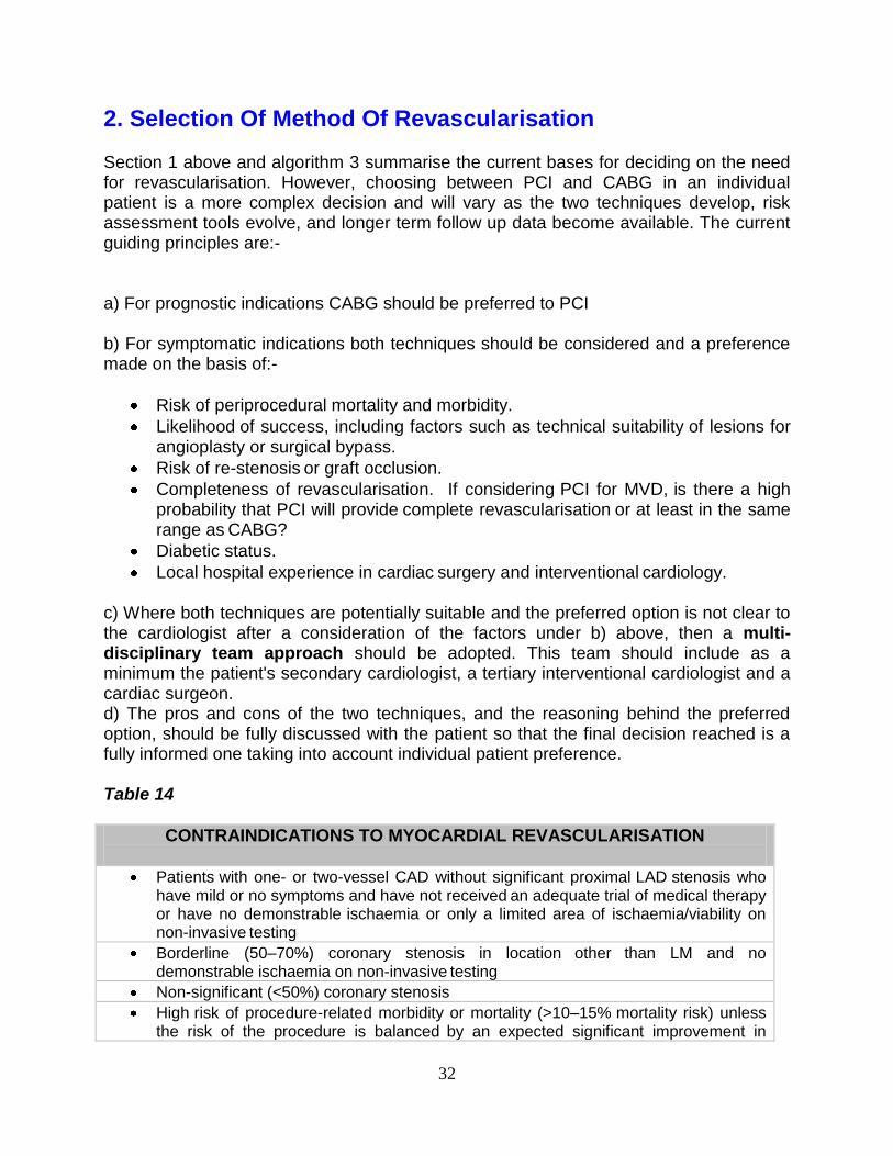
2. Selection Of Method Of Revascularisation Section 1 above and algorithm 3 summarise the current bases for deciding on the need for revascularisation. However, choosing between PCI and CABG in an individual patient is a more complex decision and will vary as the two techniques develop, risk assessment tools evolve, and longer term follow up data become available. The current guiding principles are:- a) For prognostic indications CABG should be preferred to PCI b) For symptomatic indications both techniques should be considered and a preference made on the basis of:-
Risk of periprocedural mortality and morbidity.
Likelihood of success, including factors such as technical suitability of lesions for angioplasty or surgical bypass.
Risk of re-stenosis or graft occlusion.
Completeness of revascularisation. If considering PCI for MVD, is there a high probability that PCI will provide complete revascularisation or at least in the same range as CABG?
Diabetic status.
Local hospital experience in cardiac surgery and interventional cardiology. c) Where both techniques are potentially suitable and the preferred option is not clear to the cardiologist after a consideration of the factors under b) above, then a multi-disciplinary team approach should be adopted. This team should include as a minimum the patient's secondary cardiologist, a tertiary interventional cardiologist and a cardiac surgeon. d) The pros and cons of the two techniques, and the reasoning behind the preferred option, should be fully discussed with the patient so that the final decision reached is a fully informed one taking into account individual patient preference. Table 14
CONTRAINDICATIONS TO MYOCARDIAL REVASCULARISATION
Patients with one- or two-vessel CAD without significant proximal LAD stenosis who have mild or no symptoms and have not received an adequate trial of medical therapy or have no demonstrable ischaemia or only a limited area of ischaemia/viability on non-invasive testing
Borderline (50–70%) coronary stenosis in location other than LM and no demonstrable ischaemia on non-invasive testing
Non-significant (<50%) coronary stenosis
High risk of procedure-related morbidity or mortality (>10–15% mortality risk) unless the risk of the procedure is balanced by an expected significant improvement in

33
survival or the patient's quality-of-life without the procedure is extremely poor
3. Specific Patient and Lesion Subsets
Patients in whom surgical risk is prohibitively high may benefit from revascularisation by PCI, particularly when residual viability can be demonstrated in the myocardium perfused by the target vessel(s).
Although PCI in LM stem disease is feasible, and good results have been achieved in registries comparing DES and bare metal stents, surgery should remain the preferred approach until the outcome of further trials are known.
Subgroup analyses of randomised trials have shown reduced mortality with bypass surgery compared with PCI in diabetic patients with MVD. The BARI trial was the largest of these trials and the only one in which a statistical difference in mortality of patients with diabetes was detected between the treatment groups.
These trials were conducted before the widespread use of DES stents or adjuvant periprocedural antiplatelet therapy but until ongoing trials report, PCI should be used with reservation in diabetics with MVD.
There are no randomised controlled trials comparing treatment options in patients with previous bypass surgery. Re-do surgery may be undertaken on symptomatic grounds where the anatomy is suitable. However, the operative risk of re-do bypass surgery is as high as three-fold greater than initial surgery, and for those with a patent internal mammary artery (IMA) grafts there is the additional risk of damage to this graft during surgery. On the other hand PCI can be performed following previous surgical revascularisation, either in the vein graft or arterial graft, or the native coronary tree beyond the graft which is not revascularised, and may provide a useful alternative to re-do surgery for symptomatic relief.
In the case of a chronic total occlusion that cannot be crossed in patients with MVD, failure to treat the chronic total occlusion will result in incomplete revascularisation, which could be avoided if the patient were to be referred for bypass surgery.
34
STAGE 7
LIFESTYLE AND RISK FACTOR MODIFICATION
CARDIAC REHABILITATION (CR) CR has been defined as "The co-ordinated sum of activities required to influence favourably the underlying cause of cardiovascular disease as well as to provide the best possible physical, mental and social conditions, so that the patients may, by their own efforts, preserve or resume optimal functioning in their community and through improved health behaviour, slow or reverse progression of disease". The British Association of Cardiovascular Prevention and Rehabilitation (BACPR) has just produced the 2nd edition (2012) of its standards and core components which outlines:- A) The benefits of CR
1. Reduces:
• All cause mortality by 11- 26% • Cardiac mortality by 26 – 36% • Morbidity
• Unplanned admissions by 28 -56%
2. Improves:
• Quality of life • Functional capacity
3. Supports:
• Easy return to work • The development of self-management skills
B) The 7 standards of CR The seven standards for cardiac rehabilitation are:
1. The delivery of the seven core components employing an evidence-based approach.
2. An integrated multidisciplinary team consisting of qualified and competent practitioners, led by a clinical coordinator.
3. Identification, referral and recruitment of eligible patient populations.
35
4. Early initial assessment of individual patient needs in each of the core components, ongoing assessment and reassessment upon programme completion.
5. Early provision of a cardiac rehabilitation programme, with a defined pathway of care, which meets the core components and is aligned with patient preference and choice.
6. Registration and submission of data to the National Audit for Cardiac Rehabilitation (NACR).
7. Establishment of a business case including a cardiac rehabilitation budget which meets the full service cost
C) The 7 core components of CR
1. Health behaviour change and education
2. Lifestyle risk factor management
- Physical activity and exercise
- Diet
- Smoking cessation
3. Psychosocial health
4. Medical risk factor management
5. Cardioprotective therapies
6. Long-term management
7. Audit and evaluation The BACPR considers the above 7 core components to be the "The co-ordinated sum of activities" referred to in the definition of CR above. NICE CG126 assessed the clinical/cost effectiveness of CR programmes for patients with stable angina as opposed to the other groups of patients e.g. post-MI, heart failure which currently access CR. They concluded that the "evidence did not indicate benefit for patients from comprehensive cardiac rehabilitation programmes". However they felt "people with angina are likely to need a variety of interventions geared to understanding and coping with their diagnoses and helping them to engage in activities for secondary prevention". NICE guidance recommended that a menu of health needs may need to be addressed and patients should be directed to services they individually require i.e. a "tailored" approach rather than a comprehensive programme. The CMCN Working Group consensus view was that CR was an important component of stable angina management and that is could most efficiently be delivered via the CR services already in place which currently tailor their programmes to individual patient needs. All CR services should continue to use the national data set (NACR audit database) to ensure consistent record keeping of ongoing management and patient
36
outcomes to inform future service development and comprehensive audit.
Priorities for Referral In addition to the NSF requirements of post-MI and post-revascularisation, it is recommended that the following cohorts of patients with stable CAD be offered cardiac rehabilitation in the priority order indicated:
1. All patients newly diagnosed with stable angina.
2. Patients previously diagnosed with stable angina who are experiencing severe
adaptation problems such as issues with activity levels, weight control, adherence to medication etc.
3. Patients waiting for coronary revascularisation who will benefit from input to prepare for the procedure (‘pre-hab’) and afterwards to aid recovery.
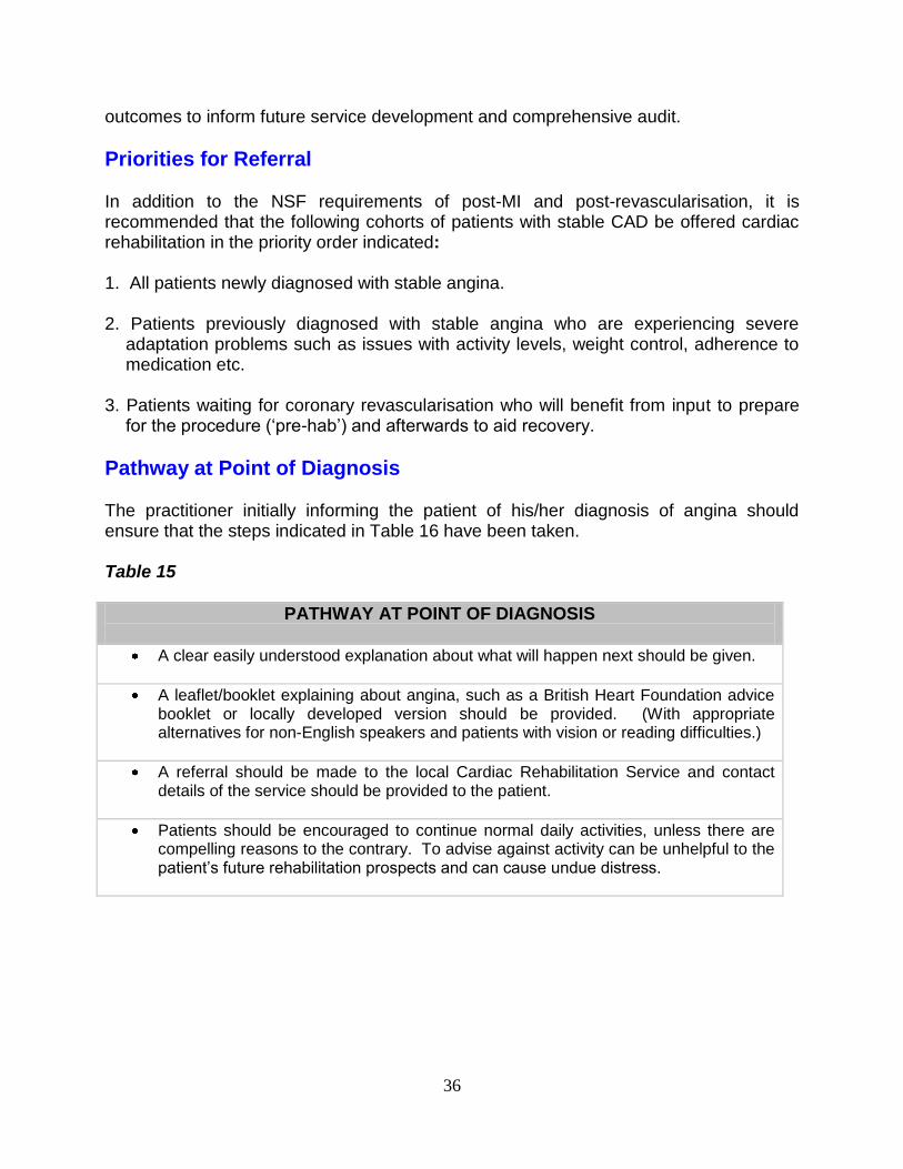
Pathway at Point of Diagnosis The practitioner initially informing the patient of his/her diagnosis of angina should ensure that the steps indicated in Table 16 have been taken. Table 15
PATHWAY AT POINT OF DIAGNOSIS
A clear easily understood explanation about what will happen next should be given.
A leaflet/booklet explaining about angina, such as a British Heart Foundation advice booklet or locally developed version should be provided. (With appropriate alternatives for non-English speakers and patients with vision or reading difficulties.)
A referral should be made to the local Cardiac Rehabilitation Service and contact details of the service should be provided to the patient.
Patients should be encouraged to continue normal daily activities, unless there are compelling reasons to the contrary. To advise against activity can be unhelpful to the patient’s future rehabilitation prospects and can cause undue distress.
37
Choice of Programme for Stable Angina Following this consultation and in discussion with the patient, an agreed decision will be made on the appropriate individualised cardiac rehabilitation pathway to be followed. Outcomes from the consultation may also reveal a need for referral onto specialist services such as smoking cessation, diabetic and psychology.
Comprehensive Rehabilitation Programme The optimal provision for patients with newly diagnosed stable angina is an individualised programme based on the needs identified at the initial consultation and which aligns with BACPR's 7 core components and includes the following:
Lifestyle advice e.g. healthy eating, smoking cessation, weight reduction
Goal setting and targeting
Medication advice to ensure adherence
Stress management
Relaxation
Education about the heart, its function and the disease process.
Individually tailored exercise programme
Monitoring and evaluation of outcomes The principles of adult learning should be adopted in order to improve patients’ understanding. Exercise programmes should include a mixture of warm-up, pulse-raising activities, strength work and cool-down exercises (as outlined in British Association Cardiac Rehabilitation [BACR]/American College of Sports Medicine [ACSM] guidelines). They should be delivered by appropriately trained cardiac rehabilitation practitioners or by suitably qualified exercise professionals (BACR Level IV), working, for example, in leisure centres. There is no current evidence on the most effective length of a supervised exercise programme. The NSF for CHD suggests that cardiac rehabilitation services should aim to provide around two supervised exercise sessions per week for at least six weeks. It is therefore reasonable to expect that angina programmes work towards this level.
Home-Based Programme Where a centre-based comprehensive programme is unavailable or unsuitable, or when a patient decides against this approach due to personal preference, a home-based programme such as The Angina Plan or equivalent locally-devised programme should be provided. For some patients, the greater flexibility and familiarity of a home-based programme will improve their compliance.
38
The patient should be assessed by a cardiac rehabilitation practitioner and a management plan agreed. The plan requires professional review and monitoring through regular contact, usually by telephone. Patients will have a copy of the plan that will be evaluated upon completion. Where patient goals have not been met, a reassessment should take place. The Angina Plan can be delivered by a variety of health professionals who have undergone the required training and assessment.
Serious Pre-existing Psychological Problems Patients identified as having serious or pre-existing psychological problems at assessment should be referred in consultation with their GP to local specialised services. If there is a significant delay between referral and consultation, regular contact should be maintained with the patient by the Cardiac Rehabilitation Service wherever possible.
Continuing Angina Cardiac rehabilitation practitioners should consider referring those patients who continue to have adaptation/coping difficulties, despite intervention from the local service, to the National Refractory Angina Centre (NRAC) for further management including motivational psychotherapy and multidisciplinary cognitive behavioural therapy (CBT). For further details about refractory angina and sample NRAC referral form, see Section 2.
Communication with Primary Care Clear communication with primary care should take place for all patients at the following key points:
Following initial diagnosis.
After initial consultation with cardiac rehabilitation practitioner, outlining the next steps which will have taken account of the patient’s preferences and the services available locally
Following the patient’s completion of the rehabilitation programme.
Long Term Management The patient’s GP practice should ensure that the patient is placed on the heart disease register and offered regular follow-ups. These follow-ups will include checks on blood pressure and cholesterol, weight management and medication review. It is an
39
opportunity to assess angina status and reinforce positive lifestyle choices. In order to help patients maintain the beneficial changes achieved by cardiac rehabilitation, referral to an exercise programme run in the patient’s local community should be made. Usually described as Phase IV cardiac rehabilitation, these programmes are often provided by local authority and voluntary organisations such as heart support groups. Personnel delivering these programmes should be qualified to BACR IV trainer level and should be in regular contact with the local Cardiac Rehabilitation Service. This will help ensure continuity of care.
Staffing Cardiac rehabilitation practitioners will have a variety of professional backgrounds but should be trained and experienced in line with the competencies identified in the NHS Knowledge and Skills Framework. In particular, they should be assessed as possessing the following: Specialist knowledge
Cardiopulmonary anatomy and physiology.
Cardiovascular disease process, major diagnoses, the implications of modifiable risk factors, frequently used drugs and their complications.
Research-based evidence of the impact of environmental, social, lifestyle and behavioural factors on the incidence of CVD and of the impact of CVD on individuals and their families.
Principles and practice of adult learning. Specialist skills
Cardiac risk stratification
Assessment of psychological, social and emotional needs
Assessment of cardiopulmonary capacity
Monitoring cardiovascular and pulmonary responses to exercise
Methods of monitoring to ensure patient safety
REFRACTORY ANGINA
Definition and Epidemiology of Refractory Angina Chronic refractory angina pectoris (CRA) is a clinical diagnosis and may be defined as the presence of symptoms of stable angina due to myocardial ischaemia resulting from advanced CAD which persists despite optimal anti-anginal medication and when revascularisation is unfeasible or where the risks are unjustified. Such patients therefore present with continuing angina which significantly impairs the quality of their lives and which is not responding to optimal anti-anginal drug therapy and is not amenable to any
40
form of coronary intervention such as PCI or CABG. It is a distressing chronic pain condition that causes severe reduction in the quality of life of both patients and their families. Typically such patients complain of a myriad of problems causing repeat and often protracted hospital admissions. These problems can be exacerbated by lack of support and understanding of their condition that in turn adds to their continuing general deterioration. Patients attending a refractory angina clinic are a heterogeneous cohort made up of sub-groups whose angina is refractory to conventional medical therapy for one or more of the following reasons:
PCI and CABG are not technically feasible e.g. because of severe distal disease.
PCI and/or CABG have already been carried out on one or more occasion and further intervention is deemed to carry unjustifiable risks.
Co-morbidity precludes coronary intervention.
Patient choice not to undergo further coronary intervention. The current prevalence of CRA is said to be one in ten thousand and its incidence one in twenty thousand with both rates increasing year on year. The average age is sixty three years and 70% are male. Generally, there is a long history of CAD (>8 years) which is usually three vessel disease (>68%) and often in association with some LV impairment.
Refractory Angina Management at National Refractory Angina Centre (NRAC) Treatment aims and the treatment contract The primary aim of therapy is to maximise the patient’s quality of life by ameliorating the effects of the condition without jeopardising quantity of life. The patient and their carers need time and help to define how angina impairs their quality of life and what level of recovery would be acceptable. These are difficult concepts in the present care system which is a largely pathology or disease-based medical treatment paradigm. The ideal doctor/patient relationship exists when the patient, their carers and doctor can openly 'negotiate' a treatment contract with clearly stated aims and objectives. In this way the choice of therapy becomes simplified for the doctor and it enables the patient to make more rational decisions about which therapy is most appropriate to his/her particular needs.
41
Core Programme Patient Education and Advice
Education and advice to promote both the patient’s and carer’s understanding of their condition and the available treatments is a core component of the NRAC care model in line with internationally accepted best practice guideline recommendations. The recent NICE Clinical guideline on Stable Angina (CG126) has recommended that patients whose angina has not responded to drug treatment and/or revascularisation should be offered comprehensive re-evaluation and advice which may include:
Recommendations and link to evidence Offer people whose stable angina has not responded to drug treatment and/or
revascularisation comprehensive re-evaluation and advice, which may include:
• Exploring the person’s understanding of their condition • Exploring the impact of symptoms on the person’s quality of life • Reviewing the diagnosis and considering non-ischaemic causes of pain • Reviewing drug treatment and considering future drug treatment and
revascularisation options • Acknowledging the limitations of future treatment • Explaining how the person can manage the pain themselves • Specific attention to the role of psychological factors in pain • Development of skills to modify cognitions and behaviours associated with pain
Therapeutic options for pain management
A broad menu of available therapeutic options has been developed for pain management. However NICE CG 126 considered the evidence for and cost-effectiveness of a number of these interventions and concluded that transcutaneous electrical nerve stimulation (TENS), enhanced external counter pulsation (EECP), and acupuncture should NOT be offered.
The following were not assessed by NICE and should still be considered :-
- Temporary sympathectomy – This is carried out using local anaesthetic infiltrated around the left stellate ganglion at level C5/6 of the cervical vertebra and in the para-vertebral region of T3/4. Usually stellate blockade is tried first with three attempts at four to six week intervals. This is the average period of remission gained by a
42
successful blockade. If this fails then a series of three para-vertebral blocks again at four to six week intervals is used.
- Opioids – Although there is limited evidence for the effectiveness of
opioids in CRA, in clinical practice oral and transdermal opioids can be effective. Furthermore, epidural or intrathecal opioids are sometimes beneficial.
- Spinal Cord Stimulation – This is a form of neuro-modulation in which
suppression of pain is induced by application of an electronic device to modify nervous system function. The underlying principles are based on the gate theory of pain control i.e. that non–destructive stimuli can interfere with the transmission of pain within the central nervous system and thereby prevent the pain messages from reaching the brain.
- Intrathecal pumps – These are used for the suppression of pain by
the infusion of prescribed medication such as hydromorphone directly into the intrathecal space. However, dose related side effects are common and many patients find these intolerable.
Psychological Support Modalities
Group cognitive behavioural therapy (CBT) – This format of CBT is felt to optimise patient empowerment and is designed to address a number of issues including demystifying angina; graduated exercise using goal setting and pacing; relaxation and stress management; optimization of medication; lifestyle modification; understanding therapeutic alternatives and dealing with setbacks.
Motivational psychotherapy – Patients in whom an appropriate psychological
assessment tool e.g. the Hospital Anxiety and Depression (HAD) score indicates the need, should undergo professional psychological assessment in order to decide whether formal psychotherapy might be beneficial.
Counselling – This enables a realistic and achievable “treatment contract” to be
decided upon so that the patient, their family and the clinical treatment team have an agreed objective.
Patient Referral Currently, access is limited to referral from consultants within secondary and tertiary cardiological care. It is hoped that access will be expanded in the future to include referral from GPs. Direct referral by letter is acceptable providing that adequate details are given including cardiac history, clinical features, relevant investigation results, risk factors and current treatment.
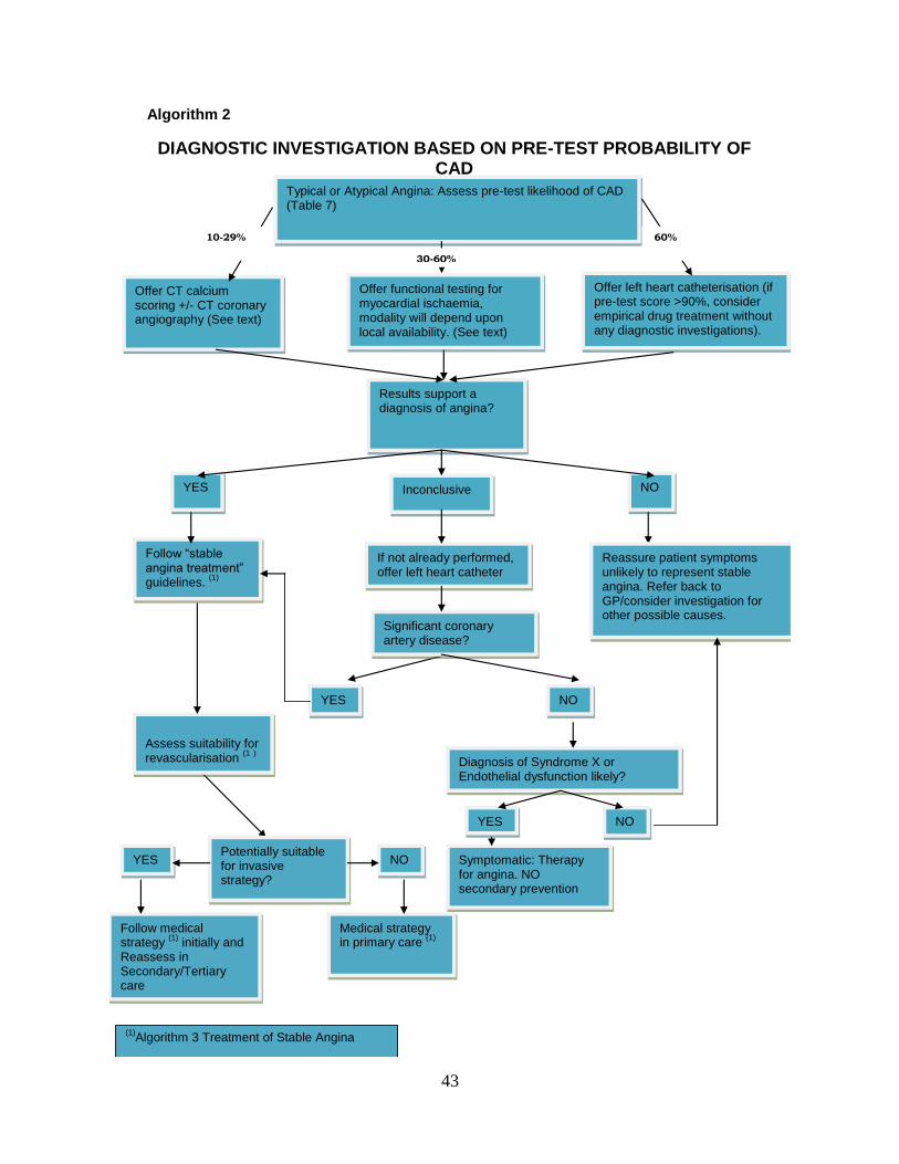
43
Algorithm 2
DIAGNOSTIC INVESTIGATION BASED ON PRE-TEST PROBABILITY OF CAD
Offer left heart catheterisation (if pre-test score >90%, consider empirical drug treatment without any diagnostic investigations).
Typical or Atypical Angina: Assess pre-test likelihood of CAD (Table 7)
Follow “stable angina treatment” guidelines.
(1)
Offer CT calcium scoring +/- CT coronary angiography (See text)
Results support a diagnosis of angina?
10-29%
Offer functional testing for myocardial ischaemia, modality will depend upon local availability. (See text)
YES Inconclusive NO
60%
If not already performed, offer left heart catheter
Reassure patient symptoms unlikely to represent stable angina. Refer back to GP/consider investigation for other possible causes.
Significant coronary artery disease?
Assess suitability for revascularisation
(1 )
YES NO
Diagnosis of Syndrome X or Endothelial dysfunction likely?
YES NO
YES Potentially suitable for invasive strategy?
NO Symptomatic: Therapy for angina. NO secondary prevention
Follow medical strategy
(1) initially and
Reassess in Secondary/Tertiary care
Medical strategy in primary care
(1)
(1)Algorithm 3 Treatment of Stable Angina
30-60%
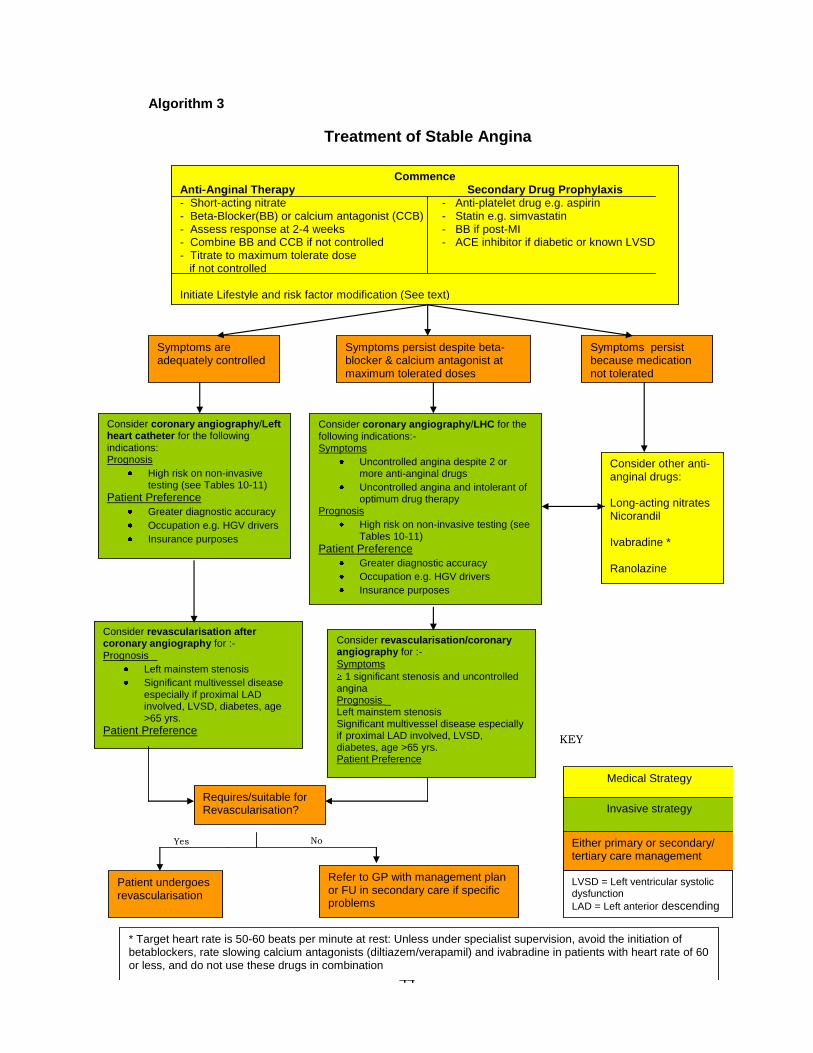
44
Algorithm 3
Treatment of Stable Angina
Commence Anti-Anginal Therapy Secondary Drug Prophylaxis - Short-acting nitrate - Anti-platelet drug e.g. aspirin - Beta-Blocker(BB) or calcium antagonist (CCB) - Statin e.g. simvastatin - Assess response at 2-4 weeks - BB if post-MI - Combine BB and CCB if not controlled - ACE inhibitor if diabetic or known LVSD - Titrate to maximum tolerate dose if not controlled Initiate Lifestyle and risk factor modification (See text)
Symptoms are adequately controlled
Symptoms persist despite beta-blocker & calcium antagonist at maximum tolerated doses
Symptoms persist because medication not tolerated
Consider coronary angiography/Left heart catheter for the following
indications: Prognosis
High risk on non-invasive testing (see Tables 10-11)
Patient Preference
Greater diagnostic accuracy
Occupation e.g. HGV drivers
Insurance purposes
Consider other anti-anginal drugs: Long-acting nitrates Nicorandil Ivabradine * Ranolazine
Refer to GP with management plan or FU in secondary care if specific problems
Patient undergoes revascularisation
Requires/suitable for Revascularisation?
Yes No
KEY
Medical Strategy
Invasive strategy
Either primary or secondary/ tertiary care management
* Target heart rate is 50-60 beats per minute at rest: Unless under specialist supervision, avoid the initiation of betablockers, rate slowing calcium antagonists (diltiazem/verapamil) and ivabradine in patients with heart rate of 60 or less, and do not use these drugs in combination
Consider revascularisation after coronary angiography for :-
Prognosis _
Left mainstem stenosis
Significant multivessel disease especially if proximal LAD involved, LVSD, diabetes, age >65 yrs.
Patient Preference
Consider coronary angiography/LHC for the
following indications:- Symptoms
Uncontrolled angina despite 2 or more anti-anginal drugs
Uncontrolled angina and intolerant of optimum drug therapy
Prognosis
High risk on non-invasive testing (see Tables 10-11)
Patient Preference
Greater diagnostic accuracy
Occupation e.g. HGV drivers
Insurance purposes
Consider revascularisation/coronary angiography for :-
Symptoms
1 significant stenosis and uncontrolled angina Prognosis _ Left mainstem stenosis Significant multivessel disease especially if proximal LAD involved, LVSD, diabetes, age >65 yrs. Patient Preference
LVSD = Left ventricular systolic dysfunction
LAD = Left anterior descending coronary artery