Embed Size (px)
Citation preview
REVIEW
Gut microbiota and colorectal cancer
R. Gao1 & Z. Gao1 & L. Huang1 & H. Qin1
Received: 21 September 2016 /Accepted: 14 December 2016 /Published online: 7 January 2017# The Author(s) 2017. This article is published with open access at Springerlink.com
Abstract The gut microbiota is considered as a forgotten or-gan in human health and disease. It maintains gut homeostasisby various complex mechanisms. However, disruption of thegut microbiota has been confirmed to be related to gastroin-testinal diseases such as colorectal cancer, as well as remoteorgans in many studies. Colorectal cancer is a multi-factorialand multi-stage involved disorder. The role for microorgan-isms that initiate and facilitate the process of colorectal cancerhas become clear. The candidate pathogens have been identi-fied by culture and next sequencing technology. Persuasivemodels have also been proposed to illustrate the complicatedand dynamic time and spatial change in the carcinogenesis.Related key molecules have also been investigated to demon-strate the pathways crucial for the development of colorectalcancer. In addition, risk factors that contribute to the tumori-genesis can also be modulated to decrease the susceptibilityfor certain population. In addition, the results of basic studieshave also translated to clinical application, which displayed acritical value for the diagnosis and therapy of colorectal can-cer. In this review, we not only emphasize the exploration ofthe mechanisms, but also potential clinical practice implica-tion in this microbiota era.
Introduction
Colorectal cancer (CRC) has been a common malignancy inthe world, especially in China, in recent years. According tothe epidemiological data, the 5-year prevalence proportion hasreached 74.6 and 58.3 per 100,000 in men and women respec-tively [1]. An updated estimation reveals that more than 376,000 new cases of CRC and 191,000 deaths occur every year inChina [2]. CRC has long been investigated and it is classifiedinto two typical types: colitis-associated colorectal cancer(CAC) and sporadic colorectal cancer (SCC), according togenomic mutation diversity. Hereditary syndrome has beenidentified with a total of fourteen mutations [3]. The innerinvolved signal pathways are totally different between thesetwo relatively independent phenotypes, but they also share afew sequential genetic mutations. CAC is always associatedwith inflammatory bowel disease, an inflamed disorder phe-notype in the young population. SCC is usually used to referto the common colorectal cancer that considered without fam-ily heredity. CRC is a malignant disease which involves mul-tiple factors during its multi-stage development. The initiatingevents of CRC have been proved to be APC mutation in SCCand TP53 mutation in CAC. The etiology of CRC has beeninvestigated using large cohorts and confirmed by animalmodels, and the consensus conclusion contains genetic back-ground and environmental risk factors such as diabetes, cho-lecystectomy, obesity, high fat diet, and processed and redmeat [4–9]. However, a large number of studies have recentlyreported that the gut microbiota may also participate as anessential contributor factor in the initiation and developmentof CRC. Here, we will focus on the potentially plausible rela-tionship between gut microbiota and colorectal cancer.
R. Gao, Z. Gao, and L. Huang contributed equally to this work.
* H. [email protected]
1 Tongji University School of Medicine affiliated Tenth People’sHospital, No.301 Middle Yanchang Road, Shanghai 200072, China
Eur J Clin Microbiol Infect Dis (2017) 36:757–769DOI 10.1007/s10096-016-2881-8
Overview of microbiota in the gut
The gut contains a complicated environment that is settled bybacteria, fungi, and viruses. The total number may reach 100trillion, and the number of microbe cells is estimated to be 10-fold more than the human cells. This densely resident micro-bial community consistently communicates with the host andalso enhances the epithelial defense against pathogens, accel-erates the maturity of the immune system, and absorbs thenutrition from ingested foods [10, 11]. Despite the mucuslayer, which consists of various macromolecules and secretedantimicrobial molecular and intercellular tight connection pro-teins, the gut microbiota also possess the capacity to defendpathogens by inducing IgG antibodies through recognition oftheir conserved antigen part of gram-negative bacteria [12,13]. The gut microbiota not only protect the local homeostasis,but also mediate the related organ. For example, an in-vivoexperiment proved that the gut microbiota was manipulatedby intestinal lectins to decrease alcohol-associatedsteatohepatitis [14]. Along with the evolution of gut microbi-ota, body cells also demonstrate effective pathways foravoiding the pathogen infection. Salmonella Typhi is a well-known pathogen that once caused great damage to humanhealth. Recently, Spano and his colleagues proposed a neo-mechanism, Rab32-dependent cell autonomous antimicrobialapproach, which was critical for the host to restrict SalmonellaTyphi infection [15]. Gut microbiota residing in infants is de-rived from the obstetric canal and mother’s skin after birth,then becomes matured and keeps relatively stable during along time of lifespan and changes in the elderly time.Despite age, a variety of factors such as diet, drugs, sports,and genotype also impact the gut microbial community[16–20]. In a healthy gut, the dominant core bacteria are com-posed of Firmucutes, Proteobacteria, Bacteroidetes andActinobacteria at the phylum level. However, the gut micro-community displays a diverse structure at the genus and spe-cies levels.
Gut microbiota and colorectal cancer
With the strong evidence displayed by multi-direction proofs,the stomach cancer associated pathogenetic bacterium(Helicobactor pylori) has been identified and recognized in-ternationally as the level one carcinogen. Likewise, with amore complicated microbial community covering the innersurface of the colon, this earlier discovery enlightens the re-searchers to seek for a similar pathogen to explain the initia-tion and development of CRC. To explore the possible role ofmicrobiota in the etiology of CRC, the researchers first sepa-rate and culture several bacteria in various media. However,less supportive evidence can illustrate the role of microbiota inthe CRC development. Along with an improvement in detec-tion technology, more and more studies utilize the next
sequencing technology to explore the candidatecarcinogenetic pathogen in the gut among population withdistinct differing disease phenotypes.
The first report that links the gut microbiota with CRC ispublished by Weisburger and his colleagues [21]. Later, moreand more studies confirm the relationships between pathoge-netic bacteria and colorectal cancer. For example, infectionwith Streptococcus bovis, a group of gram-positive cocci,has been reported to be a risky sign for colon tumors [22].Kostic and his team identify high enrichment of Fusobacteriasequence in colorectal carcinoma tissue using whole genomesequencing, and confirm the result in a large scale study ofcolorectal cancer t issue samples [23]. Similarly,Enterotoxigenic Bacteroides fragilis and Fusobacteriumnucletum are identified to be highly expressed in colorectalcancer tissue compared to the matched tissue, andFusobacterium nucletum is proved to be associated with highmicrosatellite instability [24]. Our previous study also iden-tifies a discrepancy in tissue-associated gut microbiota be-tween colorectal cancer patients and healthy volunteers [25].In addition, mucosa-associated E.coli belonging to the B2phylogroup is found to be more prevalent in CRC tissues,and is identified to encode cyclomodulin which is vital forcolon epithelia cell mutation [26].To explore subsequently,Fusobacterium nucleatum, which belongs to Fusobacteria,has been isolated from tumor tissue and proved to be invasivein the in-vitro experiments. In addition, Fusobacteriumnucleatum also has a positive correlation with lymph nodemetastasis in CRC [27].Furthermore, Zhao and his colleaguesstudy the stool samples of CRC patients in China, and foundfind that Bacteroides fragilis, Enterococcus, Escherichia/Shigella, Klebsiella, Streptococcus, and Peptostreptococcusdisplay a higher relative abundance in CRC patients, whileRoseburia- and Lachnospiraceae-related OTUs dominat ahigh load in the healthy controls [28]. In another study, re-searchers also compare stool samples and find that the CRCpatients have a lower microbiota diversity and Clostridiaabundance, but a high abundance of Fusobacterium andPorphyromonas at genus level [29]. The lumen and tissuemicrobiota are obviously different in microbial structure. Inthe t issue samples , benef ic ia l microbes such asBifidobacterium, Faecalibacterium, and Blautia were are sig-nificantly reduced, while Fusobacterium is enriched in theCRC patients [30]. However, the stool samples show a signif-icant different microbial landscape with Paraprevotella,Eubacterium, and several other bacteria enriched in CRC pa-tients [30]. Inflammation is also an important factor that con-tributed to CRC progress via gut microbiota. Arthur finds thatE.coli NC101 will increase the colon tumor load in AOMtreated IL10-/- mice. When he deletes the polyketide synthase(pks) Genotoxic Island in E.coliNC101, a significant decreaseof tumor load and invasion capacity are observed [31].Clinical study also revealeds a close connection between
758 Eur J Clin Microbiol Infect Dis (2017) 36:757–769
E.coli and advanced stages, and animal experiment shows ahigh tumor load under incubation with E.coli [32, 33]. Tobetter understand adenoma-carcinoma sequence-related gutmicrobiota and functional genes, sequential continuous detec-tion is performed in the stool samples. By metagenomic anal-ysis, the researchers find that a total of 130,000 genes aredifferent in any two-group comparison among the healthy,adenoma, and CRC patients [34]. And further analysis includ-ing the diet pattern concludes that fruit and vegetable con-sumption are related to the healthy group, while high levelof red meat consumption and C-reation protein are associatedwith the carcinoma phenotype. In addition to these find-ings, the study also show that sugar transporter and acouple of amino acids consist of histidine, lysine, methio-nine, cysteine, and leucine are enriched in the healthywhen compared with adenoma or in adenoma in compar-ison with carcinoma patients. Despite the stool microbiotachange, the architecture of gut microbiota is also altered inthe tissue samples by the sequencing. By 16S ribosomalRNA sequencing, researchers identify that Fusobacterium,Parvimonas, Gemella, and Leptotrichia are enriched andanti-inflammatory F. prausnitzii loses its abundance inearly-stage colorectal cancer [35]. Furthermore, currentstudies also demonstrates that the Fusobacterium nucletumis strongly associated with CpG island methylator pheno-type [36]. A recent study explores the gut microbiota inmatched tissue and stool samples, host genes, and immunesystem together. The results show that firstly the fecalmicrobiota only has partial similarity with the tissuemicrobiota. Then a new cluster set is proposed and namedco-abundance groups (CAGs) which is similar toenterotypes, and identified decreased Bacteroidetes cluster1 and Firmicutes cluster 1, also cluster 2 of Bacteroidetesand Firmicutes as well as pathogen and Prevotella clusterin the colorectal cancer tissue community [37]. The studyalso identifies that CAGs are also associated with humanimmune responses such as IL17a, myc, and STAT3.
To illustrate the relationships between CRC and gut micro-biota, several typical rodent models which simulate the CRCdevelopment are also performed. In a dimethylhydrazine-induced model, a obvious separated lumen gut microbiota isobserved [38]. APCmin/+ mice raised in a germ-free environ-ment display a reduced tumor load after that in the SPF con-dition [39]. When the germ-free mice are delivered with gutmicrobiota from tumor burden mice, they display more andlarger tumors. To verify that the increased tumor burden thatappears in germ-free mice are derived from the harmful mi-crobiota, antibiotics are applied to the receptor mice which, asa result, did slow down the carcinogenesis process [40]. Theseexperiments show us the critical role of gut microbiota incolorectal cancer and also plausible causality of gut microbi-ota for the rodent models. However, gut microbiota in therodent models differ significantly from the human beings, so
it is not certain whether the same ideal results will re-emerge in the human-derived gut microbiota. Nielson andhis partners transplant human donor stool into the mice,and results show that the tumor burden is apparently asso-ciated with the gut microbiota structure at baseline in thegerm-free mice [41]. These results sufficiently confirm thatdysbiosis in the gut is one of the reasons that caused colo-rectal cancer.
Pathogen identification by the immune system
When ingested microorganisms reach the gut, it is of vitalimportance for the immune system to identify them to protectthe host (Fig.1). Currently, several receptors have been recog-nized to mediate the process. The host possesses immuneinnate receptors named pattern recognition receptors (PRRs)to search the pathogen-associated molecular patterns(PAMPs) that expressed in the pathogens. We will describeand discuss these receptor-pathogen interactions based onachievements so far.
Toll-like receptors
Among the PRRs, the Toll-like receptors (TLRs) are studiedearliest to detect the signal from pathogens. The nature ofTLRs is type I membrane glycoprotein, which belongs to asuperfamily that includes interleukin-1 receptors. The struc-ture of membrane varying between them discriminate theTLRs and IL-1R. TLRs do not only locate in the membraneof epithelia cells (TLR1, TLR2, TLR4, TLR5, TLR6), theyalso express in the endosome membrane (TLR3, TLR7,TLR8, TLR9, TLR11, TLR13) [42].Pathogens always pro-duce lipoproteins to induce the host immune response, triggermonocyte apoptosis, and activate the NF-κB signal pathwayvia TLR-2 [43]. TLR-2 also acts as a mediator to promote thecell activation by peptidoglycan and recruited by macro-phages to recognize the pathogens in in-vivo experiments[44, 45]. Mammalian TLR-3 is identified to recognize thedouble-stranded RNAwhich is associated with viral infection[46]. Study on TLR-4 has proved that this protein is involvedin the cooperator for CD14 which would activate thelipopolysaccharide-induced NF-κB signaling [47]. By geneknockout mice model, TLR-3 and TLR-4 have been identifiedto function normally with an essential factor Toll/IL-1 receptordomain-containing adaptor [48]. While TLR-7 and TLR-8mediate the recognition of species-specific single-strandedRNA from virus [49, 50]. TLR-9 has been proved to helpthe cellular response to CpG DNA of infectious pathogens[51]. The TLRs has an asymmetrical distribution in humanperipheral blood mononuclear cells. According to Hornung’swork, TLR-1 and TLR-6 expresse in all the cells includingplasmacytoid dendritic cells (PDC), B cells, NK cells, T cellsand also monocytes [52]. TLR-2 is highly expressed in
Eur J Clin Microbiol Infect Dis (2017) 36:757–769 759
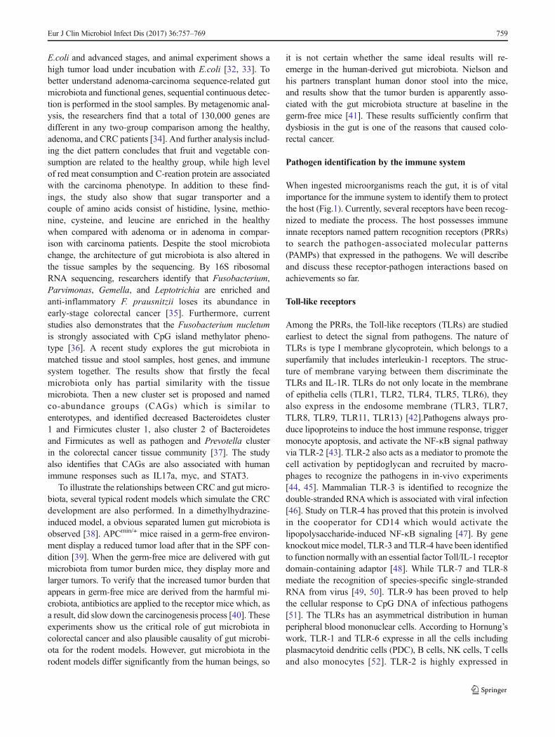
monocytes. TLR-3 has a relative high expression in NKcells and all low in other cells. TLR-4 expresses high inmonocytes. TLR-6 is mainly detected in B cells, but canalso be detected in NK cells, monocytes, and PDC. TLR-7is moderately expressed in PDC and B cells. Detectedexpression of TLR-8 is only high in monocytes. TLR-9is almost three-fold more highly expressed in PDC than inB cells. However, TLR-10 is expressed highly only in Bcells and PDC; the other cells are relatively low. All TLRs
activated the MyD-88 dependent pathways to induce thedownstream immune responses [53].
To better understand the mechanism of toll-like receptorsignaling, Akira summarizes the pathways that are involvedin detail to show us a more visible landscape [54]. In conclu-sion, after binding to the TLR/IL-1R, signals pass down totrigger a cascade. The essential molecules included during thisprocess may include adaptor molecule myeloid differentiationprimary-response protein 88 (MyD88), transforming growth
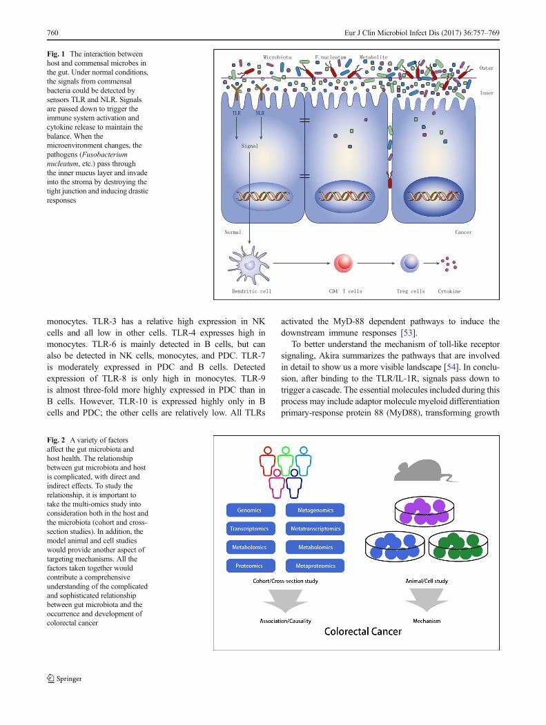
Fig. 2 A variety of factorsaffect the gut microbiota andhost health. The relationshipbetween gut microbiota and hostis complicated, with direct andindirect effects. To study therelationship, it is important totake the multi-omics study intoconsideration both in the host andthe microbiota (cohort and cross-section studies). In addition, themodel animal and cell studieswould provide another aspect oftargeting mechanisms. All thefactors taken together wouldcontribute a comprehensiveunderstanding of the complicatedand sophisticated relationshipbetween gut microbiota and theoccurrence and development ofcolorectal cancer
Fig. 1 The interaction betweenhost and commensal microbes inthe gut. Under normal conditions,the signals from commensalbacteria could be detected bysensors TLR and NLR. Signalsare passed down to trigger theimmune system activation andcytokine release to maintain thebalance. When themicroenvironment changes, thepathogens (Fusobacteriumnucleatum, etc.) pass throughthe inner mucus layer and invadeinto the stroma by destroying thetight junction and inducing drasticresponses
760 Eur J Clin Microbiol Infect Dis (2017) 36:757–769
factor-β activated kinase, and tumour-necrosis factorreceptor-associated factor 6 [54]. Despite the direct killingfunctions, the activated TLRs in the macrophages also inducethe increased expression of vitamin D receptor, and thus facil-itate the function of antimicrobial peptide on mycobacteriumtuberculosis [55].
NOD-like receptors
The innate immune system provides a rapid response to thepathogens without a memory process. Such a process relies onthe perception of conserved microbial motifs that namedPAMPs as described before. After acquiring the signals, thehost launches a series of defensive mechanisms against thepathogens. Apart from the TLRs in the cell membrane, anoth-er defense system, NOD-like receptors, has also been identi-fied by Shigella flexneri infection [56]. NOD stands fornucleotide-binding oligomerization domain inside the cells.The NOD-like receptors (NLRs) are essential for defensivearchitecture against invasive bacteria and the bacteria productsin the inner space of cells. In addition, the NLRs also showpenetrating sensor of the Bdanger signals^ from injured ordying cells. The already known NLRs include NOD1-5,NALP1-14, CII TA, Ipaf, and Naip, which constitute a bigfamily [57]. Among them, the first three proteins have beeninvestigated and are well understood. The structure of NLRsconsist of various domains such as leucine-rich repeat domainin the C-terminal, nucleotide-binding domain in the centerpart, and a protein–protein interaction domain in the N-terminal [57]. According to the N-terminal domains, NLRshave been clustered into three groups: caspase recruitmentdomain containing NODs, pyrin containing NALPs, andbaculovirus inhibitor repeat containing NAIPs [58]. TheseNLRs sense the PAMPs and subsequently trigger conforma-tional rearrangements to conduct the signal spread and finallyactivate the diverse signal pathways. For example, Salmonellahas shown the ability to inhibit the expression of NLR FamilyCARD domain containing protein 4, thus not only decreasingthe secretion of IL-1b, but also preventing the apoptosis of Bcells, maintaining the ideal niche for its persistence and repro-duction [59].
NOD1 can recognize the g-D-glutamyl-meso-dia-minopimelic acid which exists in Gram-positive and Gram-negative bacteria. It prevents the candidate pathogens by in-nate immune functions. In addition to that, the NOD1 alsoshows the potential possibility to hamper the process fromcolon inflammation to tumorigenesis. NOD2 has been showedto respond to a wide variety of bacteria. The inside type IIIsecretion system cooperates with this function of Salmonella.Knockout ofNOD2 gene in the mice displays an increased gutload of bacteria, decreased capacity to prevent the coloniza-tion of pathogens and also damaged crypt function. The mu-tation of NOD2 also triggers inflammatory diseases [60].
Double knockout of NOD1 and NOD2 causes increased gutpermeability, decreased expression of E-cadherin, and im-paired antimicrobial function in the C57BL/6 mice model[61]. NOD2 holds great importance for the balance in theintestinal microbiota [62]. Nevertheless, a recent study showsthat though NOD1-deficient mice has a weaker epithelial bar-rier in the gut, the microbiota composition does not change.The NOD2-deficient-mice show more interesting results,among which no significant alterations are observed in im-mune damage and microbiota profile [63]. In addition, themicrobiota change is associated with the housing conditionsof the mice. This result needs to be confirmed by more similarexperiments.
The other NLRs involved mechanisms also have beenoverviewed and summarized with the target of NLRP3,NLRC4, NAIP, and NLRP1 [64]. It was revealed that mito-chondria plays an important role in the activation of NLRP3inflammasome. The mitochondria provides a convenient plat-form for NLPR3 to assemble, and also effectors such as amitochondrial reactive oxygen species which derived fromitself to activate the immune process. Another two regulators,guanylate-binding protein 5 and double-stranded RNA-de-pendent protein kinase, may also contribute to this process.The NLRC4 inflammasome activates the bacteria secretionsystem, type III and IV, through detecting the bacteria pro-teins. NAIPs will be activated by binding to the bacteria fla-gellin or type III system. Lethal toxin may be the activator forNLRP1 inflammasome, and it has also been shown thatNLRP1 is associated with the viral immune responses andproteolytic function [64]. NRPL10 has been shown to be cru-cial to maintain adaptive immunity, and NRPL12 knockoutmice show a susceptibility to colitis and colon tumorgenesis,which display its important role in the gut homeostasis [65,66].
Hypothesis models associated with colorectal cancer
To better understand the role of gut microbiota in the initiationand progression of colorectal cancer, a few hypotheses areraised by the researchers. The driver–passenger model is setin an totally different respect, with the intention of illustratingthe various roles of commensal bacteria in the development ofcolorectal cancer [67]. This model classifies microbes into twodifferent groups, and shows that the driver microorganismscause DNA damage in epithelial cells which may start theprogression of CRC in the first-time spatial location, thenthe tumor microenviroment subsequently changes to favorthe blooming of passenger bacteria which may dominate inthe tumor site later. This model highlights the point that al-though the driver bacteria initiate colorectal cancer, these mi-croorganisms will not always exist as a marker, resemblinggenetic mutation, in the surroundings of tumor cells, but willdisappear from the cancerous tissue as a loss of growth
Eur J Clin Microbiol Infect Dis (2017) 36:757–769 761
advantage. This model may be effective to illustrate the dis-crepancy among various results in different studies, but it alsoincreases the challenges regarding what we can do to clarifythis ambiguous relationship, and how.
The other model proposed is the keystone hypothesis [68].This model declares that a keystone pathogen is defined as amicroorganism that supporting the disease-associated dysbioticmicrobiota. The microorganism may display a relatively lowabundance in the ecosystem. Here, this theory no longer em-phasizes the strength level in the disease-related microbiota, butthe functions that contribute to and maintain the imbalancedstate. The base of this hypothesis is the pathogenesis ofPorphyromonas gingivalis and Periodontitis. In a model, thePorphyromonas gingivalis could still induce periodontitis evenat very low abundance (less than 1%) in the whole community.In addition, the accompanying microbiota also change to firethe inflammation. If the pathogen is erased in a condition, noperiodontitis would occur even with the same commensal bac-teria. With a similar thought process,Klebsiella pneumonia andProteus mirabilis could be treated as the keystone pathogens ininflammatory bowel disease, as well as the role of enterotoxi-genic Bacteroides fragilis in colon cancer. This hypothesis pro-vides a new insight for us to reconsider the potential role of gutpathogens in the initiation and development of related disor-ders. However, logistic identification is still required to confirmthis notion with more model experiments.
Garrett and his colleagues propose their theoreticalmodels of microbe or microbial community carcinogenesison colorectal cancer [69]. One model is the specific micro-organism, the second model is the microbial community, andthe last is the sequential collaboration by the single andcommunity.
Since current studies have identified several candidatepathogenic bacteria that have a close relationship with colo-rectal cancer, the involved mechanisms also need to be inves-tigated. The first model is well understood and has been in-vestigated by many researchers. The community model maywell explain the current understandings in inflammatory bow-el disease.
Carcinogenesis mechanisms of candidate species
The clinical abnormal distribution of microbiota in colorectalcancer and matched normal tissue has been described in theformer context. Here, we will discuss mainly the mechanismof main candidate species based on current results.
Fusobacterium nucleatum
Studies of the relationship between Fusobacterium nucleatumand colorectal cancer has been reported extensively. Currently,the researchers have started to focus on the potential mecha-nisms of the pathogen. A recent study reveals that CRC cell
proliferation is stimulated through FadA binding to E-cadherin expressed in the CRC cells including HCT116,DLD1, SW480 and HT29 [70]. The APCmin/+ mice modeldemonstrates that more colon tumors exist in mice fed withFusobacterium nucletum in comparison with streptococcus[71]. In addition, Fusobacterium nucletum does not inducecolitis or accelerate the colitis-associated cancer. However, itis able to recruit the immune cells and hence provides a pro-inflammatory microenvironment for the initiation and devel-opment of colorectal cancer. Isolation from the inflamed tissueshows that the Fusobacterium nucletum diaplays a high inva-sive ability and evokes a high expression of MUC2, as well astumor necrosis factor alpha in both in-vivo and in-vitro studies[72]. Study has also shown that Fusobacterium nucleatuminhibits the NK cell function by binding to the inhibitory re-ceptor TIGIT via its Fap2 protein [73]. A trap which needs tobe avoided here is that not all the isolated Fusobacteriumnucletum exerts the same pathogenic ability. In fact, basedon current evidence, isolates of Fusobacterium nucletum fromthe inflamed parts are more invasive than the normal tissueeither from the IBD patients or the healthy controls [72, 74].The mechanisms may be due to the copy number variationamong the bacteria strains [75].
ETBF
Cumulative evidence has proposed a gut pathogenic bacteria-enterotoxigenic Bacteroides Fragilis (ETBF), which encodedB. fragilis metalloprotease toxin (BFT) to induce diarrhea inmost reports, as a carcinogenic bacteria in colorectal cancer[76]. The clinical proofs that point out the association withcolorectal cancer have been described before. Previous knowl-edge on the function of ETBF is mainly focused on its poten-tial to remodel the epithelial cytoskeleton and F-actin structureby targeting the E-cadherin [77]. Here, we will mainly discussthe involved mechanisms, based on several current discover-ies. ETBF has been shown to trigger colitis and colonictumors in multiple intestinal neoplasia mice [78–80]. Thisbacteria will induce activation of transcription-3 (Stat3)pathway with characterization of T helper type 17 response[81]. In addition, BFT has been demonstrated to trigger cellproliferation and activate c-Myc expression in vitro, as wellas increase the polyamine metabolism and induce DNAdamage [82, 83].
E.coli
Escherichia coli (E.coli), a Gram-negative, anaerobic com-mensal bacteria, is common in the gut microenvironment.Several studies have linked the E.coli with colorectal cancerrisk. Nevertheless, the involved mechanism is still unknown.Clinical study shows that cyclomodulin-producing E. coli col-onizes in most cancerous samples [26, 84]. Isolates of E.coli
762 Eur J Clin Microbiol Infect Dis (2017) 36:757–769
from colon cancer tissue show adherence and invasive poten-tial to a specific cell line and also induce interleukin-8 expres-sion [85, 86]. Currently, the pks island containing E.coli hasbeen shown to express Colibactin gene and induce DNA dam-age, chromosome aberrations, as well as increased gene mu-tations in vivo [87]. The increased gene mutation may due tothe depletion of the DNA mismatch repair system related tothe effector protein of E.coli [88]. A colibactin warhead thatdirectly binds to the duplex DNAwith spirobicyclic structureprovided more clear evidence for its potential role with regardto carcinogenesis [89]. The pks positive E.coli is first isolatedfrom inflammatory disease; however, a recent finding alsoidentifies that the E.coli also contained this pathogenic island[90]. This finding may provide new evidence that links in-flammatory disorders with colorectal cancer. In addition, in-vivo study also demonstrates that enteropathogenicEscherichia coli promotes cancer cell survival by inductionof macrophage inhibitory cytokine 1, thus activating thetransforming growth factor β-activated kinase 1 and RhoAGTPase following pathogen infection, and also the continuousexpression of COX-2 [91, 92]. When the Caco-2 cell is co-cultured with E.coli in vivo, genes that correlate with the ox-idation stress are up-expressed, suggesting a defensive re-sponse which may be due to the changed microenvironmentin the system [93]. Other candidate carcinogenetic microor-ganisms also include streptococcus bovis, H. pylori, andClostridium, which have been described in detail elsewhere[21].
Alteration of gut microbiota may also induce cytokine im-balance. The IL-17 family from the Th17 cell has been foundto be closely associated with colorectal cancer. IL-17A, IL-17 F, and IL-22 are identified to promote the tumorigenesis ofcolorectal cancer in early studies. Then IL-17C is also provedto be required in the formation of colorectal cancer in geneknockdown mice model [94]. The detailed mechanism maydue to the effect of prolonged epithelial cells by induction ofBCL-xL and BCL-2 expression [94].
Other studies of the mechanism show that activating ofEGFR-MAPK pathway is also a risk path in colon cancerprogress [95]. The ingested alcohol is transformed to highlevel of acetaldehyde in the colon, but this effect is preventedwith antibiotics that target the gut microbiota [96]. The exper-iment suggests that the gut microbiota promote colon cancerby targeting the metabolites. Short-chain fatty acids decreasein colorectal cancer patients, but if the concentration is in-creased, it stimulates the epithelia metabolism, decreases theintracellular O2 load, and protectes the tight barrier function[97].
Beneficial effects of Faecalibacterium prausnitzii
In the previous part of this paper, we summarize the candidategut microorganisms which relate to colorectal cancer. In fact,
the researches not only identify the pathogens, but also thebeneficial microorganisms which can theoretically be treatedas probiotics. As a member of the Clostridium leptum group,Faecalibacterium prausnitzii could represent the beneficialcommensal bacteria. Clinical investigation has found that thebacteria is at low abundance in ulcerative colitis patients [98].However, the ability of this potential mechanism in terms ofanti-inflammatory and colitis prevention may depend on thecapacity to induce IL-10 secretion and Treg cell modulation.The cytology experiment reveals that Faecalibacteriumprausnitzii could upregulate the ovalbumin-specific T-cellproliferation and reduce the number of IFN-γ + Tcells to yieldanti-inflammatory effects [99]. Recently, an anti-inflammatory protein of 15 kDa called the microbial anti-inflammatory molecule (MAM) is identified by the re-searchers. And in the subsequent in-vivo and in-vitro experi-ments, the MAM shows a significant function in decreasingthe NF-KB pathway and also alleviates chemically inducedmouse colitis [100].Faecalibacterium prausnitzii has shown avery promising probiotic property as a partner with the gutcommensals in inflammatory diseases and colon tumors[101]. This butyrate-producing commensal bacteria is defi-nitely worth more attention and exploration.
Clinical value of the microbiota
Almost all the candidate pathogenic microbes are identifiedfrom clinical samples, and then the carcinogenesis mechanismswere investigated in vivo and in vitro. However, many studieshalte at this point. Nevertheless, all the discoveries from basicexperiments should be closely connected with the clinical out-come. The current study shows that the gut microbiome can beused as an effective tool in early screening for colorectal cancer.When applied to three groups of patients, the gut microbiomeshows a desired ability to distinguish among the adenoma tocarcinoma sequences, in combination with several known de-mographical factors [102]. Another study demonstrates that theeffectiveness of the microbiome is analogous with FOBT indetecting colorectal cancer, but greatly increases the sensitivityby ∼45% when the two are in combination [103]. In addition,metagenome analysis not only confirmes the given dysbiosisbut also found 20 gene markers during the comparison andidentifies four gene markers that validated finally [104].Genetic mutation has been reported to be related to the prog-nostic outcome of colorectal cancer. The combination of micro-satellite stable or microsatellite instability low, CpG islandmethylator phenotype positive, BRAF mutation positive, andKRAS mutation negative have been reported to have the worstoutcome among various gene mutation groups [105]. In addi-tion, the specific pathogen could also be used as a disease stateprediction tool [74]. One study finds that not all colon lesionsare related to Fusobacterium nucletum, only high-grade dyspla-sia and colorectal cancer, not adenoma, display a higher
Eur J Clin Microbiol Infect Dis (2017) 36:757–769 763
expression in colon tissues [106]. This study also identifies theassociation between Fusobacterium nucletum and colorectalclinical outcome, such as patients with a lower abundance ofFusobacterium nucletum will have a longer overall survivaltime. A large cohort displays that the high expression ofFusobacterium nucletum is associated with high microstalliteinstability, high abundance of pks+ E.coli and a bad prognosis,similarly as before, by Cox proportional hazards analysis [24,107]. The candidate reason for observed poor prognosis maydue to the inverse association between Fusobacteriumnucletum and the infiltrated T-cell amount, which has a negativecorrelation with cancer, in the cancer sites [108].Fusobacterium abundance in cancerous tissues also shows anassociation with molecular patterns of colorectal cancer. Highexpression of Fusobacterium has a positive association withCpG island methylator phenotype positivity status, TP53wild-type, hMLH1methylation positivity, and microsatellite instabil-ity, as well as CHD7/8 mutation positivity [109].
In addition to the primary prevention role, gut microbi-ota also show great potential in cancer therapy. In immu-notherapy against cancers, specif ic bacteria ofBacteroidetes have shown better efficacy. Recently,Vetizou and his team find that intake of Bacteroidetesfragile or related Bacteroidetes or species-specific activatedT cells will enhance the treatment outcome, targetingCTLA-4 [110]. Another experiment confirms that com-mensal bifidobacteria also strengthens the anti-tumor effectin the same way as a checkpoint blockade, and almostinhibites tumor growth in combination [111]. The potentialmechanism involved targeting the CD8+ T cells in themicroenvironment. These exciting discoveries highlightthe important clinical preventive and therapeutic valuesof gut microbiota, and initially call for more clinical trialswith a larger sample size to confirm the conclusions.
Manipulation toward the microbiota
Just as described above, the microbiota has been classifiedinto different groups. Like the proverb that the same knife cutsbread and fingers, even the same strains may also functiondifferently under various conditions. So how to manipulationthe gut microbiota to a beneficial direction will be an impor-tant issue in the management of disease prevention and eventherapy. Diet is the most important factor that can be modifiedand thereby affect the cancer risk through gut microbiota. Diethad a discriminate function on the gut microbiota calledenterotypes. Long-term consumption of animal fat has beenshown to favour Bacteroides enterotype, while carbohydratefavoured the prevotella Prevotella enterotype [112]. Dietaryintervention is proved to be effective in improving gene diver-sity and clinical indexes, but have a limited role in inflamma-tion [113]. A lower component of oligosaccharides, disaccha-rides, monosaccharides, and polyol display an improved
micriobiota diversity and bacteria load compared to the con-trol [114]. Study also shows a rapid transition of gut microbi-ota in response to diet change [115]. The crosstalk mechanismbetween dietary lipids and gut microbiota is based on the TLRsignal pathway [116]. Therefore, manipulation of the gut mi-crobiota by dietary pattern alteration is considered to be aneconomical, effective, and beneficial method of dampeningcancer risk in the susceptible population. An in-vitro studyfocused on dietary fibers reveales that pH would shift andFaecalibacterium prausnitzii dominates at the species leveland an improvement created in the diversity of the gut micro-biota [117]. The effect of change in exercise pattern on the gutmicrobiota is also investigated, and shows that exercise sig-nificantly improves gut microbiota diversity and the relativeabundance of specific microbes [18].
The most ideal method for modifying the gut microbiotamay be direct consumption of probiotics. To study the com-plicated relationships among different microorganisms, re-search has been carried out on four Bifidobacteria strains toevaluate their metabolic functions [118]. Bifidobacteria func-tion on the gut microbiota has also been investigated in themurine models by multi-omics. The results show that the rel-at ive abundance of Rikenel laceae increases andLachnospiraceae decreases over time. In the group withcombinated Bifidobacteria, the relative abundance ofBacteroidaceae is increased at the family level. And the finalanalysis reveales that the composition and structure of gutmicrobiota with single probiotics are different from those withtwo or more bifidobacteria, which is also identified by Wanget al. [119]. Our clinical trial also demonstrates the benefits ofprobiotics for colorectal cancer patients, including lower in-fection and short length of stay [120]. In addition, asprobiotics can not defend against elimination by the gastricjuices, the amount of probiotics that reaches the colon finallyto carry out their function is uncertain. So researchers havealso proposed that prebiotics would certainly be more ideal topromote a bloom of gut beneficial bacteria. Prebiotics aredefined as the non-indigestive components that pass throughthe gastric and small intestinal parts and stimulate gut benefi-cial microbiota in the colon and rectal parts [121]. Onceprobiotics and prebiotics are integrated, the gut microbiotaalso shows a promising healthy condition [122]. With regardto the bioactive enzyme functions in the pathogen, recentstudy identifies a small molecule substance which holds thepromise to inhibit the genotoxic effect by binding to the activesite [123]. Thus, candidate carcinogenetic microbes will bedestroyed without the side-effects of antibiotics. In addition,as well as antibiotic and proton pump inhibition, drugs couldalso deliver a beneficial effect on the imbalanced gut micro-biota of some diseases. Berbelin, an isoquinoline alkaloidused for intestinal infection, has been proved to reverse theincreased opportunistic pathogen proportion and inhibit theactivation of related carcinogenesis pathways [124].
764 Eur J Clin Microbiol Infect Dis (2017) 36:757–769
Current predicament and future direction
In the former parts of this paper, we mainly describe and discusscurrent achievements in colorectal cancer. When we perform theexperiments, the methods chosen are critically important to ourstudy results. Just as described above, the association study be-tween microbiota and colorectal cancer is sometime based ontumor tissue and matched normal tissue. However, intestinalcleaning is a regular preparation activity for tumorectomy. Thepharmaceuticals usedwill surely have an impact on the gut tissueassociated microbiota composition. The use of enema has beenconfirmed to affect the fecal microbial load, and it was shownthat restoration to the previous gut microbiota structure took atleast 2 weeks [125]. However, based on the current perspective,the tissue samples obtained during the operation are still theoptimized sample for further colorectal cancer study. Anotherquestion that needs to be assessed is how to choose the bestway to improve our gut microbiota among the options ofprobiotics, diet, or sports, and which condition of gut microbiotacan be considered as the standard condition in the gut. The fieldof gut microbiota has become a promising new frontier thatimpacts host health and disease, and is worthy of more attentionin studying and application [126]. With the application of cohortand case–control studies in the population by use of multi-omics,as well as studies of model animals and cells (Fig. 2), we believethat the relationship between gut microbiota and colorectal can-cer will be explored more deeply and demonstrated more accu-rately. In conclusion, we need more evidence to support thecausality role of gut microbiota in colorectal cancer, and alsomore clinical practice in the management of colorectal cancer.
Compliance with ethical standards
Conflict of interest None of the authors have any conflicts of interest todeclare.
Open Access This article is distributed under the terms of the CreativeCommons At t r ibut ion 4 .0 In te rna t ional License (h t tp : / /creativecommons.org/licenses/by/4.0/), which permits unrestricted use,distribution, and reproduction in any medium, provided you give appro-priate credit to the original author(s) and the source, provide a link to theCreative Commons license, and indicate if changes were made.
References
1. Zheng R, Zeng H, Zhang S, Chen T, Chen W (2016) Nationalestimates of cancer prevalence in China, 2011. Cancer Lett 370(1):33–38. doi:10.1016/j.canlet.2015.10.003
2. ChenW, Zheng R, Baade PD, Zhang S, Zeng H, Bray F, Jemal A,Yu XQ, He J (2016) Cancer statistics in China, 2015. CA Cancer JClin 66(2):115–132. doi:10.3322/caac.21338
3. Peters U, Bien S, Zubair N (2015) Genetic architecture of colo-rectal cancer. Gut 64(10):1623–1636. doi:10.1136/gutjnl-2013-306705
4. Larsson SC, Orsini N, Wolk A (2005) Diabetes mellitus and riskof colorectal cancer: a meta-analysis. J Natl Cancer Inst 97(22):1679–1687. doi:10.1093/jnci/dji375
5. Shao T, Yang YX (2005) Cholecystectomy and the risk of colo-rectal cancer. Am J Gastroenterol 100(8):1813–1820. doi:10.1111/j.1572-0241.2005.41610.x
6. Norat T, Bingham S, Ferrari P, Slimani N, Jenab M, Mazuir M,Overvad K, Olsen A, Tjonneland A, Clavel F, Boutron-RuaultMC, Kesse E, Boeing H, Bergmann MM, Nieters A, LinseisenJ, Trichopoulou A, Trichopoulos D, Tountas Y, Berrino F, Palli D,Panico S, Tumino R, Vineis P, Bueno-de-Mesquita HB, PeetersPH, Engeset D, Lund E, Skeie G, Ardanaz E, Gonzalez C,Navarro C, Quiros JR, Sanchez MJ, Berglund G, Mattisson I,Hallmans G, Palmqvist R, Day NE, Khaw KT, Key TJ, SanJoaquin M, Hemon B, Saracci R, Kaaks R, Riboli E (2005)Meat, fish, and colorectal cancer risk: the European ProspectiveInvestigation into cancer and nutrition. J Natl Cancer Inst 97(12):906–916. doi:10.1093/jnci/dji164
7. Larsson SC, Wolk A (2006) Meat consumption and risk of colo-rectal cancer: a meta-analysis of prospective studies. Int J Cancer JInt Cancer 119(11):2657–2664. doi:10.1002/ijc.22170
8. Magalhaes B, Peleteiro B, Lunet N (2012) Dietary patterns andcolorectal cancer: systematic review and meta-analysis. Eur JCancer Prev: Off J Eur Cancer Prev Org 21(1):15–23.doi:10.1097/CEJ.0b013e3283472241
9. Bardou M, Barkun AN, Martel M (2013) Obesity and colorectalcancer. Gut 62(6):933–947. doi:10.1136/gutjnl-2013-304701
10. Kamada N, Chen GY, Inohara N, Nunez G (2013) Control ofpathogens and pathobionts by the gut microbiota. Nat Immunol14(7):685–690. doi:10.1038/ni.2608
11. Chung H, Pamp SJ, Hill JA, Surana NK, Edelman SM, Troy EB,Reading NC, Villablanca EJ, Wang S, Mora JR, Umesaki Y,Mathis D, Benoist C, RelmanDA, Kasper DL (2012) Gut immunematuration depends on colonization with a host-specific microbi-ota. Cell 149(7):1578–1593. doi:10.1016/j.cell.2012.04.037
12. Brenchley JM, Douek DC (2012) Microbial translocation acrossthe GI tract. Annu Rev Immunol 30:149–173. doi:10.1146/annurev-immunol-020711-075001
13. Zeng MY, Cisalpino D, Varadarajan S, Hellman J, Warren HS,Cascalho M, Inohara N, Nunez G (2016) Gut microbiota-induced immunoglobulin G controls systemic infection by symbi-otic bacteria and pathogens. Immunity 44(3):647–658.doi:10.1016/j.immuni.2016.02.006
14. Wang L, Fouts DE, Starkel P, Hartmann P, Chen P, Llorente C,DePew J,Moncera K, Ho SB, Brenner DA, Hooper LV, Schnabl B(2016) Intestinal REG3 lectins protect against alcoholicsteatohepatitis by reducing mucosa-associated microbiota andpreventing bacterial translocation. Cell Host Microbe 19(2):227–239. doi:10.1016/j.chom.2016.01.003
15. Spano S, Gao X, Hannemann S, Lara-Tejero M, Galan JE (2016)A bacterial pathogen targets a host Rab-family GTPase defensepathway with a GAP. Cell Host Microbe 19(2):216–226.doi:10.1016/j.chom.2016.01.004
16. Claesson MJ, Jeffery IB, Conde S, Power SE, O’Connor EM,Cusack S, Harris HM, Coakley M, Lakshminarayanan B,O’Sullivan O, Fitzgerald GF, Deane J, O’Connor M, Harnedy N,O’Connor K, O’Mahony D, van Sinderen D, Wallace M, BrennanL, Stanton C, Marchesi JR, Fitzgerald AP, Shanahan F, Hill C,Ross RP, O’Toole PW (2012) Gut microbiota composition corre-lates with diet and health in the elderly. Nature 488(7410):178–184. doi:10.1038/nature11319
17. Spor A, Koren O, Ley R (2011) Unravelling the effects of theenvironment and host genotype on the gut microbiome. Nat RevMicrobiol 9(4):279–290. doi:10.1038/nrmicro2540
18. Clarke SF, Murphy EF, O’Sullivan O, Lucey AJ, Humphreys M,Hogan A, Hayes P, O’Reilly M, Jeffery IB, Wood-Martin R,
Eur J Clin Microbiol Infect Dis (2017) 36:757–769 765
Kerins DM, Quigley E, Ross RP, O’Toole PW, Molloy MG,Falvey E, Shanahan F, Cotter PD (2014) Exercise and associateddietary extremes impact on gut microbial diversity. Gut 63(12):1913–1920. doi:10.1136/gutjnl-2013-306541
19. Marlicz W, Loniewski I (2015) The effect of exercise and diet ongut microbial diversity. Gut 64(3):519–520. doi:10.1136/gutjnl-2014-307909
20. Salonen A, de VosWM (2014) Impact of diet on human intestinalmicrobiota and health. Annu Rev Food Sci Technol 5:239–262.doi:10.1146/annurev-food-030212-182554
21. Gagniere J, Raisch J, Veziant J, Barnich N, Bonnet R, Buc E,BringerMA, Pezet D, BonnetM (2016) Gutmicrobiota imbalanceand colorectal cancer. World J Gastroenterol 22(2):501–518.doi:10.3748/wjg.v22.i2.501
22. Gupta A, Madani R, Mukhtar H (2010) Streptococcus bovis en-docarditis, a silent sign for colonic tumour. Color Dis: Off J AssocColoproctol Great Britain Ireland 12(3):164–171. doi:10.1111/j.1463-1318.2009.01814.x
23. Kostic AD, Gevers D, Pedamallu CS, Michaud M, Duke F, EarlAM, Ojesina AI, Jung J, Bass AJ, Tabernero J, Baselga J, Liu C,Shivdasani RA, Ogino S, Birren BW, Huttenhower C, GarrettWS,Meyerson M (2012) Genomic analysis identifies association ofFusobacterium with colorectal carcinoma. Genome Res 22(2):292–298. doi:10.1101/gr.126573.111
24. Viljoen KS, Dakshinamurthy A, Goldberg P, Blackburn JM(2015) Quantitative profiling of colorectal cancer-associated bac-teria reveals associations between fusobacterium spp., enterotoxi-genic Bacteroides fragilis (ETBF) and clinicopathological featuresof colorectal cancer. PLoS One 10(3):e0119462. doi:10.1371/journal.pone.0119462
25. Gao Z, Guo B, Gao R, Zhu Q, Qin H (2015) Microbiota disbiosisis associated with colorectal cancer. Front Microbiol 6:20.doi:10.3389/fmicb.2015.00020
26. Buc E, Dubois D, Sauvanet P, Raisch J, Delmas J, Darfeuille-Michaud A, Pezet D, Bonnet R (2013) High prevalence ofmucosa-associated E. coli producing cyclomodulin and genotoxinin colon cancer. PLoS One 8(2):e56964. doi:10.1371/journal.pone.0056964
27. Castellarin M, Warren RL, Freeman JD, Dreolini L, KrzywinskiM, Strauss J, Barnes R, Watson P, Allen-Vercoe E, Moore RA,Holt RA (2012) Fusobacterium nucleatum infection is prevalent inhuman colorectal carcinoma. Genome Res 22(2):299–306.doi:10.1101/gr.126516.111
28. Wang T, Cai G, Qiu Y, Fei N, Zhang M, Pang X, Jia W, Cai S,Zhao L (2012) Structural segregation of gut microbiota betweencolorectal cancer patients and healthy volunteers. ISME J 6(2):320–329. doi:10.1038/ismej.2011.109
29. Ahn J, Sinha R, Pei Z, Dominianni C, Wu J, Shi J, Goedert JJ,Hayes RB, Yang L (2013) Human gut microbiome and risk forcolorectal cancer. J Natl Cancer Inst 105(24):1907–1911.doi:10.1093/jnci/djt300
30. ChenW, Liu F, Ling Z, Tong X, Xiang C (2012) Human IntestinalLumen and Mucosa-Associated Microbiota in Patients withColorectal Cancer. PLoS One 7(6):e39743. doi:10.1371/journal.pone.0039743.t001
31. Arthur JC, Perez-Chanona E,Muhlbauer M, Tomkovich S, UronisJM, Fan TJ, Campbell BJ, Abujamel T, Dogan B, Rogers AB,Rhodes JM, Stintzi A, Simpson KW, Hansen JJ, Keku TO,Fodor AA, Jobin C (2012) Intestinal inflammation targetscancer-inducing activity of the microbiota. Science 338(6103):120–123. doi:10.1126/science.1224820
32. Bonnet M, Buc E, Sauvanet P, Darcha C, Dubois D, Pereira B,Dechelotte P, Bonnet R, Pezet D, Darfeuille-Michaud A (2014)Colonization of the human gut by E. coli and colorectal cancerrisk. Clin Cancer Res 20(4):859–867. doi:10.1158/1078-0432.CCR-13-1343
33. Kohoutova D, Smajs D, Moravkova P, Cyrany J, Moravkova M,Forstlova M, Cihak M, Rejchrt S, Bures J (2014) Escherichia colistrains of phylogenetic groupB2 andD and bacteriocin productionare associated with advanced colorectal neoplasia. BMC InfectDis 14:733. doi:10.1186/s12879-014-0733-7
34. Feng Q, Liang S, Jia H, Stadlmayr A, Tang L, Lan Z, Zhang D, XiaH, Xu X, Jie Z, Su L, Li X, Li X, Li J, Xiao L, Huber-Schonauer U,Niederseer D, Xu X, Al-Aama JY, Yang H, Wang J, Kristiansen K,Arumugam M, Tilg H, Datz C, Wang J (2015) Gut microbiomedevelopment along the colorectal adenoma–carcinoma sequence.Nat Commun 6:6528. doi:10.1038/ncomms7528
35. Nakatsu G, Li X, Zhou H, Sheng J, Wong SH, Wu WK, Ng SC,Tsoi H, Dong Y, Zhang N, He Y, Kang Q, Cao L, Wang K, ZhangJ, Liang Q, Yu J, Sung JJ (2015) Gut mucosal microbiome acrossstages of colorectal carcinogenesis. Nat Commun 6:8727.doi:10.1038/ncomms9727
36. Ito M, Kanno S, Nosho K, Sukawa Y, Mitsuhashi K, Kurihara H,Igarashi H, Takahashi T, Tachibana M, Takahashi H, Yoshii S,Takenouchi T, Hasegawa T, Okita K, Hirata K, Maruyama R,Suzuki H, Imai K, Yamamoto H, Shinomura Y (2015)Association of Fusobacterium nucleatum with clinical and molec-ular features in colorectal serrated pathway. Int J Cancer 137(6):1258–1268. doi:10.1002/ijc.29488
37. Flemer B, Lynch DB, Brown JM, Jeffery IB, Ryan FJ, ClaessonMJ, O’Riordain M, Shanahan F, O’Toole PW (2016) Tumour-associated and non-tumour-associated microbiota in colorectalcancer. Gut. doi:10.1136/gutjnl-2015-309595
38. Zhu Q, Jin Z,WuW, Gao R, Guo B, Gao Z, Yang Y, Qin H (2014)Analysis of the intestinal lumen microbiota in an animal model ofcolorectal cancer. PLoS One 9(6), e90849. doi:10.1371/journal.pone.0090849
39. Li Y, Kundu P, Seow SW, de Matos CT, Aronsson L, Chin KC,Karre K, Pettersson S, Greicius G (2012) Gut microbiota acceler-ate tumor growth via c-jun and STAT3 phosphorylation inAPCMin/+ mice. Carcinogenesis 33(6):1231–1238. doi:10.1093/carcin/bgs137
40. Zackular JP, Baxter NT, Iverson KD, Sadler WD, Petrosino JF,Chen GY, Schloss PD (2013) The gut microbiome modulates co-lon tumorigenesis. MBio 4(6):e00692-00613. doi:10.1128/mBio.00692-13
41. Baxter NT, Zackular JP, Chen GY, Schloss PD (2014) Structure ofthe gut microbiome following colonization with human feces de-termines colonic tumor burden. Microbiome 2:20. doi:10.1186/2049-2618-2-20
42. Gay NJ, Symmons MF, Gangloff M, Bryant CE (2014) Assemblyand localization of Toll-like receptor signalling complexes. NatRev Immunol 14(8):546–558. doi:10.1038/nri3713
43. Aliprantis AO, Yang RB, Mark MR, Suggett S, Devaux B, RadolfJD, Klimpel GR, Godowski P, Zychlinsky A (1999) Cell activa-tion and apoptosis by bacterial lipoproteins through toll-like re-ceptor-2. Science 285(5428):736–739
44. Schwandner R, Dziarski R, Wesche H, Rothe M, Kirschning CJ(1999) Peptidoglycan- and lipoteichoic acid-induced cell activa-tion is mediated by toll-like receptor 2. J Biol Chem 274(25):17406–17409
45. Underhill DM, Ozinsky A, Hajjar AM, Stevens A, Wilson CB,Bassetti M, Aderem A (1999) The Toll-like receptor 2 is recruitedto macrophage phagosomes and discriminates between pathogens.Nature 401(6755):811–815. doi:10.1038/44605
46. Alexopoulou L, Holt AC, Medzhitov R, Flavell RA (2001)Recognition of double-stranded RNA and activation of NF-kappaB by Toll-like receptor 3. Nature 413(6857):732–738.doi:10.1038/35099560
47. Chow JC, Young DW, Golenbock DT, Christ WJ, Gusovsky F(1999) Toll-like receptor-4 mediates lipopolysaccharide-inducedsignal transduction. J Biol Chem 274(16):10689–10692
766 Eur J Clin Microbiol Infect Dis (2017) 36:757–769
48. Yamamoto M, Sato S, Hemmi H, Hoshino K, Kaisho T, Sanjo H,Takeuchi O, Sugiyama M, Okabe M, Takeda K, Akira S (2003)Role of adaptor TRIF in theMyD88-independent toll-like receptorsignaling pathway. Science 301(5633):640–643. doi:10.1126/science.1087262
49. Heil F, Hemmi H, Hochrein H, Ampenberger F, Kirschning C,Akira S, Lipford G, Wagner H, Bauer S (2004) Species-specificrecognition of single-stranded RNA via toll-like receptor 7 and 8.Science 303(5663):1526–1529. doi:10.1126/science.1093620
50. Lund JM, Alexopoulou L, Sato A, Karow M, Adams NC, GaleNW, Iwasaki A, Flavell RA (2004) Recognition of single-strandedRNA viruses by Toll-like receptor 7. Proc Natl Acad Sci U S A101(15):5598–5603. doi:10.1073/pnas.0400937101
51. Hemmi H, Takeuchi O, Kawai T, Kaisho T, Sato S, Sanjo H,Matsumoto M, Hoshino K, Wagner H, Takeda K, Akira S(2000) A Toll-like receptor recognizes bacterial DNA. Nature408(6813):740–745. doi:10.1038/35047123
52. Hornung V, Rothenfusser S, Britsch S, Krug A, Jahrsdorfer B,Giese T, Endres S, Hartmann G (2002) Quantitative Expressionof Toll-Like Receptor 1-10 mRNA in Cellular Subsets of HumanPeripheral Blood Mononuclear Cells and Sensitivity to CpGOligodeoxynucleotides. J Immunol 168(9):4531–4537.doi:10.4049/jimmunol.168.9.4531
53. Barton GM, Medzhitov R (2003) Toll-like receptor signaling path-ways. Science 300(5625):1524–1525. doi:10.1126/science.1085536
54. Akira S, Takeda K (2004) Toll-like receptor signalling. Nat RevImmunol 4(7):499–511. doi:10.1038/nri1391
55. Liu PT, Stenger S, Li H, Wenzel L, Tan BH, Krutzik SR, OchoaMT, Schauber J, Wu K, Meinken C, Kamen DL, Wagner M, BalsR, Steinmeyer A, Zugel U, Gallo RL, Eisenberg D, Hewison M,Hollis BW, Adams JS, Bloom BR, Modlin RL (2006) Toll-likereceptor triggering of a vitamin D-mediated human antimicrobialresponse. Science 311(5768):1770–1773. doi:10.1126/science.1123933
56. Philpott DJ, Yamaoka S, Israel A, Sansonetti PJ (2000) InvasiveShige l la f lexner i ac t iva tes NF-kappa B through alipopolysaccharide-dependent innate intracellular response andleads to IL-8 expression in epithelial cells. J Immunol 165(2):903–914
57. Fritz JH, Ferrero RL, Philpott DJ, Girardin SE (2006) Nod-likeproteins in immunity, inflammation and disease. Nat Immunol7(12):1250–1257. doi:10.1038/ni1412
58. Kanneganti TD, Lamkanfi M, Nunez G (2007) Intracellular NOD-like receptors in host defense and disease. Immunity 27(4):549–559. doi:10.1016/j.immuni.2007.10.002
59. Perez-Lopez A, Rosales-Reyes R, Alpuche-Aranda CM, Ortiz-Navarrete V (2013) Salmonella downregulates Nod-like receptorfamily CARD domain containing protein 4 expression to promoteits survival in B cells by preventing inflammasome activation andcell death. J Immunol 190(3):1201–1209. doi:10.4049/jimmunol.1200415
60. Biswas A, Petnicki-Ocwieja T, Kobayashi KS (2012) Nod2: a keyregulator linking microbiota to intestinal mucosal immunity. J MolMed (Berl) 90(1):15–24. doi:10.1007/s00109-011-0802-y
61. Natividad JM, Petit V, Huang X, de Palma G, Jury J, Sanz Y,Philpott D, Garcia Rodenas CL, McCoy KD, Verdu EF (2012)Commensal and probiotic bacteria influence intestinal barrierfunction and susceptibility to colitis in Nod1-/-; Nod2-/- mice.Inflamm Bowel Dis 18(8):1434–1446. doi:10.1002/ibd.22848
62. Petnicki-Ocwieja T, Hrncir T, Liu YJ, Biswas A, Hudcovic T,Tlaskalova-Hogenova H, Kobayashi KS (2009) Nod2 is requiredfor the regulation of commensal microbiota in the intestine. ProcNatl Acad Sci U S A 106(37):15813–15818. doi:10.1073/pnas.0907722106
63. Robertson SJ, Zhou JY, Geddes K, Rubino SJ, Cho JH, GirardinSE, Philpott DJ (2013) Nod1 andNod2 signaling does not alter the
composition of intestinal bacterial communities at homeostasis.Gut Microbes 4(3):222–231. doi:10.4161/gmic.24373
64. Wen H, Miao EA, Ting JP (2013) Mechanisms of NOD-like re-ceptor-associated inflammasome activation. Immunity 39(3):432–441. doi:10.1016/j.immuni.2013.08.037
65. Eisenbarth SC, Williams A, Colegio OR, Meng H, Strowig T,Rongvaux A, Henao-Mejia J, Thaiss CA, Joly S, Gonzalez DG,Xu L, Zenewicz LA, Haberman AM, Elinav E, Kleinstein SH,Sutterwala FS, Flavell RA (2012) NLRP10 is a NOD-like receptoressential to initiate adaptive immunity by dendritic cells. Nature484(7395):510–513. doi:10.1038/nature11012
66. Zaki MH, Vogel P, Malireddi RK, Body-Malapel M, Anand PK,Bertin J, Green DR, Lamkanfi M, Kanneganti TD (2011) TheNOD-like receptor NLRP12 attenuates colon inflammation andtumorigenesis. Cancer Cell 20(5):649–660. doi:10.1016/j.ccr.2011.10.022
67. Tjalsma H, Boleij A, Marchesi JR, Dutilh BE (2012) A bacterialdriver-passenger model for colorectal cancer: beyond the usualsuspects. Nat Rev Microbiol 10(8):575–582. doi:10.1038/nrmicro2819
68. Hajishengallis G, Darveau RP, Curtis MA (2012) The keystone-pathogen hypothesis. Nat Rev Microbiol 10(10):717–725.doi:10.1038/nrmicro2873
69. Sears CL, Garrett WS (2014) Microbes, microbiota, and coloncancer. Cell Host Microbe 15(3):317–328. doi:10.1016/j.chom.2014.02.007
70. Rubinstein MR, Wang X, Liu W, Hao Y, Cai G, Han YW (2013)Fusobacterium nucleatum promotes colorectal carcinogenesis bymodulating E-cadherin/beta-catenin signaling via its FadAadhesin. Cell Host Microbe 14(2):195–206. doi:10.1016/j.chom.2013.07.012
71. Kostic AD, Chun E, Robertson L, Glickman JN, Gallini CA,Michaud M, Clancy TE, Chung DC, Lochhead P, Hold GL, El-Omar EM, Brenner D, Fuchs CS, Meyerson M, Garrett WS(2013) Fusobacterium nucleatum potentiates intestinal tumorigen-esis and modulates the tumor-immune microenvironment. CellHost Microbe 14(2):207–215. doi:10.1016/j.chom.2013.07.007
72. Dharmani P, Strauss J, Ambrose C, Allen-Vercoe E, Chadee K(2011) Fusobacterium nucleatum infection of colonic cells stimu-lates MUC2 mucin and tumor necrosis factor alpha. Infect Immun79(7):2597–2607. doi:10.1128/IAI.05118-11
73. Gur C, Ibrahim Y, Isaacson B, Yamin R, Abed J, Gamliel M, EnkJ, Bar-On Y, Stanietsky-Kaynan N, Coppenhagen-Glazer S,Shussman N, Almogy G, Cuapio A, Hofer E, Mevorach D,Tabib A, Ortenberg R, Markel G, Miklic K, Jonjic S, BrennanCA, Garrett WS, Bachrach G, Mandelboim O (2015) Binding ofthe Fap2 protein of Fusobacterium nucleatum to human inhibitoryreceptor TIGIT protects tumors from immune cell attack.Immunity 42(2):344–355. doi:10.1016/j.immuni.2015.01.010
74. Strauss J, Kaplan GG, Beck PL, Rioux K, Panaccione R,Devinney R, Lynch T, Allen-Vercoe E (2011) Invasive potentialof gut mucosa-derived Fusobacterium nucleatum positively cor-relates with IBD status of the host. Inflamm Bowel Dis 17(9):1971–1978. doi:10.1002/ibd.21606
75. Greenblum S, Carr R, Borenstein E (2015) Extensive strain-levelcopy-number variation across human gut microbiome species.Cell 160(4):583–594. doi:10.1016/j.cell.2014.12.038
76. Sears CL, Islam S, Saha A, Arjumand M, Alam NH, Faruque AS,Salam MA, Shin J, Hecht D, Weintraub A, Sack RB, Qadri F(2008) Association of enterotoxigenic Bacteroides fragilis infec-tion with inflammatory diarrhea. Clin Infect Dis 47(6):797–803.doi:10.1086/591130
77. Sears CL (2001) The toxins of Bacteroides fragilis. Toxicon39(11):1737–1746
78. Rhee KJ, Wu S, Wu X, Huso DL, Karim B, Franco AA,Rabizadeh S, Golub JE, Mathews LE, Shin J, Sartor RB,
Eur J Clin Microbiol Infect Dis (2017) 36:757–769 767
Golenbock D, Hamad AR, Gan CM, Housseau F, Sears CL(2009) Induction of persistent colitis by a human commensal,enterotoxigenic Bacteroides fragilis, in wild-type C57BL/6 mice.Infect Immun 77(4):1708–1718. doi:10.1128/IAI.00814-08
79. Housseau F, Sears CL (2014) Enterotoxigenic bacteroides fragilis(ETBF)-mediated colitis in Min (Apc+/-) mice: a humancommensal-based murine model of colon carcinogenesis. CellCycle 9(1):3–5. doi:10.4161/cc.9.1.10352
80. Hwang S, Gwon SY, KimMS, Lee S, Rhee KJ (2013) Bacteroidesfragilis toxin Induces IL-8 secretion in HT29/C1 cells throughdisruption of E-cadherin junctions. Immune Netw 13(5):213–217. doi:10.4110/in.2013.13.5.213
81. Wu S, Rhee KJ, Albesiano E, Rabizadeh S, Wu X, Yen HR, HusoDL, Brancati FL, Wick E, McAllister F, Housseau F, Pardoll DM,Sears CL (2009) A human colonic commensal promotes colontumorigenesis via activation of T helper type 17 T cell responses.Nat Med 15(9):1016–1022. doi:10.1038/nm.2015
82. Wu S, Morin PJ, Maouyo D, Sears CL (2003) Bacteroides fragilisenterotoxin induces c-Myc expression and cellular proliferation.Gastroenterology 124(2):392–400. doi:10.1053/gast.2003.50047
83. Sears CL, Geis AL, Housseau F (2014) Bacteroides fragilis sub-verts mucosal biology: from symbiont to colon carcinogenesis. JClin Invest 124(10):4166–4172. doi:10.1172/JCI72334
84. MagdyA, ElhadidyM, Abd Ellatif ME, El Nakeeb A, Abdallah E,Thabet W, Youssef M, Khafagy W, Morshed M, Farid M (2015)Enteropathogenic Escherichia coli (EPEC): Does it have a role incolorectal tumourigenesis? A prospective cohort study. Int J Surg18:169–173. doi:10.1016/j.ijsu.2015.04.077
85. Martin HM, Campbell BJ, Hart CA, El Mpofu C, Nayar M, SinghR, Englyst H, Williams HF, Rhodes JM (2004) EnhancedEscherichia coli adherence and invasion in Crohn’s disease andcolon cancer. Gastroenterol 127(1):80–93 doi: 10.1053/j.gastro.2004.03.054
86. Raisch J, Buc E, Bonnet M, Sauvanet P, Vazeille E, de Vallee A,Dechelotte P, Darcha C, Pezet D, Bonnet R, Bringer MA,Darfeuille-Michaud A (2014) Colon cancer-associated B2Escherichia coli colonize gut mucosa and promote cell prolifera-tion. World J Gastroenterol 20(21):6560–6572. doi:10.3748/wjg.v20.i21.6560
87. Cuevas-Ramos G, Petit CR, Marcq I, Boury M, Oswald E,Nougayrede JP (2010) Escherichia coli induces DNA damagein vivo and triggers genomic instability in mammalian cells.Proc Natl Acad Sci U S A 107(25):11537–11542. doi:10.1073/pnas.1001261107
88. Maddocks OD, Scanlon KM, Donnenberg MS (2013) AnEscherichia coli effector protein promotes host mutation via de-pletion of DNA mismatch repair proteins. MBio 4(3):e00152-00113. doi:10.1128/mBio.00152-13
89. Vizcaino MI, Crawford JM (2015) The colibactin warheadcrosslinks DNA. Nat Chem 7(5):411–417. doi:10.1038/nchem.2221
90. Bronowski C, Smith SL, Yokota K, Corkill JE, Martin HM,Campbell BJ, Rhodes JM, Hart CA, Winstanley C (2008) A sub-set of mucosa-associated Escherichia coli isolates from patientswith colon cancer, but not Crohn’s disease, share pathogenicityislands with urinary pathogenic E. coli. Microbiology 154(Pt 2):571–583. doi:10.1099/mic.0.2007/013086-0
91. Choi HJ, Kim J, Do KH, Park SH, Moon Y (2013)Enteropathogenic Escherichia coli-induced macrophage inhibito-ry cytokine 1 mediates cancer cell survival: an in vitro implicationof infection-linked tumor dissemination. Oncogene 32(41):4960–4969. doi:10.1038/onc.2012.508
92. Raisch J, Rolhion N, Dubois A, Darfeuille-Michaud A, BringerMA (2015) Intracellular colon cancer-associated Escherichia colipromote protumoral activities of human macrophages by inducing
sustained COX-2 expression. Lab Investig 95(3):296–307.doi:10.1038/labinvest.2014.161
93. He X, Mishchuk DO, Shah J, Weimer BC, Slupsky CM (2013)Cross-talk between E. coli strains and a human colorectaladenocarcinoma-derived cell line. Scientific Rep 3:3416.doi:10.1038/srep03416
94. Song X, Gao H, Lin Y, YaoY, Zhu S,Wang J, Liu Y, Yao X,MengG, Shen N, Shi Y, Iwakura Y, Qian Y (2014) Alterations in themicrobiota drive interleukin-17C production from intestinal epi-thelial cells to promote tumorigenesis. Immunity 40(1):140–152.doi:10.1016/j.immuni.2013.11.018
95. Centuori SM, Martinez JD (2014) Differential regulation ofEGFR-MAPK signaling by deoxycholic acid (DCA) andursodeoxycholic acid (UDCA) in colon cancer. Dig Dis Sci59(10):2367–2380. doi:10.1007/s10620-014-3190-7
96. Sharon G, Garg N, Debelius J, Knight R, Dorrestein PC,Mazmanian SK (2014) Specialized metabolites from themicrobiome in health and disease. Cell Metab 20(5):719–730.doi:10.1016/j.cmet.2014.10.016
97. Kelly CJ, Zheng L, Campbell EL, Saeedi B, Scholz CC, BaylessAJ,Wilson KE, Glover LE, KominskyDJ,MagnusonA,Weir TL,Ehrentraut SF, Pickel C, Kuhn KA, Lanis JM, Nguyen V, TaylorCT, Colgan SP (2015) Crosstalk between microbiota-derivedshort-chain fatty acids and intestinal epithelial HIF augments tis-sue barrier function. Cell Host Microbe 17(5):662–671.doi:10.1016/j.chom.2015.03.005
98. Machiels K, Joossens M, Sabino J, De Preter V, Arijs I, EeckhautV, Ballet V, Claes K, Van Immerseel F, Verbeke K, Ferrante M,Verhaegen J, Rutgeerts P, Vermeire S (2014) A decrease of thebutyra te-producing species Rosebur ia hominis andFaecalibacterium prausnitzii defines dysbiosis in patients with ul-cerative colitis. Gut 63(8):1275–1283. doi:10.1136/gutjnl-2013-304833
99. Rossi O, van Berkel LA, Chain F, Tanweer Khan M, Taverne N,Sokol H, Duncan SH, Flint HJ, Harmsen HJ, Langella P, SamsomJN, Wells JM (2016) Faecalibacterium prausnitzii A2-165 has ahigh capacity to induce IL-10 in human and murine dendritic cellsand modulates T cell responses. Scientific Rep 6:18507.doi:10.1038/srep18507
100. Quevrain E, Maubert MA, Michon C, Chain F, Marquant R,Tailhades J, Miquel S, Carlier L, Bermudez-Humaran LG,Pigneur B, Lequin O, Kharrat P, Thomas G, Rainteau D, AubryC, Breyner N, Afonso C, Lavielle S, Grill JP, Chassaing G, ChatelJM, Trugnan G, Xavier R, Langella P, Sokol H, Seksik P (2016)Identif ication of an anti-inflammatory protein fromFaecalibacterium prausnitzii, a commensal bacterium deficient inCrohn’s disease. Gut 65(3):415–425. doi:10.1136/gutjnl-2014-307649
101. Hornef MW, Pabst O (2016) Real friends: Faecalibacteriumprausnitzii supports mucosal immune homeostasis. Gut 65(3):365–367. doi:10.1136/gutjnl-2015-310027
102. Zackular JP, Rogers MA, Ruffin MT, Schloss PD (2014) Thehuman gut microbiome as a screening tool for colorectal cancer.Cancer Prev Res 7(11):1112–1121. doi:10.1158/1940-6207.CAPR-14-0129
103. Zeller G, Tap J, Voigt AY, Sunagawa S, Kultima JR, Costea PI,Amiot A, Bohm J, Brunetti F, Habermann N, Hercog R, Koch M,Luciani A, Mende DR, Schneider MA, Schrotz-King P,Tournigand C, Tran Van Nhieu J, Yamada T, Zimmermann J,Benes V, Kloor M, Ulrich CM, von Knebel Doeberitz M,Sobhani I, Bork P (2014) Potential of fecal microbiota for early-stage detection of colorectal cancer. Mol Syst Biol 10:766.doi:10.15252/msb.20145645
104. Yu J, Feng Q,Wong SH, ZhangD, Liang QY, Qin Y, Tang L, ZhaoH, Stenvang J, Li Y,WangX, XuX, Chen N,WuWK,Al-Aama J,Nielsen HJ, Kiilerich P, Jensen BA, Yau TO, Lan Z, Jia H, Li J,
768 Eur J Clin Microbiol Infect Dis (2017) 36:757–769
Xiao L, Lam TY, Ng SC, Cheng AS, Wong VW, Chan FK, Xu X,Yang H, Madsen L, Datz C, Tilg H, Wang J, Brunner N,Kristiansen K, Arumugam M, Sung JJ, Wang J (2015)Metagenomic analysis of faecal microbiome as a tool towardstargeted non-invasive biomarkers for colorectal cancer. Gut.doi:10.1136/gutjnl-2015-309800
105. Phipps AI, Limburg PJ, Baron JA, Burnett-Hartman AN,Weisenberger DJ, Laird PW, Sinicrope FA, Rosty C, BuchananDD, Potter JD, Newcomb PA (2015) Association between molec-ular subtypes of colorectal cancer and patient survival.Gastroenterology 148(1):77.e2–87.e2. doi:10.1053/j.gastro.2014.09.038
106. Flanagan L, Schmid J, Ebert M, Soucek P, Kunicka T, Liska V,Bruha J, Neary P, DezeeuwN, TommasinoM, JenabM, Prehn JH,Hughes DJ (2014) Fusobacterium nucleatum associates withstages of colorectal neoplasia development, colorectal cancer anddisease outcome. Eur J Clin Microbiol Infect Dis 33(8):1381–1390. doi:10.1007/s10096-014-2081-3
107. Mima K, Nishihara R, Qian ZR, Cao Y, Sukawa Y, Nowak JA,Yang J, Dou R, Masugi Y, Song M, Kostic AD, Giannakis M,Bullman S, Milner DA, Baba H, Giovannucci EL, GarrawayLA, Freeman GJ, Dranoff G, Garrett WS, Huttenhower C,Meyerson M, Meyerhardt JA, Chan AT, Fuchs CS, Ogino S(2015) Fusobacterium nucleatum in colorectal carcinoma tissueand patient prognosis. Gut. doi:10.1136/gutjnl-2015-310101
108. MimaK, Sukawa Y, Nishihara R, Qian ZR, YamauchiM, InamuraK, Kim SA, Masuda A, Nowak JA, Nosho K, Kostic AD,Giannakis M, Watanabe H, Bullman S, Milner DA, Harris CC,Giovannucci E, Garraway LA, Freeman GJ, Dranoff G, Chan AT,Garrett WS, Huttenhower C, Fuchs CS, Ogino S (2015)Fusobacterium nucleatum and T Cells in colorectal carcinoma.JAMA Oncol 1(5):653–661. doi:10.1001/jamaoncol.2015.1377
109. Tahara T, Yamamoto E, Suzuki H, Maruyama R, Chung W,Garriga J, Jelinek J, Yamano HO, Sugai T, An B, Shureiqi I,Toyota M, Kondo Y, Estecio MR, Issa JP (2014) Fusobacteriumin colonic flora and molecular features of colorectal carcinoma.Cancer Res 74(5):1311–1318. doi:10.1158/0008-5472.CAN-13-1865
110. VetizouM, Pitt JM, Daillere R, Lepage P, Waldschmitt N, FlamentC, Rusakiewicz S, Routy B, Roberti MP, Duong CP, Poirier-Colame V, Roux A, Becharef S, Formenti S, Golden E, CordingS, Eberl G, Schlitzer A, Ginhoux F,Mani S, Yamazaki T, JacquelotN, Enot DP, Berard M, Nigou J, Opolon P, Eggermont A,Woerther PL, Chachaty E, Chaput N, Robert C, Mateus C,Kroemer G, Raoult D, Boneca IG, Carbonnel F, Chamaillard M,Zitvogel L (2015) Anticancer immunotherapy by CTLA-4 block-ade relies on the gut microbiota. Science 350(6264):1079–1084.doi:10.1126/science.aad1329
111. Sivan A, Corrales L, Hubert N, Williams JB, Aquino-Michaels K,Earley ZM, Benyamin FW, Lei YM, Jabri B, Alegre ML, ChangEB, Gajewski TF (2015) Commensal Bifidobacterium promotesantitumor immunity and facilitates anti-PD-L1 efficacy. Science350(6264):1084–1089. doi:10.1126/science.aac4255
112. Wu GD, Chen J, Hoffmann C, Bittinger K, Chen YY, KeilbaughSA, BewtraM, Knights D,WaltersWA, Knight R, Sinha R, GilroyE, Gupta K, Baldassano R, Nessel L, Li H, Bushman FD, LewisJD (2011) Linking long-term dietary patterns with gut microbialenterotypes. Science 334(6052):105–108. doi:10.1126/science.1208344
113. Cotillard A, Kennedy SP, Kong LC, Prifti E, Pons N, Le ChatelierE, Almeida M, Quinquis B, Levenez F, Galleron N, Gougis S,Rizkalla S, Batto JM, Renault P, consortium ANRM, Dore J,Zucker JD, Clement K, Ehrlich SD (2013) Dietary interventionimpact on gut microbial gene richness. Nature 500(7464):585–588. doi:10.1038/nature12480
114. Halmos EP, Christophersen CT, Bird AR, Shepherd SJ, GibsonPR, Muir JG (2015) Diets that differ in their FODMAP contentalter the colonic luminal microenvironment. Gut 64(1):93–100.doi:10.1136/gutjnl-2014-307264
115. David LA, Maurice CF, Carmody RN, Gootenberg DB, ButtonJE, Wolfe BE, Ling AV, Devlin AS, Varma Y, Fischbach MA,Biddinger SB, Dutton RJ, Turnbaugh PJ (2014) Diet rapidly andreproducibly alters the human gut microbiome. Nature 505(7484):559–563. doi:10.1038/nature12820
116. Caesar R, Tremaroli V, Kovatcheva-Datchary P, Cani PD,Backhed F (2015) Crosstalk between gut microbiota and dietarylipids aggravates WAT inflammation through TLR signaling. CellMetab 22(4):658–668. doi:10.1016/j.cmet.2015.07.026
117. ChungWS,Walker AW, Louis P, Parkhill J, Vermeiren J, BosscherD, Duncan SH, Flint HJ (2016) Modulation of the human gutmicrobiota by dietary fibres occurs at the species level. BMCBiol 14(1):3. doi:10.1186/s12915-015-0224-3
118. Turroni F, Milani C, Duranti S, Mancabelli L, Mangifesta M,Viappiani A, Lugli GA, Ferrario C, Gioiosa L, Ferrarini A, Li J,Palanza P, Delledonne M, van Sinderen D, Ventura M (2016)Deciphering bifidobacterial-mediated metabolic interactions andtheir impact on gut microbiota by a multi-omics approach. ISMEJ 10(7):1656–1668. doi:10.1038/ismej.2015.236
119. Wang J, Tang H, Zhang C, Zhao Y, Derrien M, Rocher E, van-Hylckama Vlieg JE, Strissel K, Zhao L, Obin M, Shen J (2015)Modulation of gut microbiota during probiotic-mediated attenua-tion of metabolic syndrome in high fat diet-fed mice. ISME J 9(1):1–15. doi:10.1038/ismej.2014.99
120. Gao Z, Guo B, Gao R, Zhu Q, Wu W, Qin H (2015) Probioticsmodify human intestinal mucosa-associated microbiota in patientswith colorectal cancer. Mol Med Rep 12(4):6119–6127.doi:10.3892/mmr.2015.4124
121. Bindels LB, Delzenne NM, Cani PD, Walter J (2015) Towards amore comprehensive concept for prebiotics. Nat RevGastroenterol Hepatol 12(5) :303–310. doi :10.1038/nrgastro.2015.47
122. van Zanten GC, Krych L, Roytio H, Forssten S, Lahtinen SJ, AbuAl-Soud W, Sorensen S, Svensson B, Jespersen L, Jakobsen M(2014) Synbiotic Lactobacillus acidophilus NCFM and cellobiosedoes not affect human gut bacterial diversity but increases abun-dance of lactobacilli, bifidobacteria and branched-chain fattyacids: a randomized, double-blinded cross-over trial. FEMSMicrobiol Ecol 90(1):225–236. doi:10.1111/1574-6941.12397
123. Cougnoux A, Delmas J, Gibold L, Fais T, Romagnoli C, Robin F,Cuevas-Ramos G, Oswald E, Darfeuille-Michaud A, Prati F,Dalmasso G, Bonnet R (2015) Small-molecule inhibitors preventthe genotoxic and protumoural effects induced by colibactin-producing bacteria. Gut 65(2):278–285. doi:10.1136/gutjnl-2014-307241
124. Yu YN, Yu TC, Zhao HJ, Sun TT, Chen HM, Chen HY, An HF,Weng YR, Yu J, Li M, Qin WX, Ma X, Shen N, Hong J, Fang JY(2015) Berberine may rescue Fusobacterium nucleatum-inducedcolorectal tumorigenesis by modulating the tumor microenviron-ment. Oncotarget 6(31):32013–32026. doi:10.18632/oncotarget.5166
125. Jalanka J, Salonen A, Salojarvi J, Ritari J, Immonen O, MarcianiL, Gowland P, Hoad C, Garsed K, Lam C, Palva A, Spiller RC, deVos WM (2015) Effects of bowel cleansing on the intestinal mi-crobiota. Gut 64(10):1562–1568. doi:10.1136/gutjnl-2014-307240
126. Marchesi JR, Adams DH, Fava F, Hermes GD, Hirschfield GM,Hold G, Quraishi MN, Kinross J, Smidt H, Tuohy KM, ThomasLV, Zoetendal EG, Hart A (2016) The gut microbiota and hosthealth: a new clinical frontier. Gut 65(2):330–339. doi:10.1136/gutjnl-2015-309990
Eur J Clin Microbiol Infect Dis (2017) 36:757–769 769


![Surgical Treatment of Ovarian Cancer - IntechOpen · Thus, ovarian cancer is a challenging and complex malignancy.[4] Surgical management of ovarian cancer remains as the cornerstone](https://img.pdfslide.net/doc/110x75/5f02570e7e708231d403c953/surgical-treatment-of-ovarian-cancer-intechopen-thus-ovarian-cancer-is-a-challenging.jpg)