Embed Size (px)
Citation preview
Indian Journal of Pharmaceutical Education and Research | Vol 55 | Issue 2 | Apr-Jun, 2021 383
Original Article
www.ijper.org
Design and Investigation of Alginate Coated Solid Lipid Nanoparticles for Oral Insulin Delivery
Marina Koland1,*, Rakshitha Bhaskar Anchan1, Sawan Ghetia Mukund2, Sindhoor Shridharan Mulleria1
1Department of Pharmaceutics, NGSM Institute of Pharmaceutical Sciences, NITTE (Deemed to be University), Deralakatte, Mangalore, Karnataka, INDIA.2Department of Pharmacology, NGSM Institute of Pharmaceutical Sciences, NITTE (Deemed to be University), Deralakatte, Mangalore, Karnataka, INDIA.
ABSTRACTIntroduction: The conventional subcutaneous administration of insulin has been associated with several limitations leading to poor patient compliance. The poor oral bioavailability of insulin due to degradation by gastrointestinal enzymes and secretions can be countered by the use of protective carriers such as solid lipid nanoparticles that are capable of being taken up by the Peyer’s patches. The aim of the investigation was to design and investigate alginate coated solid lipid nanoparticles (SLN) of insulin for oral administration. Materials and Methods: The SLN were prepared from glyceryl behenate and glyceryl monostearate and coated with mucoadhesive polymer, sodium alginate. The SLN were evaluated for size, shape, zeta potential, drug content, in vitro release and ex vivo drug permeation through goat intestinal mucosa and Caco-2 cell monolayer model. Results and Discussion: Transmission electron microscopy revealed spherical particles of uniform size distribution. In vitro drug release using the reverse dialysis method revealed that the alginate coating maintained the potency of insulin in simulated GI fluids and also provided sustained release. Absorption enhancement was demonstrated in ex vivo permeation studies in the goat intestinal mucosal model as well as in the Caco-2 cell monolayer model. The oral administration of alginate-coated insulin SLN in streptozotocin induced diabetic rats resulted in a significant hypoglycemic effect as compared to that of uncoated insulin-loaded SLN. The percentage glycemia at the end of 10 h was statistically significant (p<0.05) to oral insulin and the hypoglycemic levels reached were comparable to that of the conventional subcutaneous insulin. Conclusion: Alginate coated SLN has the potential of improving the absorption of insulin through intestinal mucosa and possibly its bioavailability.
Key words: Solid lipid nanoparticle, Insulin, Oral delivery, Sodium alginate, Glycerol monostearate, Hypoglycemia.
DOI: 10.5530/ijper.55.2.76Correspondence:Dr. Marina KolandNGSM Institute of Pharmaceutical Sciences, Derelakatte, Mangalore-575018, Karnataka, INDIA.Phone no: +91 9886042035Email id: [email protected]
Submission Date: 22-06-2020;Revision Date: 02-12-2020;Accepted Date: 01-03-2021
INTRODUCTIONDiabetes Mellitus is a metabolic disorder in which there is an increase in blood glucose level caused by impaired glucose metabolism. It is followed by altered carbohydrate, fat and protein metabolism. There are mainly two types of diabetes mellitus. Type 1 diabetes mellitus which is caused by a decrease in insulin secretion due to atrophy of pancreatic β cells. Thus the patients suffering from Type 1 diabetes require an exogenous supply of insulin for their survival. Type 2 diabetes is caused
due to the resistance of peripheral cell for the secretion of insulin. For few select individuals and in the later stage of Type 2 diabetes, an exogenous supply of insulin may be required.1
Insulin is commonly given through subcutaneous route, however the subcutaneous administration of insulin has several limitations such as local discomfort, pain, allergic reactions, inconvenience associated with multiple injection and hypoglycemia due to overdose. To overcome
Koland, et al.: Alginate Coated Solid Lipid Nanoparticles for Oral Insulin Delivery
384 Indian Journal of Pharmaceutical Education and Research | Vol 55 | Issue 2 | Apr-Jun, 2021
these problems several approaches for the novel drug delivery of insulin was explored.2,3
Among the various routes of administration of a drug to the systemic circulation, the oral route is found to be most convenient, safe and acceptable route of administration. But insulin as a protein has poor bioavailability due to degradation by enzymes in the gasto-intestinal tract and poor penetration due to higher molecular size and hydrophobicity. Thus it is necessary to develop an oral delivery system that will protect the drug from degradation till it is absorbed into the systemic circulation.4
Several investigations report the use of carriers such as polymeric nanoparticles, liposomes and a lipid-based system were developed in an effort to increase the bioavailability of insulin through oral administration by protecting the incorporated drug from the effects of enzymes and gastrointestinal fluids. Moreover, it has been reported that nanoparticles of biodegradable polymers show better control over the oral delivery of proteins and peptides.5 The main advantage of nanoparticles is that they can pass through the gastrointestinal (GI) tract and be taken up by M cells of Peyer’s patches, a type of Lymphatic Island in the intestine which is the main site through which nanoparticles get absorbed when compared to microparticles.5
In fact studies have shown that nanoparticles can temporarily and reversibly open the tight junctions in Caco-2 cell monolayers, thus increasing their para cellular permeability.6
Therefore there is a possibility that insulin entrapped nanoparticles could reach the systemic circulation intact, but the properties of the carrier could strongly influence the degree of uptake. Solid lipid nanoparticles (SLN) are submicron colloidal particles particle size ranging from 50-1000 nm and are considered as suitable carriers for both hydrophobic and hydrophilic drugs. Unlike polymeric nanoparticles, they possess the advantages of good tolerability, biodegradability and it can be produced in industrial scale. They are also known to improve the bioavailability, prolong their residence time and modification in the bio distribution of protein drugs.7,8 It has been reported that SLN can improve protein stability by protecting against proteolytic degradation, as well as provide sustained release of the incorporated molecules. Several peptide drugs such as cyclosporine A, insulin, calcitonin and somatostatin incorporated into SLN have been investigated.9
SLN are notoriously known to show a low entrapment efficiency for hydrophilic drugs including proteins, due to the partitioning of the latter into the aqueous phase during preparation.10 However, by suitable combination
of lipids and surfactants it is possible to improve hydrophilic drug loading efficiency of SLN.11
Mucoadhesion of the nanoparticles to the intestinal mucosa is an important requirement for promoting uptake by the intestinal mucosal cells via an endocytosis mechanism.12 In the past decade, there have been several investigations wherein chitosan nanoparticles or chitosan coated nanoparticles have been used as carriers for protein drugs for oral administration because of several benefits such as capacity to enhance mucosa absorption and good compatibility with the incorporated protein drug. Besides, they are also biocompatible, biodegradable, non-toxic and inexpensive.13,14 It has also been reported that the absorption-promoting effect of chitosan is due to the improved mucoadhesion and temporary opening of tight junctions in the mucosal cell membrane which is responsible for the targeting of the drug and uptake by the Peyer’s patches.13,15 However chitosan has certain limitations. Since chitosan is poorly soluble in neutral and alkaline pH, for GI applications, its mucoadhesion and permeation enhancer properties are strongest in the duodenal area.16 It has been reported that at higher pH (6-6.5) chitosan has a tendency to precipitate and therefore in the distal parts of the gastrointestinal tract, chitosan may lose its mucoadhesive and permeation enhancing property.17,18
Sodium alginate is an anionic polymer with properties very similar to that of chitosan in terms of biodegradation, low immunogenicity, mucoadhesiveness and non-toxicity.19 Mucoadhesive polysaccharides such as chitosan and alginate are known to improve the permeation of protein drugs through the intestinal mucosa due to the absorption enhancing properties of the former.20 A variety of alginate carriers ranging from beads to micro and nanoparticles for the oral delivery of insulin have been investigated for sustained release while protecting the drug from enzymatic degradation.21-23 But, such approaches resulted in low encapsulation efficiency due to the rapid release of hydrophilic insulin into the cross linking medium. To counteract this problem, many studies reported the use of the formation of polyelectrolyte complex between chitosan and alginate with β-cyclodextrin for entrapping the drug.24 Others have prepared microspheres of alginate reinforced with chitosan and/or dextran sulphate to minimize insulin release at gastric pH.25 However such methods were complicated with the need for pH adjustment which affected encapsulation efficiency. We have used alginate as a mucoadhesive coating around the drug incorporated solid lipid matrix rather than the particle itself. To the best of our knowledge, only one study by Andreani T et al. described the use of
Koland, et al.: Alginate Coated Solid Lipid Nanoparticles for Oral Insulin Delivery
Indian Journal of Pharmaceutical Education and Research | Vol 55 | Issue 2 | Apr-Jun, 2021 385
alginate coating of silica nanoparticles for oral insulin delivery.26 Unlike the biodegradable nature of solid lipid nanoparticles, silica nanoparticles have the potential of accumulating in the liver or spleen.27 In light of these facts, we hypothesized that sodium alginate may be a better alternative to chitosan as a coating polymer for the insulin containing SLN, since its mucoadhesiveness will be unaffected at the neutral to alkaline pH of the intestine without compromising on its ability to promote uptake by the intestinal epithelial cells. It has been demonstrated by Borges et al. that sodium alginate-coated nanoparticles were readily taken up by rat Peyer’s patches.15 Moreover, the alginate coating would also provide further protection to the entrapped insulin from the degrading effects of acid and enzymes of the gastric fluids.
MATERIALS AND METHODS
MaterialsHuman lyophilized insulin (100 mg per vial) was procured from Sigma Aldrich, Mumbai. The SLN were prepared from lipids such as Glycerol monostearate (GMS) and Glyceryl behenate (GB). GMS was procured from Lobo Chemie Pvt. Ltd, Mumbai while GB was obtained from Himedia lab Pvt, Ltd and Mumbai. The initial emulsions obtained were stabilized by the use of surfactants such as Poloxamer 407 and Tween 80 which were purchased from Yarrow Chem Products Pvt. Ltd., Mumbai. All other chemicals were of analytical grade and obtained from Lobo Chemie Pvt Ltd, Mumbai.
Methods
Preparation of SLN of insulinSLN of human insulin were prepared using the modified solvent emulsification-evaporation method. The lipid was dissolved in the organic solvent in a glass test tube
by using a bath sonicator to form the oil phase. To avoid solvent evaporation during handling, closed tubes were used. The insulin was dissolved in hydrochloric acid solution (0.1M) by slight shaking to form an internal water phase. The internal water phase was added to an oil phase and sonicated at 70% amplitude for 30 sec to form w/o emulsion. To avoid solvent evaporation due to increase in temperature during sonication, the beaker was placed in an ice bath. This emulsion was then poured into a beaker containing external aqueous phase with the surfactant and sonicated at 70% amplitude for 30 sec. This resulted in the formation of a w/o/w emulsion. The prepared emulsion was magnetically stirred at 300 rpm for the evaporation of the organic solvent, forming the SLN dispersion.On the basis of results of particle size, Polydispersity Index (PDI) and zeta potential, optimal formulations of SLN were selected. They were coated with alginate by mixing equal volumes of the insulin loaded nanoparticle dispersion and a 1% w/v solution of sodium alginate in phosphate buffer with constant agitation. After a stirring period of 20 min the suspension was then centrifuged for 10 min at 1600 rpm. The supernatant was discarded and the SLN were re- suspended in 0.262 mM calcium chloride in 50 mM HEPES buffer solution and further agitated for another 10 min.28 The compositions of the aqueous and oil phase used for various formulations is shown in Table 1.
Evaluation of alginate coated SLN of insulin
Particle size and size distribution and Zeta potential measurement
The particle size and polydispersity index of SLN’s as well as the zeta potential was determined by dynamic light scattering, using Zetasizer (Nano ZS Malvern, UK). The nanoparticle dispersion was appropriately diluted with ultra-purified water before measurement.
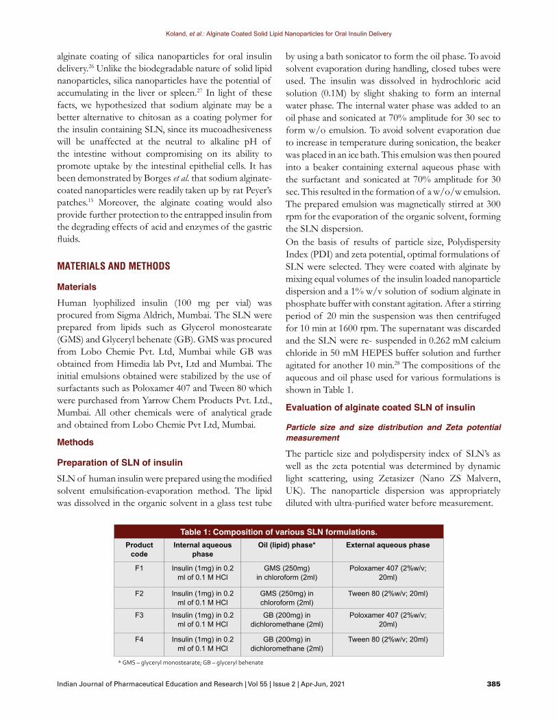
Table 1: Composition of various SLN formulations.Product
codeInternal aqueous
phaseOil (lipid) phase* External aqueous phase
F1 Insulin (1mg) in 0.2ml of 0.1 M HCl
GMS (250mg)in chloroform (2ml)
Poloxamer 407 (2%w/v;20ml)
F2 Insulin (1mg) in 0.2ml of 0.1 M HCl
GMS (250mg) inchloroform (2ml)
Tween 80 (2%w/v; 20ml)
F3 Insulin (1mg) in 0.2ml of 0.1 M HCl
GB (200mg) indichloromethane (2ml)
Poloxamer 407 (2%w/v;20ml)
F4 Insulin (1mg) in 0.2ml of 0.1 M HCl
GB (200mg) indichloromethane (2ml)
Tween 80 (2%w/v; 20ml)
* GMS – glyceryl monostearate; GB – glyceryl behenate
Koland, et al.: Alginate Coated Solid Lipid Nanoparticles for Oral Insulin Delivery
386 Indian Journal of Pharmaceutical Education and Research | Vol 55 | Issue 2 | Apr-Jun, 2021
Transmission Electron Microscopy (TEM) of coated SLNParticle size analysis was also carried out by TEM (Jeol/JEM 2100 High Resolution). A drop of the diluted selected alginate coated SLN formulation was placed on carbon-coated copper grids and dried under a lamp before the microscopic images were recorded.
Insulin association efficiencyThe association efficiency (AE) was determined by the indirect method as reported by Fonte et al.29 The amount of insulin entrapped into SLN was calculated by the difference between the total amount used to prepare the SLN and the amount of insulin that remained in the aqueous phase. The aqueous phase was separated by centrifugation and filtration. The sample was centrifuged at 4000 rpm for 1 hr. The separated supernatant was diluted and passed through a 0.2 µm syringe filter. The insulin concentration in the filtrate was determined by HPLC-UV method. The equation used to determine AE is given as follows: Total amount of insulin – Free insulin in filtrateA.E = x 100 (1) Total amount of insulinThe loading efficiency (LE) was determined from the difference between the total amount of insulin initially used to prepare the nanoparticles and the amount of residual un-associated or free insulin after particle separation as a percentage of total nanoparticle dry mass. As described above for AE, the free insulin present in the filtrate after separation of the SLN was measured by HPLC-UV. The total dry weight of the nanoparticles was determined. The residue remaining after centrifugation was combined with the rinsing from the syringe filter and the dispersion so obtained was subjected to freeze drying. The LE was then calculated using the formula below.29
Total amount of insulin – Free insulin in filtrateL.E = x 100 (2) Total weight of nanoparticles
In vitro release studiesDrug release studies were carried out by a small modification of the reverse dialysis sac method reported by Xu et al.30 The study was carried out under simulated GI conditions in two steps. In the first step, 5 ml of the SLN dispersion was mixed with sufficient simulated gastric fluid (SGF) of pH 1.2 (2.0 g of sodium chloride, 3.2 g of pepsin in 7.0 ml of hydrochloric acid and water to make 1000 ml) to produce 100 ml. This dispersion was used as the external medium and therefore has
the opportunity to release the drug content under maximum dilution and perfect sink conditions.31 For the purpose of sampling the drug released at predetermined time intervals, two dialysis bags (Himedia, molecular weight cut off: 12000-14000 Daltons; pore size: 2.4 nm) each containing 5 ml of SGF was immersed in the external medium (placed in a 250 ml beaker) which was maintained at 37°C and agitated by a magnetic stirrer at 50 rpm. The immersed dialysis bags were previously filled and equilibrated with the sink solution in the external compartment. The two bags which were fitted with glass tubes to provide for alternative sampling, make it possible to allow equilibration prior to the next sampling time. The released drug from the nanoparticles is expected to diffuse through the dialysis membrane into the internal compartments. The study was carried out for 2 h to mimic the in vivo gastric conditions. In the second step, the same procedure was repeated with the exception that simulated intestinal fluid (SIF) or phosphate buffer of pH 6.8 was used in the external compartment and dialysis bags with a fresh sample of 5 ml of the SLN dispersion. During the 6 h study, aliquots of 500 µl removed from the dialysis bags were filtered through syringe filters and analysed for drug concentration using HPLC-UV. The permeation ability of the drug in solution through the dialysis membrane was also determined by repeating the study in SGF and SIF with 5 ml of insulin solution in dilute acetic acid in the external medium.
Ex vivo permeability studies using Caco-2 cell monolayer modelEx vivo permeability studies of the drug from SLN were performed using Caco-2 cell monolayer model. Caco-2 cells (passage 70-75) were cultured at 37°C in an atmosphere of 5% CO2/95% air and 90% relative humidity in Dulbecco’s modified Eagle medium (DMEM) supplemented with 1% penicillin–streptomycin, 2 mM L-glutamine, 1% nonessential amino acids and 10% heat-inactivated foetal bovine serum.32,33
The cultured cells were seeded on 24 well (Trans well) plates in DMEM medium at a density of 10,000 cells/ well. Trans epithelial electrical resistance (TEER) measurements were carried out to monitor the growth and viability of the monolayer. After 20 days from initial seeding, aliquots of 100 µl each of insulin solution and SLN dispersions equivalent to 50 µg/ml are applied to the apical side of the cells.34 Then the permeation study was carried out for 3 h at 37°C. At the end of the period, samples of the medium were removed from the basolateral side and insulin content was analyzed by HPLC-UV.
Koland, et al.: Alginate Coated Solid Lipid Nanoparticles for Oral Insulin Delivery
Indian Journal of Pharmaceutical Education and Research | Vol 55 | Issue 2 | Apr-Jun, 2021 387
Ex vivo permeation studies using intestinal mucosa of goat35
Ex vivo permeation studies were also carried out using the freshly excised intestinal mucosa of a goat obtained from the slaughter house. The goat intestinal mucosa which was trimmed off to remove any extraneous tissue was rinsed with phosphate buffered saline (pH 6.8) to remove mucous and lumen contents. The tissue was mounted between the donor and the receptor compartment of a Franz diffusion cell with the mucosal surface facing the donor compartment. Formulation equivalent to 50 µg/ml of insulin was placed in the donor compartment, while the receptor compartment was filled with 25 ml of phosphate buffer pH 6.8 and its temperature was maintained at 37°C. The content of the receptor compartment was stirred using a magnetic stirrer at 100 rpm. An aliquot of 2 ml was withdrawn from the receptor compartment at suitable time intervals and replaced with the same volume of fresh medium. These samples were analyzed using HPLC-UV.
In vivo anti-diabetic studies using albino ratsThe animal experiments were carried out in adherence to the CPCSEA guidelines and after obtaining permission from the Institution Animal Ethics Committee of the NGSM Institute of Pharmaceutical Sciences. Male albino rats of body weight, 200-250 g were housed in controlled environmental conditions of temperature (22 ± 2°C) and relative humidity (45-65%). The rats were fed with standard diet feed and were provided fresh water. Lighting was on standard 12 h on/12 h off cycle.Total of six groups of animals was taken and treatment was given as follows: Group 1: Normal control, are treated with distilled water.Group 2: Negative control without any treatment.Group 3: Positive control with subcutaneous insulin (2.5 IU/kg)Group 4: Oral insulin solution (20 IU/kg, in Phosphate Buffered Saline). Group 5: Uncoated solid lipid nanoparticles (25 IU/kg)Group 6: Alginate coated solid lipid nanoparticles (25 IU/kg)Diabetes was induced in Groups 2 - 6 as per the method described. A single intra-peritoneal injection of streptozotocin (10mg/ml in citrate buffer, pH 4.5) at a concentration of 60 mg/kg of body weight was administered to each rat. After 3 days, rats presented fasted blood glucose levels above 250 mg/dL. These animals were used and were fasted 12 h before the
experiment and remained fasted during the experiment. But the animals had free access to water.Respective treatments were given to Groups 3 - 6. Blood samples were collected from the tail vein after needle puncture. The blood glucose level was measured using a glucometer. The samples were collected at different time intervals for a period of 12 h and the results obtained were statistically evaluated by one-way analysis of variance (ANOVA). The significant difference (p<0.05) between the groups were compared using post-hoc test (Scheffe). All statistical analysis was performed using the Statistical Package for the Social Sciences (SPSS) software.36-38 Plasma glucose levels were plotted as percentage of base line glucose levels against time.The pharmacological availability (PA) of the formulations administered to the animals was also determined to establish the efficacy of the alginate coated SLN. Taking into consideration that subcutaneous insulin would produce 100 % bioavailability, the PA was determined from the area above the plasma glucose curve (AAC) below the 100% cut-off line using the trapezoidal method and applying the following formula.20
AACoral / Doseoral PA = (3) AACSC / Dose SC
RESULTS AND DISCUSSIONWe have coated the optimized SLN formulations with sodium alginate by ionotropic gelation in calcium chloride solution so as to provide mucoadhesive characteristics to the particles. The anionic nature of the alginate coating will also increase the charge density on the surface of the SLN thereby giving better physicochemical stability and improve the mucoadhesive properties of the polymer.39
This is essential since, the mucoadhesiveness provided by this coating prolongs the drug residence time in gastrointestinal mucosal tissues, including at the Peyer’s patches.
Particle size, PDI and zeta potential measurementThe particle size of the uncoated SLN of GB was significantly greater than those of GMS. The use of the two surfactants poloxamer 407 and tween 80 did not really make much of a difference to the particle sizes for each lipid. The increase in particle size could be due to the fact that the increase in hydrophobic chain length of lipid of GB when compared to GMS could have a role to play.40
Ideally, particle size below 500 nm enhances the absorption through intestinal mucosa.41-43 Therefore SLN prepared using GMS was selected for the coating with alginate. Interestingly after coating, the choice of
Koland, et al.: Alginate Coated Solid Lipid Nanoparticles for Oral Insulin Delivery
388 Indian Journal of Pharmaceutical Education and Research | Vol 55 | Issue 2 | Apr-Jun, 2021
the surfactant did make a difference to the final particle size. Coated SLN formulation F5 using Poloxamer 407 as surfactant were significantly smaller than those of F6 with Tween 80. This was probably due to the increased deposition of alginate on the SLN surface in the case of F6 brought about by the poorer solubilizing capacity of Tween 80 on sodium alginate as compared to Poloxamer 407.44
The lower value of zeta potential is normal for particles containing non-ionic surfactants like Tween 80 and Poloxamer 407. After alginate coating, the surface charges reverted to a negative value. This shows the deposition of negatively charged alginate on the surface of SLN.45,46 The values of zeta potential of all formulations are within the stability range of ± 25 meV.With the exception of F3 and F4, the PDI of all formulations were found to be less than 0.3 indicating a narrow size distribution.
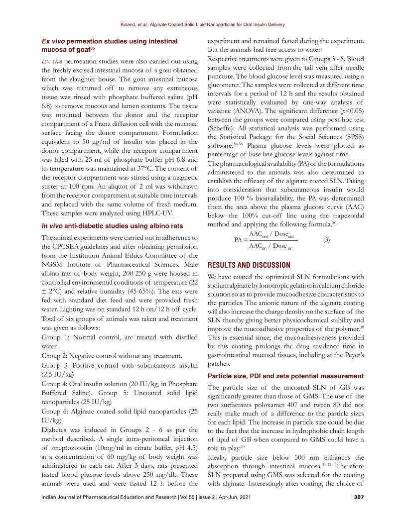
Transmission Electron Microscopy (TEM) of coated SLNThe TEM Images (Figure 1) of F1 and F5 confirm that the coated and uncoated SLN were spherical in shape and indicated a uniform size distribution. The average size of particles in the micrograph for F1 was found to be 217 ± 4 nm and that of F5 was 305 ± 2 nm.47
Obviously the larger size of F5 would be the result of coating.
Drug association efficiency and drug loading capacityThe data for association efficiency (AE) and loading efficiency (LE) is given in Table 2. The association efficiency reflects the fraction of the total insulin that is actually entrapped in the lipid. The composition of the lipids can influence both association efficiency and drug loading efficiency. Since hydrophilic drugs such as
insulin tend to get expelled from the lipid matrix during the solidification of the SLN, drug loading can be a problem.10
We have found that the choice of the lipid and surfactant can make a difference to both AE and LE. Some of the factors that determine the drug loading capacity of SLNs are the solubility of drug in lipid phase and the chemical or physical structure of solid lipid matrix.48 GMS is a good emulsifier and a suitable combination with a surfactant such as Tween 80 or Poloxamer 407 can improve the AE and LE for hydrophilic drug, insulin by enhancing solubilization in the lipid matrix. Another factor that may be considered is the molecular size and chain length that may affect the polarity of the lipid. The greater chain length of GB makes it more hydrophobic and hence amount of insulin entrapped was significantly reduced. Therefore, the SLN made from GMS when combined with similar surfactants produced greater AE and LE. It was also observed that when Poloxamer 407 was used as the surfactant, there was a substantial increase in drug entrapment as compared to the SLN formulations prepared with Tween 80, because of the greater solubilizing properties of the former. On the basis of this observation, F1 was considered as optimal for subsequent coating with alginate. Between the two coated formulations, F5 and F6, the former was considered optimal in terms of not only AE and LE but also particle size and PDI and therefore was subjected further to drug release studies.
In vitro drug release studiesIn vitro release of insulin from the solid lipid nano- particles of F1 and F5 was investigated in simulated GI fluids. This evaluation is crucial for predicting drug release behavior in the GI tract after oral administration.The method selected for the study is the reverse dialysis sac technique, which enables physical separation of the SLN from the sampling compartment by the usage of a dialysis membrane and allows for ease of sampling at periodic intervals. Moreover, the introduction of the SLN into the external release medium which is agitated ensures not only direct contact with the simulated GI fluids but also provides the sink conditions necessary for continuous diffusion of the drug into the dialysis bag.30 Thus there is a greater degree of predictability in drug release due to closer simulation of in vivo conditions. The release rate of the drug and its appearance in the dissolution medium within the dialysis bag depends on the partitioning the drug between the lipid phase and the external aqueous environment followed by diffusion of the drug across the membrane.49
Figure 1: Transmission Electron Microscopic image of F1 (A) and F5 (B).
Koland, et al.: Alginate Coated Solid Lipid Nanoparticles for Oral Insulin Delivery
Indian Journal of Pharmaceutical Education and Research | Vol 55 | Issue 2 | Apr-Jun, 2021 389
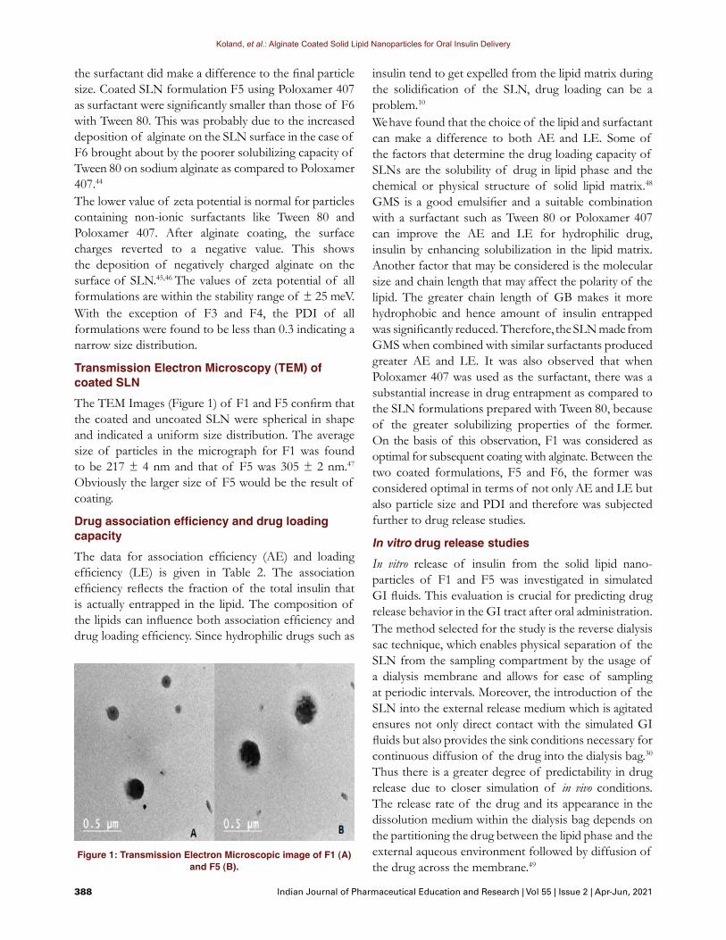
This study has been carried out in two stages, whereby in the first stage, the SLN dispersion was introduced into (SGF) in the external release compartment and samples were taken from the dialysis bag also containing the same medium. Since it was not possible to replace the medium with SIF for the second step, the entire study was repeated in a second assembly where the SLN formulation was added to SIF in the external compartment. The drug release profiles of insulin from the formulations, in SGF and SIF, are shown in Figures 2 and 3 respectively.The release profiles in SGF show that the amounts of insulin released from F5 were negligible compared to that from F1. This was to be expected since the alginate coating is poorly soluble in acidic pH due to the protonation of the pendant carboxylic moieties and thereby provides a protective barrier to the entrapped drug in the SLN matrix.50 In the absence of such a restrictive barrier in the uncoated SLN formulation, F1, an initial burst release of insulin in the first 30 min of the study was followed by a slow and sustained release that reached a maximum of just about 15 % at the end of two hours. The burst release could be attributed to a small percentage of the hydrophilic drug being expelled from the lipid matrix as it crystallizes during the formation of the SLN. However, as mentioned earlier,
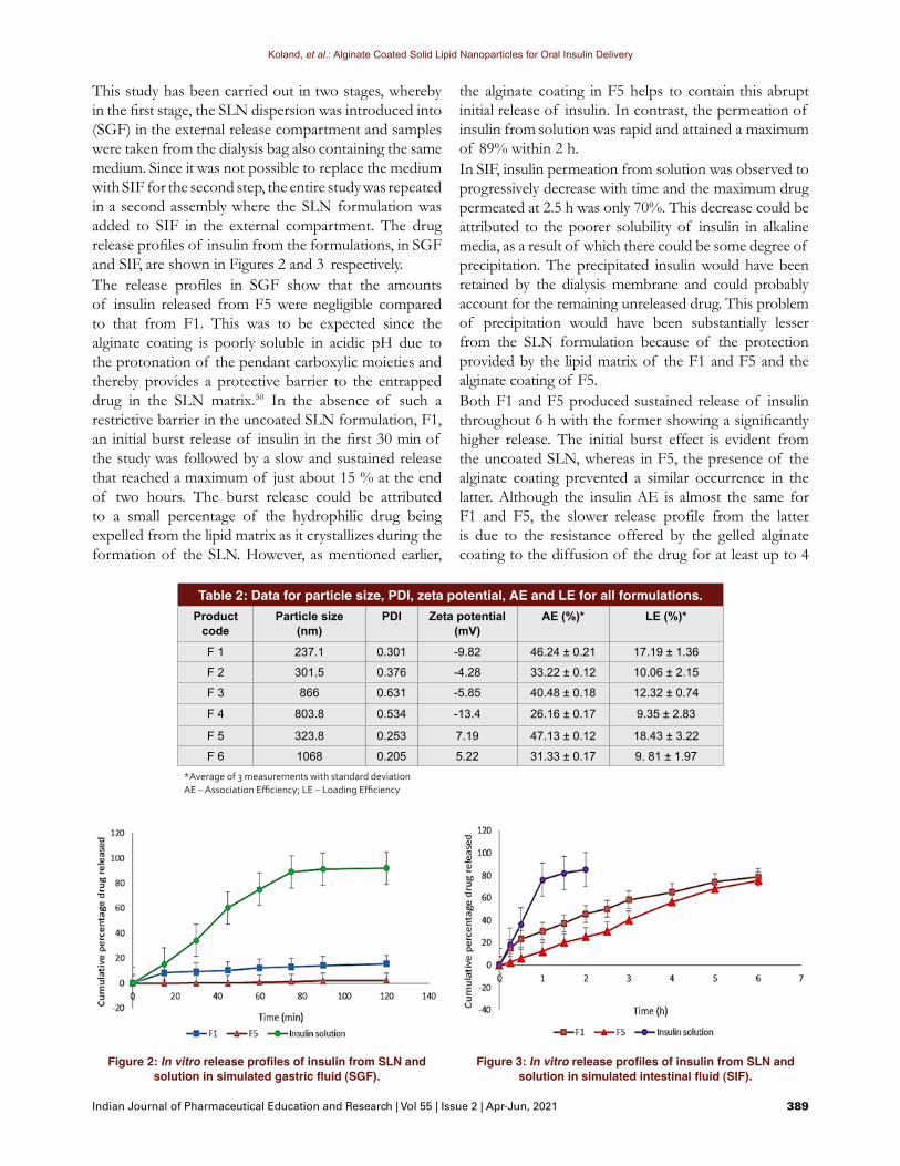
the alginate coating in F5 helps to contain this abrupt initial release of insulin. In contrast, the permeation of insulin from solution was rapid and attained a maximum of 89% within 2 h.In SIF, insulin permeation from solution was observed to progressively decrease with time and the maximum drug permeated at 2.5 h was only 70%. This decrease could be attributed to the poorer solubility of insulin in alkaline media, as a result of which there could be some degree of precipitation. The precipitated insulin would have been retained by the dialysis membrane and could probably account for the remaining unreleased drug. This problem of precipitation would have been substantially lesser from the SLN formulation because of the protection provided by the lipid matrix of the F1 and F5 and the alginate coating of F5.Both F1 and F5 produced sustained release of insulin throughout 6 h with the former showing a significantly higher release. The initial burst effect is evident from the uncoated SLN, whereas in F5, the presence of the alginate coating prevented a similar occurrence in the latter. Although the insulin AE is almost the same for F1 and F5, the slower release profile from the latter is due to the resistance offered by the gelled alginate coating to the diffusion of the drug for at least up to 4
Figure 2: In vitro release profiles of insulin from SLN and solution in simulated gastric fluid (SGF).
Figure 3: In vitro release profiles of insulin from SLN and solution in simulated intestinal fluid (SIF).
Table 2: Data for particle size, PDI, zeta potential, AE and LE for all formulations.Product
codeParticle size
(nm)PDI Zeta potential
(mV)AE (%)* LE (%)*
F 1 237.1 0.301 -9.82 46.24 ± 0.21 17.19 ± 1.36
F 2 301.5 0.376 -4.28 33.22 ± 0.12 10.06 ± 2.15
F 3 866 0.631 -5.85 40.48 ± 0.18 12.32 ± 0.74
F 4 803.8 0.534 -13.4 26.16 ± 0.17 9.35 ± 2.83
F 5 323.8 0.253 7.19 47.13 ± 0.12 18.43 ± 3.22
F 6 1068 0.205 5.22 31.33 ± 0.17 9. 81 ± 1.97*Average of 3 measurements with standard deviationAE – Association Efficiency; LE – Loading Efficiency
Koland, et al.: Alginate Coated Solid Lipid Nanoparticles for Oral Insulin Delivery
390 Indian Journal of Pharmaceutical Education and Research | Vol 55 | Issue 2 | Apr-Jun, 2021
h. After that, the gradual dissolution or erosion of the alginate coating at the intestinal pH would produce a surge in drug release to be almost the same as that of F1. It can be seen that the release of insulin from both coated and the uncoated SLN is substantially enhanced in SIF as compared to SGF. F1 showed only 15.36 % release after 2 h in SGF whereas, in SIF, the release was 45.65 %. The reduction in drug release from F1 in the gastric fluid means that insulin released from SLN was probably degraded by the proteolytic enzyme, pepsin and the acid. Studies show that insulin degrades immediately in the presence of pepsin.51 However, in our investigation, the proteolytic degradation of insulin was not immediate, which could be attributed to the slow release of insulin from the lipid matrix in both F1 and F5 and therefore, less exposure. Moreover, the additional protection offered by the alginate coating in F5 may further protect the drug from deactivation. This study, therefore, confirms the importance of the alginate coating in maintaining the potency of orally administered insulin.
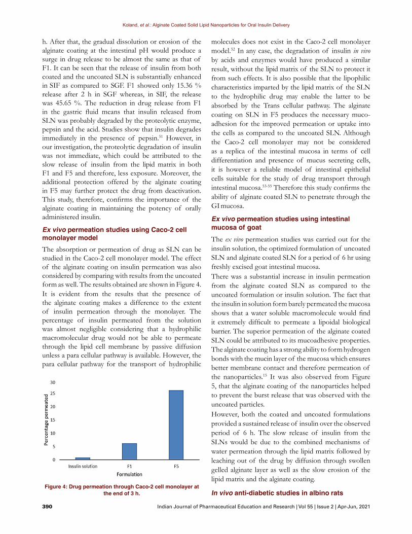
Ex vivo permeation studies using Caco-2 cell monolayer modelThe absorption or permeation of drug as SLN can be studied in the Caco-2 cell monolayer model. The effect of the alginate coating on insulin permeation was also considered by comparing with results from the uncoated form as well. The results obtained are shown in Figure 4.It is evident from the results that the presence of the alginate coating makes a difference to the extent of insulin permeation through the monolayer. The percentage of insulin permeated from the solution was almost negligible considering that a hydrophilic macromolecular drug would not be able to permeate through the lipid cell membrane by passive diffusion unless a para cellular pathway is available. However, the para cellular pathway for the transport of hydrophilic
molecules does not exist in the Caco-2 cell monolayer model.52 In any case, the degradation of insulin in vivo by acids and enzymes would have produced a similar result, without the lipid matrix of the SLN to protect it from such effects. It is also possible that the lipophilic characteristics imparted by the lipid matrix of the SLN to the hydrophilic drug may enable the latter to be absorbed by the Trans cellular pathway. The alginate coating on SLN in F5 produces the necessary muco-adhesion for the improved permeation or uptake into the cells as compared to the uncoated SLN. Although the Caco-2 cell monolayer may not be considered as a replica of the intestinal mucosa in terms of cell differentiation and presence of mucus secreting cells, it is however a reliable model of intestinal epithelial cells suitable for the study of drug transport through intestinal mucosa.53-55 Therefore this study confirms the ability of alginate coated SLN to penetrate through the GI mucosa.
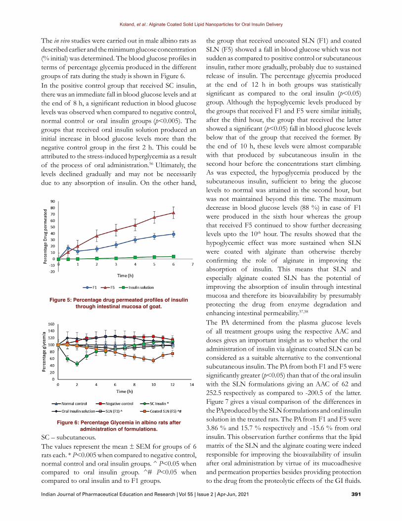
Ex vivo permeation studies using intestinal mucosa of goatThe ex vivo permeation studies was carried out for the insulin solution, the optimized formulation of uncoated SLN and alginate coated SLN for a period of 6 hr using freshly excised goat intestinal mucosa.There was a substantial increase in insulin permeation from the alginate coated SLN as compared to the uncoated formulation or insulin solution. The fact that the insulin in solution form barely permeated the mucosa shows that a water soluble macromolecule would find it extremely difficult to permeate a lipoidal biological barrier. The superior permeation of the alginate coated SLN could be attributed to its mucoadhesive properties. The alginate coating has a strong ability to form hydrogen bonds with the mucin layer of the mucosa which ensures better membrane contact and therefore permeation of the nanoparticles.15 It was also observed from Figure 5, that the alginate coating of the nanoparticles helped to prevent the burst release that was observed with the uncoated particles.However, both the coated and uncoated formulations provided a sustained release of insulin over the observed period of 6 h. The slow release of insulin from the SLNs would be due to the combined mechanisms of water permeation through the lipid matrix followed by leaching out of the drug by diffusion through swollen gelled alginate layer as well as the slow erosion of the lipid matrix and the alginate coating.
In vivo anti-diabetic studies in albino ratsFigure 4: Drug permeation through Caco-2 cell monolayer at
the end of 3 h.
Koland, et al.: Alginate Coated Solid Lipid Nanoparticles for Oral Insulin Delivery
Indian Journal of Pharmaceutical Education and Research | Vol 55 | Issue 2 | Apr-Jun, 2021 391
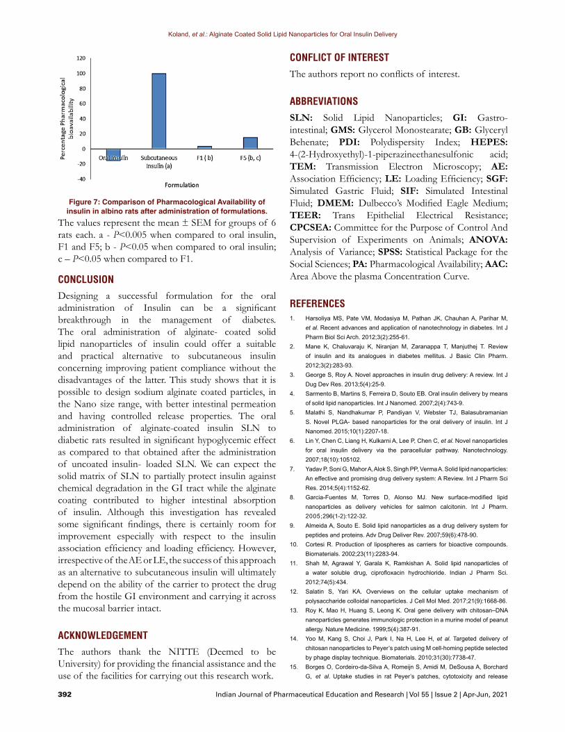
The in vivo studies were carried out in male albino rats as described earlier and the minimum glucose concentration (% initial) was determined. The blood glucose profiles in terms of percentage glycemia produced in the different groups of rats during the study is shown in Figure 6.In the positive control group that received SC insulin, there was an immediate fall in blood glucose levels and at the end of 8 h, a significant reduction in blood glucose levels was observed when compared to negative control, normal control or oral insulin groups (p<0.005). The groups that received oral insulin solution produced an initial increase in blood glucose levels more than the negative control group in the first 2 h. This could be attributed to the stress-induced hyperglycemia as a result of the process of oral administration.56 Ultimately, the levels declined gradually and may not be necessarily due to any absorption of insulin. On the other hand,
the group that received uncoated SLN (F1) and coated SLN (F5) showed a fall in blood glucose which was not sudden as compared to positive control or subcutaneous insulin, rather more gradually, probably due to sustained release of insulin. The percentage glycemia produced at the end of 12 h in both groups was statistically significant as compared to the oral insulin (p<0.05) group. Although the hypoglycemic levels produced by the groups that received F1 and F5 were similar initially, after the third hour, the group that received the latter showed a significant (p<0.05) fall in blood glucose levels below that of the group that received the former. By the end of 10 h, these levels were almost comparable with that produced by subcutaneous insulin in the second hour before the concentrations start climbing. As was expected, the hypoglycemia produced by the subcutaneous insulin, sufficient to bring the glucose levels to normal was attained in the second hour, but was not maintained beyond this time. The maximum decrease in blood glucose levels (88 %) in case of F1 were produced in the sixth hour whereas the group that received F5 continued to show further decreasing levels upto the 10th hour. The results showed that the hypoglycemic effect was more sustained when SLN were coated with alginate than otherwise thereby confirming the role of alginate in improving the absorption of insulin. This means that SLN and especially alginate coated SLN has the potential of improving the absorption of insulin through intestinal mucosa and therefore its bioavailability by presumably protecting the drug from enzyme degradation and enhancing intestinal permeability.57,58
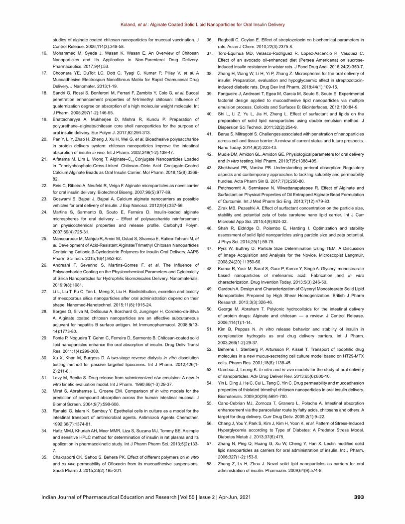
The PA determined from the plasma glucose levels of all treatment groups using the respective AAC and doses gives an important insight as to whether the oral administration of insulin via alginate coated SLN can be considered as a suitable alternative to the conventional subcutaneous insulin. The PA from both F1 and F5 were significantly greater (p<0.05) than that of the oral insulin with the SLN formulations giving an AAC of 62 and 252.5 respectively as compared to -200.5 of the latter. Figure 7 gives a visual comparison of the differences in the PA produced by the SLN formulations and oral insulin solution in the treated rats. The PA from F1 and F5 were 3.86 % and 15.7 % respectively and -15.6 % from oral insulin. This observation further confirms that the lipid matrix of the SLN and the alginate coating were indeed responsible for improving the bioavailability of insulin after oral administration by virtue of its mucoadhesive and permeation properties besides providing protection to the drug from the proteolytic effects of the GI fluids.
Figure 6: Percentage Glycemia in albino rats after administration of formulations.
SC – subcutaneous.The values represent the mean ± SEM for groups of 6 rats each. * P<0.005 when compared to negative control, normal control and oral insulin groups. ^ P<0.05 when compared to oral insulin group. ^# P<0.05 when compared to oral insulin and to F1 groups.
Figure 5: Percentage drug permeated profiles of insulin through intestinal mucosa of goat.
Koland, et al.: Alginate Coated Solid Lipid Nanoparticles for Oral Insulin Delivery
392 Indian Journal of Pharmaceutical Education and Research | Vol 55 | Issue 2 | Apr-Jun, 2021
CONCLUSIONDesigning a successful formulation for the oral administration of Insulin can be a significant breakthrough in the management of diabetes. The oral administration of alginate- coated solid lipid nanoparticles of insulin could offer a suitable and practical alternative to subcutaneous insulin concerning improving patient compliance without the disadvantages of the latter. This study shows that it is possible to design sodium alginate coated particles, in the Nano size range, with better intestinal permeation and having controlled release properties. The oral administration of alginate-coated insulin SLN to diabetic rats resulted in significant hypoglycemic effect as compared to that obtained after the administration of uncoated insulin- loaded SLN. We can expect the solid matrix of SLN to partially protect insulin against chemical degradation in the GI tract while the alginate coating contributed to higher intestinal absorption of insulin. Although this investigation has revealed some significant findings, there is certainly room for improvement especially with respect to the insulin association efficiency and loading efficiency. However, irrespective of the AE or LE, the success of this approach as an alternative to subcutaneous insulin will ultimately depend on the ability of the carrier to protect the drug from the hostile GI environment and carrying it across the mucosal barrier intact.
ACKNOWLEDGEMENTThe authors thank the NITTE (Deemed to be University) for providing the financial assistance and the use of the facilities for carrying out this research work.
Figure 7: Comparison of Pharmacological Availability of insulin in albino rats after administration of formulations.
The values represent the mean ± SEM for groups of 6 rats each. a - P<0.005 when compared to oral insulin, F1 and F5; b - P<0.05 when compared to oral insulin; c – P<0.05 when compared to F1.
CONFLICT OF INTERESTThe authors report no conflicts of interest.
ABBREVIATIONSSLN: Solid Lipid Nanoparticles; GI: Gastro-intestinal; GMS: Glycerol Monostearate; GB: Glyceryl Behenate; PDI: Polydispersity Index; HEPES: 4-(2-Hydroxyethyl)-1-piperazineethanesulfonic acid; TEM: Transmission Electron Microscopy; AE: Association Efficiency; LE: Loading Efficiency; SGF: Simulated Gastric Fluid; SIF: Simulated Intestinal Fluid; DMEM: Dulbecco’s Modified Eagle Medium; TEER: Trans Epithelial Electrical Resistance; CPCSEA: Committee for the Purpose of Control And Supervision of Experiments on Animals; ANOVA: Analysis of Variance; SPSS: Statistical Package for the Social Sciences; PA: Pharmacological Availability; AAC: Area Above the plasma Concentration Curve.
REFERENCES1. Harsoliya MS, Pate VM, Modasiya M, Pathan JK, Chauhan A, Parihar M,
et al. Recent advances and application of nanotechnology in diabetes. Int J Pharm Biol Sci Arch. 2012;3(2):255-61.
2. Mane K, Chaluvaraju K, Niranjan M, Zaranappa T, Manjuthej T. Review of insulin and its analogues in diabetes mellitus. J Basic Clin Pharm. 2012;3(2):283-93.
3. George S, Roy A. Novel approaches in insulin drug delivery: A review. Int J Dug Dev Res. 2013;5(4):25-9.
4. Sarmento B, Martins S, Ferreira D, Souto EB. Oral insulin delivery by means of solid lipid nanoparticles. Int J Nanomed. 2007;2(4):743-9.
5. Malathi S, Nandhakumar P, Pandiyan V, Webster TJ, Balasubramanian S. Novel PLGA- based nanoparticles for the oral delivery of insulin. Int J Nanomed. 2015;10(1):2207-18.
6. Lin Y, Chen C, Liang H, Kulkarni A, Lee P, Chen C, et al. Novel nanoparticles for oral insulin delivery via the paracellular pathway. Nanotechnology. 2007;18(10):105102.
7. Yadav P, Soni G, Mahor A, Alok S, Singh PP, Verma A. Solid lipid nanoparticles: An effective and promising drug delivery system: A Review. Int J Pharm Sci Res. 2014;5(4):1152-62.
8. Garcia-Fuentes M, Torres D, Alonso MJ. New surface-modified lipid nanoparticles as delivery vehicles for salmon calcitonin. Int J Pharm. 2005;296(1-2):122-32.
9. Almeida A, Souto E. Solid lipid nanoparticles as a drug delivery system for peptides and proteins. Adv Drug Deliver Rev. 2007;59(6):478-90.
10. Cortesi R. Production of lipospheres as carriers for bioactive compounds. Biomaterials. 2002;23(11):2283-94.
11. Shah M, Agrawal Y, Garala K, Ramkishan A. Solid lipid nanoparticles of a water soluble drug, ciprofloxacin hydrochloride. Indian J Pharm Sci. 2012;74(5):434.
12. Salatin S, Yari KA. Overviews on the cellular uptake mechanism of polysaccharide colloidal nanoparticles. J Cell Mol Med. 2017;21(9):1668-86.
13. Roy K, Mao H, Huang S, Leong K. Oral gene delivery with chitosan–DNA nanoparticles generates immunologic protection in a murine model of peanut allergy. Nature Medicine. 1999;5(4):387-91.
14. Yoo M, Kang S, Choi J, Park I, Na H, Lee H, et al. Targeted delivery of chitosan nanoparticles to Peyer’s patch using M cell-homing peptide selected by phage display technique. Biomaterials. 2010;31(30):7738-47.
15. Borges O, Cordeiro-da-Silva A, Romeijn S, Amidi M, DeSousa A, Borchard G, et al. Uptake studies in rat Peyer’s patches, cytotoxicity and release
Koland, et al.: Alginate Coated Solid Lipid Nanoparticles for Oral Insulin Delivery
Indian Journal of Pharmaceutical Education and Research | Vol 55 | Issue 2 | Apr-Jun, 2021 393
studies of alginate coated chitosan nanoparticles for mucosal vaccination. J Control Release. 2006;114(3):348-58.
16. Mohammed M, Syeda J, Wasan K, Wasan E. An Overview of Chitosan Nanoparticles and Its Application in Non-Parenteral Drug Delivery. Pharmaceutics. 2017;9(4):53.
17. Choonara YE, DuToit LC, Dott C, Tyagi C, Kumar P, Pillay V, et al. A Mucoadhesive Electrospun Nanofibrous Matrix for Rapid Oramucosal Drug Delivery. J Nanomater. 2013;1-19.
18. Sandri G, Rossi S, Bonferoni M, Ferrari F, Zambito Y, Colo G, et al. Buccal penetration enhancement properties of N-trimethyl chitosan: Influence of quaternization degree on absorption of a high molecular weight molecule. Int J Pharm. 2005;297(1-2):146-55.
19. Bhattacharyya A, Mukherjee D, Mishra R, Kundu P. Preparation of polyurethane–alginate/chitosan core shell nanoparticles for the purpose of oral insulin delivery. Eur Polym J. 2017;92:294-313.
20. Pan Y, Li Y, Zhao H, Zheng J, Xu H, Wei G, et al. Bioadhesive polysaccharide in protein delivery system: chitosan nanoparticles improve the intestinal absorption of insulin in vivo. Int J Pharm. 2002;249(1-2):139-47.
21. Alfatama M, Lim L, Wong T. Alginate–C18 Conjugate Nanoparticles Loaded in Tripolyphosphate-Cross-Linked Chitosan–Oleic Acid Conjugate-Coated Calcium Alginate Beads as Oral Insulin Carrier. Mol Pharm. 2018;15(8):3369-82.
22. Reis C, Ribeiro A, Neufeld R, Veiga F. Alginate microparticles as novel carrier for oral insulin delivery. Biotechnol Bioeng. 2007;96(5):977-89.
23. Goswami S, Bajpai J, Bajpai A. Calcium alginate nanocarriers as possible vehicles for oral delivery of insulin. J Exp Nanosci. 2012;9(4):337-56.
24. Martins S, Sarmento B, Souto E, Ferreira D. Insulin-loaded alginate microspheres for oral delivery – Effect of polysaccharide reinforcement on physicochemical properties and release profile. Carbohyd Polym. 2007;69(4):725-31.
25. Mansourpour M, Mahjub R, Amini M, Ostad S, Shamsa E, Rafiee-Tehrani M, et al. Development of Acid-Resistant Alginate/Trimethyl Chitosan Nanoparticles Containing Cationic β-Cyclodextrin Polymers for Insulin Oral Delivery. AAPS Pharm Sci Tech. 2015;16(4):952-62.
26. Andreani F, Severino S, Martins-Gomes F, et al. The Influence of Polysaccharide Coating on the Physicochemical Parameters and Cytotoxicity of Silica Nanoparticles for Hydrophilic Biomolecules Delivery. Nanomaterials. 2019;9(8):1081.
27. Li L, Liu T, Fu C, Tan L, Meng X, Liu H. Biodistribution, excretion and toxicity of mesoporous silica nanoparticles after oral administration depend on their shape. Nanomed-Nanotechnol. 2015;11(8):1915-24.
28. Borges O, Silva M, DeSousa A, Borchard G, Junginger H, Cordeiro-da-Silva A. Alginate coated chitosan nanoparticles are an effective subcutaneous adjuvant for hepatitis B surface antigen. Int Immunopharmacol. 2008;8(13-14):1773-80.
29. Fonte P, Nogueira T, Gehm C, Ferreira D, Sarmento B. Chitosan-coated solid lipid nanoparticles enhance the oral absorption of insulin. Drug Deliv Transl Res. 2011;1(4):299-308.
30. Xu X, Khan M, Burgess D. A two-stage reverse dialysis in vitro dissolution testing method for passive targeted liposomes. Int J Pharm. 2012;426(1-2):211-8.
31. Levy M, Benita S. Drug release from submicronized o/w emulsion: A new in vitro kinetic evaluation model. Int J Pharm. 1990;66(1-3):29-37.
32. Miret S, Abrahamse L, Groene EM. Comparison of in vitro models for the prediction of compound absorption across the human intestinal mucosa. J Biomol Screen. 2004;9(7):598-606.
33. Ranaldi G, Islam K, Sambuy Y. Epethelial cells in culture as a model for the intestinal transport of antimicrobial agents. Antimicrob Agents Chemother. 1992;36(7):1374-81.
34. Hafiz MMJ, Khuriah AH, Meor MMR, Liza S, Suzana MJ, Tommy BE. A simple and sensitive HPLC method for determination of insulin in rat plasma and its application in pharmacokinetic study. Int J Pharm Pharm Sci. 2013;5(2):133-7.
35. Chakraborti CK, Sahoo S, Behera PK. Effect of different polymers on in vitro and ex vivo permeability of Ofloxacin from its mucoadhesive suspensions. Saudi Pharm J. 2015;23(2):195-201.
36. Ragbetli C, Ceylan E. Effect of streptozotocin on biochemical parameters in rats. Asian J Chem. 2010;22(3):2375-8.
37. Toro-Equihua MD, Velasco-Rodriguez R, Lopez-Ascencio R, Vasquez C. Effect of an avocado oil-enhanced diet (Persea Americana) on sucrose-induced insulin resistance in wistar rats. J Food Drug Anal. 2016;24(2):350-7.
38. Zhang H, Wang W, Li H, Yi P, Zhang Z. Microspheres for the oral delivery of insulin: Preparation, evaluation and hypoglycaemic effect in streptozotocin-induced diabetic rats. Drug Dev Ind Pharm. 2018;44(1):109-15.
39. Fangueiro J, Andreani T, Egea M, Garcia M, Souto S, Souto E. Experimental factorial design applied to mucoadhesive lipid nanoparticles via multiple emulsion process. Colloids and Surfaces B: Biointerfaces. 2012;100:84-9.
40. Shi L, Li Z, Yu L, Jia H, Zheng L. Effect of surfactant and lipids on the preparation of solid lipid nanoparticles using double emulsion method. J Dispersion Sci Technol. 2011;32(2):254-9.
41. Barua S, Mitragotri S. Challenges associated with penetration of nanoparticles across cell and tissue barrier: A review of current status and future prospects. Nano Today. 2014;9(2):223-43.
42. Mudie DM, Amidon GL, Amidon GE. Physiological parameters for oral delivery and in vitro testing. Mol Pharm. 2010;7(5):1388-405.
43. Shekhawat PB, Varsha PB. Understanding perioral absorption: Regulatory aspects and contemporary approaches to tackling solubility and permeability hurdles. Acta Pharm Sin B. 2017;7(3):260-80.
44. Petchsomrit A, Sermkaew N, Wiwattanapatapee R. Effect of Alginate and Surfactant on Physical Properties of Oil Entrapped Alginate Bead Formulation of Curcumin. Int J Med Pharm Sci Eng. 2013;7(12):479-83.
45. Zirak MB, Pezeshki A. Effect of surfactant concentration on the particle size, stability and potential zeta of beta carotene nano lipid carrier. Int J Curr Microbiol App Sci. 2015;4(9):924-32.
46. Shah R, Eldridge D, Polambo E, Harding I. Optimization and stability assessment of solid lipid nanoparticles using particle size and zeta potential. J Phys Sci. 2014;25(1):59-75.
47. Pyrz W, Buttrey D. Particle Size Determination Using TEM: A Discussion of Image Acquisition and Analysis for the Novice. Microscopist Langmuir. 2008;24(20):11350-60.
48. Kumar R, Yasir M, Saraf S, Gaur P, Kumar Y, Singh A. Glyceryl monostearate based nanoparticles of mefenamic acid: Fabrication and in vitro characterization. Drug Invention Today. 2013;5(3):246-50.
49. Gardouh A. Design and Characterization of Glyceryl Monostearate Solid Lipid Nanoparticles Prepared by High Shear Homogenization. British J Pharm Research. 2013;3(3):326-46.
50. George M, Abraham T. Polyionic hydrocolloids for the intestinal delivery of protein drugs: Alginate and chitosan — a review. J Control Release. 2006;114(1):1-14.
51. Kim B, Peppas N. In vitro release behavior and stability of insulin in complexation hydrogels as oral drug delivery carriers. Int J Pharm. 2003;266(1-2):29-37.
52. Behrens I, Stenberg P, Artursson P, Kissel T. Transport of lipophilic drug molecules in a new mucus-secreting cell culture model based on HT29-MTX cells. Pharm Res. 2001;18(8):1138-45
53. Gamboa J, Leong K. In vitro and in vivo models for the study of oral delivery of nanoparticles. Adv Drug Deliver Rev. 2013;65(6):800-10.
54. Yin L, Ding J, He C, Cui L, Tang C, Yin C. Drug permeability and mucoadhesion properties of thiolated trimethyl chitosan nanoparticles in oral insulin delivery. Biomaterials. 2009;30(29):5691-700.
55. Cano-Cebrian MJ, Zornoza T, Granero L, Polache A. Intestinal absorption enhancement via the paracellular route by fatty acids, chitosans and others: A target for drug delivery. Curr Drug Deliv. 2005;2(1):9–22.
56. Chang J, You Y, Park S, Kim J, Kim H, Yoon K, et al. Pattern of Stress-Induced Hyperglycemia according to Type of Diabetes: A Predator Stress Model. Diabetes Metab J. 2013;37(6):475.
57. Zhang N, Ping Q, Huang G, Xu W, Cheng Y, Han X. Lectin modified solid lipid nanoparticles as carriers for oral administration of insulin. Int J Pharm. 2006;327(1-2):153-9.
58. Zhang Z, Lv H, Zhou J. Novel solid lipid nanoparticles as carriers for oral administration of insulin. Pharmazie. 2009;64(9):574-8.
Koland, et al.: Alginate Coated Solid Lipid Nanoparticles for Oral Insulin Delivery
394 Indian Journal of Pharmaceutical Education and Research | Vol 55 | Issue 2 | Apr-Jun, 2021
Cite this article: Koland M, Anchan RB, Mukund SG, Sindhoor SM. Design and Investigation of Alginate Coated Solid Lipid Nanoparticles for Oral Insulin Delivery. Indian J of Pharmaceutical Education and Research. 2021;55(2): 383-94.
PICTORIAL ABSTRACT
About Authors
SUMMARYAlginate coated, insulin-loaded solid lipid nanoparticles (SLN) for oral administration were developed and investigated for physical properties, in vitro release, ex vivo permeation through Caco-2-cell monolayer and goat intestinal mucosa and in vivo efficacy in controlling blood glucose levels in streptozotocin induced diabetic rats. The SLN were prepared from glyceryl behenate (GB) and glyceryl monostearate (GMS) and coated with mucoadhesive polymer, sodium alginate. The particle size of the uncoated SLN of GB was significantly greater than those of GMS. Scanning electron microscopy and transmission electron microscopy revealed spherical particles of uniform size distribution. When Poloxamer 407 was used as the surfactant, there was a substantial increase in drug entrapment as compared to the SLN formulations prepared with Tween 80. The in vitro release of insulin at the end of 6 h using the reverse dialysis sac technique, was considerably enhanced in simulated intestinal fluid as compared to simulated gastric fluid from both coated and the uncoated SLN. Ex vivo permeability studies through Caco-2 cell monolayer and goat’s intestinal mucosa revealed a substantial increase in insulin permeation from the alginate coated SLN as compared to the uncoated formulation or insulin solution. The oral administration of alginate-coated insulin SLN to streptozotocin induced diabetic rats resulted in a significant hypoglycemic effect (P<0.05) when compared to the groups that received uncoated insulin-loaded SLN or the oral insulin solution. The hypoglycemic levels reached with coated SLN were comparable to that of the conventional subcutaneous insulin at the end of a 12 h study. The Pharmacological Availability (PA) from both coated and uncoated formulations were significantly greater (p<0.05) than that of the oral insulin. This means that SLN and especially alginate coated SLN has the potential of improving the absorption of insulin through intestinal mucosa and therefore its bioavailability.
Ms. Rakshitha B. Anchan was a PG scholar at the time of carrying out this project. She is currently working as a Drug Safety Associate in Bioclinica Safety and Regulatory Solutions, Mysore, India for the past 2 years.
Dr. Marina Koland, is Professor and Head of the Department of Pharmaceutics at the NGSM Institute of Pharmaceutical Sciences, Nitte (Deemed to be University), Mangalore. Her area of specialization in research is mucoadhesive and nanoparticulate drug delivery. She has published 50 research papers in peer reviewed journals, presented papers at 10 National and 3 International Conferences and worked on several Nitte University funded projects. She is currently working on solid lipid nanoparticles and micelles as drug carriers for targeting to the CNS.
Mr. Sawan Mukund Ghetia, is currently working as a Research Scholar in the Department of Pharmacology at NGSM Institute of Pharmaceutical Sciences. His research mainly addresses Diabetes Associated Depression. Other areas of his research include neuroprotection, polyherbal formulation and Psoriasis. Based on the research carried out, 4 research articles have been published in indexed journals.
Mr. Sindhoor S. M is a Ph.D research scholar in the Department of Pharmaceutics at NGSM Institute of Pharmaceutical Sciences. His areas of specialization include nanotechnology based drug delivery system, lipid based formulations and gastroretentive in situ gel systems. He has published 3 papers based on nanotechnology and lipid based drug delivery system in peer reviewed indexed journals. Presently he is involved in the development of lipid based delivery systems such as nanostructured lipid carriers and nano-emulsions for the management of skin diseases.