Embed Size (px)
Citation preview

DISEASES OF AQUATIC ORGANISMSDis Aquat Org
Vol. 127: 163–175, 2018https://doi.org/10.3354/dao03205
Published March 8
INTRODUCTION
The genus Crassicauda, comprised of large parasiticnematodes, is endemic to both mysticete and odonto-cete cetaceans (Lambertsen 1985, 1986, Lambertsenet al. 1986, Geraci & St. Aubin 1987, reviewed by
Keenan-Bateman et al. 2016). Cras si caudids infect theurogenital system of mysticetes and odontocetes andthe cranial pterygoid sinuses of odontocetes (Ridg-way 1972, Dailey & Stroud 1978, Geraci et al. 1978,Perrin & Powers 1980, Gibson et al. 1998, Lopez et al.1999). Crassicauda infection has been directly linked
© Inter-Research 2018 · www.int-res.com*Corresponding author: [email protected]
Habitat use pattern of the giant parasitic nematodeCrassicauda magna within the pygmy sperm whale
Kogia breviceps
Tiffany F. Keenan-Bateman1,*, William A. McLellan1, Alex M. Costidis1,2, Craig A. Harms3, D. Mark Gay1, David S. Rotstein4, Sentiel A. Rommel1,
Charles W. Potter5, D. Ann Pabst1
1University of North Carolina Wilmington, Department of Biology and Marine Biology, Wilmington, NC 28403, USA2Virginia Aquarium & Marine Science Center, Virginia Beach, VA 23451, USA
3North Carolina State University, College of Veterinary Medicine and Center for Marine Sciences and Technology, Morehead City, NC 28557, USA
4Marine Mammal Pathology Services, Olney, MD 20832, USA5Smithsonian Institution, National Museum of Natural History, Department of Vertebrate Zoology, Washington, DC 20560, USA
ABSTRACT: The giant (>3 m) parasitic nematode Crassicauda magna infects kogiid whales, al -though only 3 studies to date have provided detailed descriptions of these worms, all based uponfragmented specimens. These fragments were found within the neck region of kogiids, an unusualanatomic site for this genus of parasites. C. magna is a species-specific parasite among kogiids,infecting only pygmy sperm whales Kogia breviceps, and with a primarily cervico-thoracic distri-bution. To date, however, the pattern of habitat use within the host and transmission path of thisparasite remain unknown. We used detailed dissections (n = 12), histological examination of hosttissues (n = 2), and scanning electron microscopy of excised nematodes (n = 7) to enhance ourunderstanding of this host−parasite relationship. Results revealed that a critical habitat for the par-asite is an exocrine gland in the whale’s ventral cervical region. C. magna male and female tailswere found intertwined within the glandular lumen, and eggs were observed within its presumedsecretion, illuminating the transmission path out of the host. The cephalic ends of these wormswere often meters away (curvilinearly), embedded deeply within epaxial muscle. A single worm’scomplete, tortuous 312 cm course, from the gland to its termination in the contralateral epaxialmuscle, is described for the first time. This study also provides the first scanning electron micro-graphs of C. magna, which illustrate taxonomically important features of the heads and tails ofboth male and female worms.
KEY WORDS: Parasitic nematode · Crassicauda magna · Kogia breviceps · Cetacean · Stranding ·Morbidity · Scanning electron microscopy
Resale or republication not permitted without written consent of the publisher

Dis Aquat Org 127: 163–175, 2018
to morbidity and mortality in both suborders of ceta -ceans (Lambertsen 1985, 1986, 1992, Geraci & St.Aubin 1987, Gibson et al. 1998, Oliveira et al. 2011).
Crassicaudids were first described within membersof the family Kogiidae by Johnston & Mawson (1939)and next by Dollfus (1966) as infecting the ‘connec-tive tissues’ and ‘subcutaneous musculature’ of theneck, an unusual site of infection for this genus ofnematodes. Both studies identified the host speciesas Kogia breviceps, although both were publishedbefore Handley’s (1966) delineation of kogiids into 2separate species, K. breviceps and K. sima.
Subsequently, several surveys of metazoan para-sites of cetaceans, from multiple geographic regions,have identified crassicaudids within K. breviceps,generally infecting cervical tissues (Abollo et al. 1998,Mignucci-Giannoni et al. 1998, Cardona- Maldonado& Mignucci-Giannoni 1999, Carvalho et al. 2010).These studies, in total, report on 6 infected K. brevi-ceps specimens.
Keenan-Bateman et al. (2016) carried out a large-scale, retrospective analysis of over 100 records ofkogiids that had stranded along the US mid-Atlanticcoast. Those authors demonstrated crassicaudids tobe a species-specific parasite among kogiid whalesin this region, infecting only pygmy sperm whalesK. breviceps and not dwarf sperm whales K. sima.This work also confirmed the parasite’s general cer-vico-thoracic distribution reported by earlier authors,and identified a specific site of infection as a previ-ously undescribed exocrine gland, located at the ven-tral terminus of the pigmented ‘false gill slit’ in thewhale’s cervical region. This gland and its associatedpigmentation pattern, like crassicaudid infection, arefeatures unique to K. breviceps (Keenan-Bateman etal. 2016).
To date, only 3 studies have provided detailed de -scriptions of the nematodes that infect K. breviceps,all of which were based upon fragmented specimens(Johnston & Mawson 1939, Dollfus 1966, Jabbar et al.2015). For example, the determination of the speciesC. magna by Johnston & Mawson (1939) was basedupon a partial worm, collected from the cervical tis-sues of an adult female K. breviceps stranded in SouthAustralia. Based upon similarities in general headmorphology (e.g. rounded head, strongly chitinizedbuccal cavity, and 2 simple lateral lips), the large sizeof the specimen relative to C. boopis de scribed fromthe humpback whale Megaptera novae angliae (Baylis1920, 1922), and the specificity of the genus Crassi-cauda to cetaceans, these authors named and de-scribed the sixth crassicaudid species, C. magna, fromthis anterior fragment of a female worm. Johnston &
Mawson (1939) did not, however, observe the 2 lateraland 4 submedial papillae de scribed by Baylis (1920)as characteristic of the genus Crassicauda.
Dollfus (1966) later named C. duguyi based solelyupon the morphological features of a posterior frag-ment of a male specimen recovered from cervical tissues of a K. breviceps stranded in southwesternFrance. Unable to identify this specimen as that of C.magna as described by Johnston & Mawson (1939),Dollfus instead compared the posterior fragment toavailable descriptions of other known spiculatedforms of the genus. Dollfus (1966) found all previ-ously described forms to be incompatible with hisspecimen and therefore erected and described indetail the new species C. duguyi.
Recently Jabbar et al. (2015) suggested that these2 species are synonymous. Those authors providednew morphological information on the heads of cras-sicaudids from museum collections, and on the tailsof male crassicaudids from both museum collectionsand from a 2014 stranding of K. breviceps fromMoreton Bay, Australia, the same location as the hostspecimen examined by Johnston & Mawson (1939).Jabbar et al. (2015) identified their specimen as C.magna based upon the similar (1) cephalic morphol-ogy of the parasite to that described by Johnston &Mawson (1939), (2) occurrence of the parasite in thesubcutaneous tissues of the neck, (3) host species(K. breviceps), and (4) geographic locality of the hoststranding. Those authors found their description ofthe male tail to be very similar to that of Dollfus(1966), except for minor differences in the placementand number of caudal papillae, and, thus, suggestedthat C. duguyi was a synonym of C. magna.
Jabbar et al. (2015) also provided an illustration ofthe apical portion of the anterior end of this nema-tode, which was not included in the original descrip-tion by Johnston & Mawson (1939); and describedan anterior cuticular plate (ptilina) unique to thegenus Crassicauda, but, like Johnston & Mawson(1939), were unable to confirm the presence of the 2lateral and 4 submedial papillae described by Baylis(1920) as characteristic of the genus. Jabbar et al.(2015) suggested that scanning electron micro scopywould be a valuable tool to confirm the presence orabsence of these characteristic features and couldserve to increase our understanding of crassicaudidmorphology.
While there exists new information on the identityof the Crassicauda nematode that infects K. brevicepsand on its general sites of infection, to date, very littleis known about how the parasite specifically uses itshost tissues. Due to the tortuous course these nema-
164

Keenan-Bateman et al.: Crassicauda magna infection in Kogia breviceps
todes take through its host, no study has yet includedan intact Crassicauda specimen. Heads and tails ofthe worm have been described, but where they arespecifically found in the host body is still un known.Thus, to date, no study has identified the pattern of‘habitat’ use of this very large worm within its kogiidhost. Habitat is defined by Bush et al. (1997), as thehost organs and tissues that make up the local envi-ronment of endoparasites. The goal of this study, whichbuilds upon previous work (Keenan- Bateman et al.2016), was to use multiple methods, in cluding grossand micro-dissection, histological exa mination of spe-cific host tissues, and scanning electron micro scopy ofcephalic and tail ends of both male and female nema-todes, to describe the pattern of habitat use of C.magna within its K. breviceps host. The complete, tor-tuous 312 cm course of a single worm through the hostbody is also described for the first time.
MATERIALS AND METHODS
To identify the pattern of habitat use of Crassi-cauda magna within Kogia breviceps, we used sys-tematic gross dissections carried out on 12 K. brevi-ceps (Table 1), and histological analyses of selectedhost tissues (n = 2). In addition, morphological exam-ination of isolated crassicaudid nematodes (n = 7),collected from multiple sites within the host, wascompleted using scanning electron microscopy (SEM).
All Kogia breviceps specimens used in this studywere necropsied in fresh to moderate carcass condi-tion (Smithsonian Institution codes 1−3; Geraci &Lounsbury 1993). Individual body condition rangedfrom emaciated to robust, and no individual displayedevidence of human interactions that may have con-tributed to mortality. Whales were considered posi-tive for infection if crassicaudids were observed grosslyand/or histologically. When crassicaudids were pres-ent, details regarding their anatomic position withinthe host were documented.
Gross descriptions of C. magna habitat use
In each K. breviceps specimen, detailed gross dis-sections were carried out to identify specifically thehost tissues used by C. magna. Dissections beganwith an external examination of the cervical region ofthe whale to identify the position of the opening ofthe duct into the ‘gill slit’ gland. Once the duct waslocated, an Argyle 14.0 cm long, open-end catheter(1.17 mm diameter) (Covidien) was inserted into thelumen to mark the location of the gland. The leftgland was routinely approached via tangential sec-tioning through the blubber (superficial to deep), andthe right gland was approached via cross-sectioning(cranial to caudal). As each blubber section andunderlying muscle layer was removed, it was investi-gated for nematodiasis caused by crassicaudid para-sites. When present, the location of the crassi cau -did(s) was documented, and selected host tissueswere collected for histological examination. Once the‘gill slit’ gland was exposed, its large central lumenwas examined for the presence of crassicaudids. In 4K. breviceps infected with C. magna, the contents ofthe gland — presumed to be the secretion — were col-lected and examined using standard wet mount tech-niques and the Fecasol flotation technique (specificgravity 1.200) (Vetoquinol). A subsample of the secre-tion was also archived (at −80°C) for future analyses.
Following the removal of the entire blubber layer,the cervical, shoulder, and epaxial musculature wasexamined grossly. Due to their large size (over 2.5 min length in adults), the epaxial muscles were system-atically serially cross-sectioned every 5−10 cm andexcised to search for the presence of crassicaudids.When either a cephalic or tail end of a crassicaudidwas recovered within host tissues, its position wasdocumented. The crassicaudid specimen was thenremoved and preserved in either acidified formolalcohol or 70% ethanol for later examination usingSEM (see below).
165
Field ID Strand date Length (cm) Sex
KLC 210
16-Sep-14 155.0 MaleNCARI 024a,b
19-Jan-14 232.0 MaleVAQS 20141093a
18-Nov-14 240.0 MaleVAQS 20131067a,b,c
21-Jun-13 268.7 MaleKLC 212a,c 3-Oct-14 293.5 MaleCAHA 207a
5-Nov-13 316.5 MaleGA 2013040
15-Oct-13 335.0 Male
VAQS 20141094a,c 1-Dec-14 236.9 Female
VAQS 20071081a,d 4-Oct-07 258.0 Female
VAQS 20131386a,b,e 1-Oct-13 289.8 Female
CAHA 267a,b,e 6-Jul-14 293.0 Female
KLC 211a,b,c 16-Sep-14 301.0 Female
aUsed for C. magna habitat utilization; bUsed for scan-ning electron microscopy of C. magna; cUsed for identifi-cation of eggs in ‘gill slit’ gland fluid; dArchived (frozen)specimen made available and investigated during thisstudy; eUsed for ‘gill slit’ gland histology
Table 1. Kogia breviceps specimens used in this study (n = 12)specifically to investigate Crassicauda magna infection (seefootnotes) and to describe the ‘gill slit’ gland (gross morphol-
ogy: all specimens; histology: 2 specimens)
Dis Aquat Org 127: 163–175, 2018
In 1 kogiid specimen (CAHA 267, adult female,293 cm), the entire course of a single crassicaudidworm through host tissues was investigated usingboth gross and micro-dissection techniques. This dis-section began in the ‘gill slit’ gland because it is acommon site of infection and presents the tail end ofthe parasite (Keenan-Bateman et al. 2016), which canbe followed retrograde. During a 2 d dissection, thecourse of this worm was followed until both itscephalic and tail ends were identified. All dissectionswere documented using digital photography andvideo graphy (either a Nikon DX camera or NikonD600 with a Nikkor 28−30 mm lens).
Histological examination of the ‘gill slit’ gland
A preliminary histological examination of the ‘gillslit’ gland was undertaken using samples collectedfrom 2 adult female K. breviceps (VAQS 20131386,CAHA 267) infected with C. magna. Tissues werecollected from the deepest portions of the gland, tosample an area not in direct contact with the ne -matode. Serial paraffin sections (10−15 µm) werestained with hematoxylin and eosin, to examine thegeneral anatomy of the tissue. Slides were examinedusing an Olympus BX60 microscope, and micro-graphs were collected utilizing a SPOT RT color cam-era (Olympus Scientific Solutions Americas; SPOTImaging Solutions).
Description of C. magna using SEM
Preserved cephalic and tail ends of recovered cras-sicaudids (n = 7) were prepared for SEM by firstrehydrating in deionized water. These specimenswere then examined and photographed using a Zeissdissecting scope and SPOT RT color camera to verifythe identity of the recovered segments. Once identi-fied, the heads and tails were cut to approximately0.5−1 cm lengths and dehydrated in ascending con-centrations of ethanol (EtOH) (50, 70, 90, 95, and 2times at 100%) in 1 h increments. The specimenswere then transferred to a 1:1 concentration of 100%EtOH and hexamethyldisilazane (HMDS) for 1 h, andthen to 100% HMDS for an additional hour. Speci-mens were then placed on filter paper and allowed todry for 1−2 h. Once dry, specimens were mounted onaluminum stubs using carbon tabs. Mounted speci-mens were placed in a Cresington 208 HR High Res-olution Sputter Coater for coating with a 10 nm thickconducting layer of platinum/palladium, and ana-
lyzed using a Philips XL 305 FEG Scanning ElectronMicroscope (FEI) to provide detailed surface imagesof cephalic and tail structures.
RESULTS
Necropsy findings
The cause of stranding was not determined in allcases and could have been multifactorial. Suspect con-tributing factors included pregnancy-related meta bolicderangements, lymphadenitis related to viral and/ orinfectious disease, and hepatic congestion. Seven in -dividuals were screened for morbillivirus using PCRand all tested negative for infection. Of note, all but 1female investigated in this study were pregnant, andthe 1 non-pregnant female was with calf.
Gross description of Crassicauda magna habitatuse and the course of a single worm
The habitat of C. magna includes the epaxial mus-culature and vasculature, the cervical ‘gill slit’ gland,and multiple tissues between these 2 anatomicallydistant sites within the host (Fig. 1). The most con -spicuous site of C. magna infection was within thecervico-thoracic epaxial musculature, extending fromthe back of the skull to the end of the ribcage. Nema-todes were discovered during dissections at multiplepositions within the muscle. On the superficial surfaceof the muscle, hardened and flattened tortuous tubesof tissue were often observed. These worm ‘sheaths’most often did not contain viable worms. Whetherthese sheaths were secreted by the worm, or producedas a host tissue response, is not yet known. Wormsthat appeared to be in good condition (i.e. circularcross-section, turgid, light cream to pink in color)were often observed deep within the epaxial muscle.A common site of infection within the epaxial musclewas near the atlanto-occipital joint, although crassi-caudids were observed throughout the cervico-tho-racic region (Fig. 2). These worms displayed the ca-pacity to alter grossly the morphology of the skeletalmuscle in which they were em bedded. Coiled lengthsof worms were routinely re covered from within cav-ernous spaces, surrounded by seemingly unaffectedmuscle. These smooth-walled, trabeculated chambers,containing multiple loops of worm, were a character-istic feature of C. magna infection within the epaxialmusculature. Despite the vast length (>1 m) of wormthat could be removed from within some of these
166

Keenan-Bateman et al.: Crassicauda magna infection in Kogia breviceps
chambers in the epaxial muscles, only 1 end of theworm — the anterior end — was recovered there. Sevenends of crassicaudid worms were dissected from theepaxial muscles of 5 K. breviceps specimens, andeach was identified as a cephalic end.
The tails of crassicaudids were discovered during dissections in an en tirely different anatomic locationwithin the host, the cervical ‘gill slit’gland. Externally, the duct of the ‘gillslit’ gland opens at the cranial tip ofthe ventral arm of the false ‘gill slit’pigmentation pattern (see Fig. 1A).Internally, the ‘gill slit’ gland is locatedbetween 2 skeletal landmarks — ven-tral to the mastoid process of the skulland extending dorsocaudad to lie justbeneath the cranial margin of thescapula (see Fig. 1B). The duct of thegland extends from its external open-ing at the skin surface caudo-mediallythrough the blubber layer at an ap -proximate 45° angle to the centralchamber of the gland, which lies be -tween 2, vertically oriented, layers ofcutaneous muscles. The muscle layersuperficial to the gland is richly in -vested with fat and a loose connectivetissue matrix, and is separated fromthe gland by a fascial layer. The super-ficial muscle layer continues dorsallyto insert into a large fat pad that is sit-uated between the mastoid process ofthe skull and leading edge of the sca -pula. The cutaneous muscle layer deepto the gland has a more typical skeletalmuscle appearance, and overlays thesternomastoid and sterno humeralismuscles. Both cutaneous muscle layerssurrounding the gland appear to bedistinct from, but very similar inappearance to, the panniculus muscledescribed by Shulte et al. (1918).
Male and female tails have onlybeen observed hanging freely and en -twined within the lumen of the ‘gillslit’ exocrine gland of infected Kogiabreviceps (Fig. 3A). To date, adultmale tails have also been found in thegland without female tails being pres-ent, but no adult female tails havebeen found without being entwined bya male tail. The presumed secretion of
the gland, collected from 4 K. breviceps parasitized byC. magna, contained eggs that were extremely abun-dant, 40− 44 µm × 20−26 µm, thick-shelled and ovoid,and contained larvae (Fig. 3B). Additional histologi-cal and molecular analyses of the kogiid ‘gill slit’
167
A
B
Fig. 1. Illustrations depicting the position of the cervical ‘gill slit’ gland (shownin green) and the epaxial locomotor muscles (shown in pink) within Kogia bre-viceps. (A) Externally, the duct of the ‘gill slit’ gland is located at the cranial tipof the ventral arm of the false ‘gill slit’ pigmentation pattern (see inset photo).(B) Internally, the gland is situated between 2 skeletal landmarks (shown in gray);the gland lies ventral to the mastoid process of the skull and extends dorsally
and caudally to lie just deep to the scapula
Fig. 2. Crassicauda magna within the epaxial musculature of its host, Kogiabreviceps. In these images, cranial is to the top of the page. (A) Dorso-cranialview of the cervical epaxial muscle at the level of the occipital condyles(marked by asterisks) (specimen TKB 002). Note the tissue damage surround-ing the nematode, which has been cut at multiple positions along its length.(B) Dorsal view of multiple loops of a single coiled nematode within a sac-like
cavernous space within the cervical epaxial muscle (VAQS 20141094)
Dis Aquat Org 127: 163–175, 2018
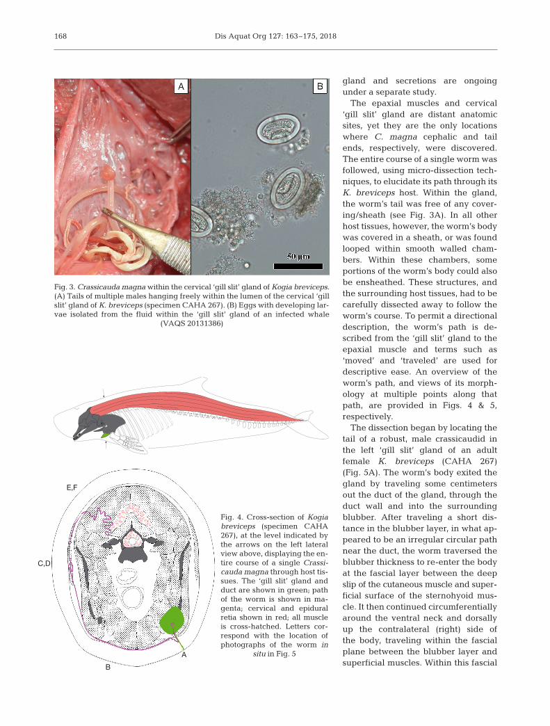
gland and secretions are ongoingunder a separate study.
The epaxial muscles and cervical‘gill slit’ gland are distant anatomicsites, yet they are the only locationswhere C. magna cephalic and tailends, re spectively, were discovered.The entire course of a single worm wasfollowed, using micro-dissection tech-niques, to elucidate its path through itsK. breviceps host. Within the gland,the worm’s tail was free of any cover-ing/ sheath (see Fig. 3A). In all otherhost tissues, however, the worm’s bodywas covered in a sheath, or was foundlooped within smooth walled cham-bers. Within these chambers, someportions of the worm’s body could alsobe en sheathed. These structures, andthe surrounding host tissues, had to becarefully dissected away to follow theworm’s course. To permit a directionaldescription, the worm’s path is de -scribed from the ‘gill slit’ gland to theepaxial muscle and terms such as‘moved’ and ‘traveled’ are used fordescriptive ease. An overview of theworm’s path, and views of its morph -ology at multiple points along thatpath, are provided in Figs. 4 & 5, respectively.
The dissection began by locating thetail of a robust, male crassicaudid inthe left ‘gill slit’ gland of an adultfemale K. breviceps (CAHA 267)(Fig. 5A). The worm’s body exited thegland by traveling some centimetersout the duct of the gland, through theduct wall and into the surroundingblubber. After traveling a short dis-tance in the blubber layer, in what ap -peared to be an irregular circular pathnear the duct, the worm traversed theblubber thickness to re-enter the bodyat the fascial layer between the deepslip of the cutaneous muscle and super -ficial surface of the sternohyoid mus-cle. It then continued circumferentiallyaround the ventral neck and dorsallyup the contralateral (right) side ofthe body, traveling within the fascialplane be tween the blubber layer andsuperficial muscles. Within this fascial
168
Fig. 3. Crassicauda magna within the cervical ‘gill slit’ gland of Kogia breviceps.(A) Tails of multiple males hanging freely within the lumen of the cervical ‘gillslit’ gland of K. breviceps (specimen CAHA 267). (B) Eggs with developing lar-vae isolated from the fluid within the ‘gill slit’ gland of an infected whale
(VAQS 20131386)
E,F
C,D
BA
Fig. 4. Cross-section of Kogiabreviceps (specimen CAHA267), at the level indicated bythe arrows on the left lateralview above, displaying the en-tire course of a single Crassi-cauda magna through host tis-sues. The ‘gill slit’ gland andduct are shown in green; pathof the worm is shown in ma-genta; cervical and epiduralretia shown in red; all muscleis cross-hatched. Letters cor-respond with the location ofphotographs of the worm in
situ in Fig. 5

Keenan-Bateman et al.: Crassicauda magna infection in Kogia breviceps
plane, the worm’s body maintained a very regularsinusoidal path with an average peak to peak ampli-tude of 1.5 cm and period of approximately 2 peaksper 5 cm (Fig. 5B).
At the level of the right sternomas-toid, the worm’s body was arrangedinto several knot-like loops alongits path, which were encased withina thin walled sac-like extension ofthe sheath surrounding the worm(Fig. 5C,D). Within the sac, the worm’sbody was looped upon itself in a con-voluted fashion, giving the appear-ance of a knot. These pseudo-knotsappeared to function to anchor theworm in place, as they were often ob -served at positions where the worm’strajectory changed, or where it entereda new tissue type or muscle. As theworm’s body continued dorsally, itentered the superficial cutaneousmuscle where its path became lesssinusoidal and more straightened, atrajectory that was similar to the circumferentially oriented (i.e. dorso-ventrally vertical) muscle fibers throughwhich it traveled. At the level of inser-tion of the rhomboideus capitis at thecaudal skull, the worm’s body turneddeep, and entered the fascial planebetween the medial surface of therhom boideus capitis and an extensiveoccipital ve nous plexus. Within thisfascial plane, the path of the wormagain became sinusoidal, althoughwith a more gently curved and widerpath. These ob servations suggestedthat the trajectory of the worm wasdependent, at least in part, upon thetissue through which it was traveling.Through fascial planes, the worm’spath was sinusoidal, while within themuscle, its path appeared more con-strained to spaces be tween fascicles.
At the deep surface of the rhom-boideus capitis, the worm’s body,which had been oriented generallywithin a transverse body plane as ittraversed around the circumference ofthe whale, took a virtual 90° turn cau-dad. The worm then entered a richvascular space dorsolateral to thesemispinalis muscle and continued
caudally. At approximately 15 cm caudal to the backof the skull, the worm’s body again appeared tobecome knotted within a thin-walled sac. Upon finemicro-dissection of the sac, these knots were simi-
169
Fig. 5. In situ photographs displaying the pattern of habitat use of Crassicaudamagna within Kogia breviceps (specimen CAHA 267). The letters correspondto specific positions along the path of the worm shown in Fig. 4. (A) Crassicau-did tails hanging freely within the central lumen of the ‘gill slit’ gland. (B) Si-nusoidal path of the worm in the subcutaneous fascial plane. (C) Knot-likeloops along the worm’s path covered by a sheath and (D) with the sheath cov-ering removed. (E) Final chamber within the epaxial musculature where the
worm terminated, and (F) its anterior end (tip of the white arrow)

Dis Aquat Org 127: 163–175, 2018
larly determined to be multiple, tightly wound loopswithin a sac that appeared to be a dilation and thin-ning of the sheath. From this pseudo-knot, theworm’s body burrowed deeply into the epaxial mus-cles (Fig. 4E,F) and traveled mediad, in a path per-pendicular to the orientation of the epaxial musclefibers. The worm’s body then moved obliquely cra-niad and entered into a third, larger, encapsulatedpseudo-knot deep within the caudal semispinalismuscle. This smooth-walled chamber was near thelevel of the vertebral articulation with the first rib(capitulum), just caudal to the transverse processes ofthe first 2 cervical vertebrae, and very near to a spe-cialized vascular structure known as the cervicalrete. Upon opening, this third sac differed in appear-ance from those observed earlier. It was a larger andmore open trabeculated chamber. Within this cham-ber, vast lengths of the worm’s body were observed,loosely coiled in multiple layers of loops rather thanforming a knot-like structure (Fig. 5E). The worm’sce ph alic end was found in this chamber, facing cra-nially (Fig. 5F).
Histological anatomy of the ‘gill slit’ exocrine gland
Preliminary histological analyses indicate that the‘gill slit’ gland is a compound tubuloalveolar exocrinegland with a large central chamber (see Figs. 3A& 5A) and many branching ducts, surrounded by afibrous connective tissue capsule (Fig. 6). The epi -thelial lining of the ducts appears to vary between
stratified columnar and stratified cuboidal. The glan-dular portions, which take the form of both branchedtubules and alveoli, maintain a double cuboidal epi -thelium and contain an eosinophilic luminal product(Fig. 6B).
Description of C. magna using SEM
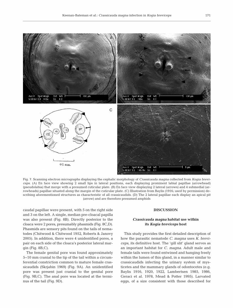
The descriptions below follow the format of bothJohnston & Mawson (1939) and Jabbar et al. (2015).The maximum diameter of preserved material was3×4 mm. The head was rounded, and the stoma wassurrounded by 2 small lateral lips (Fig. 7). Each lipdisplayed a prominent labial papilla (pseudolabium)that merged laterally with a presumed surroundingcuticular plate (ptilina). The apical surface of thepseudolabia did not possess papillae as suggestedby Jabbar et al. (2015), but, rather, had a porous,pit-like appearance (Fig. 7B). The 2 lateral and 4submedial papillae described by Baylis (1916, 1922)as characteristic of Crassicauda (Fig. 7A,B) were ob -served, al though these features appeared somewhatreduced in size relative to Baylis’ (1916, 1920, 1922)illustrations (Fig. 7C) and descriptions. Each of the 2lateral papillae displayed an apical pit, which is pre-sumably an amphid (Fig. 7B,D). Amphids are sen-sory pits found on the anterior end of nematodes(Chitwood & Chitwood 1952, Roberts & Janovy2005).
The male tail possessed prominent caudal alae,and 2 spicules of unequal length that protruded froma cloaca (Fig. 8A,B). Submedial to the cloaca, lateral
170
Fig. 6. Micrographs of the ‘gill slit’ exocrine gland of Kogia breviceps (specimen VAQS 20131386) stained with hematoxylinand eosin. (A) The ‘gill slit’ gland is a compound tubuloalveolar exocrine gland. (B) The glandular portions, which take theform of both branched tubules and alveoli, maintain a double cuboidal epithelium and contain an eosinophilic luminal product
Keenan-Bateman et al.: Crassicauda magna infection in Kogia breviceps
caudal papillae were present, with 5 on the right sideand 3 on the left. A single, median pre-cloacal papillawas also present (Fig. 8B). Directly posterior to thecloaca were 2 pores, presumably phasmids (Fig. 8C,D).Phasmids are sensory pits found on the tails of nema-todes (Chitwood & Chitwood 1952, Roberts & Janovy2005). In addition, there were 4 unidentified pores, apair on each side of the cloaca’s posterior lateral mar-gin (Fig. 8B,C).
The female genital pore was found approximately5− 10 mm cranial to the tip of the tail within a circum-ferential constriction common to mature female cras-sicaudids (Skrjabin 1969) (Fig. 9A). An unidentifiedpore was present just cranial to the genital pore(Fig. 9B,C). The anal pore was located at the termi-nus of the tail (Fig. 9D).
DISCUSSION
Crassicauda magna habitat use within its Kogia breviceps host
This study provides the first detailed description ofhow the parasitic nematode C. magna uses K. brevi-ceps, its definitive host. The ‘gill slit’ gland serves asan important habitat for C. magna. Adult male andfemale tails were found entwined and hanging freelywithin the lumen of this gland, in a manner similar tocrassicaudids infecting the urinary system of mys-ticetes and the mammary glands of odontocetes (e.g.Baylis 1916, 1920, 1922, Lambertsen 1985, 1986,Geraci et al. 1978, Mead & Potter 1995). Larvatedeggs, of a size consistent with those described for
171
Fig. 7. Scanning electron micrographs displaying the cephalic morphology of Crassicauda magna collected from Kogia brevi-ceps. (A) En face view showing 2 small lips in lateral positions, each displaying prominent labial papillae (arrowhead)(pseudolabia) that merge with a presumed cuticular plate. (B) En face view displaying 2 lateral (arrows) and 4 submedial (ar-rowheads) papillae situated along the margin of the cuticular plate. (C) Illustration from Baylis (1916; used by permission) de-scribing aforementioned structures as characteristic of all crassicaudids. (D) The 2 lateral papillae each display an apical pit
(arrow) and are therefore presumed amphids

Dis Aquat Org 127: 163–175, 2018
other crassicaudids (Johnston & Mawson 1939, Mar-golis & Pike 1955, Lambertsen 1986, Geraci et al.1978), have also been observed in the presumedsecretion of the gland, illuminating the likely trans-mission path out of the host.
The cephalic ends of these parasitic nematodes areoften found meters away (curvilinearly), embeddeddeeply within the host’s epaxial musculature, oftennear large vascular beds that feed the central nerv-ous system. Thus, C. magna is capable of penetratingthrough multiple host tissues, including epithelium,blubber, skeletal muscle, connective tissue, fascia,and vasculature. These worms also appear capable ofsignificantly altering the morphology of the tissues inwhich they are embedded. Specifically, dissectionsrevealed extensive tissue damage to the whale’s
cervico-thoracic epaxial muscles, the primary loco-motor muscles that power the upstroke (Pabst 1993).Multiple loops of worm were observed within cav-ernous spaces that appeared ‘excavated’ or ‘digested’out of the surrounding muscle. The absence of mus-cle within these chambers suggests that the wormsare consuming skeletal muscle, and potentially vas-cular tissue and blood.
Additionally, along its length within the K. brevicepshost, the crassicaudid was surrounded by a sheath.Empty sheaths were also observed within host tissues,apparently devoid of living worm tissue. Whether thesheath is secreted by the parasite or is a host reactionto the parasite is not yet known. Similar structures, as-sociated with Crassicauda infection in other cetaceans,have been described. For example, Geraci & St. Aubin
172
Fig. 8. Scanning electron micrographs displaying the tail morphology of a male Crassicauda magna collected from the ‘gill slit’gland. (A) Prominent caudal alae (arrowheads) and 2 spicules of unequal length; submedial to the cloaca are lateral caudalpapillae, with 5 on the right side and 3 on the left (arrows). (B) The smaller of the 2 spicules protrudes only slightly past thecloacal opening (arrowhead). Note also that there is a single median pre-cloacal papilla (arrow). (C) Directly posterior to thecloaca are 2 pores, presumably phasmids (arrows). Note that there are also 4 unidentified pores, a pair on each side of the cloaca’sposterior-lateral margin (arrowheads). (D) Higher magnification view of 1 of the presumed phasmids directly caudal to the cloaca

Keenan-Bateman et al.: Crassicauda magna infection in Kogia breviceps
(1979) reported finding tortuous Crassicauda ‘tracts’in the blubber of the lateral body wall of delphinids,with no worm in situ. Blaxter et al. (1992) provided areview of the role the nematode surface coat plays inevading host immunity in parasitic species. These re-searchers suggest that while the surface coat secretedby the excretory-secretory system of migrating parasiticnematodes can be used to directly evade the host’simmune mechanisms, they can also be shed and leftbehind as deposits to divert the host’s immune responseaway from the parasite as it progresses forward.
At multiple points along the worm’s progressionthrough host tissues, its movement appeared tochange. At these positions, pseudo-knots were oftenob served and resembled anchoring or stabilizingpoints. The sac-like structure covering the pseudo-
knots appeared to be continuous with, and composedof, the same material as the sheath surrounding theworm. Similar structures have been described byother authors in cetaceans from both suborders. Forexample, Baylis (1916) described the disposition ofcrassicaudid heads embedded in ‘masses of connec-tive tissue’ within the kidneys of fin whales. Theauthor described the worm as having a very tortuouscourse within these masses, and later provided illus-trations that were similar in appearance to what isde scribed here as pseudo-knots, suggesting theymay be homologous structures. Similar masses con-taining coiled worms have been described in mys-ticetes by several authors (Hamilton 1916, Rees 1953,Cockrill 1960, Skrjabin 1966, Lambertsen 1986,Geraci & St. Aubin 1987).
173
Fig. 9. Scanning electron micrographs displaying the tail morphology of a female Crassicauda magna collected from the ‘gillslit’ gland. The anal pore is located at the terminus of the tail, which is beyond the edge of the micrograph in (A). (A) Genitalpore (red circle) is found on the presumed ventral surface, approximately 5−10 mm cranial to tail tip, within a circumferentialconstriction (arrows) of the tail common to mature female crassicaudids. (B) Higher magnification view of female genital pore.An unidentified pore (arrowhead) is present just cranial to the genital pore. (C) Higher magnification view of unidentified pore
shown in (B). (D) The anal pore is located at the terminus of the tail

Dis Aquat Org 127: 163–175, 2018
Thus, C. magna appears to use its K. breviceps hostin much the same way as has been described for cras-sicaudids in other cetacean species. They consumehost tissues (likely muscle and vascular tissue); oc -cupy an excretory organ through which their eggscan exit the body (the ‘gill slit’ gland); are covered ina sheath of tissue that surrounds their body; and peri-odically, along their impressive length, arrange them-selves into complicated, looped masses, surroundedby distinct, but as of yet unidentified tissue. Howthese crassicaudid nematodes initially infect K. bre-viceps, how they successfully traverse their path,what cues they use to navigate, and how they progressthrough host tissues are unanswered questions thatwarrant further attention.
Crassicauda magna SEM studies and taxonomic implications
This study provided the first detailed scanningelectron micrographs of taxonomically important fea-tures of the cephalic and tail ends of male and femaleC. magna infecting K. breviceps. It also provided thefirst description of any female tail of this nematodespecies.
The presence of cephalic papillae, which have beenidentified as a diagnostic characteristic of the genusCrassicauda by Baylis (1920), was confirmed in C.magna infecting K. breviceps from the mid-Atlantic.These structures were not reported in descriptions byearlier authors using light microscopy (Johnston &Mawson 1939, Jabbar et al. 2015).
SEM also illuminated several taxonomically im -portant features of the male tail, including caudalpapillae and caudal alae. The males investigated inthis study possessed asymmetry in the number oflateral caudal papillae (Fig. 8A). This pattern differsfrom the most recent light microscopy study by Jab-bar et al. (2015), who reported 5 pairs of lateral cloa-cal papi llae. Caudal alae are used to differentiatebe tween the 2 genera within the subfamily Crassi-caudinae — Crassicauda and Placentonema. Pla cen -to nema are very large nematodes (greater than 8 min length) that infect the placenta of the sperm whalePhyseter macrocephalus and are identified by thepresence of caudal alae, while Crassicauda lack thisfeature (Gubanov 1951). The male Crassicauda in -fecting K. breviceps in this study, and those illus-trated by Dollfus (1966) and Jabbar et al. (2015),clearly possessed caudal alae, unlike any otherCrassicauda species described to date in any ceta -cean species. This result suggests that either the
crassicaudid nematodes that infect K. breviceps maybelong to the genus Placentonema, or that this fea-ture is not diagnostic of the genus. This morphologi-cal observation, and molecular data from Jabbar etal. (2015) that put Crassicauda within a new family,the Acuariidae, suggest that the taxonomy of thisgroup of nematodes re quires additional clarification.Correct phylogenetic placement of these nematodescould also provide insight into the life cycle of thisinadequately studied genus of parasites (Anderson2000).
CONCLUSION
This study provides the first detailed description ofthe use of the Kogia breviceps host habitat by theparasitic nematode Crassicauda magna. This nema-tode appears to feed on skeletal muscle and vasculartissues that are meters away from the exocrine glandused as its site of reproduction and egg transmissionto the environment. SEM revealed taxonomicallyimportant features on the head of C. magna, not pre-viously observed in this K. breviceps-dependent spe-cies. The presence of caudal alae on the male tail, incontrast, bring into question the genus identity of thisparasite. The gross and histological morphology ofthe ‘gill slit’ gland suggests that it produces a secre-tion that may be stored and actively expressed intothe environment, and that further investigation intoits function is warranted. Although the function ofthe ‘gill slit’ gland is not yet known in K. breviceps,this study has identified its crucial role for its crassi-caudid parasite. We hope that the basic description ofthe parasite’s habitat provided in this work provesuseful for future studies of the pathogenesis of crassi-caudosis in this kogiid whale.
Acknowledgements. We thank all members, past and pres-ent, of the North Carolina and Virginia Marine MammalStranding Programs who assisted in the response and inves-tigation of the specimens used in this study. Special thanksto Emily Singleton, Sue Barco, Sarah Mallette, Kristy Phillips,Dr. Vicky Thayer, Jill Sullivan, Karen Clark, Paul Doshkov,Marina Piscitelli, and Clay George at Georgia Departmentof Natural Resources for generously sharing their time andexpertise to help collect specimens for this project. We thankDr. Stephanie Kamel and Dr. Zachary Long for their timeand expertise. We thank the Journal of Natural History forgranting us permission to reproduce Baylis' (1916) originalillustration of the genus Crassicauda within this work. Workwas carried out under UNCW IACUC Protocols A0809-019and A1112-013, and under multiple Stranding Agreementsfrom NOAA SE to UNC Wilmington. This work was sup-ported in part by NOAA Prescott Stranding Grants to UNCW.
174

Keenan-Bateman et al.: Crassicauda magna infection in Kogia breviceps
LITERATURE CITED
Abollo E, López A, Gestal C, Benavente P, Pascual S (1998)Macroparasites in cetaceans stranded on the northwest-ern Spanish Atlantic coast. Dis Aquat Org 32: 227−231
Anderson R (2000) Nematode parasites of vertebrates: theirdevelopment and transmission, 2nd edn. CABI Publish-ing, Cambridge, MA
Baylis HA (1916) VIII. − On Crassicauda crassicauda (Crepl.)[Nematoda] and its hosts. J Nat Hist 17: 144−148
Baylis HA (1920) LV. − Observations on the genus Crassi-cauda. Ann Mag Nat Hist 5: 410−419
Baylis HA (1922) Note on the habitat and structure of Cras-sicauda (Nematoda). Parasitology 14: 9−12
Blaxter ML, Page AP, Rudin W, Maizels RM (1992) Nema-tode surface coats: actively evading immunity. ParasitolToday 8: 243−247
Bush AO, Lafferty KD, Lotz JM, Shostak AW (1997) Para-sitology meets ecology on its own terms: Margolis et al.revisited. J Parasitol 83:575–583
Cardona-Maldonado MA, Mignucci-Giannoni AA (1999)Pygmy and dwarf sperm whales in Puerto Rico and theVirgin Islands, with a review of Kogia in the Caribbean.Caribb J Sci 35: 29−37
Carvalho VL, Bevilaqua CM, Iñiguez AM, Mathews- CasconH and others (2010) Metazoan parasites of ceta ceans offthe northeastern coast of Brazil. Vet Parasitol 173: 116−122
Chitwood BG, Chitwood MB (1952) An introduction tonematology. Monumental Printing, Baltimore, MD
Cockrill WR (1960) Pathology of the Cetacea a veterinarystudy on whales — Part I. Br Vet J 116: 133−144
Dailey M, Stroud R (1978) Parasites and associated pathol-ogy observed in cetaceans stranded along the Oregoncoast. J Wildl Dis 14: 503−511
Dollfus RP (1966) Helminthofaune de Kogia breviceps (Blain -ville, 1838) cètace odontocète. Recoltes du Dr. Duguy.Ann Soc Sci Nat Charente Mar 5: 3−6
Geraci JR, Lounsbury VJ (1993) Marine mammals ashore: afield guide for strandings, 2nd edn. National Aquariumin Baltimore, Baltimore, MD
Geraci JR, St. Aubin DJ (1979) Stress and disease in the marineenvironment: insights through strandings. Biology of mar-ine mammals: insights through strandings. NOAA/ NMFSNat Tech Info Serv Rep No. 2100(77), Washington, DC
Geraci JR, St Aubin DJ (1987) Effects of parasites on marinemammals. Int J Parasitol 17: 407−414
Geraci JR, Dailey MD, St Aubin DJ (1978) Parasitic mastitisin the Atlantic white-sided dolphin (Lagenorhynchusacutus) as a probable factor in herd productivity. J FishRes Board Can 35: 1350−1355
Gibson DI, Harris EA, Bray RA, Jepson PD, Kuiken T, BakerJR, Simpson VR (1998) A survey of the helminth para-sites of cetaceans stranded on the coast of England andWales during the period 1990–1994. J Zool 244: 563−574
Gubanov A (1951) Gigantic nematode from the placenta ofcetaceans – Placentonema gigantissima nov. gen. nov.sp. (translated from Russian). Dokl Akad Nauk SSSR 77:1123–1125
Hamilton JE (1916) Biological problems incidental to theBelmullet whaling station. Report of the 85th meeting ofthe British Association for the Advancement of Science,London, p 124–146
Handley CO (1966) A synopsis of the genus Kogia (pygmysperm whales). In: Norris KS (eds) Whales, dolphins, andporpoises. University of California Press, Berkley, CA,p 62–69
Jabbar A, Beveridge I, Bryant MS (2015) Morphological andmolecular observations on the status of Crassicaudamagna, a parasite of the subcutaneous tissues of the pygmysperm whale, with a re-evaluation of the systematic rela-tionships of Crassicauda. Parasitol Res 114: 835−841
Johnston TH, Mawson PM (1939) Internal parasites of thepigmy sperm whale. Rec South Aust Mus 6: 263−274
Keenan-Bateman TF, McLellan WA, Harms CA, PiscitelliMA and others (2016) Prevalence and anatomic site ofCrassicauda sp. infection, and its use in species identifi-cation, in kogiid whales from the mid-Atlantic UnitedStates. Mar Mamm Sci 32: 868−883
Lambertsen RH (1985) Taxonomy and distribution of a Cras-sicauda species (Nematoda, Spirurida) infecting the kid-ney of the common fin whale (Balaenoptera physalusLinnè, 1758). J Parasitol 71: 485−488
Lambertsen RH (1986) Disease of the common fin whale(Balaenoptera physalus) — crassicaudiosis of the urinarysystem. J Mammal 67: 353−366
Lambertsen RH (1992) Crassicaudosis: a parasitic diseasethreatening the health and population recovery of largebaleen whales. Rev Sci Tech 11: 1131−1141
Lambertsen RH, Birnir B, Bauer JE (1986) Serum chemistryand evidence of renal failure in the North Atlantic finwhale population. J Wildl Dis 22: 389−396
López A, Valeiras X, Pascual S, Abollo E, Díaz JI (1999) Skulllesions associated with Crassicauda spp. (Nematoda,Crassicaudidae) in cetaceans stranded on the Galiciancoast (NW Spain). Proc Twelfth Annu Conf EuropeanCetacean Society, Monaco. Eur Res Cet 12:342–347
Margolis L, Pike GC (1955) Some helminth parasites of Cana-dian Pacific whales. J Fish Res Board Can 12: 97−120
Mead JG, Potter CW (1995) Recognizing two populations ofthe bottlenose dolphin (Tursiops truncatus) off the At -lantic coast of North America: morphologic and ecologicconsiderations. IBI Rep 5: 31−44
Mignucci-Giannoni AA, Hoberg EP, Siegel-Causey D, Wil l -iams EH (1998) Metazoan parasites and other symbiontsof cetaceans in the Caribbean. J Parasitol 84: 939−946
Oliveira JB, Morales JA, Gonzalez-Barrientos RC, Hernan-dez-Gamboa J, Hernandez-Mora G (2011) Parasites ofcetaceans stranded on the Pacific coast of Costa Rica. VetParasitol 182: 319−328
Pabst DA (1993) Intramuscular morphology and tendongeometry of the epaxial swimming muscles of dolphins.J Zool 230: 159−176
Perrin WF, Powers JE (1980) Role of a nematode in the naturalmortality of spotted dolphins. J Wildl Manag 44: 960−963
Rees G (1953) A record of some parasitic worms from whalesin the Ross Sea area. Parasitology 43: 27−34
Ridgway SE (1972) Mammals of the sea: biology and medi-cine. Charles H. Thomas Press, Springfield, IL
Roberts LS, Janovy J Jr (2005) Foundations of parasitology,7th edn. McGraw and Hill, New York, NY
Schulte HVW, Smith M, Andrews RC (1918) The externalcharacters, skeletal muscles and peripheral nerves ofKogia breviceps (Blainville). Bull Am Mus Nat Hist 38:Article 2
Skrjabin AS (1966) A new Crassicauda (Crassicauda dela mu -reana n. sp.), a parasite of the sei whale. Problemy Paraz-itologii, Kraevaya Parazitologiya i Prirodnaya Ochagov-ost Transmissivnikh Boleznei. Dokl Akad Nauk Ukr SSSR,p 100–107
Skrjabin KI (1969) Key to parasitic nematodes, Vol 1: Spiru-rata and Filariata. Israel Program for Scientific Transla-tions, Jerusalem
175
Editorial responsibility: Stephen Raverty, Abbotsford, British Columbia, Canada
Submitted: July 25, 2017; Accepted: December 19, 2017Proofs received from author(s): February 26, 2018