Embed Size (px)
Citation preview

Haematuria: from identification to treatment
King, K. 1 MSc, RN (Adult), PG Dip, BSc (Hons).Research Associate Nurse, UCL (Department of
Urology, Medical School, Rockefeller building, 74 Huntley Street, London, WC1E 6AU)
Steggall, M. 2 PhD, MSc, BSc (Hons), RN (Adult), FHEA, Associate Dean, Director of Undergraduate
Studies, City University London; Clinical Nurse Specialist (ED/PE) Barts Health NHS Trust
Corresponding author:
K King
UCL School of Pharmacy,
Department of Practice and Policy,
Mezzanine Floor, BMA House,
Tavistock Square, London, WC1H 9JP
Abstract
Haematuria has a prevalence of 0.1% - 2.6%. Potential diagnoses may include infection,
kidney stones, trauma, exercise or spurious causes, such as foods, drugs or menstruation,
and a tumour. Approximately 40% of patients with haematuria are found to have a
significant underlying pathology, with half of these having a urological malignancy.
Haematuria is subsequently known as the “classic presentation” of bladder cancerwith 70 -
80% of patients experiencing painless, gross (visible) haematuria. However, in all cases of
visible haematuria, a tumour should be suspected until proven otherwise.
A patient with visible haematuria requires urgent, stringent investigation warranting
specialist assessment and subsequent selective referral through a series of patient centred
investigations at a Haematuria clinic. One-stop clinics have shown to improve the patient
experience in early diagnosis of potentially life threatening conditions although such clinics
are less common in Urology settings, despite morbidity and mortality from bladder cancer
increasing. This paper will discuss the tests and investigations that need to be undertaken in
an individual with either visible or non-visible haematuria, and outline the care that is
needed to support patients through the investigation process.
Key words: haematuria; diagnosis; investigations; management; haematuria clinics; nursing.
Introduction
Blood in the urine, known as haematuria, is classified as microscopic or macroscopic, or
more recently, (based upon joint guidelines from the Renal Association and the British
Association of Urological Surgeons) visible haematuria (VT) and non-visible haematuria
(NVH) (BAUS/RA, 2007; Lotan, et al,2009) and can indicate significant underlying disease. In
visible haematuria, which has also been referred to as gross haematuria, urine is coloured
pink or red and is reported by the patient or seen by the healthcare practitioner during
urinalysis. Non-visible haematuria has also been referred to as dipstick-positive haematuria,
is subdivided into symptomatic NVH (where the symptoms include urinary hesitancy,
urgency or dysuria) and asymptomatic NVH (where detection is incidental and in the
absence of symptoms).
Haematuria is a common urological condition, occurring in up to 10-20% of individuals,
presenting in any age group, and accounting for up to 20% of Urology referrals (Miah and
Catto, 2010). Causes of haematuria vary by age, gender, ethnic group, duration or a
combination of these (Ng, et al.2012). Irrespective of the cause, haematuria requires
investigation due to its potential to indicate malignancy. Although haematuria is not always
a herald of malignancy (Watkins, 2007), between 85 - 90% of patients that present with
painless haematuria have a tumour of the urinary tract.
Transient causes of haematuria, for example urinary tract infection (UTI), exercise induced
haematuria and menstruation, need to be excluded before the extent of the severity of
haematuria can be established (Henning, et al. 2013). Vigorous exercise, for example long-
distance running and swimming, can cause transient haematuria that usually resolves within
24-48 hours (Rees, et al. 2008). Women are at greater risk than men of experiencing
haematuria, as they are more likely to develop UTI, for example cystitis, which are the most
common causes of haematuria (Fernandes, et al, 2012). This is because the female urethra
is shorter and straighter, and closer to the anus (and gut flora) compared to males, hence
the increased occurrence of urinary tract infection in women. This frequency of UTI/cystitis
may result in failure to appropriately investigate females, resulting in delayed diagnosis
(Watkins, 2007; Johnson, et al.2008). In view of this potential to under- or misdiagnose
females, Kelly, et al. (2009) contends that urinalysis should only be performed after
menstruation has finished.
Although haematuria is more common in the elderly (particularly males, over 60 years) than
in the young, it is important to gather clues as to the underlying cause of the haematuria,
for example, loin pain suggests renal origin; colicky pain indicates stones/calculi; and
painless haematuria may indicate a tumour (Ng et al., 2012). Visible haematuria conveys a
higher risk of urological malignancy (14.7%-21.8% of cases) compared with non-visible
haematuria (2% - 11% of cases) (O’Regan et al, 2009), although the additional factors of
positive smoking history, age >40 years, previous malignancies and occupational exposure
to rubber, dyes and chemicals, significantly increase the risk of developing bladder cancer.
Although, when these risks are eliminated or reduced, for example, following the removal of
aromatic amines from dyes, bladder cancer incidence decreases (Christiani, 2011).
The aim of this paper is to outline the tests and investigations that are undertaken once a
patient is referred to a haematuria clinic, and to discuss the role of the nurse in supporting
the patient and improving patient satisfaction.
Referral
To balance the need for unnecessary investigations, Loo, et al. (2009) recommend urgent
referral for specialist opinion or investigations for all male and female patients with visible
haematuria on a single urinalysis, irrespective of age (and irrespective of whether anti-
coagulants or anti-platelet therapy is present). For those experiencing non-visible
haematuria, both asymptomatic and symptomatic patients should be referred if they
provide at least two of three positive urinalysis tests, again, irrespective of age. This is in line
with the referral criteria by NICE (2005) for suspected cancer and follows the ‘two week
wait’ directive within Primary Care (Department of Health, 1997).
Despite previous research showing evidence for unequal referral patterns for the urological
evaluation of haematuria (Johnson et al, 2008) the NICE guidelines do not discriminate
between genders. Patients with haematuria need to be monitored and reviewed from a
medical and economic perspective (Mostafid et al, 2010), with investigations being specific
and accurate to prevent over-investigating, avoid under or misdiagnosing cancer,whilst
simultaneously maximising patient satisfaction and reducing patient anxiety.
Investigations
Once the patient has been referred to the haematuria clinic, patients will undergo a series
of investigations (refer to Figure 1 and Table 1).
Insert Figure 1
Insert Table 1
Within both referral and initial investigations, the patient will be asked to provide their
background history (see Table 2), to identify possible causes of haematuria.
Insert table 2
At the clinical visit, the potential causes, along with any associated risk factors, will be
established. Patients will be informed of the range of causes of haematuria, for example,
some foods (e.g. beetroot) can indicate the presence of blood, but this is ‘normal’
discolouration of the urine. These samples would create ‘false positive’ results where a test
incorrectly indicates a positive result, but the disease is not present. Normally, the cause of
these cases is not serious or does not require further investigation.
For most patients however further tests will be needed. Local policies and corresponding
investigation protocols may dictate the tests which patients receive (Loo et al, 2009). It is
important that the rationale for each test is given to the patient, as well as explaining what
the test may (or may not) indicate, and which further investigations may be required.
Patients should also be advised that not all tests will show urological or renal pathology and
a negative result does not always mean absence of disease.
Blood tests
Blood profile (full blood count) is needed to detect the presence of anaemia, and serum
urea, creatinine and electrolytes are needed to identify any renal impairment (Miah and
Catto, 2010). If there is evidence of a declining glomerular filtration rate (GFR) (by 10ml/min
at any stage within the previous 5 years or by >5ml/min with the last year) and if
urea/electrolyte/creatinine indicate stage 4 or 5 kidney disease, a referral to a nephrologist
will be needed (BAUS/AR, 2007).
Urine tests
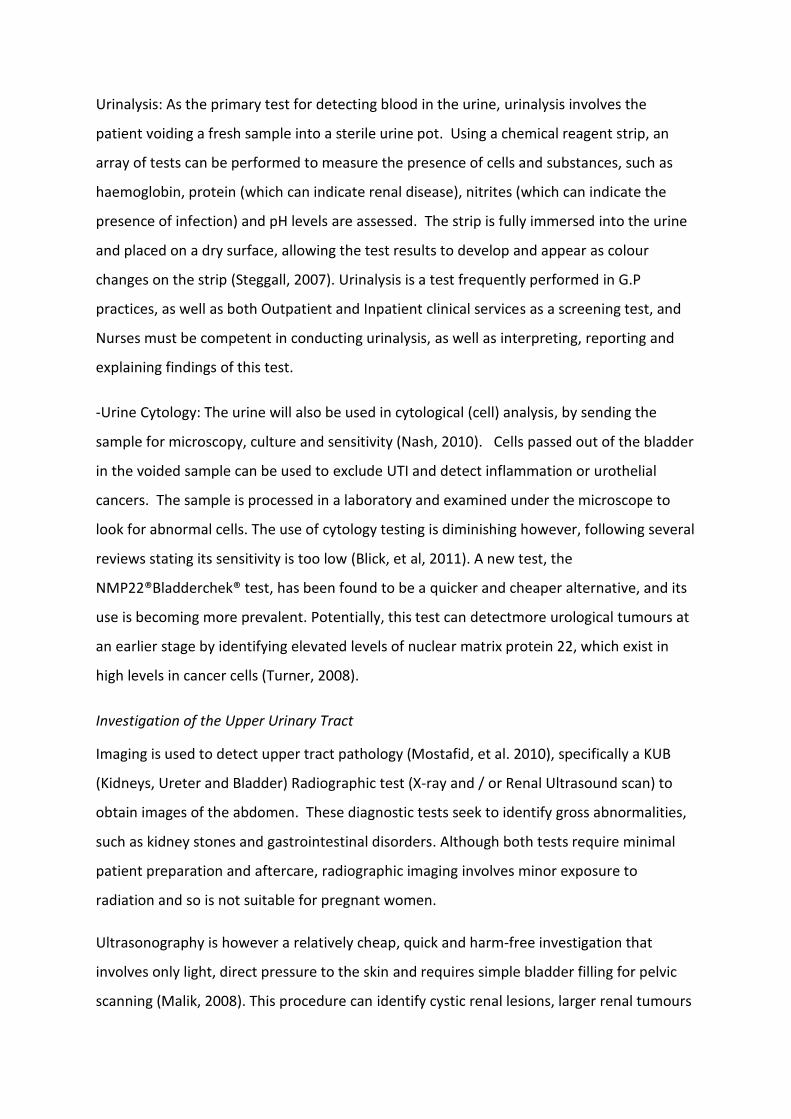
Urinalysis: As the primary test for detecting blood in the urine, urinalysis involves the
patient voiding a fresh sample into a sterile urine pot. Using a chemical reagent strip, an
array of tests can be performed to measure the presence of cells and substances, such as
haemoglobin, protein (which can indicate renal disease), nitrites (which can indicate the
presence of infection) and pH levels are assessed. The strip is fully immersed into the urine
and placed on a dry surface, allowing the test results to develop and appear as colour
changes on the strip (Steggall, 2007). Urinalysis is a test frequently performed in G.P
practices, as well as both Outpatient and Inpatient clinical services as a screening test, and
Nurses must be competent in conducting urinalysis, as well as interpreting, reporting and
explaining findings of this test.
-Urine Cytology: The urine will also be used in cytological (cell) analysis, by sending the
sample for microscopy, culture and sensitivity (Nash, 2010). Cells passed out of the bladder
in the voided sample can be used to exclude UTI and detect inflammation or urothelial
cancers. The sample is processed in a laboratory and examined under the microscope to
look for abnormal cells. The use of cytology testing is diminishing however, following several
reviews stating its sensitivity is too low (Blick, et al, 2011). A new test, the
NMP22®Bladderchek® test, has been found to be a quicker and cheaper alternative, and its
use is becoming more prevalent. Potentially, this test can detectmore urological tumours at
an earlier stage by identifying elevated levels of nuclear matrix protein 22, which exist in
high levels in cancer cells (Turner, 2008).
Investigation of the Upper Urinary Tract
Imaging is used to detect upper tract pathology (Mostafid, et al. 2010), specifically a KUB
(Kidneys, Ureter and Bladder) Radiographic test (X-ray and / or Renal Ultrasound scan) to
obtain images of the abdomen. These diagnostic tests seek to identify gross abnormalities,
such as kidney stones and gastrointestinal disorders. Although both tests require minimal
patient preparation and aftercare, radiographic imaging involves minor exposure to
radiation and so is not suitable for pregnant women.
Ultrasonography is however a relatively cheap, quick and harm-free investigation that
involves only light, direct pressure to the skin and requires simple bladder filling for pelvic
scanning (Malik, 2008). This procedure can identify cystic renal lesions, larger renal tumours
and is used for testing renal morphology and structure. However, ultrasound may fail to
identify small lesions, and in these situations further investigation may be required with an
X-ray of the Urinary system (Intravenous Urography or IVU). However IVU can also fail to
diagnose small renal or bladder masses, or other causes of haematuria such as calculi.
McDonald, et al. (2006) contends that the only reliable way of detecting bladder cancer is
via the lower urinary tract, by flexible cystoscopy.
Investigation of the Lower Urinary Tract
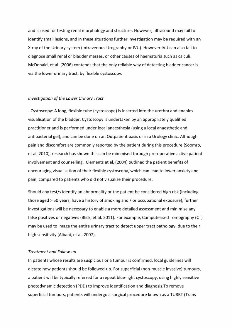
- Cystoscopy: A long, flexible tube (cystoscope) is inserted into the urethra and enables
visualisation of the bladder. Cystoscopy is undertaken by an appropriately qualified
practitioner and is performed under local anaesthesia (using a local anaesthetic and
antibacterial gel), and can be done on an Outpatient basis or in a Urology clinic. Although
pain and discomfort are commonly reported by the patient during this procedure (Soomro,
et al. 2010), research has shown this can be minimised through pre-operative active patient
involvement and counselling. Clements et al, (2004) outlined the patient benefits of
encouraging visualisation of their flexible cystoscopy, which can lead to lower anxiety and
pain, compared to patients who did not visualise their procedure.
Should any test/s identify an abnormality or the patient be considered high risk (including
those aged > 50 years, have a history of smoking and / or occupational exposure), further
investigations will be necessary to enable a more detailed assessment and minimise any
false positives or negatives (Blick, et al. 2011). For example, Computerised Tomography (CT)
may be used to image the entire urinary tract to detect upper tract pathology, due to their
high sensitivity (Albani, et al. 2007).
Treatment and Follow-up
In patients whose results are suspicious or a tumour is confirmed, local guidelines will
dictate how patients should be followed-up. For superficial (non-muscle invasive) tumours,
a patient will be typically referred for a repeat blue-light cystoscopy, using highly sensitive
photodynamic detection (PDD) to improve identification and diagnosis.To remove
superficial tumours, patients will undergo a surgical procedure known as a TURBT (Trans

Urethral Resection of the Bladder), whereby any unusual growth is removed from the
bladder wall. To prevent progression, and in conjunction with surgery, additional therapies
such as BCG (Bacillus Calmette-Guerin) intravesical therapy (a vaccination) or mitomycin C
(chemotherapy) will be given.
For more aggressive tumours of the bladder (i.e. muscle invasive cancer) suitable patients
will be given chemotherapy to shrink the cancer, followed by either radiotherapy or surgery.
Current radiotherapy aims to maximise the delivery of radiation to tumour tissue, whilst
minimising that to healthy tissues.
When the tumour is advanced and invasive, or if the patient has had repeated recurrence of
non-muscle invasive cancer, a cystectomy may be required. This is the surgical removal of all
or part of the bladder, with the formation of an ileostomy (if the entire bladder is removed).
Once treatment is complete, the patient will receive regular follow-up appointments to
monitor their health status, due to the high risk of recurrence in bladder cancer. A typical
follow-up plan includes repeated physical examination, blood tests, urine cytology and
imaging tests, as well as regular cystoscopy (assuming the bladder has not been removed).
Any recurrence of haematuria would warrant further/repeat investigation.
Support for Patients
Irrespective of the investigations that the patient undergoes, recommendations for lifestyle
changes may be appropriate along their journey to minimise, or if possible prevent, any
current or future risks to the patient’s health.
Treatment, combined with a diagnosis of any condition associated with haematuria, can
involve symptoms and side effects, as well as being physically exhausting and emotionally
stressful for the patient. The focus of healthcare is to provide compassionate and sensitive
care based around six values, the ‘Six C’s’ care, compassion, courage, communication,
competence and commitment (Department of Health, 2012), and to deliver compassionate
care, patients will need the opportunity to talk and raise concerns that they might have. It is
important that patients feel listened to, understood and actively involved with their care
(Gourdji, et al.2003).
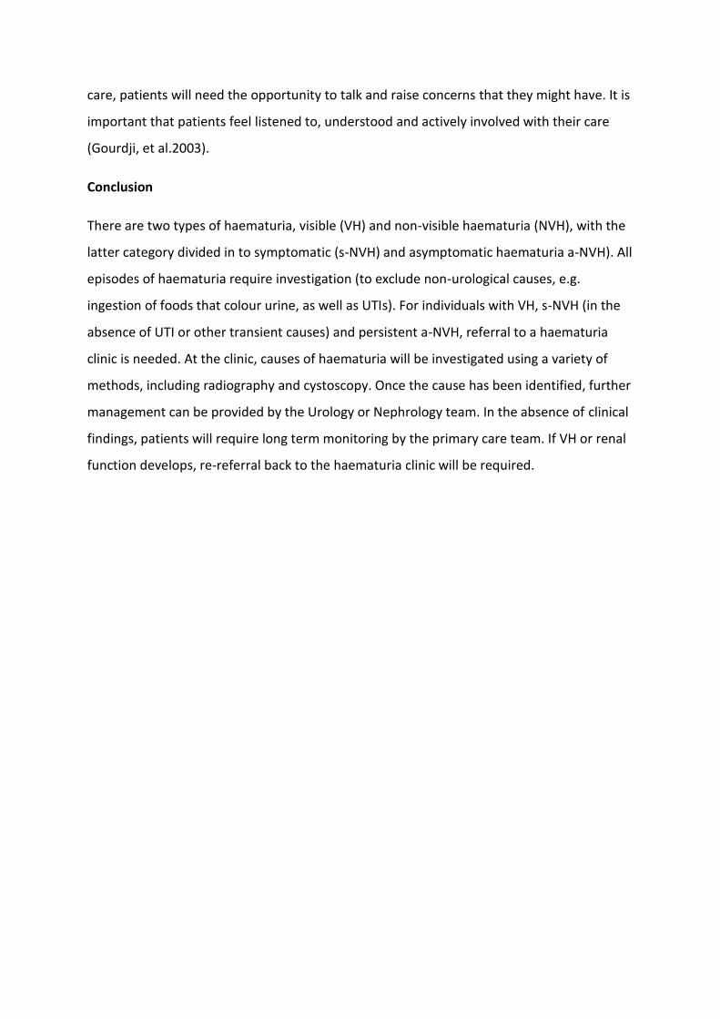
Conclusion
There are two types of haematuria, visible (VH) and non-visible haematuria (NVH), with the
latter category divided in to symptomatic (s-NVH) and asymptomatic haematuria a-NVH). All
episodes of haematuria require investigation (to exclude non-urological causes, e.g.
ingestion of foods that colour urine, as well as UTIs). For individuals with VH, s-NVH (in the
absence of UTI or other transient causes) and persistent a-NVH, referral to a haematuria
clinic is needed. At the clinic, causes of haematuria will be investigated using a variety of
methods, including radiography and cystoscopy. Once the cause has been identified, further
management can be provided by the Urology or Nephrology team. In the absence of clinical
findings, patients will require long term monitoring by the primary care team. If VH or renal
function develops, re-referral back to the haematuria clinic will be required.

References Albani,J.M., Ciaschini, M.W., Streem, S.B., Herts, B.R. &Angermeier, K.W. (2007). The role of computerised tomographic urography in the initial evaluation of hematuria.Journal of Urology. 177, 2, 644 – 648.
Blick, C.G.T.,Nazir, S.A., Mallet, S., Turney, B.W., Onwu, N.N, Roberts, I.S.D., Crew, J.P & Cowan, N.C. (2011). Evaluation of diagnostic strategies for bladder cancer using computed tomography (CT) urography, flexible cystoscopy and voided urine cytology: results from 778 patients from a hospital haematuria clinic. British Journal of Urology.110, 84 – 94.
British Association of Urological Surgeons/Renal Association (2008) Joint Consensus Statement on the Initial Assessment of Haematuria. http://www.baus.org.uk/AboutBAUS/publications/haematuria-guidelines
Clements, S.D, Sells, H. & Wright, M.P.J. (2004). Use of video in flexible cystoscopy: a prospective randomized study of effect on patient experience. Journal of Ambulatory Surgery.11:45-6.
Christiani, D. (2011). Combating Environmental Cause of Cancer.The New England Journal of Medicine.364: 791- 793.
Department of Health (1997).The New NHS, Modern and Dependable. London: The Stationary Office. Department of Health.(1998).The NHS, modern and dependable; a national framework for assessing performance. London: NHS Executive.
Department of Health (2012).Compassion in Practice.Nursing, Midwifery and Care Staff.Our Vision and Strategy. Available: www.commissiongboard.nhs.uk Accessed 16 April 2013. Fernandes, J.P; Amorim, R., Gomes, M.J., Oliveira, V., Reis, A. &Ribeiro-Castro, J.(2012), Posterior Nutcracker syndrome with left Renal Vein Duplication: A Rare Case of Haematuria in a 12 year Old Boy. Case Reports in Urology.Vol. 2012, 1 – 4.
Gourdji, I., McVey, L. & Carmen, L. (2003).Patients’ Satisfaction and Importance Ratings of Quality in an Outpatient Oncology Center.Journal of Nursing Care Quality.18: (1), 43 -55. Henning,A.,Wehrberg,M.,Madersbacher,S.,Pycha,A.,Martini,T.,Comploj,E.,Jeschke,K.,Tripolt,C.&Rauchenwald,M. (2013). Do differences in clinical symptoms and referral patterns contribute to the gender gap in bladder cancer? British Journal of Urology International. doi:10.1111/j.1464-410X2012.11661.x

Johnson, E.K., Daignault, S., Zhang, Y. & Lee, C.T. (2008). Patterns of haematuria referral to Urologists: does a gender disparity exist? Urology. 72: 498 – 502.
Kelly, J.D.; Fawcett, D.P. & Goldberg, L.C. (2009).“Assessment and management of non-visible haematuria in primary care.Clinical Review.”British Medical Journal.338, 227 – 338. Loo, R., Whittaker, J.&Rabrenovich, V. (2009). National Practice Recommendations for Haematuria: How to Evaluate in the Absence of Strong Evidence. The Permanente Journal.13 (1): 37 – 46. Lotan, Y., Elias, K., Svatek, R.S., Bagrodia, A., Nuss, G., Moran, B. &Sagalowsky, A.I. (2009). Bladder Cancer Screening in a High Risk Asymptomatic Population Using a Point of Care Urine Based Protein Tumour Marker. The Journal of Urology.182, 52 – 58.
Malik, T. (2008).Ultrasonography.The Professional Medical Journal.15, (01), 26.
McDonald, M.M., Swagerty, D. & Wetzel, L. (2006).Assessment of microscopic hematuria in adults. American Family Physician.73, 10, 1748 – 1754.
Miah, S. &Catto, J. W. F. (2010) Haematuria. Renal and Urology II Surgery, 28:12.
Mostafid, H., Persad, R., Kockelbergh, R. & Fawcett, D. (2010). Is it time to re-design the Haematuria Clinic? British Journal of Urology International.1: 585 – 587. Nash, E. (2010). Haematuria. InnovAiT: The RCGP Journal for Associates in Training.3: 30.
National Institute for Clinical Excellence. (2005) Referral Guidelines for Suspected Cancer.Quick Reference Guide. NICE, London.
Ng, L.K., Htun, T.H., Dublin, N., Ong, T.A. and Razack, A.H. (2012).Assessment and Clinical Significance of Haematuria in Malaysian Patients –Relevance to Early Cancer Diagnosis.Research Communication Asian Pacific Journal of Cancer Prevention, 13, 2515 – 2518.
O’Regan, K.N., O’Connor, O.J., McGloughlin, P. and Maher, M. (2009).The role of imaging in the investigation of painless haematuria in adults. Semin. Ultrasound CT MR.30 (4), 258 – 70. Rao, P.K & Jones, J.S. (2008). Screening for Bladder Cancer in Primary Care: A Comprehensive Review. Medscape Urology.1: 2. Rees, J., Patel, B. and Persad, R. (2008) Asymptomatic dipstick haematuria. Trends in Urology, Gynaecology and Sexual Health 13(1):14-16

Soomro, K.Q., Nasir, A.R. and Ather, M.H.(2010). Impact of Patient’s Self-Viewing of Flexible Cystoscopy on Pain using a Visual Analog Scale in a Randomised Controlled Trial. Urology 77,(1), 21 – 23. Steggall, M. (2007).Urine samples and urinalysis. Nursing Standard, 22(14-16), 42-45. Turner, B. (2008). Haematuria: Causes and Management. Nursing Standard.23, (1), 50 – 56. Watkins, J. (2007) Haematuria. Practice Nursing 18(8):398

Figure 1: An Investigatory Flowchart for patients referred to the Haematuria clinic
History, examination, urinalysis, bloods
Ultrasound scan
Flexible
cystoscopy (if
needed)
(Kelly, Fawcett & Goldberg, 2009)
Normal 1st line
1st line abnormality
Dipstick positive
Dipstick negative
2nd line investigation
(Biopsy, Bladder resection)
Discharge
Intravenous urogram
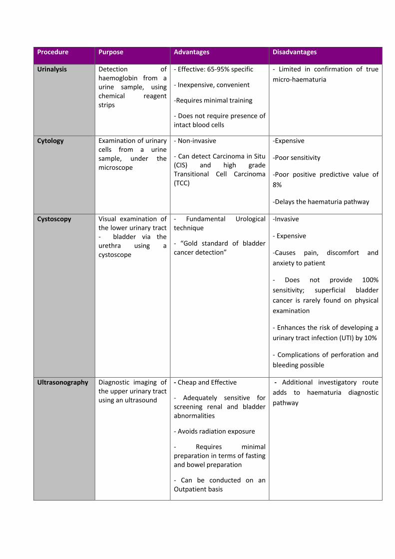
Procedure Purpose Advantages Disadvantages
Urinalysis Detection of haemoglobin from a urine sample, using chemical reagent strips
- Effective: 65-95% specific
- Inexpensive, convenient
-Requires minimal training
- Does not require presence of intact blood cells
- Limited in confirmation of true
micro-haematuria
Cytology Examination of urinary cells from a urine sample, under the microscope
- Non-invasive
- Can detect Carcinoma in Situ (CIS) and high grade Transitional Cell Carcinoma (TCC)
-Expensive
-Poor sensitivity
-Poor positive predictive value of
8%
-Delays the haematuria pathway
Cystoscopy Visual examination of the lower urinary tract - bladder via the urethra using a cystoscope
- Fundamental Urological technique
- “Gold standard of bladder cancer detection”
-Invasive
- Expensive
-Causes pain, discomfort and
anxiety to patient
- Does not provide 100%
sensitivity; superficial bladder
cancer is rarely found on physical
examination
- Enhances the risk of developing a
urinary tract infection (UTI) by 10%
- Complications of perforation and
bleeding possible
Ultrasonography Diagnostic imaging of the upper urinary tract using an ultrasound
- Cheap and Effective
- Adequately sensitive for screening renal and bladder abnormalities
- Avoids radiation exposure
- Requires minimal preparation in terms of fasting and bowel preparation
- Can be conducted on an Outpatient basis
- Additional investigatory route
adds to haematuria diagnostic
pathway

Table 1: Diagnostic tests for Haematuria (Albani et al, 2007; Malik, 2008; Rao and Jones, 2008; O’Regan et al, 2009; Soomro, et al, 2010).

Background Information Rationale
Age Haematuria is more common in the older adult. Potential diagnoses
include bladder cancer, prostate cancer, etc.
Sex Females have an increased risk of haematuria due to Urinary Tract
Infections (UTIs) and menstruation.
Occupation Occupational exposure to dyes, rubbers, textiles and chemicals
have found to be associated with bladder cancer.
Family history An onset of persistent microscopic hematuria, typically during
childhood is indicative of “familial haematuria;” a group of genetic
kidney disorders for example.
Medical History For example, a history of previous urological conditions may
indicate recurrence.
Physical Activity
Vigorous exercise, mild trauma or sexual intercourse can all be a
cause of transient haematuria.
Diet Foods such as beetroot, blackberries and rhubarb can cause urine
to look red or brown.
Medication Haematuria may be drug-induced, by non-steroidal anti-
inflammatory drugs, antibiotics or anticoagulants.
Smoking history Smoking significantly increases the risk of bladder cancer and
subsequent likelihood of haematuria.
Drug use Ketamine dependence for example is associated with urinary tract
effects.
Haematuria history Number and type of episodes of haematuria, associated pain,
urological symptoms, and point in the urinary stream when the
haematuria is visible, will all enable the clinician to help identify
potential causes
Table 2: Information required for the Haematuria Patient’s History (Adapted from Turner, 2008;
Nash, 2010).