Embed Size (px)
Citation preview

CHHS18/026
Canberra Hospital and Health ServicesClinical ProcedureHaemodialysis for Adults Contents
Contents....................................................................................................................................1
Purpose.....................................................................................................................................2
Scope........................................................................................................................................ 2
Section 1 – Haemodialysis/Haemodiafiltration.........................................................................2
Section 2 – Medication Administration during Haemodialysis..................................................4
Section 3 – Dialysis related procedures....................................................................................8
Section 4 – Complications during Haemodialysis....................................................................17
Section 5 – Anticoagulant Free Haemodialysis........................................................................21
Section 6 – Monthly Blood Review..........................................................................................27
Section 7 – Administration of Blood and Blood products on dialysis......................................28
Section 8 – Calcium Chloride Infusions during Regional Citrate Anticoagulated Filtration based Plasma Exchange..........................................................................................................29
Definition of Terms................................................................................................................. 33
Implementation...................................................................................................................... 34
Related Policies, Procedures, Guidelines and Legislation.......................................................34
References.............................................................................................................................. 35
Search Terms.......................................................................................................................... 37
Doc Number Version Issued Review Date Area Responsible PageCHHS18/026 1 19/01/2018 01/12/2020 Medicine 1 of 37Do not refer to a paper based copy of this policy document. The most current version can be found on the ACT Health Policy Register

CHHS18/026
Purpose
This procedure outlines the clinical processes for managing patients with kidney failure.
Back to Table of Contents
Scope
Applies to Renal nurses who are trained and deemed competent and practice under the governance of the Renal Network (ACT Health and Southern NSW Local Health District -SNSWLHD)
Back to Table of Contents
Section 1 – Haemodialysis/Haemodiafiltration
This section covers techniques required to deliver a Haemodialysis or Haemodiafiltration treatment.Patients will receive a Haemodiafiltration treatment except those who cannot achieve a 21 Litre substitution volume (after three attempts). In this case a conventional haemodialysis treatment will be delivered.
Standard Treatment1. New chronic dialysis patients : Follow chronic pathway for first six treatments (see
attachment “Chronic Pathway”)2. Acute dialysis patients: Obtain an individualized prescription for each treatment3. Chronic dialysis patients (after the pathway is completed)
When determining dialysis prescription, refer to relevant standing orders and chart below:
A. 12- 21 hours per weekBFR 150 – 500 mL/minDialysate flow 500 mL/min Dialyser Polyflux Dialyser surface area ≥ to 75% body surface areaMaximum net Ultra Filtration (UF) rate
1000 mL/hr (with nurse or carer) or 500mL/hr no assistant
Maximum isolated UF rate Not to exceed 1 Litre /30 minutes (in centre only)Dialysate temperature 36° CVenous pressure Upper limit 250 mmHgArterial pressure Lower limit - 250 mmHgIonic clearance target ≥ 1.1Kt/v ≥ 1.3Haemodiafiltration HDF >21 L Substitution volume achievedTarget Dialysis Dose Determined by ATR
Doc Number Version Issued Review Date Area Responsible PageCHHS18/026 1 19/01/2018 01/12/2020 Medicine 2 of 37Do not refer to a paper based copy of this policy document. The most current version can be found on the ACT Health Policy Register

CHHS18/026
B. Nocturnal > 21 hours dialysis per weekBFR 200 - 250 mL/minDialysate flow 300 mL/minDialyser Polyflux Dialyser surface area 140H – 170H or as prescribed
by physicianTarget Dialysis Dose Physician assessedDialysate temp 36° CMaximum UF rate with carer 1000 mL/hrMaximum UF rate without carer 500 mL/hr
Correction of low phosphate: addition of fleet enema to SoftpacTM
1. Equipment Fleet Ready-to-Use Enema® 133mL Gambro SoftpacTM concentrate for Haemodialysis Alcohol swab Measuring cup 18 g Terumo drawing up blunt needle 30mL syringe
2. ProceduresUsual dose 15-30mL of Fleet enema added to 3.5L SoftpacTM As serum phosphate levels are affected by variables such as dialyser clearance, dialysis time, blood pump speed, dialysate flow, access efficiency and diet, the amount of enema added to the dialysis solution will vary. This should be prescribed by a Nephrologist, and based on pre and post dialysis phosphate levels performed once a month.
1. Hold SoftpacTM upside-down.2. Break frangible opening of SoftpacTM concentrate.3. Draw up prescribed amount of phosphate enema into syringe with blunt needle.4. Inject phosphate slowly into the SoftpacTM concentrate solution via opening using blunt
needle and syringe.
3. Storage Once the enema has been opened it may be refrigerated and reused for up to one week.
4. Patient TrainingHome haemodialysis patients are trained by home haemodialysis nurses to ensure competent addition of phosphate to dialysate for use in the home setting.
Back to Table of Contents
Section 2 – Medication Administration during Haemodialysis
Doc Number Version Issued Review Date Area Responsible PageCHHS18/026 1 19/01/2018 01/12/2020 Medicine 3 of 37Do not refer to a paper based copy of this policy document. The most current version can be found on the ACT Health Policy Register

CHHS18/026
Medications that are dialysed off are administered post haemodialysis or at end of dialysis. Non dialysed medications could be given anytime during the treatment as prescribed. Refer to Medication Handling Policy.
Equipment Medication to be administered Dilutant as required Chlorhexidine swab
ProcedureMedications are given on dialysis using the following procedure.
2.1 AnticoagulantsAnticoagulants are administered during commencement of dialysis procedure or at other specified times
Refer to CV5 prescription/National Inpatient Medication Chart (NIMC)
Heparin Sodium Injection 5000 International Units/5mLFollow instructions on how to attach syringe to the heparin pump and setting the heparin values in the machine as per Dialysis machine Operator’s Manual 500 units = 0.5mL at start of treatment (load dose) then 500 units = 0.5mL/hour (infuse
rate) native arteriovenous fistula or PTFE graft cease 30 – 60 minutes before the end of
treatment Central Venous Catheter (CVC) access continue heparin till end of dialysis if clotting occurs in extracorporeal circuit, e.g. in venous chamber or dialyser, the stat
and then hourly dosage may be increased by 500 unit increments to a maximum of 2000 units stat and 2000 units hourly. Further increases require consultation with the patient’s treating renal physician or registrar in which case, documentation is required in CV5 Renal Electronic Medical Record (EMR) or NIMC
end times may be adjusted if prolonged bleeding occurs (in excess of 15 minutes bleeding time post cannula removal at the end of dialysis)
Enoxaparin Sodium (Clexane) Enoxaparin may occasionally be indicated for alopecia, non-Heparin Induced Thrombotic
Thrombocytopenia Syndrome (HITTS) thrombocytopenia, dyslipidaemia or convenience. It cannot be monitored using activated partial thromboplastin time (APTT), but only by the anti-Xa activity or clinically; under anticoagulation can be manifested as clotting of the dialyser. Use is not suitable where there is a high bleeding risk.
Enoxaparin has an initial half-life of 5 hours in those with normal renal function (4 hours in haemodialysis), and a terminal half-life of 9 hours (16 hours post haemodialysis). This makes it more suitable for long dialysis as the half-life of dalteparin during dialysis is 1.9 hours.
Doc Number Version Issued Review Date Area Responsible PageCHHS18/026 1 19/01/2018 01/12/2020 Medicine 4 of 37Do not refer to a paper based copy of this policy document. The most current version can be found on the ACT Health Policy Register
CHHS18/026
Short Dialysis Sessions: dialysis sessions up to 6 hours Commence Enoxaparin as per the Medication Standing Order: Enoxaparin for Home
Haemodialysis (Dialysis Session up to 6 hours) Check dialyser in accordance with standard procedures post-dialysis to assess for
stranding/clotting. Increase subsequent session dose of Enoxaparin if necessary as per the Medication
Standing Order: Enoxaparin for Home Haemodialysis (Dialysis Session up to 6 hours). If doses above those in the standing order are required, consult a Nephrologist. If minor excessive bleeding occurs at the puncture site (bleeding controlled with
prolonged local pressure), reduce the dose of Enoxaparin as per the standing order. If major bleeding occurs (bleeding not controlled with prolonged local pressure, or
bleeding from any site that requires admission to hospital or blood transfusion), consult medical staff immediately for advice.
Long Dialysis Sessions: dialysis sessions more than 6 – 8 hours (low bleeding risk) Commence Enoxaparin as per the Medication Standing Order: Enoxaparin for Home
Haemodialysis (Dialysis Session more than 6 – 8 hours) Low Bleeding Risk (Attachment B). Check dialyser in accordance with standard procedures post-dialysis to assess for stranding/clotting.
Increase subsequent session dose of Enoxaparin if necessary as per the Medication Standing Order: Enoxaparin for Home Haemodialysis (Dialysis Session more than 6 – 8 hours) Low Bleeding Risk (Attachment B).
If doses above those in the standing order are required, consult a Nephrologist.
Long Dialysis Sessions: dialysis sessions > than 6 – 8 hours (increased bleeding risk) Consult the Nephrologist to commence Enoxaparin. If under-anticoagulation is manifest within the first four hours of dialysis then consult
the Nephrologist for an increase of the bolus dose. Under anticoagulation manifest after 4 hours dialysis requires an increase in the infusion rate.
Subsequent dose adjustment will require discussion with the Nephrologist.
Lepirudin 50mg vial in powder form: Draw up 5 mL Sodium Chloride 0.9% solution. Reconstitute Lepirudin with sodium
chloride 0.9% solution. Draw up appropriate amount as ordered. Swab arterial port of arterial blood line then inject Lepirudin dose
Orgaran 375 units/mL Swab arterial port of arterial blood line then inject Orgaran dose
2.2 Antibiotics Administered during or at completion of HD (refer to dialysis of drugs chart) via service line of venous drip chamber. 1. Prepare antibiotic to be administered as per instructions2. Ensure service line of the venous drip chamber is clamped
Doc Number Version Issued Review Date Area Responsible PageCHHS18/026 1 19/01/2018 01/12/2020 Medicine 5 of 37Do not refer to a paper based copy of this policy document. The most current version can be found on the ACT Health Policy Register

CHHS18/026
3. Attach syringe to the service line. While maintaining pressure to the plunger, open the service line clamp and administer the antibiotic slowly over 30- 60 seconds. Clamp service line once completed.
4. If using an infusion pump, attach infusion line to the service line. Open the clamp and run medication through as prescribed. Clamp service line once completed
2.3 Erythropoietin (EPO)EPO comes in different doses in pre filled syringes. Check order and check with second Registered Nurse or endorsed Enrolled Nurse. Swab venous port of venous blood line then inject EPO dose. Given towards end of dialysis session
2.4 IronEquipment1st and 2nd doses of Iron (can also be given in Hospital in the Home (HITH) for satellite patients; Note: reduced dose test) Iron Polymaltose 100mg/ 2mL Sodium Chloride 0.9% 10 mL x two Drawing up needle 30 mL luer lock syringe Flow control tubing for springfusor Springfusor (one hr)
Consecutive Doses Iron Polymaltose 100mg/ 2mL 10 mL luer lock syringe Drawing up needle
ProcedureAdminister infusion in the first hour of dialysis for first and second doses.Observe the patient closely during the infusion for: Pruritus, headache, hypotension, tachycardia, flushing, nausea, vomiting, chest
tightness, shortness of breath, dizziness or joint and muscle pain Observe patient during infusion. At the first sign of any adverse reaction, discontinue
infusion, inform doctor and request patient review Doctor should be informed before commencing infusion and be in close proximity to the
acute unit for the first 15 minutes of the infusion Record BP and Pulse every 5 minutes for 15 minutes
First and Second Doses Iron Infusion: Use springfusor to administer. Dilute iron dose with NS solution to 20 mL using a 30mL luer lock syringe Disconnect saline line and connect stopcock to reinfusion line of arterial line. Connect
saline and iron to the stopcock inlets Unclamped reinfusion line and open iron infusion. Ensure saline is closed
Doc Number Version Issued Review Date Area Responsible PageCHHS18/026 1 19/01/2018 01/12/2020 Medicine 6 of 37Do not refer to a paper based copy of this policy document. The most current version can be found on the ACT Health Policy Register

CHHS18/026
Consecutive Iron Infusions: If heparin pump not in use, Iron can be administered anytime during HD otherwise give at the last hour after adjustment of anticoagulant Draw up 2 mL iron in 10 mL luer lock syringe Administer the last hour of anticoagulant dose to patient by bolus before connecting
iron infusion to heparin pump Clamp heparin line, discard heparin syringe then connect iron infusion syringe to the
heparin pump on machine. Note amount of anticoagulant patient received for documentation
Run iron (Fe) infusion at 2mL/hr
2.5 Glucose 50% in 50mL See CHHS Diabetes Management including Hypoglycaemia, IV insulin Infusions and Insulin Pumps (Adults only)
2.6 Calcium Chloride 100 mmol in 250 mL Sodium chloride 0.9%Refer to RCT standing order
2.7 Sodium chloride 0.9%Equipment Sodium chloride 0.9% (1L bag solution and 10 mL ampoule)
Procedure1. Priming and rinsing of the extracorporeal circuit – 500- 1000 mL Sodium chloride 0.9% is
used as per Operator’s Manual for Dialysis Machine 2. Discontinuation of haemodialysis 300 mL Sodium Chloride 0.9% is used to return
patient’s blood3. Flushing of the following:
Arterial and venous lumens of CVC - 10mL of Sodium Chloride 0.9% used Arterial and Venous Cannula post insertion of Arteriovenous Fistula (AVF)/ Accessory
Vein Ligation(AVL) – 5mL of Sodium Chloride 0.9% in each cannula Management of hypotensive episode during dialysis – 100 to 200 mL bolus of Sodium
Chloride 0.9% administered through the saline infusion line depending on the severity of the hypotension and the patient’s symptoms
Extracorporeal circuit during anticoagulant free haemodialysis. Refer to anticoagulant free haemodialysis
Doc Number Version Issued Review Date Area Responsible PageCHHS18/026 1 19/01/2018 01/12/2020 Medicine 7 of 37Do not refer to a paper based copy of this policy document. The most current version can be found on the ACT Health Policy Register

CHHS18/026
2.8 CVC locks: Southern New South Wales Local Health District (SNSWLHD) Heparin locks 5000 units/mL after each dialysis Topical application of Mupirocin ointment (once a week) for Blood Stream Infection
prevention Patients with heparin locked catheters require nose, groin, vascath site swabs for
MSSA/MRSA at week 4 and then every 8 weeks till catheter is removed.
ACT Gentamicin10 mg, sodium citrate 93.9 mg in 5mL sodium chloride locks In patients with documented HITTS use trisodium citrate locks (to be authorised by
treating renal physician)
2.9 Lignocaine Intradermal administration of lignocaine 1% sodium chloride (0.5-1mL) prior to insertion
of cannulas under aseptic technique Emla cream can be applied by the patient on the arteriovenous fistula/PTFE graft 60
minutes prior to presenting for a dialysis session. This should be removed prior to cannulation
Back to Table of Contents
Section 3 – Dialysis related procedures Note: Return of patients’ blood is always done using online fluid or sodium chloride 0.9%. Runoff on air is not permitted.
3.1 Single needle haemodialysis treatmentEquipment Haemodialysis machine with Single Needle capability Single Needle Cannula Y connector for CVC
ProcedureRefer to Haemodialysis Single Needle treatment in Operator’s Manual
3.2 Double needle haemodialysis treatmentRefer to Haemodialysis Machine Operator’s Manual
3.3 Haemodiafiltration (HDF)Refer to Haemodialysis Machine Operator’s Manual
3.4 Isolated ultrafiltration (UF)Follow Haemodialysis machine Operators manual
Doc Number Version Issued Review Date Area Responsible PageCHHS18/026 1 19/01/2018 01/12/2020 Medicine 8 of 37Do not refer to a paper based copy of this policy document. The most current version can be found on the ACT Health Policy Register
CHHS18/026
3.5 Blood volume monitoring (BVM)Follow Haemodialysis machine Operators manual
3.6 HemocontrolPhysician order is required Refer to Haemodialysis Machine Operator’s Manual
3.7 Calculation of distribution volume (Watson’s formula)Refer to Refer to Haemodialysis Machine Operator’s ManualRefer to Standard HD Prescription for target ionic clearance value
3.8 Cannulation of an Arterio-Venous Access (AVF or Arteriovenous Graft (AVG))Note: Cannulate in direction of flow
Equipment Haemodialysis Machine set up and primed ready for use 2 x cannula cannulation equipment tape tourniquet for AVF Personal Protective Equipment (PPE) 70% Alcohol and 2% Chlorhexidine swab stick or Betadine swab stick or solution
Considerations Assess haemodialysis access for patency (using stethoscope), infection, haematoma,
oedema or aneurysms before cannulation Two cannulation attempts per staff member to a maximum of four attempts. Avoid cannulating through haematomas, aneurysms, and painful and/or inflamed areas. Note other vessels which cross the fistula
Procedure1. Ensure patient has washed hands and access site with Triclosan2. Position loose tourniquet if required3. Perform hand hygiene4. Clean trolley with detergent impregnated wipes allowing to dry completely5. Open cannulating equipment on to sterile field6. Perform hand hygiene and don sterile gloves7. Prepare and check equipment including local anaesthetic if required8. Place sterile drape under access limb9. Clean access with appropriate antiseptic 10. If required, inject local anaesthetic and allow time to work
Arterio-Venous Graft (AVG)
Doc Number Version Issued Review Date Area Responsible PageCHHS18/026 1 19/01/2018 01/12/2020 Medicine 9 of 37Do not refer to a paper based copy of this policy document. The most current version can be found on the ACT Health Policy Register

CHHS18/026
1. Determine flow direction by consulting operation record/CV5 2. Grasp cannula by wings, bevel facing up (black dot uppermost)3. Locate and stabilize graft with fingers4. Insert needle into the graft at a 45° angle until bevel is no longer visible5. Flatten angle of insertion and advance. 6. Check flashback and tape7. Secure cannula with steri-strips, covering insertion site8. Loosen cap from cannula allowing blood to fill to end of tubing and clamp 9. For blood collections attach vacuette, unclamp cannula and fill required blood tubes.
Clamp cannula10. Attach sodium chloride 0.9% in 5mL syringe to cannula. Unclamp cannula11. Flush cannula. Leave syringe in situ and clamp cannula 12. Check for extravasation (pain or swelling)13. Insert second cannula using same technique
Arterio-Venous Fistula (AVF):1. Tighten tourniquet using gauze to maintain sterility of gloves2. Grasp cannula by wings with bevel facing up (black dot uppermost)3. Ensure cap on firmly, but not too tight4. Locate and stabilize vessel with fingers5. Insert needle through the fistula at 25% angle and tape 6. Secure cannula with steri-strips, covering insertion site7. Loosen cap from cannula allowing blood to fill to end of tubing and clamp 8. For blood collections attach vacuette, unclamp cannula and fill required blood tubes.
Clamp cannula9. Attach sodium chloride 0.9% in 5mL syringe to cannula. Unclamp cannula10. Flush cannula. Leave syringe in situ and clamp cannula 11. Check for extravasation (pain or swelling)12. Insert second cannula using same technique
3.9 Accessing the CVC using TEGO® needle free connector
NOTE:For blood collection other than Coags, remove locks (3 mL) from each lumen, then collect the blood using BD Vacutainer Luer-Lok DeviceWhen taking blood for coagulation studies (APTT, INR) remove locks from both lumens, flush lumens with 10 mL of Sodium Chloride 0.9%, and wait 1 minute. Withdraw 5mL of blood from 1 lumen and discard. Attach Vacutainer and obtain blood
Note:Collecting of Blood Cultures is a STANDARD Aseptic Non Touch Technique using sterile glovesFor Blood culture collection refer to Blood Culture Collection Policy (CHHS16/004) Section
Doc Number Version Issued Review Date Area Responsible PageCHHS18/026 1 19/01/2018 01/12/2020 Medicine 10 of 37Do not refer to a paper based copy of this policy document. The most current version can be found on the ACT Health Policy Register

CHHS18/026
2. Take blood cultures first then other blood tests as required.
3.10 Accessing the CVC using TEGO® needle free connector for dialysis
Note:This is STANDARD Aseptic Non Touch Technique using Non sterile gloves.
Equipment Bedside table1x dressing pack2x Chlorhexidine 2% Alcohol 70% swabs (large)PPE including safety glasses or face shieldNon sterile gloves2x 10 mL ampoules of Sodium Chloride 0.9%1x drawing up needle2x 10 mL luer syringes2x 3mL luer syringes1x protective barrier pad (Bluey)Sharps container
Procedure 1. Perform Hand Hygiene(HH)2. Position patient in a supine position and place BLUEY under the CVC lumens3. Instruct the patient to turn head away from the CVC site and not to talk during the
procedure4. Perform Hand Hygiene5. Clean patient’s bedside table6. Prepare aseptic field and place Chlorhexidine and Alcohol swabs onto tray7. Open 2x ampoules of Sodium Chloride 0.9%8. Open 3mL and 10 mL syringes and place onto aseptic field9. Perform Hand Hygiene10. Don PPE and non-sterile gloves11. Draw up 2x10 mL Sodium Chloride 0.9% and place syringes onto aseptic field12. Place drape under lumens13. Using 1 x sterile gauze hold CVC lumen and clean the hub of the TEGO® connector
vigorously for at least 15 seconds, allow to dry 14. Attach 3mL syringe into Tego® connector. You can now place syringe onto drape as you
have protected key site with key part15. Repeat steps 13 and 14 for other lumen16. Withdraw 3mL of blood from each lumen17. Replace and flush both lumens with saline18. Remove syringe and connect dialysis line to CVC lumen using straight on connect
approach 19. Commence dialysis as per Intermittent Haemodialysis policy
Doc Number Version Issued Review Date Area Responsible PageCHHS18/026 1 19/01/2018 01/12/2020 Medicine 11 of 37Do not refer to a paper based copy of this policy document. The most current version can be found on the ACT Health Policy Register

CHHS18/026
3.11 Disconnection of dialysis line from TEGO® needle free connector
NOTE:This is STANDARD Aseptic Non Touch Technique using Non sterile gloves.
Equipment Bedside table 1x dressing pack 2x Chlorhexidine 2% Alcohol 70% swabs PPE including safety glasses or face shield Non sterile gloves 2x Gentamycin and Citrate locking solution 2x 10 mL ampoules of Sodium Chloride 0.9% 1x drawing up needle 2x 10 mL luer syringes 2x3mL luer syringes 1x protective barrier pad (Bluey) 1x catheter lock label Sharps container
Procedure 1. Collect equipment2. Run back patient’s blood as per Completion of Haemodialysis Protocol3. Place Bluey under CVC 4. Perform Hand Hygiene5. Clean patient’s bedside table6. Prepare aseptic field and place 2 x Chlorhexidine and Alcohol swab onto tray7. Open Gentamycin/Citrate lock onto aseptic field8. Open 2x ampoules of Sodium Chloride 0.9% 9. Perform Hand Hygiene10. Don PPE and non-sterile gloves11. Draw up 2x10 mL Sodium Chloride 0.9% and place syringes onto aseptic field12. Place drape under lumens13. Using sterile gauze (1 to hold TEGO® and 1 to hold dialysis patient line) disconnect
dialysis line and place onto drape Continue holding TEGO.14. Using 1x Chlorhexidine swab clean the hub of the TEGO® connector vigorously for at
least 15 seconds, allow to dry15. Connect saline flush to TEGO® and flush. You can place syringe down onto drape as you
have protected key site with key part16. Repeat steps 13,14 and 15 for other line17. Draw up correct amount of locking solution into 3mL syringes18. Administer lock into lumens of CVC19. Remove 3mL syringes from TEGO20. Attach catheter lock label
Doc Number Version Issued Review Date Area Responsible PageCHHS18/026 1 19/01/2018 01/12/2020 Medicine 12 of 37Do not refer to a paper based copy of this policy document. The most current version can be found on the ACT Health Policy Register

CHHS18/026
3.12 Weekly TEGO® needle free connector changeDone at same time as weekly dressing changeIf CVC not to be used for more than one week revert to non TEGO® caps
Note:Change of Dressing and Needleless Injection Cap is a STANDARD Aseptic Non Touch Technique using sterile gloves
Equipment Bedside table 1 x dressing pack 2 x Chlorhexidine 2% Alcohol 70% swabs 2 x Chlorhexidine 2% Alcohol 70% swab stick 1 x clear transparent occlusive dressing PPE including safety glasses or face shield Non sterile gloves 1 x sterile gloves 2 x Gentamycin and Citrate locking solution (if dialysis not required) 2 x 10 mL ampoules of Sodium Chloride 0.9% 1 x drawing up needle 2 x 10 mL luer syringes 2 x 3mL luer syringes 2 extra 3mL syringes (for lock solution if dialysis not required) 1 x protective barrier pad (Bluey) 2 x TEGO® needle free connectors Sharps container
Procedure 1. Perform Hand Hygiene2. Don non sterile gloves and remove old dressing. Discard dressing3. Perform Hand Hygiene4. Open dressing pack5. Open 2 x TEGO® connectors and place onto sterile field6. Place 4 x Chlorhexidine and Alcohol swabs onto tray7. Open 2 x Chlorhexidine and Alcohol swab sticks onto field8. Open 1 x dressing and place onto field9. Open 2 x Sodium Chloride 0.9% ampoules10. HH and don sterile gloves11. Place drape under CVC lumens12. Using gauze x 2 (1 to hold CVC line, 1 to remove TEGO® needle free connector) remove
connector, discard and clean vigorously around the end of lumen with 1x Chlorhexidine 2% alcohol 70% swab for at least 15 seconds to remove debris then repeat with 2nd swab. Allow to dry
13. Attach new TEGO® connector. Repeat for other lumen14. Perform dressing as per Intermittent Haemodialysis protocol.
Doc Number Version Issued Review Date Area Responsible PageCHHS18/026 1 19/01/2018 01/12/2020 Medicine 13 of 37Do not refer to a paper based copy of this policy document. The most current version can be found on the ACT Health Policy Register

CHHS18/026
15. If dialysis is required follow procedure 1, Steps 14-1916. If dialysis not required attach 3mL syringes to TEGO® connector and remove locks.17. Replace and flush both lumens with saline.18. Draw up correct amount of locking solution into 3 mL syringes19. Administer lock into lumens of CVC20. Remove syringes21. Attach catheter label
3.13 Commencement of haemodialysis via AVF or AVGOnce access is cannulated dialysis can begin
Procedure1. Stop blood pump 2. Clamp saline infusion line with both clamps3. Clamp venous and arterial lines on either side of recirculator and remove4. Remove syringes from arterial and venous cannulae and attach venous and arterial lines 5. Unclamp venous and arterial line6. Start blood pump and gradually increase blood flow slowly to prescribed rate7. Check that venous and arterial pressure readings are satisfactory and commence dialysis8. Fasten lines securely, eliminating kinks9. Perform machine check and record appropriate information on dialysis record10. Ensure that patient has the emergency call bell11. Discard waste in an appropriate manner12. Perform Hand Hygiene
3.14 Disconnection from haemodialysis - AVF or AVGEquipment Coming off equipment PPE Tape Sharps bin
Procedure1. Run back patient as per Haemodialysis Machine Operator’s Manual2. Disconnect arterial and venous lines from arterial and venous cannula and attach a cap
to the end of each cannula3. Ensure sharps container within reach with lid open4. Perform hand hygiene and don PPE5. Clean work surface with detergent impregnated wipe 6. Prepare coming off equipment7. If patient holding spots provide alcohol rub for them to clean hands prior to cannula
removal. Patient to don non-sterile glove and place sterile drape under limb to protect clothing
Doc Number Version Issued Review Date Area Responsible PageCHHS18/026 1 19/01/2018 01/12/2020 Medicine 14 of 37Do not refer to a paper based copy of this policy document. The most current version can be found on the ACT Health Policy Register

CHHS18/026
8. Remove tapes from venous needle and pull back cannula a fraction to make sure it will move freely. Place a folded piece of gauze over needle entry site without any pressure to prevent inadvertently pushing needle tip through the vessel
9. Gently remove cannula putting pressure on gauze directly over cannula insertion site, immediately after cannula removed. Discard used cannula into sharps container immediately
10. Maintain pressure until bleeding has ceased 11. Apply IV Pressure Pad (spot). Never leave cannula site uncovered even after bleeding
stopped. 12. Repeat process for arterial cannula.
3.15 Button-Hole Technique for cannulation of arteriovenous fistulaThe preferred cannulation method for haemodialysis is “rope ladder”. An information sheet will be given to the patient describing the button-hole technique and the risk of potentially lethal fistula infections.
Button-hole cannulation has some advantages and these may outweigh the added risks in some patients. Some patients may choose to undertake button-hole cannulation for specific reasons which they need to discuss with their Nephrologist and nurses. Buttonhole Cannulation of an AVF is performed using aseptic technique for the purpose of repeated haemodialysis.
Patient selection is an essential part of reducing the risk of infection for patients using this procedure.
Consideration of the following should be included when the decision to use button holing technique is considered: Patients with poor wound recovery time Patients on immunosuppressant drugs Patients who have nasal carriage of Staphylococcus Patients who are known “pickers” Patients who have dental infections
The following are considered exclusion criterion: Patients with a mechanical heart valve Patients with any stenotic or regurgitant heart valve lesion, that is graded moderate or
worse Patients with a past history of endocarditis
The decision to use button-hole technique should be made by the Nephrologist and the Nursing team and the decision and reasons discussed with the patient
Nephrologist and Nursing team are responsible for assessing the patient for suitabilityClose monitoring of the button-holes and the patients general health is essential and will be carried out by the Nursing team and the Nephrologist
Doc Number Version Issued Review Date Area Responsible PageCHHS18/026 1 19/01/2018 01/12/2020 Medicine 15 of 37Do not refer to a paper based copy of this policy document. The most current version can be found on the ACT Health Policy Register

CHHS18/026
Procedure Establishing the button-holes is done by cannulating the native AVF through the same
entry point, at the same angle, 8-10 dialysis sessions in a row This technique is not to be used for synthetic AV grafts The cannulation should be done by the patient to avoid introduction of a different tract
due to a different angle being used The patient should keep a record of the angle and direction Use the same equipment and techniques for routine cannulation. (See Access for Renal
Replacement Therapy operational guideline). Two non-bevelled drawing up needles should be included to remove each of the two scabs which form over the button-hole site
Once the button-holes are created the patient should move to using blunt needles and should cease using local anaesthetic
Under no circumstances should sharp needles be used in the tract once the switch to blunt needles has been made
If the tract cannot be found a new area should be cannulated using a sharp cannula
Requirements for established button –holes cannulation Cannulation equipment (see Access for Renal Replacement Therapy operational
guideline) 2 x 18 gauge non bevelled drawing up needles Two blunt cannulae
Principles for cannulation using the button-hole technique Infection is a major risk and meticulous attention to aseptic technique should be
maintained throughout the procedure Use of a new needle to completely remove the scab overlying each button-hole tract Gentle removal of any scab, without digging or vigorous scratching Cleansing of the access as described in the Access for Renal Replacement Therapy
operational guideline. Ensure that the button-hole site is cleaned using the product described in the Access for Renal Replacement Therapy operational guideline. Once the scabs have been removed a second cleaning of the button-hole should be undertaken with fresh swabs sticks as per the Access for Renal Replacement Therapy operational guideline. Follow the manufacturer’s recommendations in relation to drying time
Do not flip the cannula Prevent “hubbing” by leaving a space between the needle and the insertion site, i.e. do
not push the cannula up against the skin
Section 4 – Complications during Haemodialysis
Potential complications and management of these complications on dialysis are listed in alphabetical order
4.1 Air EmbolismCommon causes
Doc Number Version Issued Review Date Area Responsible PageCHHS18/026 1 19/01/2018 01/12/2020 Medicine 16 of 37Do not refer to a paper based copy of this policy document. The most current version can be found on the ACT Health Policy Register

CHHS18/026
Separation of patient lines from patient cannula or CVC A crack in the line connectors Inadvertent displacement of central venous catheter (e.g. falling out) Malfunction of central venous catheter clamps
Procedure 1. Turn blood pump off and clamp venous cannula as close to the patient as possible 2. Turn patient on left side with the head lower than the heart. This position traps air in
the right ventricle and away from the pulmonary valve 3. Call for urgent medical help. (Medical Emergency Team (MET) in a hospital setting or
0000 in a community dialysis setting)4. Administer high flow oxygen via face mask 5. Monitor Blood Pressure (BP), level of consciousness (Refer to MEWS escalation)6. Notify renal physician team leader and CNC7. Prepare patient for transfer as ordered by physician8. Initiate cardio-pulmonary resuscitation if necessary9. Document all events in patient’s progress notes10. Assess and determine cause of air embolism11. Report faulty equipment to CNC for notification to product supplier
4.2 Allergic reactionCommon causes Blood transfusion Drug allergies
Procedure 1. Cease dialysis immediately2. Assess patient MEWS and initiate response as per CHHS Management of Anaphylaxis in
Adults and Children Procedure3. Clamp lines and cannulae and disconnect patient from machine.4. Withdraw 5 mL of blood from cannulae or CVC line to remove affected blood and flush
with Sodium Chloride 0.9%5. Keep cannulae patent by flushing with Sodium Chloride 0.9%.6. For patients with CVC, refer to disconnection of haemodialysis via CVC 7. Discard the blood lines and dialyser8. Notify ATR/ Renal physician by phone 9. Reassure patient and request assistance from fellow team members 10. Administer oxygen via mask in severe case11. Continue MEWS and escalate as indicated
4.3 Blood leak alarmCommon causes Faulty or damaged dialyser False alarms caused by air bubbles in the dialysate compartment or dirty optical sensor
Doc Number Version Issued Review Date Area Responsible PageCHHS18/026 1 19/01/2018 01/12/2020 Medicine 17 of 37Do not refer to a paper based copy of this policy document. The most current version can be found on the ACT Health Policy Register

CHHS18/026
Procedure Major (Macro) blood leak There will be visible blood in dialysate outlet port on the dialyser Cease dialysis immediately and DO NOT RETURN THE BLOOD Flush cannulae with sodium chloride 0.9% Set up new machine and recommence dialysis Disinfect affected machine as per Machine Operator’s manual for blood leak Notify CNC and company (record filter batch number of dialyser)
Minor (Micro) blood leak No red cells are visible in the dialysate outlet port but alarm won’t clear Test dialysate with haemastix
o If positive return blood to patient as per Machine Operator’s manual. Replace lines and filter and resume dialysis
o If negative continue dialysis
False alarm Alarms can be cleared and dialysis continued
4.4 Clotted linesSymptoms Increase in venous pressure reading Increasing trans-membrane (TMP) pressure Dark blood in blood lines and/or venous bubble trap Fibrin in venous bubble trap Visible clots in the venous bubble trap
Procedure 1. Reduce blood flow rate to 100 mL/min and try to flush with sodium chloride 0.9% to
return blood back to patient and recommence dialysis with new lines and as per Machine Operator’s manual
2. If circuit completely clotted:a. Cease dialysis b. Clamp lines and cannulae and disconnect patient from machine as per machine
operator’s manual c. Keep cannulae or CVC patent by flushing with Sodium Chloride 0.9%d. Discard the blood lines and dialyser.e. Recommence dialysis using a new blood line and dialyserf. Check patient’s Haemoglobin level before the incident and only repeat the test if
haemoglobin level was less than 100 or the patient is symptomatic lowg. Review anti-coagulation regiment
4.5 CrampsCommon causes Excessive ultrafiltration
Doc Number Version Issued Review Date Area Responsible PageCHHS18/026 1 19/01/2018 01/12/2020 Medicine 18 of 37Do not refer to a paper based copy of this policy document. The most current version can be found on the ACT Health Policy Register

CHHS18/026
Dry weight too low Electrolyte imbalance Hypocalcaemia
Procedure 1. Turn off UF2. Administer 100 -200 mL Sodium Chloride 0.9% to ease cramping3. Gently massage area of cramps4. Heat pack may be applied to painful area (see CH&HS clinical procedure)5. Review Ultra Filtration (UF) rate, fluid removal total, IBW and alter if necessary6. Consider medical assessment if cramps are a recurring issue
4.6 DisequilibriumCommon causes Marked and rapid change in blood chemistry can cause a shift in electrolytes and pH of the cerebrospinal fluid leading to cerebral oedema. This is more likely to occur when highly efficient dialysis is applied in a patient with severe chronic uraemia.
Symptoms Headache Nausea and vomiting Restlessness Slurred speech Seizure, myoclonus and/or coma
Procedure 1. If convulsions occur cease dialysis immediately and call for MET in a hospital setting/ 0
000 in a community dialysis setting)2. For lesser reactions notify Renal ATR/ Renal physician to address symptoms which can
be reduced or relieved
4.7 Extravasation (Bombing) managementSymptoms Pain and swelling at cannula site and surrounding areas High venous pressure (high venous alarm)
Procedure 1. Stop blood pump immediately2. Assess possibility of inserting new cannula in a timely manner and continue dialysis
using new cannula OR3. Return patient’s blood using the remaining good cannula or recirculate blood as per
Machine Operator’s manual4. Remove the offending cannula5. Apply pressure to stop bleeding and apply ice pack to the area as required
Doc Number Version Issued Review Date Area Responsible PageCHHS18/026 1 19/01/2018 01/12/2020 Medicine 19 of 37Do not refer to a paper based copy of this policy document. The most current version can be found on the ACT Health Policy Register

CHHS18/026
6. Re-cannulate (as per Peripheral Intravenous Cannula, Adults and Children (Not neonates) procedure). Ensure the new cannula does not feed into a bombed venous site
7. Recommence dialysis8. Give patient Hirudoid cream to apply at home
4.8 Symptomatic Hypotension on haemodialysis (Hypotension)Procedure 1. Lay patient flat 2. Turn UF off3. Administer 100 -200mL fluid bolus 4. Reassess BP following fluid bolus – repeat bolus if necessary to a total of 500mL5. Commence O2 therapy if indicated6. Review UF rate, total fluid removal and alter if necessary7. When patient recovers consider commencing UF again in incremental amounts and
consider reduction in total UF8. Continue to monitor patient closely9. If not recovering cease dialysis and follow MEWS escalation plan10. Review medication list for blood pressure medications and consult ATR
Note: For patients who do not respond to adjustments to treatment regimes (Ideal Body Weight and reduction in antihypertensive medication) and continue to experience hypotension, consider restricting the patient’s food intake during or just before dialysis
4.9 HaemolysisCommon causes trauma to the red blood cells due to flow of blood at high rate through partially
obstructed (kinked) lines Dialysate is overheated or hypotonic or contains chloramines
SymptomsA port wine appearance of blood in the venous line and the blood becomes more translucent due to the loss of red cells
Procedure 1. Stop the blood pump immediately Do not return the blood 2. Call for medical assistance or send patient to ED3. Flush the cannula with Sodium Chloride 0.9% to maintain patency4. Re test water for total chlorine levels
Emergency cessation of dialysisWhere total chlorine exceeds acceptable limits stop dialysis for entire unit where a central RO is in use or individual patient where a portable RO is in use 1. Access patients and respond to Mews appropriately2. Notify Director and technician3. Notify Assistant Director of Nursing (ADON)
Doc Number Version Issued Review Date Area Responsible PageCHHS18/026 1 19/01/2018 01/12/2020 Medicine 20 of 37Do not refer to a paper based copy of this policy document. The most current version can be found on the ACT Health Policy Register

CHHS18/026
4.10 HypertensionCommon causes Fluid overload Issues with anti-hypertensive medications
Procedure 1. Follow MET criterion2. If BP greater than 200 systolic or 120 diastolic medical review is required3. Seek review of blood pressure medications with ATR4. Reassess target weight and fluid removal goal5. Reassurance if patient is experiencing anxiety
4.11Catheter Malfunction on Haemodialysis See Access for Renal Replacement Therapy Clinical Guideline
Back to Table of Contents
Section 5 – Anticoagulant Free Haemodialysis
Anticoagulation free dialysis is recommended for patients who are actively bleeding, are at risk of bleeding and for patients with heparin induced thrombocytopenia with thrombosis ‐syndrome (HITTS). The nurse should choose the most appropriate method, which is within their scope of practice, from the techniques listed below
5.1 Line-change only methodProcedure Deliver dialysis with pump speed ≥ 300mL/ min Change lines and dialyser after two hours dialysis (earlier if evidence of clotting) Consider accepting shorter dialysis for one treatment
5.2 Saline Flush methodEquipment Sodium Chloride 0.9% one Litre bag Renal Clamp PPE
Procedure 1. Run blood pump at a rapid rate - aim for > 300 mL/min blood flow rate2. Half hourly flushes of Sodium Chloride 0.9% - 100 to 200 mL bolus. Ensure planned
Sodium Chloride 0.9% flushes volumes are added to total UF volumeTo administer:a. Open roller clamp on Sodium Chloride solution line. Unclamp infusion lineb. Clamp arterial line of the extracorporeal circuit before the infusion line
Doc Number Version Issued Review Date Area Responsible PageCHHS18/026 1 19/01/2018 01/12/2020 Medicine 21 of 37Do not refer to a paper based copy of this policy document. The most current version can be found on the ACT Health Policy Register

CHHS18/026
c. Administer 100 mL bolus. Observe dialyser and circuit for streaks or clots in the extracorporeal circuit. If moderate to heavy amount of streaks or clots continue with run off procedure.
d. Change dialyser and circuit3. If concerned about disequilibrium, hypotensive episodes or intolerance during dialysis
due to fast blood flow rate, use a smaller size dialyser.
5.3 Regional Citrate Therapy (RCT) method
Refer to Calcium Chloride Infusion during Regional Citrate Intermittent Haemodialysis Procedure
5.3.1 Citrate Haemodialysis: Nursing WorkflowRefer to Calcium chloride during regional citrate haemodialysis medication standing orderFluids required: Calcium chloride 100mmol calcium in 250mL 0.9% sodium chloride pre-mix minibag ACD-A: 22g/L Sodium Citrate Dihydrate & 7.5g/L citric acid monohydrate [Total citrate
112.8mmol/L] Dialysate part A: Zero calcium dialysate
1. Set-up dialysis machine using zero Calcium part A solution. Protocol is not for use with ISO-UF
2. Check the LFT. All patients must have an LFT at the commencement of their first citrate dialysis session and at least monthly thereafter, if they remain on citrate dialysis. See Table B.
3. Check the most recent serum calcium (Cacorr). All patients receiving citrate dialysis must have serum calcium measured at the commencement of citrate dialysis, and at least monthly while they remain on the therapy. If the Cacorr is <1.95mmol/L then it must be discussed with a renal physician or advanced trainee registrar as a higher calcium replacement rate may be needed.
4. Use a dual lumen venous return cannula set for blood return line.5. Connect the calcium chloride bag via infuser pump to the 2nd lumen of the dual lumen
venous return cannula. Expel air and clamp. 6. Connect ACD-A bag via an infuser pump to piggy back on the standard saline infusion
line.7. Use a dual lumen venous return cannula set for blood return line.8. Connect the calcium chloride bag via infuser pump to the 2nd lumen of the dual lumen
venous return cannula. Expel air and clamp. 9. Connect ACD-A bag via an infuser pump to piggy back on the standard saline infusion
line. 10. Determine starting BFR, UF goal, reason for use of citrate and enter into CV5.
Doc Number Version Issued Review Date Area Responsible PageCHHS18/026 1 19/01/2018 01/12/2020 Medicine 22 of 37Do not refer to a paper based copy of this policy document. The most current version can be found on the ACT Health Policy Register

CHHS18/026
11. Set infusion rate for ACD-A and Calcium as per CV5 prompt. Prompts can be confirmed by looking at Table 1 (For the first change only). (ACD-A is given in mL/hour as BFR*1.25, Ca given as ACD-A rate/8.73) Do not commence infusions until dialysis commences.
12. Commence dialysis at the determined BFR at a bicarbonate of 28a. Commence ACD-A and Calcium infusions and set the hourly UF rate add-on as per the
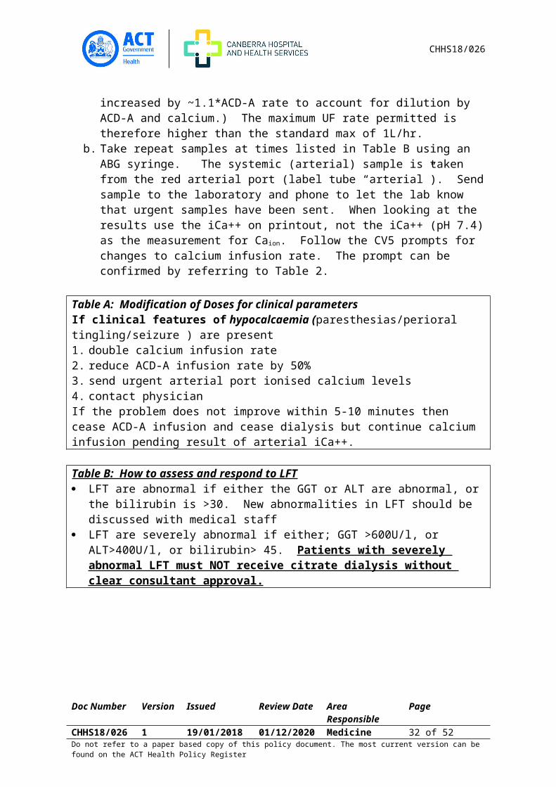
CV5 prompt. The UF add-on prompt can be confirmed by looking at Table 1. (UF is increased by ~1.1*ACD-A rate to account for dilution by ACD-A and calcium.) The maximum UF rate permitted is therefore higher than the standard max of 1L/hr.
b. Take repeat samples at times listed in Table B using an ABG syringe. The systemic (arterial) sample is taken from the red arterial port (label tube “arterial”). Send sample to the laboratory and phone to let the lab know that urgent samples have been sent. When looking at the results use the iCa++ on printout, not the iCa++ (pH 7.4) as the measurement for Caion. Follow the CV5 prompts for changes to calcium infusion rate. The prompt can be confirmed by referring to Table 2.
Table A: Modification of Doses for clinical parametersIf clinical features of hypocalcaemia (paresthesias/perioral tingling/seizure ) are present1. double calcium infusion rate 2. reduce ACD-A infusion rate by 50% 3. send urgent arterial port ionised calcium levels4. contact physicianIf the problem does not improve within 5-10 minutes then cease ACD-A infusion and cease dialysis but continue calcium infusion pending result of arterial iCa++.
Table B: How to assess and respond to LFT LFT are abnormal if either the GGT or ALT are abnormal, or the bilirubin is >30. New
abnormalities in LFT should be discussed with medical staff LFT are severely abnormal if either; GGT >600U/l, or ALT>400U/l, or bilirubin> 45.
Patients with severely abnormal LFT must NOT receive citrate dialysis without clear consultant approval.
Doc Number Version Issued Review Date Area Responsible PageCHHS18/026 1 19/01/2018 01/12/2020 Medicine 23 of 37Do not refer to a paper based copy of this policy document. The most current version can be found on the ACT Health Policy Register

CHHS18/026
Table C: When to re-test systemic (arterial) Calcium 30 min after starting dialysis in all patients 30 min after altering calcium infusion in response to a systemic (arterial) iCa++ level <=
0.95 mmol/L 90 min after commencing dialysis in patients with normal LFT Every 60minutes until completion of dialysis in patients with abnormal LFT or with an
Hb <90g/L or an Hb >139g/L.
Table D: Additional Considerations When systemic iCa++ result is >1.39 or <0.81 A high arterial iCa++ that fails to normalise 30min after a reduction in the calcium infusion is likely due to inadvertent use of calcium containing dialysate, or infusion rate error. For high iCa++>1.39 do the following: Check that the calcium and citrate infusions are correct Check that calcium free dialysate is being used If there are no apparent errors then repeat the systemic (arterial) iCa++, and in
addition take an ABG sample from the post dialyser (blue dialysis line port- labelled “post dialyser”), and a sample from the heparin infusion line (labelled “recirc test”). These samples are to permit determination of the source of the problem. After these samples are taken discuss ceasing citrate dialysis with the renal consultant or advanced trainee registrar.
Any iCa++ result >1.5 must be discussed urgently with a renal advanced trainee registrar, or renal consultant.
A low arterial iCa++ <0.81 is likely due to poor liver function, or to an infusion error. Discuss with a renal consultant or advanced trainee registrar whether to cease Citrate dialysis for any arterial Ca++<0.81. Citrate dialysis should be ceased if the iCa++ is not >0.90mmol/L) when next checked (30minutes).
Table E: How to Cease Regional Citrate Dialysis but Continue Dialysing in the presence of hypocalcaemiaTo avoid worsening of hypocalcaemia, perform steps in the following order1. Stop ACD-A infusion2. Slow Calcium Infusion to 29mL/hour3. Switch to a Part A bath that contains calcium4. Check arterial iCa++5. Cease calcium infusion if arterial iCa++ result is >=1.0mmol/L
5.3.2 Post Parathyroidectomy using RCTFluids Required Calcium (as chloride) 100mmol in 0.9% sodium chloride 250mL (0.4mmol/mL) pre-mix
minibag. 100 mmol calcium/250mL sodium chloride 0.9% (0.4mmol/mL) orders need to be
prescribed using the Calcium Chloride Infusion post parathyroidectomy using “Calcium chloride infusion post parathyroidectomy during Regional Citrate intermittent Haemodialysis” Refer to Medication Standing Order. If the pre-mix minibag is not available, during pharmacy opening hours, infusions may be prepared in the Pharmacy
Doc Number Version Issued Review Date Area Responsible PageCHHS18/026 1 19/01/2018 01/12/2020 Medicine 24 of 37Do not refer to a paper based copy of this policy document. The most current version can be found on the ACT Health Policy Register

CHHS18/026
Department IV room. Orders need to be scanned down to the Pharmacy Department between the hours of 8:30am – 4pm. After 7pm, if the pre-mix minibag is unavailable, the on-call pharmacist can be contacted.
ACD-A: 22g/L Sodium Citrate Dihydrate & 7.5g/L citric acid monohydrate [Total citrate 112.8mmol/L].
Dialysate part A: zero calcium dialysate.
Dialysis Preparation: Protocol for 500mL/min dialysate flow only, not isolated UF Set-up dialysis machine using zero Calcium part A solution. Check the most recent liver function tests (LFT). All patients must have an LFT at the
commencement of their first citrate dialysis session and at least monthly thereafter, if they remain on citrate dialysis. An abnormal LFT is defined as a GGT and/or ALT above the laboratory reference range, or a bilirubin over 30. Severely abnormal LFT are defined as a GGT over 600U/l, an ALT over 400, or a bilirubin over 45. Patients with severely abnormal LFT must NOT receive citrate dialysis.
Check the most recent ionized blood calcium (iCa++) or serum calcium (Cacorr). All patients receiving citrate dialysis post parathyroidectomy must have a serum calcium or Ca++ result available that is no more than 2 hours old, prior to commencement of citrate dialysis. In patients where this is not available take an ABG for ionized calcium during run-on process.
Use a dual lumen venous return cannula set for blood return line. Connect the calcium chloride bag via infuser pump to the 2nd lumen of the dual lumen
venous return cannula. Expel air and clamp. Connect ACD-A bag via an infuser pump to piggy back on the standard saline infusion
line.
Setting Infusion Ratesa. Determine starting BFR, UF goal, reason for use of citrate and record in progress notes. b. Set the infusion rate for ACD-A and calcium Table 1: ACD-A, Calcium chloride and add-on
UF rates at commencement of dialysis [Attachment B]. ACD-A is given in mL/hour as [BFR x1.25]. Calcium is given as [ACD-A rate/8.73].
c. Do not commence infusion until dialysis commences.
Commencing Dialysisa. Commence dialysis at the determined BFR at bicarbonate of 28. b. Commence infusion of ACD-A piggy backed to saline infusion line. c. Commence calcium infusion into 2nd lumen of dual lumen venous return set.d. Set the hourly UF rate add-on as per the database prompt. The UF add-on prompt can be
confirmed by looking at Table 1a or 1b as appropriate UF is increased by [~1.1 x ACD-A rate] to account for dilution by ACD-A and calcium.
The maximum UF rate permitted is therefore higher than the standard max of 1L/hr.
Modification of Infusion Rates
Doc Number Version Issued Review Date Area Responsible PageCHHS18/026 1 19/01/2018 01/12/2020 Medicine 25 of 37Do not refer to a paper based copy of this policy document. The most current version can be found on the ACT Health Policy Register
CHHS18/026
ACD-A and calcium infusion rates should be adjusted immediately if the delivered blood flow rate is altered. Such BFR driven changes are not based on the most recent iCa++ result, and can be determined by entering the new BFR into the database.
Timing of iCa++ blood samples:a. 30 minutes after starting dialysis and again every 60minute during therapy. b. 30 minutes after altering calcium infusion rates in response to a systemic (arterial)
iCa++ level
Method for Sampling blood for iCa++:a. Take a systemic blood sample from the red arterial dialysis port using an ABG
syringes and label the tube “arterial”. b. Send ABG syringe to the laboratory and phone to let the lab know that urgent
samples have been sent. When looking at the results, use the “iCa++ on printout”, not the “iCa++ (pH
7.4)” as the measurement for Caion. c. Calcium infusion is to be altered according to Standing Order
Clinical Parameter Monitoring: Modification of Dose Watch for features of hypocalcaemia (paresthesias/perioral tingling/seizure). If present:
a. Double calcium infusion rate,b. Reduce ACD-A infusion rate by 50%,c. Send urgent systemic (arterial port) ionized calcium levels,d. Contact renal physician.
If the problem does not improve within five to ten minutes then cease ACD-A infusion and cease dialysis but continue calcium infusion pending result of arterial iCa++.
When systemic iCa++ results are low, or higha. A high arterial iCa++, particularly if it fails to normalise following a reduction in the
calcium infusion, is likely due to inadvertent use of calcium containing dialysate, or incorrect setting of the calcium infusion. In this situation do the following: Check that the calcium and citrate infusions are correct Check that calcium free dialysate is being used If there are no apparent errors then repeat the systemic (arterial) iCa++, and in
addition take an ABG sample from the post dialyser (blue dialysis line port- labelled “post dialyser”), and a sample from the heparin infusion line (labelled “recirc test”). These samples are to permit determination of the source of the problem. After these samples are taken discuss whether citrate dialysis should be ceased with the renal consultant or advanced trainee registrar.
Any iCa++ result over 1.5 must be discussed urgently with a renal advanced trainee registrar, or renal consultant.
b. A low arterial iCa++ is likely due to poor liver function, or to an infusion error. Citrate dialysis must be ceased if no infusion errors are detected and the iCa++ remains <0.9 at the next (30min) calcium measurement.
Doc Number Version Issued Review Date Area Responsible PageCHHS18/026 1 19/01/2018 01/12/2020 Medicine 26 of 37Do not refer to a paper based copy of this policy document. The most current version can be found on the ACT Health Policy Register

CHHS18/026
How to Cease Regional Citrate Dialysis but Continue Dialysing in the presence of hypocalcaemiaTo avoid worsening hypocalcaemia, perform steps in the following order: Stop ACD-A infusion. Slow calcium infusion to 29mL/hour (11.6mmol/hr). Switch to a Part A bath that contains calcium. Check arterial iCa++. Cease calcium infusion if arterial iCa++ result is ≥ 1.15 mmol/L.
DocumentationDocumentation of altered ACD-A, calcium chloride and UF infusion rates must occur on the Inpatient Haemodialysis Medical Orders form.
Dose: The rate of infusion (dose) will vary according to the patient’s iCa ++. The Calcium Chloride Infusion during Regional Citrate Intermittent Haemodialysis Post parathyroidectomy SO must be followed
Back to Table of Contents
Section 6 – Monthly Blood Review
Monthly blood and haemodialysis prescription review are managed by the Renal Advanced Trainee Registrar (ATR) under the guidance of the Renal Physician
Blood specimen collection during haemodialysis Blood can be collected for routine testing from the dialysis machine lines to reduce the taking of peripheral samples. Specimen should be collected before dialysis begins. Routine blood samples are collected on the day which falls in the middle of the patient’s dialysis week using the schedule below:
Month Test Month TestJanuary BGL,RUP (UEC, Ca, Mg, PO4,
urate), FBC, CRP, Fe StudiesT/T for patients on transplant list
July BGL, RUP (UEC, Ca, Mg, PO4, urate), FBC, CRP, Fe StudiesT/T for patients on transplant list
February BGL RUP (UEC, Ca, Mg, PO4, urate), FBC, CRP,PTHT/T for patients on transplant list
August BGL, RUP (UEC, Ca, Mg, PO4, urate), FBC, CRP,PTHT/T for patients on transplant list
March BGL, RUP (UEC, Ca, Mg, PO4, urate), FBC, CRP, Fe StudiesT/T for patients on transplant listHbA1C for known diabetics
September BGL, RUP (UEC, Ca, Mg, PO4, urate), FBC, CRP, Fe StudiesT/T for patients on transplant list HbA1C for known diabetics
April BGL, RUP UEC, Ca, Mg, PO4, October BGL, RUP (UEC, Ca, Mg, PO4,
Doc Number Version Issued Review Date Area Responsible PageCHHS18/026 1 19/01/2018 01/12/2020 Medicine 27 of 37Do not refer to a paper based copy of this policy document. The most current version can be found on the ACT Health Policy Register
CHHS18/026
Month Test Month Testurate), FBC, CRP,PTHT/T for patients on transplant list
urate), FBC, CRP,PTHT/T for patients on transplant list
May BGL, RUP (UEC Ca, Mg, PO4, urate), FBC, CRP, Fe Studies, LFT’sT/T for patients on transplant list
November BGL, RUP (UEC, Ca, Mg, PO4, urate), FBC, CRP, Fe Studies, LFT’sT/T for patients on transplant list
June BGL, RUP (UEC, Ca, Mg, PO4, urate), FBC, CRP, Aluminium, PTH, HbA1C for known diabeticsT/T for patients on transplant listAl, HDL,LDL, TG’s, Hep B, Hep C &HIV
December BGL, RUP (UEC, Ca, Mg, PO4, urate), FBC, CRP, Aluminium, PTH, HbA1C for known diabeticsT/T for patients on transplant listAl, HDL,LDL, TG’s, Beta 2 Micro globulin
Note: Patients on RCT should have monthly LFT’s
Back to Table of Contents
Section 7 – Administration of Blood and Blood products on dialysis
Refer to: CHHS Fresh Blood Products Administration (Adults, paediatrics and Neonates) procedure for: Ordering and Delivery of Blood Products Patient information and Consent Identity check of Patient and Product Administration of Blood Platelet Transfusion Procedure and Fresh Frozen Plasma and Cryoprecipitate
Equipment Infusion Pump Infusion line Blood Product
Procedure Prime Infusion line with blood or blood product and attach to infusion pump as required Attach to side line port of arterial chamber Program rate and volume to be infused on infusion pump.
o Blood transfusion can be given over 30mins o Transfused with care alert must be given over 60 minuteso FFP can be administered directly into side line port of arterial chamber without an
infusion pump o Albumin as per physician order
Document in CV5 (blood transfusion assessment)
Doc Number Version Issued Review Date Area Responsible PageCHHS18/026 1 19/01/2018 01/12/2020 Medicine 28 of 37Do not refer to a paper based copy of this policy document. The most current version can be found on the ACT Health Policy Register

CHHS18/026
Back to Table of Contents
Section 8 – Calcium Chloride Infusions during Regional Citrate Anticoagulated Filtration based Plasma Exchange
Fluids Required Calcium (as chloride) 100mmol in sodium chloride 0.9% 250mL (0.4mmol/mL) ACD-A: 22g/L Sodium Citrate Dihydrate & 7.5g/L citric acid monohydrate [Total citrate
112.8mmol/L]. 4% albumin and/or Fresh Frozen Plasma (volume as per prescription) 4 x 1 Litre sodium chloride 0.9%
Equipment required PRISMAFLEX® machine 1 x PRISMAFLEX® TPE 2000 Set Gloves (non-sterile) 1x Accessory drain bag
Equipment for patient connection 1x cannulation pack with one SN fistula needle for venous return or 1x CVC pack with one SN-Adapter Luer-Lock for venous return 3x ampoule of sodium Chloride 0.9% 10mL 1x Accessory spike 1x infusion line
Equipment for patient disconnection depending on access type
Note:Call for Albumin first then Fresh Frozen Plasma when 2nd last bottle commenced. Give Phenergan 10mg oral administration an hour prior to commencing FFP
Blood Tests required Pre filterPre TPE: FBC, CPM (Calcium, Magnesium and Phosphate), EUC, ABG ionised Ca++, Coags & Fibrinogen. Use gold top pathology tube for Anti GBM if ordered.
During: ABG ionised Ca++ (half hour then hourly if no change to ionised Calcium result)
Prior to completion: ABG ionised Ca++, CPM & CoagsTotal Calcium from plasma effluent mix then collect and label “body fluid calcium” use a new form, Write plasma exchange on clinical details
ObservationsPre TPE and at completion and Filtration Fraction: MEWS and follow up MEWS
During: BP/HR and TPE assessment half hourly
Doc Number Version Issued Review Date Area Responsible PageCHHS18/026 1 19/01/2018 01/12/2020 Medicine 29 of 37Do not refer to a paper based copy of this policy document. The most current version can be found on the ACT Health Policy Register

CHHS18/026
Ionised Calcium range: 0.96 – 1.32Filtration Fraction range: < 28%
Procedure:1. Plasma exchange Preparation: As per product manual with these additional steps
a. All patients must have a; haematocrit, serum creatinine, total calcium, ionized calcium, phosphate, magnesium, LFT and coagulation profile (prothrombin time, activated partial thromboplastin time, fibrinogen) within 12 hours of the commencement of each plasma exchange session. The exchange can commence prior to these results being available with specialist approval.
b. Severe renal impairment (eGFR <30mL/min/1.73m2) or severely deranged LFT (defined as a GGT over 600units/L, an ALT over 400 units/L, or a bilirubin over 45micromol/L) denote an increased risk of citrate toxicity.
c. Use a dual lumen venous return cannula set for blood return line.d. Connect the calcium chloride bag via infuser pump to the 2nd lumen of the dual
lumen venous return cannula. Expel air and clamp. e. Connect ACD-A bag to the PBP line on PrismaFlex
2. Equation for calculation of patient plasma volumePlasmavolume=0.065∗Weight (1−haematocrit )If patient weight is >100kg then set weight at 100kg.
3. Selection of replacement fluid
a. When albumin is used as the replacement fluid bleeding due to depletion of clotting factors is a risk.
b. In those with a high risk of bleeding it is desirable to target an APTT and INR ratio of <1.5 and a fibrinogen >1g/L by use of FFP as the complete or partial replacement fluid. This increases the risk of allergic reactions and possibly the rise of hypocalcaemia.
c. Most patients will have clotting factor derangements that may increase bleeding risk immediately after a plasma exchange with albumin and recovery of clotting factors occurs over the subsequent 2 days. Use of 50% FFP as replacement fluid is recommended for patients at increased risk of bleeding.
d. When FFP is used as the replacement fluid allergic reaction are more common with urticarial reactions seen in 17%. Premedication with an antihistamine should be considered.
4. Blood flow ratea. Citrate requirement (and hence the risk of toxicity) is determined by plasma flow, not
whole blood flow. PrismaFlex requires a minimum whole blood flow of 100mL/minb. Faster blood flows reduce the risk of filter clotting (primarily by reducing the required
filtration fraction).
Doc Number Version Issued Review Date Area Responsible PageCHHS18/026 1 19/01/2018 01/12/2020 Medicine 30 of 37Do not refer to a paper based copy of this policy document. The most current version can be found on the ACT Health Policy Register

CHHS18/026
c. An increased blood flow at the same citrate dose leads to more rapid citrate infusion and hence a greater risk of citrate accumulation (with subsequent hypocalcaemia).
d. Patients with relatively normal liver function generally tolerate plasma citrate doses of 0.8mmol/minute without toxicity and there is evidence that a plasma citrate concentration of approximately 5.5 mmol/L may be sufficient to inhibit clotting1–3. Based on this the target plasma flow rate is 145mL/min to achieve reasonable circuit patency, safe citrate dosage and minimize treatment time.
e. Utilize the formulae below or see Table 1 for the initial machine settings.
5. Setting Infusion Rates at commencement of exchange
Whole blood flow setting= 145
1−Haemoglobin300
Citrate dose=5.5∗(1−Haemoglobin300
)
Where; Whole blood flow is in mL/minHaemoglobin is in g/LCitrate dose is in mmol/L of whole bloodCalcium dose is in mL/hour
Initial Calcium dose is 17 mL/hour when albumin is the replacement fluid and 18mL/min when FFP is the replacement fluid.
Patient Haemtocrit
Approximate Patient Hb (g/L)
Whole blood flow rate
Citrate dose/L blood
0.2 67 160 40.25 83 170 40.3 100 185 40.35 117 200 40.4 133 215 30.45 150 235 30.5 167 260 3
6. Modification of Infusion RatesThe calcium infusion rate should be adjusted immediately if the delivered blood flow rate is altered. (PrismaFlex will automatically adjust the citrate infusion if this occurs.) Such BFR driven changes are not based on the most recent iCa++ result. The citrate and calcium infusion rate settings at commencement of each session may be adjusted by the treating physician based on circuit patency and the perceived risk of citrate toxicity. During treatment, the calcium infusion rates are to be adjusted according the Table 2.
7. Timing of calcium measurements
Doc Number Version Issued Review Date Area Responsible PageCHHS18/026 1 19/01/2018 01/12/2020 Medicine 31 of 37Do not refer to a paper based copy of this policy document. The most current version can be found on the ACT Health Policy Register
CHHS18/026
iCa++ and total serum calcium within 12 hours prior to commencement of plasma exchange
iCa++ 30minutes after commencing plasma exchange and at least hourly thereafter iCa++ and total serum calcium at the completion of plasma exchange iCa++ 30 minutes after altering calcium infusion rates in response to a systemic
(arterial) iCa++ level ≤0.95 or ≥1.40 mmol/L.
8. Method for Sampling blood for iCa++: Take a systemic blood sample from the red arterial dialysis port using an ABG
syringes and label the tube “arterial”. a. Send ABG syringe to the laboratory and phone to let the lab know that urgent
samples have been sent (if taken in 8A), in ICU run sample through local analyzer without delay. When looking at the results, use the “iCa++ on printout”, not the “iCa++ (pH
7.4)” as the measurement for Caion. b. Enter the decision support framework or see Table 2 [Attachment A]: Response to
“Arterial” iCa++ (mmol/L) to determine the correct response to the systemic (arterial) iCa++ results.
9. Clinical Parameter Monitoring: Modification of Dose Watch for features of hypocalcaemia (paresthesias/perioral tingling/seizure). If
present:a. Double calcium infusion rate,b. Halve ACD-A (citrate) infusion ratec. Obtain urgent systemic (arterial port) ionized calcium levels,d. Contact treating physician.If there is a delay in obtaining the ionized calcium the problem fails to improve within five to ten minutes then cease ACD-A infusion and cease plasma exchange but continue calcium infusion pending result of arterial iCa++.
Watch for features of hypercalcaemia (stupor, new nausea or new vomiting). If present1. Obtain urgent systemic (arterial port) ionized calcium level2. Contact treating physician3. If:
there is a delay in obtaining the ionized calcium or the problem fails to improve within five to ten minutes and
- the physician does not provide alternate instructions then cease ACD-A infusion, cease calcium infusion and cease plasma exchange
pending result of arterial iCa++. Urgent haemodialysis may be required in cases of severe hypercalcaemia
10. When systemic iCa++ results are low, or high
Doc Number Version Issued Review Date Area Responsible PageCHHS18/026 1 19/01/2018 01/12/2020 Medicine 32 of 37Do not refer to a paper based copy of this policy document. The most current version can be found on the ACT Health Policy Register

CHHS18/026
a. A high arterial iCa++, particularly if it fails to normalise following a reduction in the calcium infusion, is likely due to incorrect setting of the calcium or citrate infusion but may be due to recirculation.
b. In this situation do the following:o Check that the calcium and citrate infusions are correcto If there are no apparent errors then repeat the systemic (arterial) iCa++, plasma total
serum calcium and urgently discuss with the treating physician whether plasma exchange should be ceased. If treatment is ceased then note the effluent volume and, after mixing the bag, send a sample of effluent in a red top tube for total calcium. These samples are to help determination the source of the problem at a later date.
o Any iCa++ result over 1.5mmol/L must be discussed urgently with the treating physician.
c. A low arterial iCa++ is likely due to poor liver function, or to an infusion error. Citrate anticoagulated plasma exchange under this standing order must be ceased if no infusional errors are detected and the iCa++ remains <0.9 mmol/L at the next (30min) calcium measurement.
11. Documentation Alterations to ACD-A and calcium chloride rates, the total treatment time, the total
calcium chloride volume infused the total effluent volume and any suspected adverse events must be documented in the medical record. A sample of mixed effluent must be sent in a red-top tube for measurement of total calcium at the completion of each exchange.
Back to Table of Contents
Definition of Terms
ATR: Advanced Trainee Registrar. For the purpose of this document this refers to the Renal Advanced Trainee
HITTS: Heparin Induced Thrombotic Thrombocytopenia Syndrome PPE: Personal Protective Equipment UF: Ultra filtration is the total amount of fluid removed on dialysis CVC: Central Venous Catheter used for haemodialysis QB: Blood flow on dialysis VAN: Vascular Access Nurse Fluid bolus: Sodium Chloride 0.9% or online fluid given to prime haemodialysis circuit or
treat hypotension RO: Reverse Osmosis machine
Back to Table of Contents
Implementation
Doc Number Version Issued Review Date Area Responsible PageCHHS18/026 1 19/01/2018 01/12/2020 Medicine 33 of 37Do not refer to a paper based copy of this policy document. The most current version can be found on the ACT Health Policy Register

CHHS18/026
New colleagues who join the haemodialysis team are required to undergo hospital orientation and orientation to Renal Services. This consists of orientation to the individual unit they are working on as well as the service as a whole.
Each colleague is required to complete the Haemodialysis training manual, the Company Dialysis Machine manual and the Cannulation SDLP
Those who are on call are required to complete the Acute team SDLP Yearly review of skills is attended by the CDN and CNC
Back to Table of Contents
Related Policies, Procedures, Guidelines and Legislation
Policies: CHHS Medication Policy CHHS Patient Identification and Procedure Matching Policy
Procedures:CHHS Healthcare Associated InfectionsCHHS Diabetes Management Including Hypoglycaemia, IV Insulin Infusion and Insulin Pumps (Adults Only)CHHS Fresh Blood Products Administration (Adults, Paediatric and Neonates)CHHS Access for Renal Replacement Therapy Clinical GuidelineCHHS Access for Renal Replacement Therapy Operational GuidelineCHHS Calcium Chloride Infusion during Regional Citrate Intermittent HaemodialysisCHHS Patient Identification and Procedure Matching procedure
LegislationEnrolled Nurse Scope of Practice
ManualsHaemodialysis Single Needle treatment in Operator’s ManualHaemodialysis Machine Operator’s Manual
Medication Standing OrdersStandard Haemodialysis PrescriptionCalcium Chloride during regional citrate haemodialysis
Back to Table of Contents
References
1. Akoh JA. Prosthetic arteriovenous grafts for hemodialysis. The Journal Of Vascular Access. 2009;10(3): 137-147.
2. Ball, L.K., & Mott, S. (2010). How do you prevent indented buttonhole sites? Nephrology Nursing Journal, 37(4), 427-428, 431.
3. Ball, L. (2010). The buttonhole technique: Strategies to reduce infections. Nephrology Nursing Journal, 37(5), 473-478.
Doc Number Version Issued Review Date Area Responsible PageCHHS18/026 1 19/01/2018 01/12/2020 Medicine 34 of 37Do not refer to a paper based copy of this policy document. The most current version can be found on the ACT Health Policy Register

CHHS18/026
4. Macrae JM, Ahmed SB, Atkar R, Hemmelgarn BR. A randomized trial comparing buttonhole with rope ladder needling in conventional hemodialysis patients. Clin J Am Soc Nephrol. 2012;7(10): 1632-1638.
5. Pergolotti A, Rich E, Lock K. The effect of the buttonhole method vs. the traditional method of AV fistula cannulation on hemostasis, needle stick pain, pre-needle stick anxiety, and presence of aneurysms in ambulatory patients on hemodialysis. Nephrol Nurs J. 2011;38(4): 333-336.
6. van Loon MM, Goovaerts T, Kessels AG, van der Sande FM, Tordoir JH. Buttonhole needling of haemodialysis arteriovenous fistulae results in less complications and interventions compared to the rope-ladder technique. Nephrol Dial Transplant. 2010;25(1): 225-230.
7. Silva Gdos S, Silva RA, Nicolino AM, Pavanetti LC, Alasmar VL, Guzzardi R, et al. Initial experience with the buttonhole technique in a Brazilian hemodialysis center. J Bras Nefrol. 2010;32(3): 257-262.
8. Struthers J, Allan A, Peel RK, Lambie SH. Buttonhole needling of ateriovenous fistulae: a randomized controlled trial. ASAIO J. 2010;56(4): 319-322.
9. Verhallen AM, Kooistra MP, van Jaarsveld BC. Cannulating in haemodialysis: rope-ladder or buttonhole technique? Nephrology Dialysis Transplantation. 2007;22(9): 2601-2604.
10. St. George Hospital – Haemodialysis Unit SOP on Treatment of ARF from Multiple Myeloma Cast Nephropathy with HCO dialysis
11. Hutchison, C., Bradwell, B., Cook, M., Basnayake, K, Basu, S., Harding, S., Hattersley, J., 12. Evans, D., Chappel, M., Sampson, P.,Foggensteiner, ., Adu, D., and Cockwell, P,
Treatment of Acute Renal Failure Secondary to Multiple Myeloma with Chemotherapy and Extended High Cut-Off Hemodialysis Clin J Am Soc Nephrol 4: 745–754, 2009. doi: 10.2215/CJN.04590908
13. Hutchison, C., Cook, M., Heyne, N., Weisel, K., Billingham, L., Bradwell, A., and Cockwell, P., European trial of free light chain removal by extended haemodialysis in cast nephropathy (EuLITE): A randomised controlled trial, Trials 2008, 9:55 doi:10.1186/1745-6215-9-55
14. Shavit L, Lifschitz M, Plaksin J, Grenader T, Slotki I. High dose urokinase for restoration of patency of occluded permanent central venous catheters in hemodialysis patients. Clin Nephrol. 2010 Oct;74(4):297-302
15. Tumlin J, Goldman J, Spiegel DM, Roer D, Ntoso KA, Blaney M, Jacobs J, Gillespie BS, Begelman SM. A phase III, randomized, double-blind, placebo-controlled study of tenecteplase for improvement of hemodialysis catheter function: TROPICS 3. Clin J Am Soc Nephrol. 2010 Apr;5(4):631-6
16. Power A, Duncan N, Singh SK, Brown W, Dalby E, Edwards C, Lynch K, Prout V, Cairns T, Griffith M, McLean A, Palmer A, Taube D. Sodium citrate versus heparin catheter locks for cuffed central venous catheters: a single-center randomized controlled trial. Am J Kidney Dis. 2009 Jun;53(6):1034-41
17. Ash SR. Advances in tunneled central venous catheters for dialysis: design and performance. Semin Dial. 2008 Nov-Dec;21(6):504-15
18. Moran JE, Ash SR; ASDIN Clinical Practice Committee. Locking solutions for hemodialysis catheters; heparin and citrate--a position paper by ASDIN. Semin Dial. 2008 Sep-Oct;21(5):490-2
Doc Number Version Issued Review Date Area Responsible PageCHHS18/026 1 19/01/2018 01/12/2020 Medicine 35 of 37Do not refer to a paper based copy of this policy document. The most current version can be found on the ACT Health Policy Register

CHHS18/026
19. Davies J, Casey J, Li C, Crowe AV, McClelland P. Restoration of flow following haemodialysis catheter thrombus. Analysis of rt-PA infusion in tunnelled dialysis catheters. J Clin Pharm Ther. 2004 Dec;29(6):517-20
20. Zacharias JM, Weatherston CP, Spewak CR, Vercaigne LM. Alteplase versus urokinase for occluded hemodialysis catheters. Ann Pharmacother. 2003 Jan;37(1):27-33
21. James MT, Conley J, Tonelli M, Manns BJ, MacRae J, Hemmelgarn BR; Alberta Kidney Disease Network. Meta-analysis: antibiotics for prophylaxis against hemodialysis catheter-related infections. Ann Intern Med. 2008 Apr 15;148(8):596-605
22. Macrae JM, Loh G, Djurdjev O, Shalansky S, Werb R, Levin A, Kiaii M. Short and long alteplase dwells in dysfunctional hemodialysis catheters. Hemodial Int. 2005 Apr;9(2):189-95
23. Clase CM, Crowther MA, Ingram AJ, Cinà CS. Thrombolysis for restoration of patency to haemodialysis central venous catheters: a systematic review. J Thromb Thrombolysis. 2001 Apr;11(2):127-36
24. Alteplase product information on Medical Index Medicus Service (MIMS)25. McFarland H. F., Dinwiddie, L., Ferrell, J.,& Forloines-Lynn, S. (2002). Lytic Therapy in
central Venous Catheters for Haemodialysis. Nephrology Nursing Journal; Aug.2002; 29, 4: Health Module pg. 355
26. McFarland H. F., Dinwiddie, L., Ferrell, J.,& Forloines-Lynn, S. (2002). Lytic Therapy in central Venous Catheters for Haemodialysis. Nephrology Nursing Journal; Aug.2002; 29, 4: Health Module pg. 355
27. Webb, A., Abdalla, M., & Russell, G.,(2001). A protocol of Urokinase infusion and warfarin for the management of the thrombosed haemodialysis catheter, Nephrology Dialysis Transplantation; (2201) 16:2075-2078
28. Deitcher, S., Fraschini, G.,Himmelfarb,J., Schuman, E., Smith, T., Schultz, G., Firszt, C., MouginisT., (2004). Dose-Ranging Trial with a Recombinant Urokinase (Urokinase Alfa) for Occluded Central venous Catheter in Oncology Patients.;, Journal Vascular Intervention Radiology; 2004; 15: 575-579
29. Urokinase product information on Micromedics 30. The Canberra Hospital Oncology Department protocol on clearing vascular catheter using
Urokinase31. Adapted from St. George Hospital – Haemodialysis Unit SOP on Treatment of ARF from
Multiple Myeloma Cast Nephropathy with HCO dialysis.
Back to Table of Contents
Search Terms
Intermittent, Haemodialysis, Renal, Replacement, dialysis, AVF, AVG, Arteriovenous, fistula, graft, calcium, chloride, blood,
Policy Team ONLY to complete the following:Date Amended Section Amended Divisional Approval Final Approval 15/11/2018 Complete Review ED Medicine CHHS Policy Committee
Doc Number Version Issued Review Date Area Responsible PageCHHS18/026 1 19/01/2018 01/12/2020 Medicine 36 of 37Do not refer to a paper based copy of this policy document. The most current version can be found on the ACT Health Policy Register

CHHS18/026
This document supersedes the following: Document Number Document NameCHHS15/151 Intermittent Haemodialysis for Adults Clinical Procedure
Doc Number Version Issued Review Date Area Responsible PageCHHS18/026 1 19/01/2018 01/12/2020 Medicine 37 of 37Do not refer to a paper based copy of this policy document. The most current version can be found on the ACT Health Policy Register