Embed Size (px)
Citation preview
Series on Biomechanics, Vol.27, No. 1-2 (2012), 76- 83
76
Haemorheology in the Foetus and the Neonate
M.W. Rampling, M.A.Anwar
Bioengineering Department, Imperial College of Science Technology and Medicine, London SW7 2AZ, UK: [email protected]
1. Introduction
The foetus undergoes enormous anatomical development during the relatively short time of gestation. Blood starts to form during the embryonic stage, but there are still substantial changes in the haematology accompanying the foetal anatomical development which lead to large alterations in the rheology of foetal blood. One aim of this paper is to relate these anatomical, haematological and haemorheological variations to one another. Birth is a cataclysmic event that results in dramatic changes in the very short term in the neonatal cardiovascular system and is normally accompanied by rapid and large haematological and haemorheological adjustments. These will also be documented
These phenomena should be of interest to anyone concerned with human haemorheology. However, the area is now of considerable clinical importance because therapeutic manipulation of the blood of the neonate and of the in-utero foetus is an increasing aspect of the clinical handling of these immature humans [1, 2, 3]. There is a need to understand the haemorheological changes that accompany normal gestation and birth and to determine whether and to what extent they can be altered without deleterious physiological effects.
2. Anatomical development during gestation
Shortly after fertilisation the conceptus begins to differentiate and within a week the rudimentary yolk sac, the amniotic sac and the beginnings of the embryonic structure are apparent. Within 3 weeks the brain, the spinal cord, the heart and the gastro-intestinal tract start to develop. Also during his time haematopoietic cells start to appear in the yolk sac. By 4-5 weeks the arm and leg buds have begun to form, as have the eyes and ears and the tissue that will eventually form the skeleton. During this time the heart starts to beat, and rudimentary blood begins to flow in the major blood vessels. By 6 weeks the hands and feet start to form, as do the lungs.
It is conventional to call week 8 the end of the embryonic period. This period is not of great importance to this paper which is concerned primarily with haemorheology, and at this stage the blood system is very primitive and no attempt at therapeutic or diagnostic in utero manipulation is possible or reasonable. At this stage the embryo is a mere 2-3cm long and weighs only a few grams.
The foetal period is now entered. By 9 to 12 weeks the face has become well formed and the genitals have appeared, but the head still makes up about half the size of the foetus. The liver now takes over the production of red blood cells. By the 16th week meconium is produced by the GI tract, and the baby begins to move. By 19 weeks the movements of the baby may be detected by the mother. This period is clinically important as diagnostic sampling is practicable. The foetus should be approaching 300 g in weight.
The period from 21 weeks to term is of greatest importance clinically because therapeutic circulatory manipulation is now possible and premature babies born in this period are able, in principle, to survive. Between 20 and 23 weeks the bone marrow starts producing blood cells, and by a few weeks before normal birth is the main producer. During this period the lower respiratory airways start to develop (but with little surfactant at this stage) and storage of fat starts. Development continues until birth, usually at 37 - 40 weeks.
So in the short space of some 9 months the conceptus has developed into a small human weighing something in the region of 3.5kg. 3. Physiological development during gestation
The early embryo is sufficiently small for its nutrient supply and waste product loss to be adequate by simple
M.W. Rampling et al./ Haemorheology in the Foetus and the Neonate
77
diffusion to and from its surroundings. However, in a few weeks it has increased sufficiently in size that it is vital that the foetal circulatory and blood systems have become competent to perform these tasks. These systems have to cope with a variety of potentially serious problems:-
The very rapid rate of increase in size of the embryo/foetus. The development of the various organs which may have differing circulatory requirements. The requirement for adequate exchange with the maternal circulation through the placenta. The hypoxia normally present in foetal blood (for reasons to be discussed later); the foetal arterial blood
is only about 70% saturated with oxygen. The blood pressure is, compared with the adult, very low (see Table 1).
Table 1
Typical foetal systolic blood pressure during gestation
Gestational age (weeks)
25
32
40
Adults
Systolic blood pressure (mmHg)
45
55
65
120
Up to the time of birth the foetal lungs are collapsed and receive relatively little blood, just enough to support
their living requirements, and the blood is oxygenated at the placenta. However after leaving the placental circulation it is very soon mixed with deoxygenated blood from the foetal tissues and then enters the heart. (This is why the foetal ‘arterial’ blood is relatively hypoxic, ~70% saturated) A little is pumped to the lungs by the right heart but the majority passes through the foramen ovale directly to the left side of the heart from which it is pumped to the rest of the body. The deflated lungs have a high resistance at this stage and this is mainly what limits their blood supply.
Shortly after birth the circulatory arrangement undergoes a sudden and dramatic change. The baby starts to breathe so the lungs become inflated with air which causes their circulatory resistance to fall. The result is that the pressure in the right heart also falls so that the foramen ovale spontaneously closes and this is the main reason that the baby switches in a very short time to the neonatal/adult blood flow pattern. The arterial blood now, of course, suddenly becomes fully saturated with oxygen.
These circulatory arrangements in the foetus/neonate make it particularly vulnerable to problems related to the circulation e.g.:-
There is an ever present risk of serious hypoxia because even in the normal foetus the arterial blood is substantially unsaturated with oxygen.
Foetal transfusion from or to the placenta can cause serious haematocrit and blood viscosity disturbances in the foetus. This is particularly so at birth and is very dependent on the position of the baby with respect to the placenta and the speed with which the umbilicus is clamped.
In a similar fashion, transfusions in utero between twins may be mutually problematic. These problems when they arise are potentially treatable by therapeutic transfusion, haemodilution etc. Furthermore, there are a variety of other conditions for which circulatory manipulation by means of in utero transfusion can be clinically useful, e.g. protection against alloimmune reactions.
It is, therefore, important that the question be answered:– Are the changes in haematology and haemorheology during foetal life and immediately thereafter contributory to allowing the foetal circulation to cope with the stress and strains of normal foetal and neonatal existence or are they merely an epiphenomenon?
If the former then hemorheological changes must be taken into account in defining criteria for transfusion and other circulatory manipulations in the foetus and neonate. Equally they must be taken into account in defining the degree of haematological change that is considered to be pathological and, therefore, deserving of treatment. In fact this paper will not answer the question. However, the major changes (and their variability) in the haematology and haemorheology which accompany foetal and early neonatal life will be documented. A knowledge of these is essential to answering the question.
M.W. Rampling et al./ Haemorheology in the Foetus and the Neonate
78
4. Haematology and haemorheology of the intrauterine foetus
The data for this section were taken from Reference 4. The foetuses inspected for inclusion in the study were from referrals for assessment of suspected foetal structural abnormalities detected by routine ultrasound, or for maternal conditions, e.g. suspected alloimmune reactions, maternal infection, suspected growth retardation, or because of a previous child with a condition diagnosable by in-utero sampling e.g. haemoglobinopathy. The blood samples were only included in this study if, on inspection, the foetuses were considered to be normal.
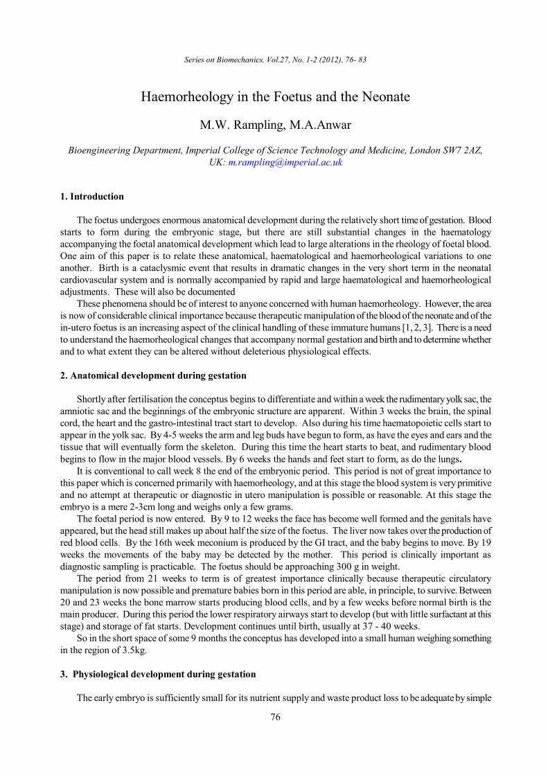
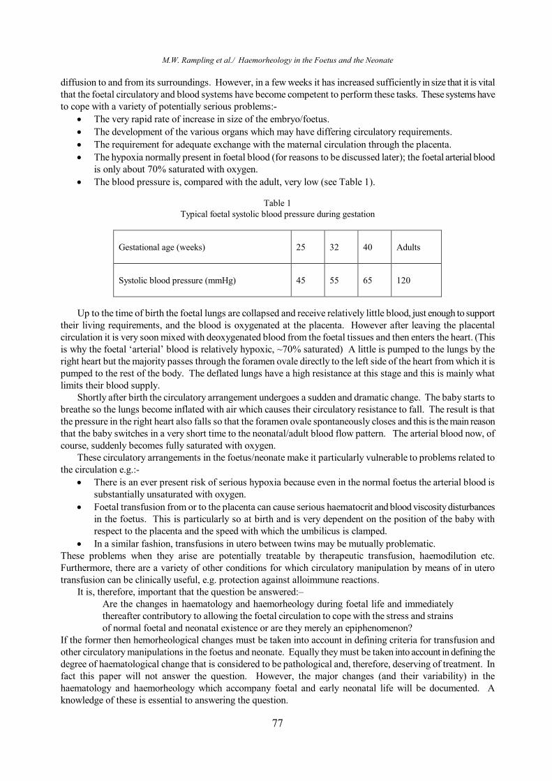
Fig 1 shows that the foetal haematocrit is very substantial and rises throughout later intra-uterine life. Even at 18 weeks it averages some 32%, rising to an average of 38% at 35 weeks. These values are not far from the normal range for adults (37-50%). What is particularly noteworthy is the relatively low level of variation at any week (the 95% confidence range is shown in Fig 1). The plasma protein concentration also increases throughout later intrauterine life (see Fig 2), but is generally well below the normal adult range (60-90g/l).
Fig.1. The variation of foetal haematocrit with gestational age. [4]
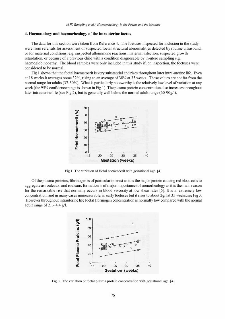
Of the plasma proteins, fibrinogen is of particular interest as it is the major protein causing red blood cells to aggregate as rouleaux, and rouleaux formation is of major importance to haemorheology as it is the main reason for the remarkable rise that normally occurs in blood viscosity at low shear rates [5]. It is in extremely low concentration, and in many cases immeasurable, in early foetuses but it rises to about 2g/l at 35 weeks, see Fig 3. However throughout intrauterine life foetal fibrinogen concentration is normally low compared with the normal adult range of 2.1- 4.4 g/l.
Fig. 2. The variation of foetal plasma protein concentration with gestational age. [4]
M.W. Rampling et al./ Haemorheology in the Foetus and the Neonate
79
Fig.3. The variation of foetal plasma fibrinogen concentration with gestational age. [4]
Fig.4. The variation of foetal plasma viscosity with gestational age. [4] Moving now to the haemorheology of the foetus; Fig 4 shows that the plasma viscosity increases steadily, in
line with the plasma protein concentration, from about 0.85 to about 1.05mPa.s between 18 and 35 weeks. Throughout this time the viscosity is generally below the normal adult range (1.2-1.5mPas).
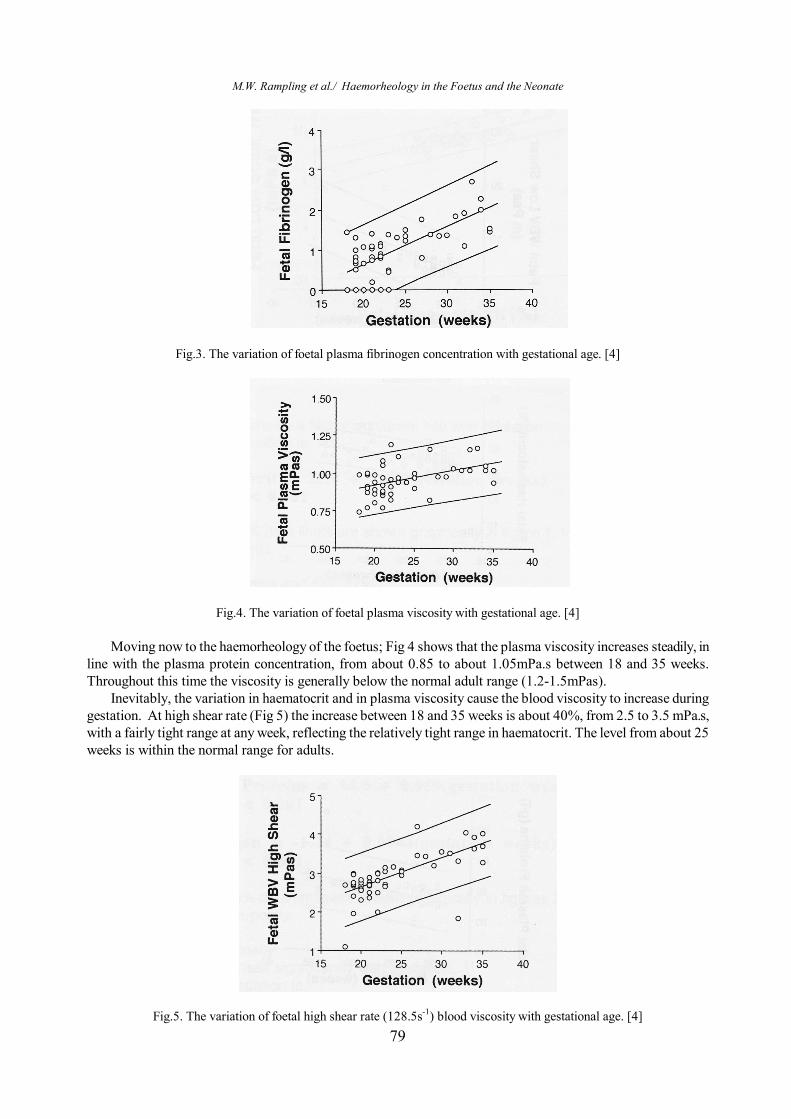
Inevitably, the variation in haematocrit and in plasma viscosity cause the blood viscosity to increase during gestation. At high shear rate (Fig 5) the increase between 18 and 35 weeks is about 40%, from 2.5 to 3.5 mPa.s, with a fairly tight range at any week, reflecting the relatively tight range in haematocrit. The level from about 25 weeks is within the normal range for adults.
Fig.5. The variation of foetal high shear rate (128.5s-1) blood viscosity with gestational age. [4]

M.W. Rampling et al./ Haemorheology in the Foetus and the Neonate
80
Fig.6. The variation of foetal low shear rate (0.277s-1) blood viscosity with gestational age. [4] The increase in foetal blood viscosity at low shear rate (see Fig 6) over the same period is much bigger, from
about 2.5 to 15mPa.s. This greater increase compared to that at high shear rate is due to the added effect of the increasing fibrinogen level on rouleaux formation. But even at 35 weeks the low shear rate viscosity is generally lower than that of the average adult (15-55mPas) – this is because the fibrinogen level is lower in the foetus. However the higher sialic acid levels on the foetal fibrinogen [6] probably makes it less rouleaugenic than the adult form because of the increased negative charge that it induces in the molecule. The result of all this is that the blood viscosity in early foetuses shows almost Newtonian characteristics, i.e. shows almost no variation with shear rate. This is very different from adult blood the viscosity of which increases greatly in going from high to low shear rate. Even at 35 weeks of gestation the level of non-Newtonianism in foetal blood is low compared with the adult. 5. Haematology and haemorheology of the early and term neonate
Attention is now turned to the neonate: it will have recently gone through the process of leaving the uterus,
having the umbilicus clamped, starting to breathe air through the lungs, of the foramen ovale closing and for the arterial blood to have increased its oxygen level to almost full saturation. The data used here are from reference 7, where the samples were obtained from the umbilical vein of the neonate immediately after cord clamping. Although some of the neonates were pre-term, all were healthy, of normal weight for gestational age and born vaginally.
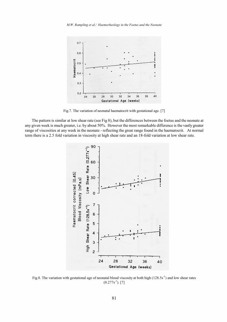
Fig 7 shows that, as for the foetus, there is a small but steady increase in haematocrit with gestational age at birth. However the values are consistently higher than of the similarly aged foetus - by an average of about 10% of haematocrit. This reflects placental transfusion to the foetus at birth and subsequent haemoconcentration. It is also noteworthy that there is a greatly increased spread in haematocrit at any week. This reflects the random nature of the placental transfusion, which is affected by the relative positions of the neonate and the placenta immediately after birth and the speed of clamping of the cord. At birth about half the term neonates have haematocrits above the upper end of the normal adult range (50%), as do many of the preterm infants. This indicates the potential for polycythaemia in the neonate. It should also be noted that even after birth rapid changes take place in the blood composition, for example Shohat et al [8] have reported that the haematocrit usually rises to a peak at two hours after birth and then steadily falls over the next 12 hours.
The plasma protein concentration and plasma viscosity in the neonates show similar rising trends with gestational age to those of the in-utero foetus and, although these levels are slightly higher in the neonate, they are still below adult levels.
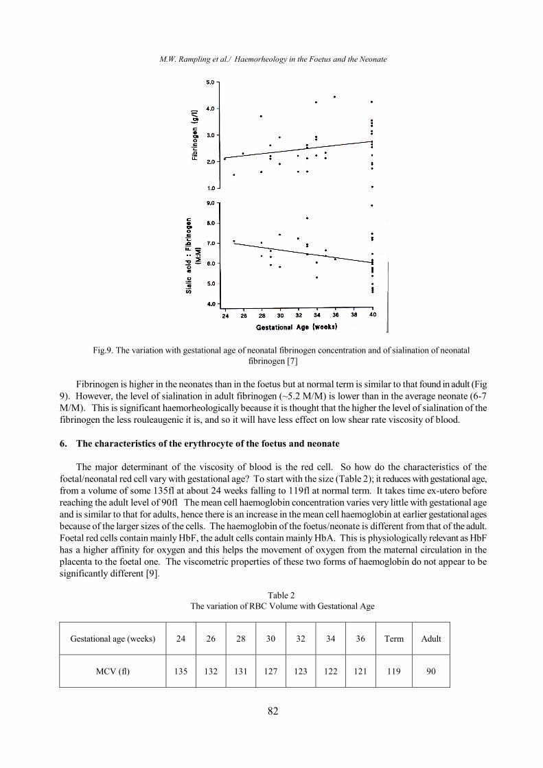
The characteristics of whole blood viscosity at high shear rate of the neonate (see Fig 8) once again follow a similar pattern to that of the in-utero foetus, i.e. a linear rise with gestational age. But, in line with the haemoconcentration effects, in the neonate the values are some 1mPas higher across the board. Also note the remarkable number of term neonates with very high viscosities compared to the normal adult (i.e. above 5mPs), reflecting again the wide variation in haematocrit in the neonates.
M.W. Rampling et al./ Haemorheology in the Foetus and the Neonate
81
Fig.7. The variation of neonatal haematocrit with gestational age. [7]
The pattern is similar at low shear rate (see Fig 8), but the differences between the foetus and the neonate at any given week is much greater, i.e. by about 50%. However the most remarkable difference is the vastly greater range of viscosities at any week in the neonate - reflecting the great range found in the haematocrit. At normal term there is a 2.5 fold variation in viscosity at high shear rate and an 18-fold variation at low shear rate.
Fig.8. The variation with gestational age of neonatal blood viscosity at both high (128.5s-1) and low shear rates (0.277s-1). [7]
M.W. Rampling et al./ Haemorheology in the Foetus and the Neonate
82
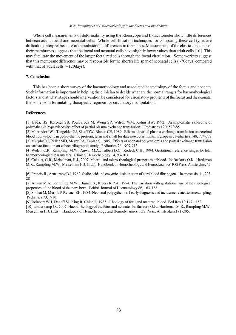
Fig.9. The variation with gestational age of neonatal fibrinogen concentration and of sialination of neonatal fibrinogen [7]
Fibrinogen is higher in the neonates than in the foetus but at normal term is similar to that found in adult (Fig
9). However, the level of sialination in adult fibrinogen (~5.2 M/M) is lower than in the average neonate (6-7 M/M). This is significant haemorheologically because it is thought that the higher the level of sialination of the fibrinogen the less rouleaugenic it is, and so it will have less effect on low shear rate viscosity of blood.
6. The characteristics of the erythrocyte of the foetus and neonate
The major determinant of the viscosity of blood is the red cell. So how do the characteristics of the
foetal/neonatal red cell vary with gestational age? To start with the size (Table 2); it reduces with gestational age, from a volume of some 135fl at about 24 weeks falling to 119fl at normal term. It takes time ex-utero before reaching the adult level of 90fl The mean cell haemoglobin concentration varies very little with gestational age and is similar to that for adults, hence there is an increase in the mean cell haemoglobin at earlier gestational ages because of the larger sizes of the cells. The haemoglobin of the foetus/neonate is different from that of the adult. Foetal red cells contain mainly HbF, the adult cells contain mainly HbA. This is physiologically relevant as HbF has a higher affinity for oxygen and this helps the movement of oxygen from the maternal circulation in the placenta to the foetal one. The viscometric properties of these two forms of haemoglobin do not appear to be significantly different [9].
Table 2
The variation of RBC Volume with Gestational Age
Gestational age (weeks)
24
26
28
30
32
34
36
Term
Adult
MCV (fl)
135
132
131
127
123
122
121
119
90
M.W. Rampling et al./ Haemorheology in the Foetus and the Neonate
83
Whole cell measurements of deformability using the Rheoscope and Ektacytometer show little differences between adult, foetal and neonatal cells. Whole cell filtration techniques for comparing these cell types are difficult to interpret because of the substantial differences in their sizes. Measurement of the elastic constants of their membranes suggests that the foetal and neonatal cells have slightly lower values than adult cells [10]. This may facilitate the movement of the larger foetal red cells through the foetal circulation. Some workers suggest that this membrane difference may be responsible for the shorter life span of neonatal cells (~70days) compared with that of adult cells (~120days).
7. Conclusion
This has been a short survey of the haemorheology and associated haematology of the foetus and neonate. Such information is important in helping the clinician to decide what are the normal ranges for haemorheological factors and at what stage should intervention be considered for circulatory problems of the foetus and the neonate. It also helps in formulating therapeutic regimen for circulatory manipulation.
References [1] Bada, HS, Korones SB, Pourcyrous M, Wong SP, Wilson WM, Kolini HW, 1992. Asymptomatic syndrome of polycythemic hyperviscosity: effect of partial plasma exchange transfusion. J Pediatrics 120, 579-85 [2] Maertzdorf WJ, Tangelder GJ, Slaaf DW, Blanco CE, 1989. Effects of partial plasma exchange transfusion on cerebral blood flow velocity in polycythemic preterm, term and small for date newborn infants. European J Pediartics 148, 774-778 [3] Murphy DJ, Reller MD, Meyer RA, Kaplan S, 1985. Effects of neonatal polycythemia and partial exchange transfusion on cardiac function an echocardiographic study. Pediatrics 76, 909-913. [4] Welch, C.R., Rampling, M.W., Anwar M.A., Talbert D.G., Rodeck C.H., 1994. Gestational reference ranges for fetal haemorheological parameters. Clinical Hemorheology 14, 93-103 [5] Cokelet, G.R., Meiselman, H.J., 2007. Macro- and micro-rheological properties of blood. In: Baskurk O.K., Hardeman M.R., Rampling M.W., Meiselman H.J. (Eds), Handbook of Hemorheology and Hemodynamics. IOS Press, Amsterdam, 45-71. [6] Francis JL, Armstrong DJ, 1982. Sialic acid and enzymic desialination of cord blood fibrinogen. Haemostasis, 11, 223-28 [7] Anwar M.A,. Rampling M.W., Bignall S., Rivers R.P.A., 1994. The variation with gestational age of the rheological properties of the blood of the new-born. British Journal of Haematology 86, 163-168. [8] Shohat M, Merlob P Reisner SH, 1984. Neonatal polycythemia: I early diagnosis and incidence related to time sampling. Pediatrics 73, 7-10. [9] Reinhart WH, Danoff SJ, King R, Chien S, 1985. Rheology of fetal and maternal blood. Ped Res 19 147 - 153 [10] Linderkamp O., 2007. Haemorheology of the fetus and neonate. In: Baskurk O.K., Hardeman M.R., Rampling M.W., Meiselman H.J. (Eds), Handbook of Hemorheology and Hemodynamics. IOS Press, Amsterdam,191-205.