Embed Size (px)
Citation preview
Haemorrhagic stroke in children
Finbar O’Callaghan Ins8tute of Child Health, UCL
& Great Ormond Street Hospital, London
Defini8on
• Acute neurological deficit secondary to focal haemorrhage in the brain.
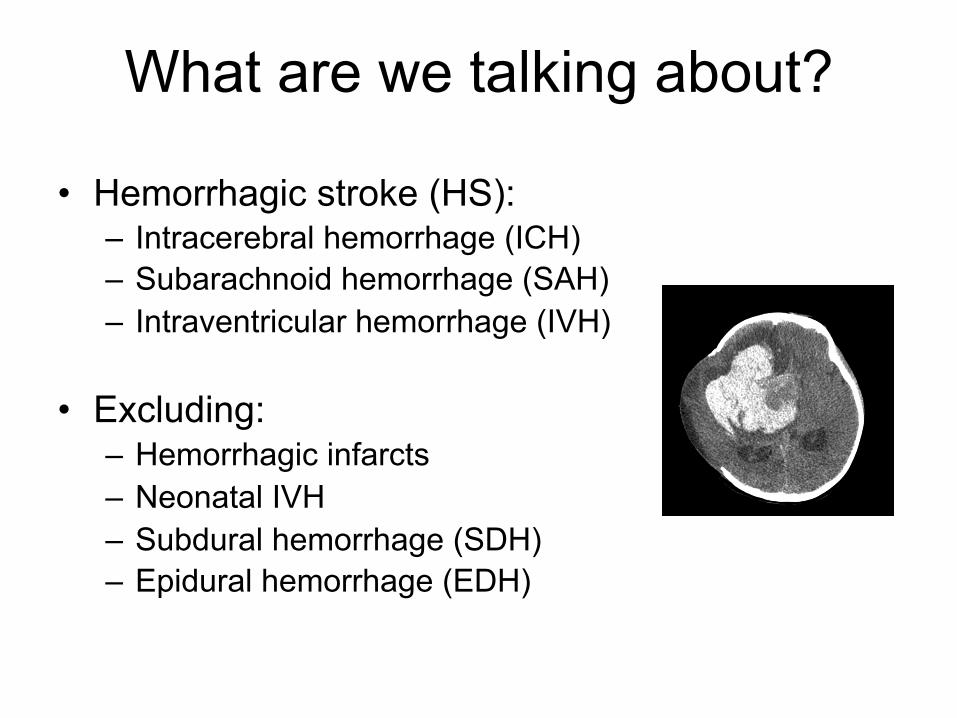
What are we talking about?
• Hemorrhagic stroke (HS): – Intracerebral hemorrhage (ICH) – Subarachnoid hemorrhage (SAH) – Intraventricular hemorrhage (IVH)
• Excluding: – Hemorrhagic infarcts – Neonatal IVH – Subdural hemorrhage (SDH) – Epidural hemorrhage (EDH)
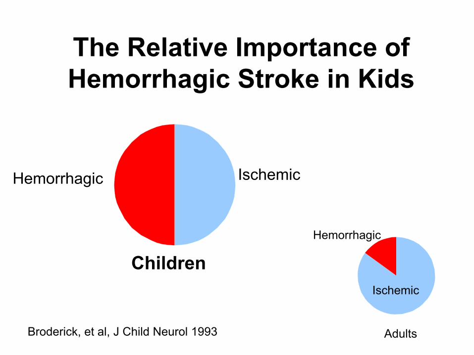
The Relative Importance of Hemorrhagic Stroke in Kids
Hemorrhagic Ischemic
Children
Adults
Ischemic
Hemorrhagic
Broderick, et al, J Child Neurol 1993
Background • In adults haemorrhagic stroke (HS) – 15% strokes
• Children HS : 45% -‐ 50% stroke
• Mortality rate HS x5 higher than ischaemic stroke
• High risk of serious life-‐long disability – Affec8ng mul8ple domains
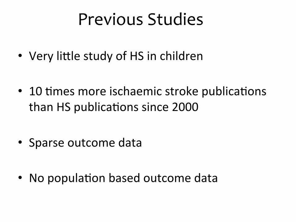
Previous Studies
• Very liUle study of HS in children
• 10 8mes more ischaemic stroke publica8ons than HS publica8ons since 2000
• Sparse outcome data
• No popula8on based outcome data
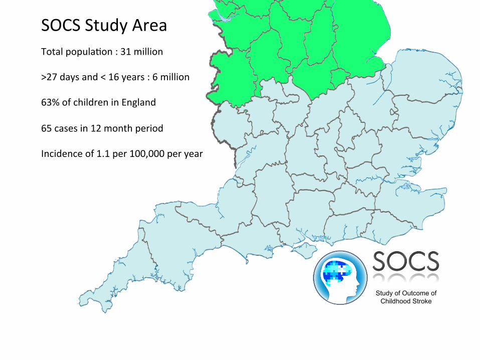
SOCS Study Area
Total popula8on : 31 million >27 days and < 16 years : 6 million 63% of children in England 65 cases in 12 month period Incidence of 1.1 per 100,000 per year
Study of Outcome of Childhood Stroke
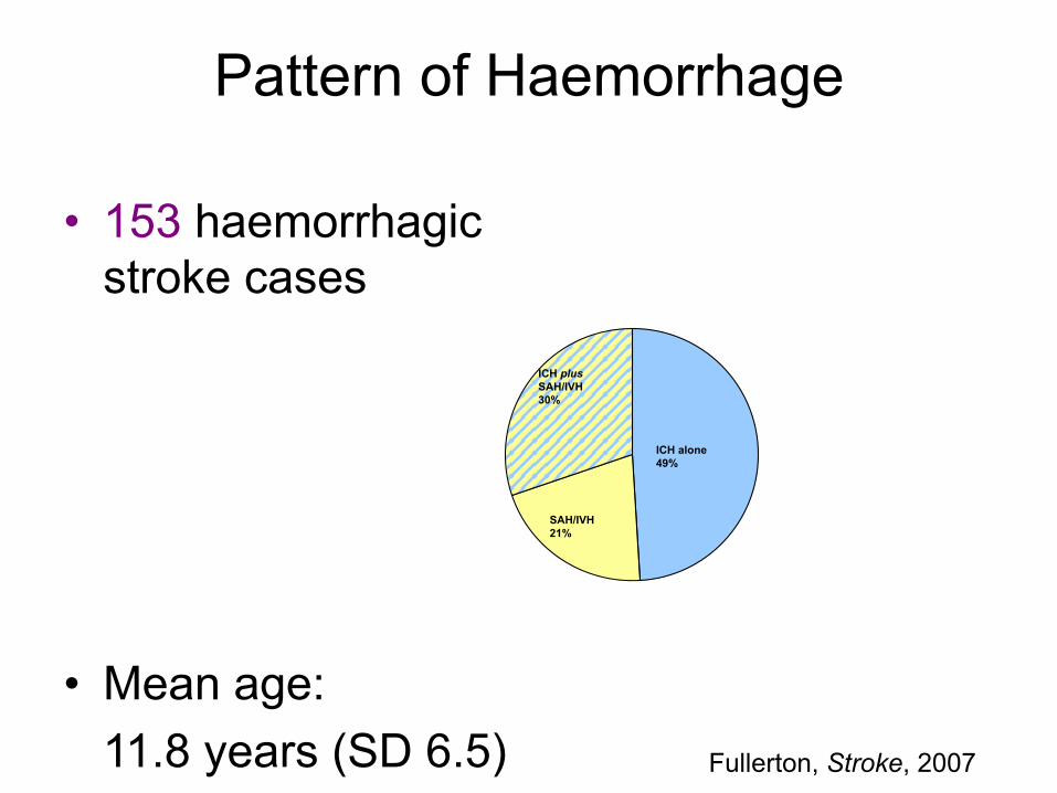
ICH alone 49%
SAH/IVH 21%
ICH plus SAH/IVH 30%
Pattern of Haemorrhage
• 153 haemorrhagic stroke cases
• Mean age: 11.8 years (SD 6.5) Fullerton, Stroke, 2007
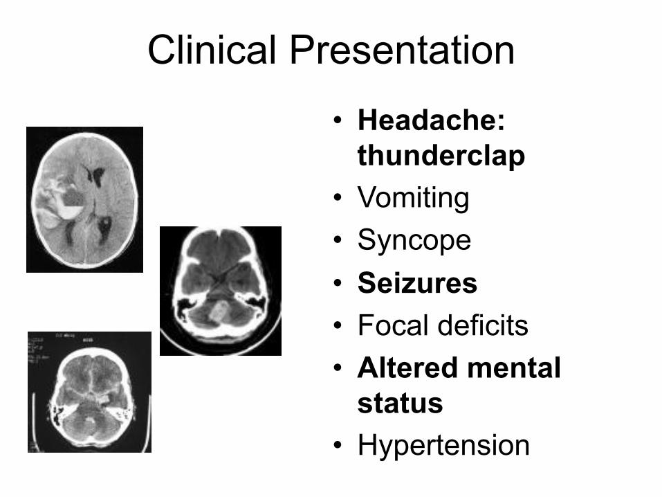
Clinical Presentation • Headache:
thunderclap • Vomiting • Syncope • Seizures • Focal deficits • Altered mental
status • Hypertension
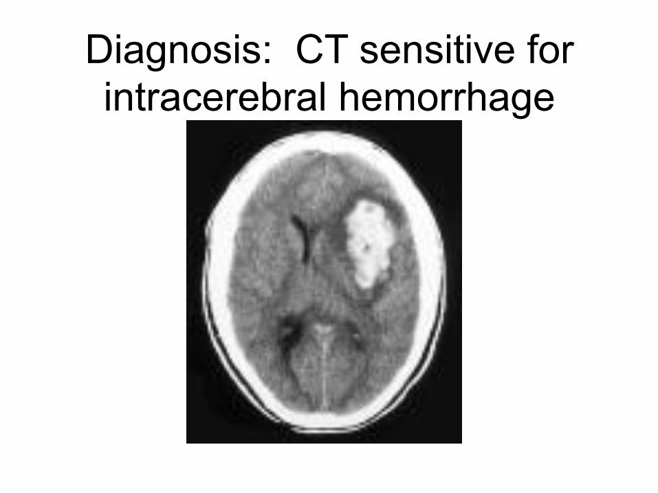
Diagnosis: CT sensitive for intracerebral hemorrhage
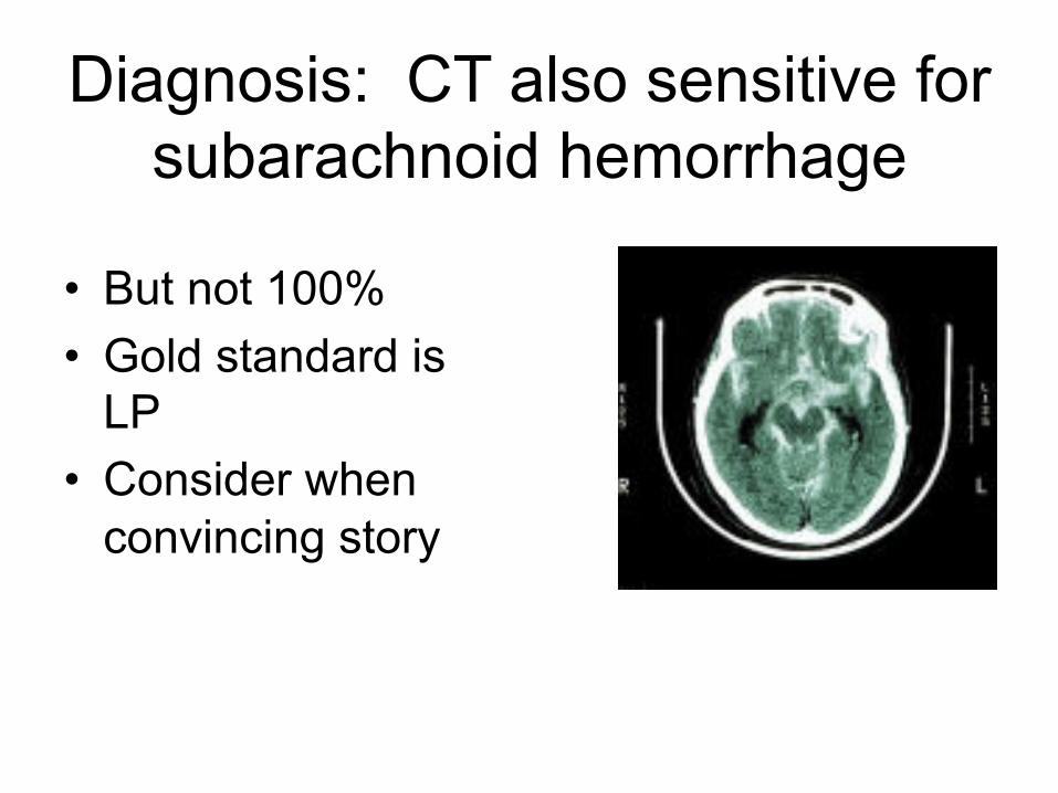
Diagnosis: CT also sensitive for subarachnoid hemorrhage
• But not 100% • Gold standard is
LP • Consider when
convincing story
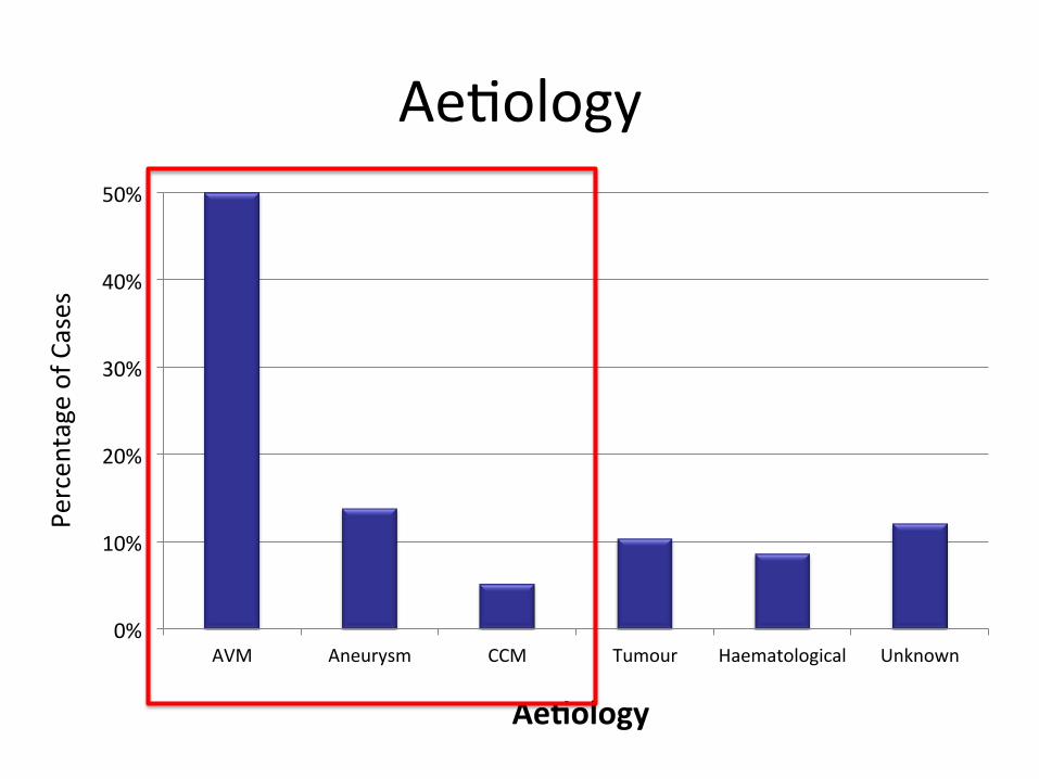
Ae8ology
0%
10%
20%
30%
40%
50%
AVM Aneurysm CCM Tumour Haematological Unknown
Percen
tage of C
ases
Ae#ology

Etiologies of Pediatric HS • Structural 53%
– Other 10% • Hypertension, drug use,
thrombocytopenia, hemophilia, leukemia
AVM 31%
Aneurysm 12%
Cav Mal 8%
Tumor 2%
Trauma 21%
Other 10%
Idiopathic 16%
Fullerton, Stroke, 2007
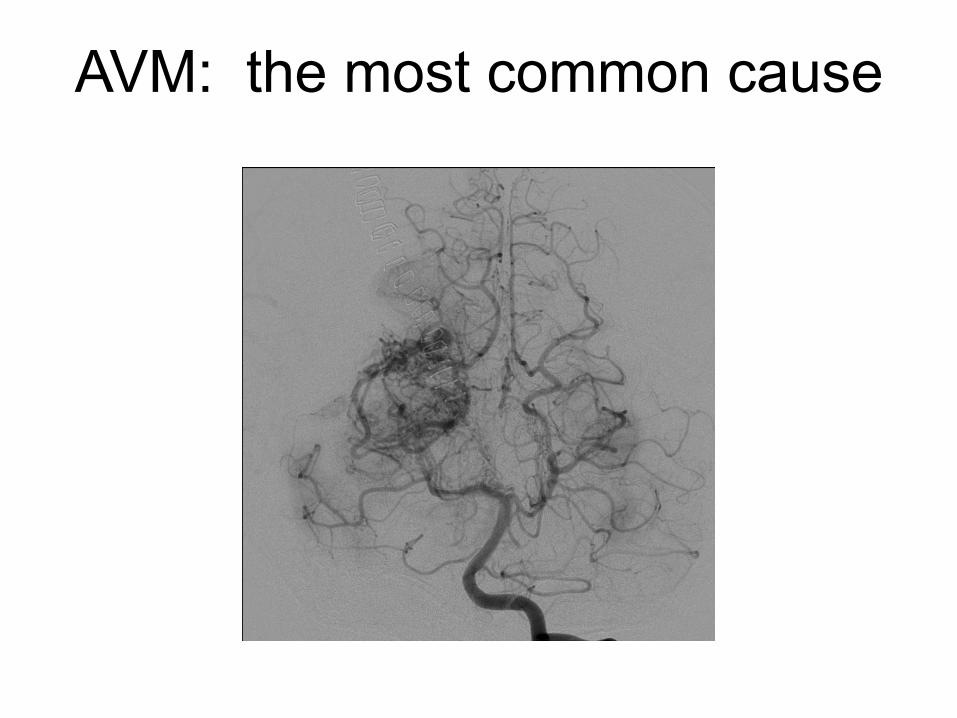
AVM: the most common cause
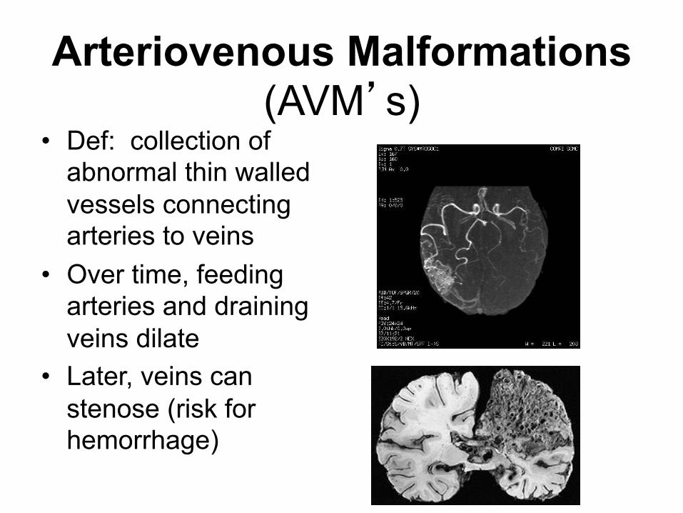
Arteriovenous Malformations (AVM’s)
• Def: collection of abnormal thin walled vessels connecting arteries to veins
• Over time, feeding arteries and draining veins dilate
• Later, veins can stenose (risk for hemorrhage)
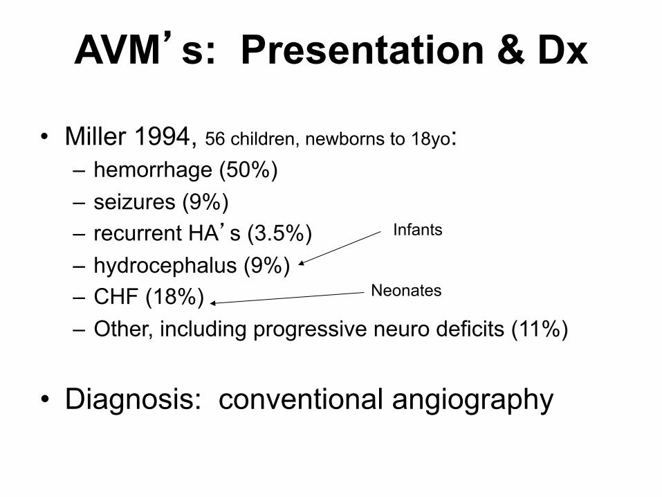
AVM’s: Presentation & Dx
• Miller 1994, 56 children, newborns to 18yo: – hemorrhage (50%) – seizures (9%) – recurrent HA’s (3.5%) – hydrocephalus (9%) – CHF (18%) – Other, including progressive neuro deficits (11%)
• Diagnosis: conventional angiography
Infants
Neonates
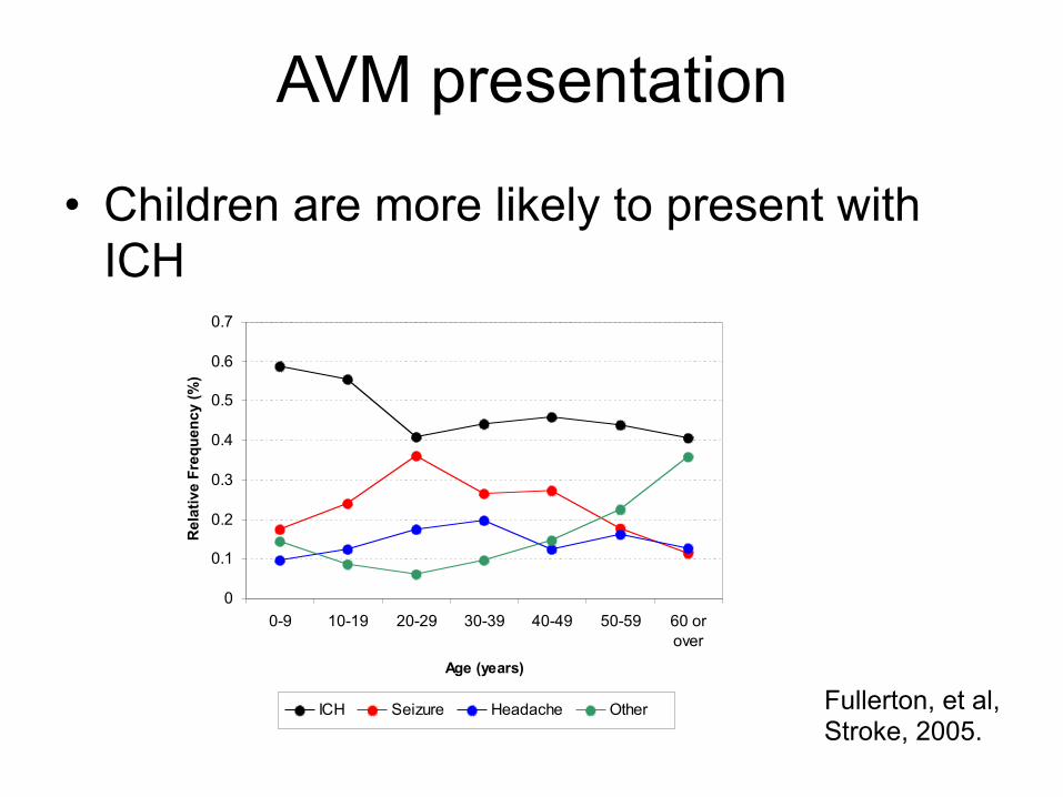
AVM presentation
• Children are more likely to present with ICH
0
0.1
0.2
0.3
0.4
0.5
0.6
0.7
0-9 10-19 20-29 30-39 40-49 50-59 60 orover
Age (years)
Rel
ativ
e Fr
eque
ncy
(%)
ICH Seizure Headache Other Fullerton, et al, Stroke, 2005.
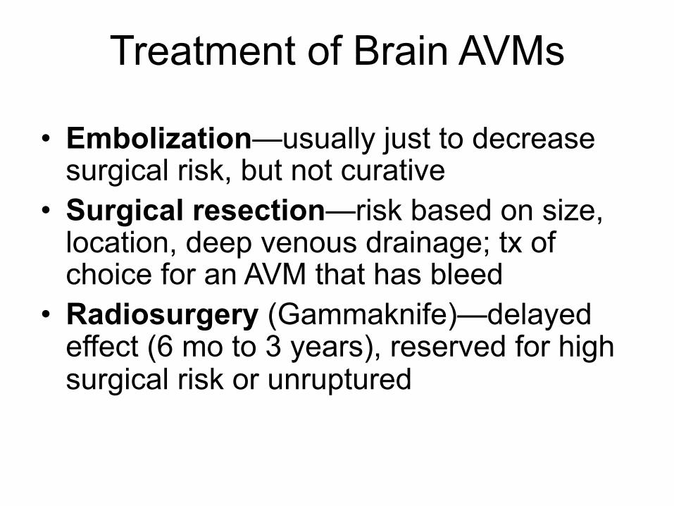
Treatment of Brain AVMs
• Embolization—usually just to decrease surgical risk, but not curative
• Surgical resection—risk based on size, location, deep venous drainage; tx of choice for an AVM that has bleed
• Radiosurgery (Gammaknife)—delayed effect (6 mo to 3 years), reserved for high surgical risk or unruptured
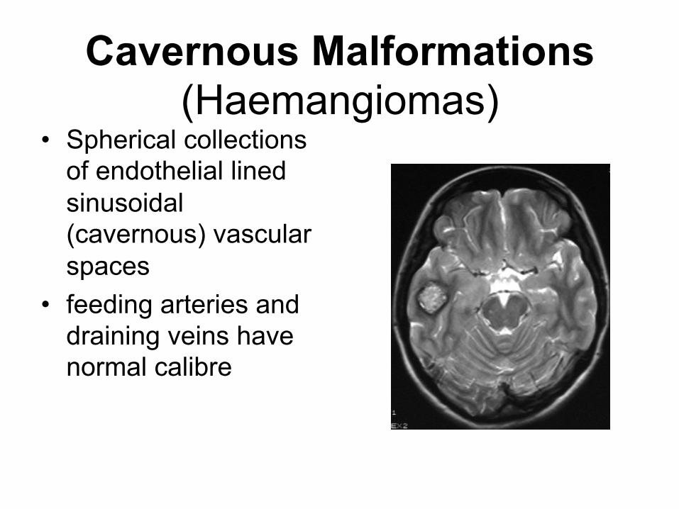
Cavernous Malformations (Haemangiomas)
• Spherical collections of endothelial lined sinusoidal (cavernous) vascular spaces
• feeding arteries and draining veins have normal calibre
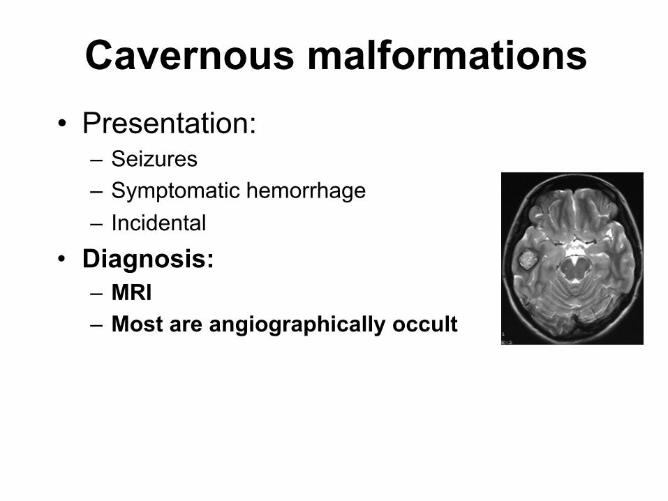
Cavernous malformations • Presentation:
– Seizures – Symptomatic hemorrhage – Incidental
• Diagnosis: – MRI – Most are angiographically occult
Cav mal: Management
• Management: – Surgical – Observation – Radiosurgery not
effective • Goals of surgery:
– Prevention of hemorrhage
– (Seizure control)
• Indications for tx: – Symptomatic
hemorrhage – (Uncontrolled
epilepsy)
• Risks of surgery vs natural history risk

Cav mal: Natural Hx Annual Hemorrhage Risk
• LIMITED data: retrospective, ? definition of hemorrhage
• Depends on Presentation (Komata 95; Kondziolka 95) – Incidental: 0.4%/year – Hemorrhagic presentation: 4.5-23%/year
• Depends on location (Wallace 97; Zabramski 99) – “Deep” (BS, cerebellar nuclei, deep grey; mostly sx):
5-11%/year – “Superficial”: 0%
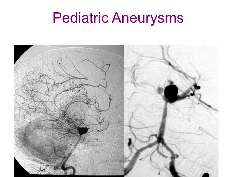
Pediatric Aneurysms
in the 2 cases. Case 1 has a collagenoma on the cheek ipsilateralto the aneurysm. Case 3 had an aneurysm in the right internalcarotid artery detected at 38 years of age, which was diagnosedand treated in another institution, and brain images were notavailable for review. A female patient (case 4) with probabletuberous sclerosis complex had an aneurysm in the left middlecerebral artery (figure 3). She had lymphangioleiomyomatosisand hepatic and adrenal angiomyolipomas, but no tubers, sub-ependymal nodules, or other manifestations of tuberous sclero-sis complex.
Discussion
The prevalence of intracranial aneurysms in this cohort withdefinite tuberous sclerosis complex is 3 of 404 (0.74%; 95%
confidence interval, 0.19%-2.34%), which seems slightly higherthan the incidental finding of aneurysms on brain MRI in thegeneral population (0.35%; 95% confidence interval, 0.13%-0.67%).9 To the authors’ knowledge, 31 cases of intracranialarteriopathy in patients with tuberous sclerosis complex havebeen reported in the literature (Table 2). Most of them are aneur-ysms, but other arterial abnormalities, such as stenosis, ectasia,tortuosity, or moyamoya syndrome, have been reported.10,11
Assessment by conventional MRI can lead to the underesti-mation of intracranial arteriopathy in tuberous sclerosis com-plex, and in studies in the general population as well, becausemagnetic resonance angiography, which provides more sensi-tivity in the detection of more small lesions, is not usually per-formed as a routine examination. At the moment, there is notenough information to warrant a change in this practice, but
Figure 1. (a) In patient 1, an aneurysm at the junction of the right posterior cerebral artery with the right posterior communicant artery isalready seen at 21 months of age (arrow). (b) The aneurysm (arrow) and 2 duplicated bilateral superior cerebellar arteries (arrowheads) areseen in the magnetic resonance angiogram performed at 10 years of age.
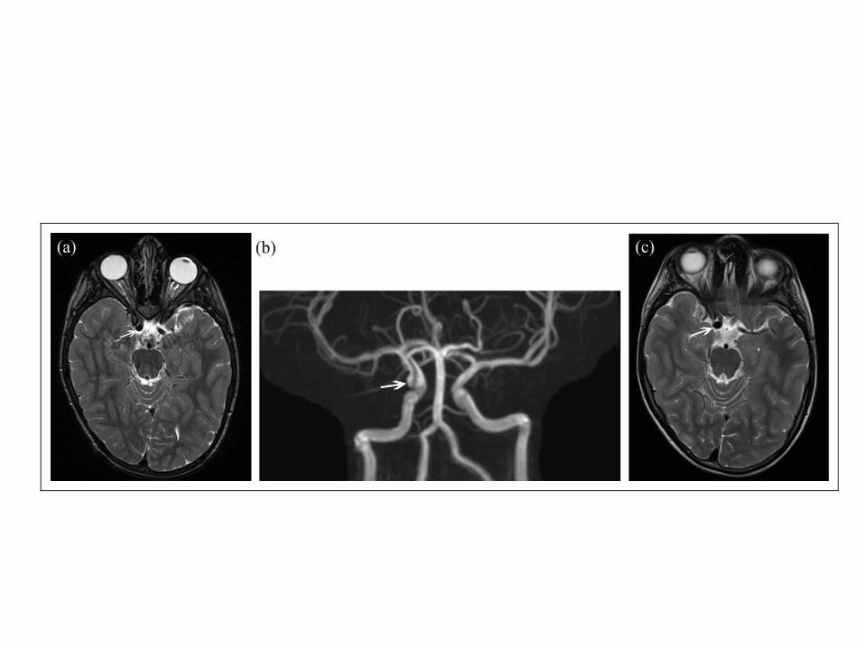
Figure 2. (a) In patient 2, slight dilation of the right internal carotid artery consistent with a developing fusiform aneurysm (arrow) is observed at3 years of age. (b) Magnetic resonance angiogram and (c) axial T2-weighted image show the enlarged fusiform aneurysm at 9 years of age.
914 Journal of Child Neurology 29(7)
at University College London on March 26, 2015jcn.sagepub.comDownloaded from
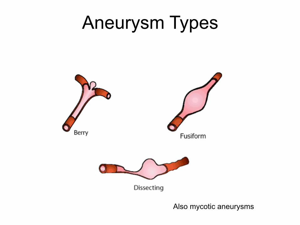
Aneurysm Types
Also mycotic aneurysms
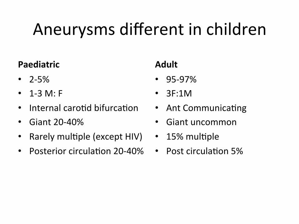
Aneurysms different in children
Paediatric • 2-‐5% • 1-‐3 M: F • Internal caro8d bifurca8on • Giant 20-‐40% • Rarely mul8ple (except HIV) • Posterior circula8on 20-‐40%
Adult • 95-‐97% • 3F:1M • Ant Communica8ng • Giant uncommon • 15% mul8ple • Post circula8on 5%
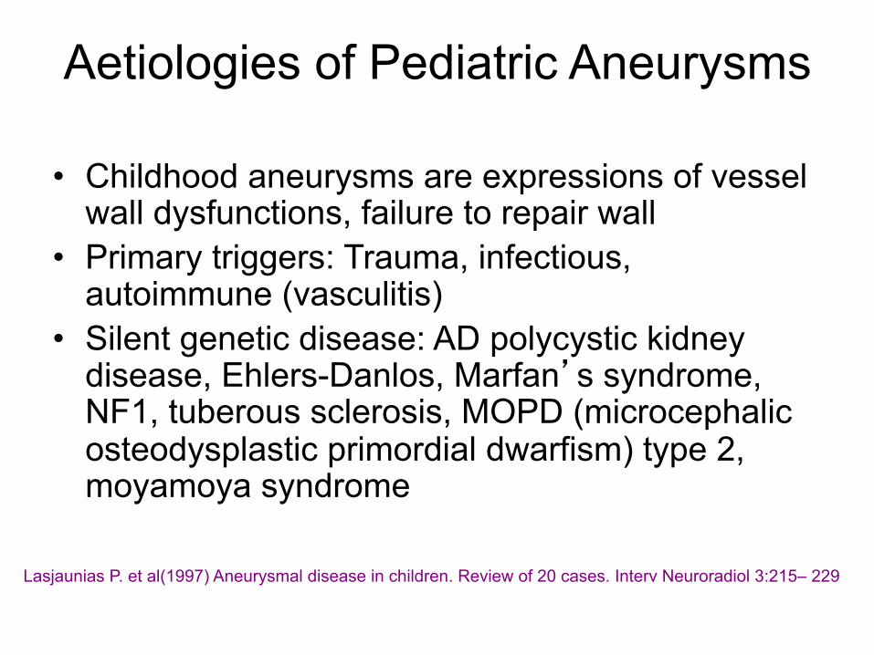
Aetiologies of Pediatric Aneurysms
• Childhood aneurysms are expressions of vessel wall dysfunctions, failure to repair wall
• Primary triggers: Trauma, infectious, autoimmune (vasculitis)
• Silent genetic disease: AD polycystic kidney disease, Ehlers-Danlos, Marfan’s syndrome, NF1, tuberous sclerosis, MOPD (microcephalic osteodysplastic primordial dwarfism) type 2, moyamoya syndrome
Lasjaunias P. et al(1997) Aneurysmal disease in children. Review of 20 cases. Interv Neuroradiol 3:215– 229
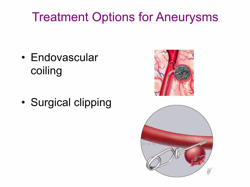
Treatment Options for Aneurysms
• Endovascular coiling
• Surgical clipping
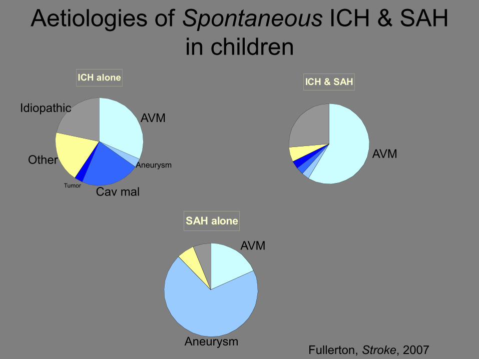
Aetiologies of Spontaneous ICH & SAH in children
ICH alone ICH & SAH
SAH alone
AVM
AVM
AVM
Cav mal
Aneurysm
Aneurysm
Other
Idiopathic
Tumor
Fullerton, Stroke, 2007
Recurrence risk
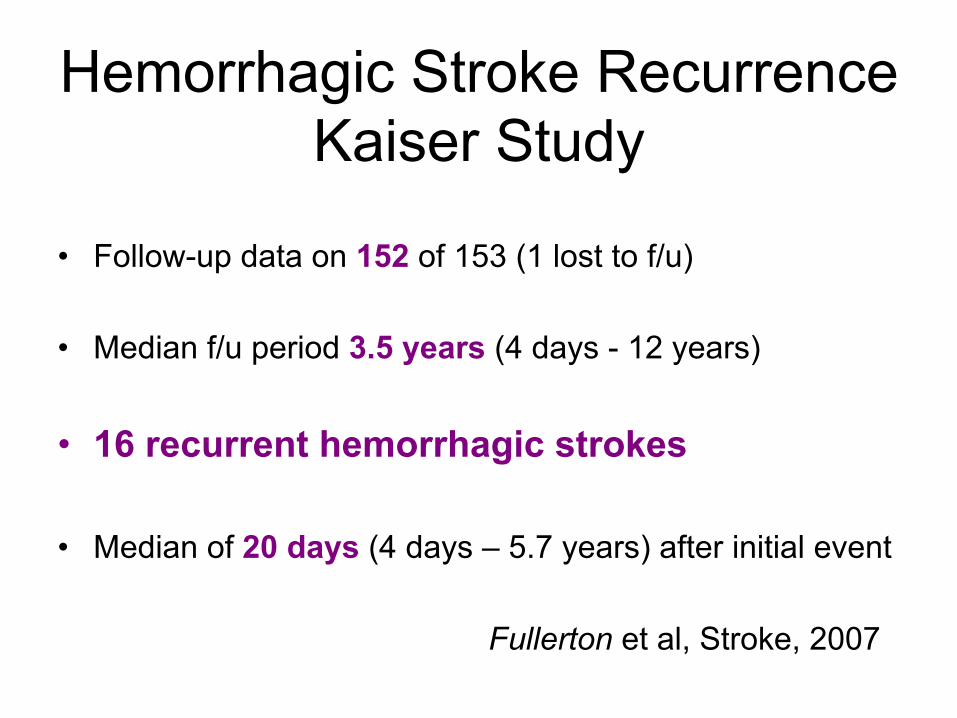
Hemorrhagic Stroke Recurrence Kaiser Study
• Follow-up data on 152 of 153 (1 lost to f/u)
• Median f/u period 3.5 years (4 days - 12 years)
• 16 recurrent hemorrhagic strokes
• Median of 20 days (4 days – 5.7 years) after initial event
Fullerton et al, Stroke, 2007
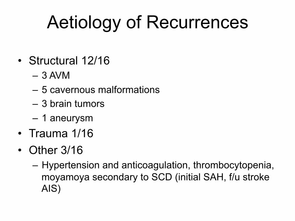
Aetiology of Recurrences
• Structural 12/16 – 3 AVM – 5 cavernous malformations – 3 brain tumors – 1 aneurysm
• Trauma 1/16 • Other 3/16
– Hypertension and anticoagulation, thrombocytopenia, moyamoya secondary to SCD (initial SAH, f/u stroke AIS)
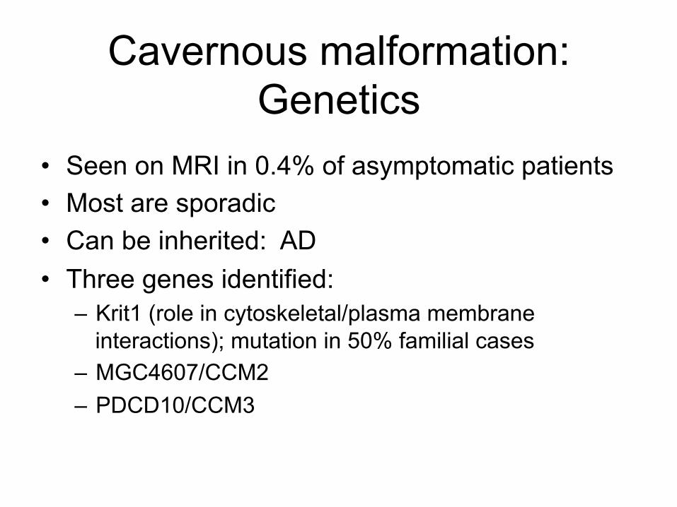
Cavernous malformation: Genetics
• Seen on MRI in 0.4% of asymptomatic patients • Most are sporadic • Can be inherited: AD • Three genes identified:
– Krit1 (role in cytoskeletal/plasma membrane interactions); mutation in 50% familial cases
– MGC4607/CCM2 – PDCD10/CCM3
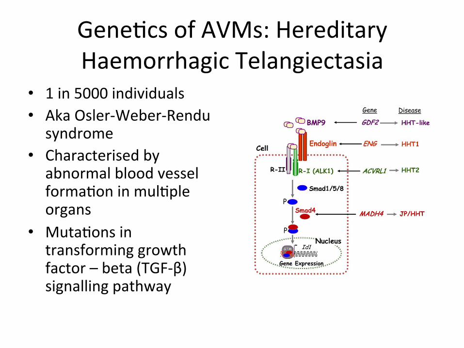
Gene8cs of AVMs: Hereditary Haemorrhagic Telangiectasia
• 1 in 5000 individuals • Aka Osler-‐Weber-‐Rendu
syndrome • Characterised by
abnormal blood vessel forma8on in mul8ple organs
• Muta8ons in transforming growth factor – beta (TGF-‐β) signalling pathway
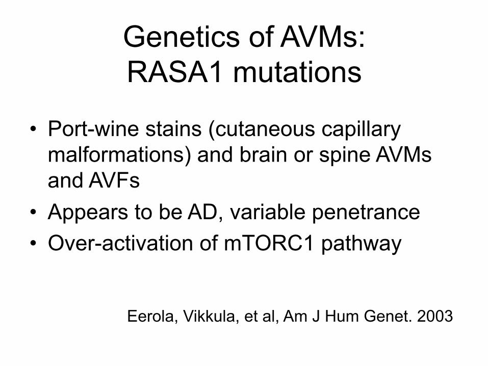
Genetics of AVMs: RASA1 mutations
• Port-wine stains (cutaneous capillary malformations) and brain or spine AVMs and AVFs
• Appears to be AD, variable penetrance • Over-activation of mTORC1 pathway
Eerola, Vikkula, et al, Am J Hum Genet. 2003
Outcome
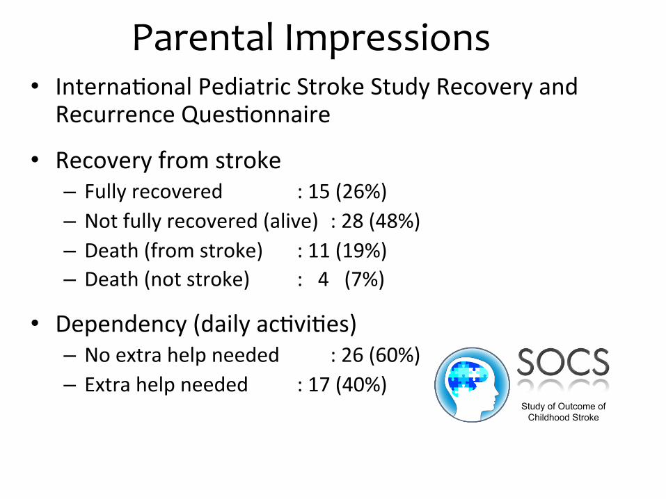
Parental Impressions • Interna8onal Pediatric Stroke Study Recovery and Recurrence Ques8onnaire
• Recovery from stroke – Fully recovered : 15 (26%) – Not fully recovered (alive) : 28 (48%) – Death (from stroke) : 11 (19%) – Death (not stroke) : 4 (7%)
• Dependency (daily ac8vi8es) – No extra help needed : 26 (60%) – Extra help needed : 17 (40%)
Study of Outcome of Childhood Stroke
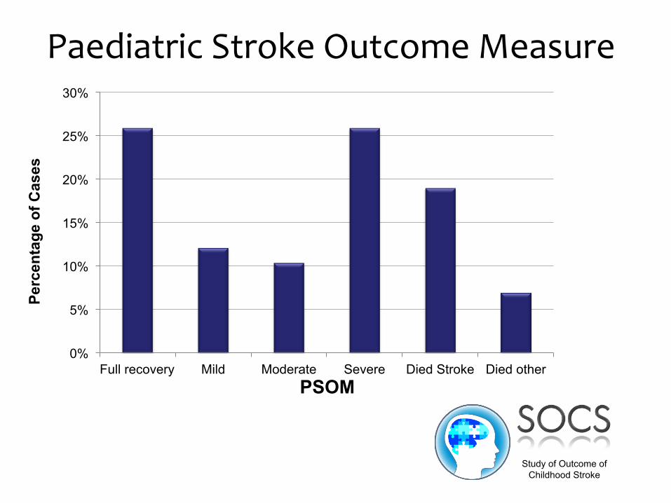
Paediatric Stroke Outcome Measure
0%
5%
10%
15%
20%
25%
30%
Full recovery Mild Moderate Severe Died Stroke Died other
Perc
enta
ge o
f Cas
es
PSOM
Study of Outcome of Childhood Stroke
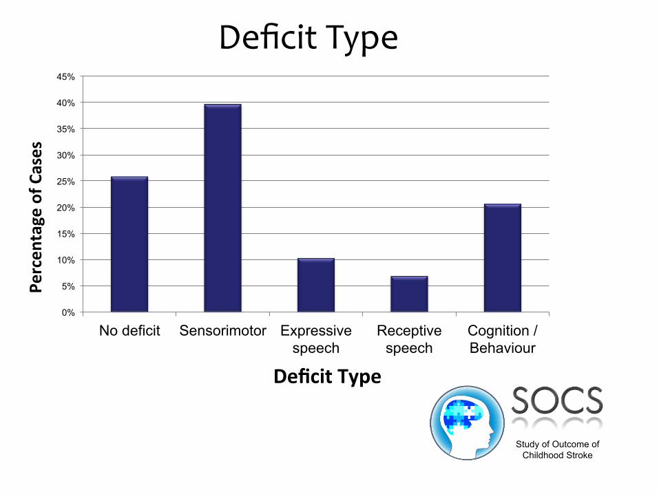
Deficit Type
0%
5%
10%
15%
20%
25%
30%
35%
40%
45%
No deficit Sensorimotor Expressive speech
Receptive speech
Cognition / Behaviour
Percen
tage of C
ases
Deficit Type
Study of Outcome of Childhood Stroke
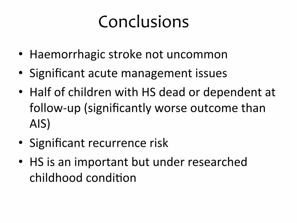
Conclusions
• Haemorrhagic stroke not uncommon • Significant acute management issues • Half of children with HS dead or dependent at follow-‐up (significantly worse outcome than AIS)
• Significant recurrence risk • HS is an important but under researched childhood condi8on
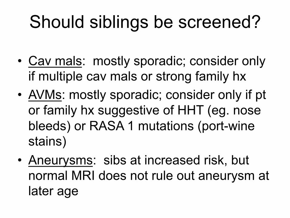
Should siblings be screened?
• Cav mals: mostly sporadic; consider only if multiple cav mals or strong family hx
• AVMs: mostly sporadic; consider only if pt or family hx suggestive of HHT (eg. nose bleeds) or RASA 1 mutations (port-wine stains)
• Aneurysms: sibs at increased risk, but normal MRI does not rule out aneurysm at later age
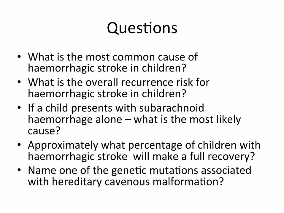
Ques8ons • What is the most common cause of haemorrhagic stroke in children?
• What is the overall recurrence risk for haemorrhagic stroke in children?
• If a child presents with subarachnoid haemorrhage alone – what is the most likely cause?
• Approximately what percentage of children with haemorrhagic stroke will make a full recovery?
• Name one of the gene8c muta8ons associated with hereditary cavenous malforma8on?


![Hemorrhagic Stroke Size by Optimization of - cureus.com · hemorrhagic stroke, and are associated with a higher mortality risk than ischemic strokes [3-4]. An IPH can lead to secondary](https://img.pdfslide.net/doc/110x75/5e0958916e06c4432d031ac7/hemorrhagic-stroke-size-by-optimization-of-hemorrhagic-stroke-and-are-associated.jpg)