Embed Size (px)
Citation preview
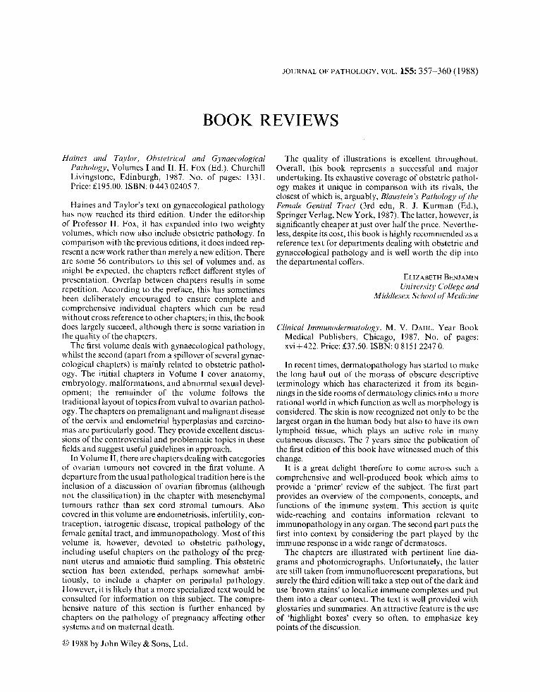
JOURNAL OF PATHOLOGY, VOL. 155: 357-360 ( 1 988)
BOOK REVIEWS
Haines and Taylor, Obstetrical and Gynaecological Pathology, Volumes I and 11. H. FOX (Ed.). Churchill Livingstone, Edinburgh, 1987. No. of pages: 1331. Pricc: E195.00. ISBN: 0 443 02405 7.
Haines and Taylor’s text on gynaecological pathology has now reached its third edition. Under the editorship of Professor H. Fox, it has expanded into two weighty volumes, which now also include obstetric pathology. In comparison with the previous editions, it does indeed rep- resent a new work rathcr than merely a new edition. There are some 56 contributors to this set of volumes and, as might be expected, the chapters reflect different styles of presentation. Overlap between chapters results in some repetition. According to the preface, this has sometimes been deliberately encouraged to ensure complete and comprehensive individual chapters which can be read without cross reference to other chapters; in this, the book does largely succeed, although there is some variation in the quality of the chapters.
The first volume deals with gynaecological pathology, whilst the second (apart from a spillover of several gynae- cological chapters) is mainly related to obstetric pathol- ogy. The initial chapters in Volume I cover anatomy, embryology, malformations, and abnormal sexual devel- opment: the remainder of the volume follows the traditional layout of topics from vulva1 to ovarian pathol- ogy. The chapters on premalignant and malignant disease of the cervix and cndometrial hyperplasias and carcino- mas are particularly good. They provide excellent discus- sions of the controversial and problematic topics in these fields and suggest useful guidelines in approach.
In Volume 11, there are chapters dealing with categories of ovarian tumours not covered in the first volume. A departure from the usual pathological tradition here is the inclusion of a discussion of ovarian fibromas (although not the classification) in the chapter with mesenchymal tumours rather than sex cord stromal tumours. Also covered in this volume are endometriosis, infertility, con- traception, iatrogenic disease, tropical pathology of the female genital tract, and immunopathology. Most of this volume is, however, devoted to obstetric pathology, including useful chapters on the pathology of the preg- nant uterus and amniotic fluid sampling. This obstetric section has been extended, perhaps somewhat ambi- tiously, to include a chapter on perinatal pathology. However, it is likely that a more specialized text would be consulted for information on this subject. The compre- hensive nature of this section is further enhanced by chapters on the pathology of pregnancy affecting other systems and on maternal death.
0 1988 by John Wiley & Sons, Ltd.
The quality of illustrations is excellent throughout. Overall, this book represents a successful and major undertaking. Its exhaustive coverage of obstetric pathol- ogy makes it unique in comparison with its rivals, the closest of which is, arguably, Blaustein’s Pathology qf the Female Genital Tract (3rd edn, R. J. Kurman (Ed.), Springer Verlag, New York, 1987). The latter, however, is significantly cheaper at just over half the price. Ncverthe- less, despite its cost, this book is highly recommended as a reference text for departments dealing with obstetric and gynaecological pathology and is well worth thc dip into the departmental coffers.
ELIZA~ETH BENJAMIN University College and
Middlesex School of Mdicine
Clinical Immunodermatology. M. V. DAHL. Year Book Medical Publishers, Chicago, 1987. No. of pages: xvi t422. Price: E37.50. ISBN: 0 8151 2247 0.
In recent times, dermatopathology has startcd to make the long haul out of the morass of obscure descriptive terminology which has characterized it from its begin- nings in the side rooms of dermatology clinics into a more rational world in which function as well as morphology is considered. The skin is now recognized not only to be the largest organ in the human body but also to have its own lymphoid tissue, which plays an active role in many cutaneous diseases. The 7 years since the publication of the first edition of this book have witnessed much of this change.
It is a great delight therefore to come across such a comprehensive and well-produced book which aims to provide a ‘primer’ review of the subject. The first part provides an overview of the components, concepts, and functions of the immune system. This section is quite wide-reaching and contains information relevant to immunopathology in any organ. The second part puts the first into context by considering the part played by the immune response in a wide range of dermatoses.
The chapters are illustrated with pertinent line dia- grams and photomicrographs. Unfortunately, the latter are still taken from immunofluorescent preparations, but surely the third edition will take a step out of the dark and use ‘brown stains’ to localize immune complexes and put them into a clear context. The text is well provided with glossaries and summaries. An attractive feature is the use of ‘highlight boxes’ every so often, to emphasize key points of the discussion.


![Obstetrical & Gynaecological Society of Malaysia · 2018. 3. 23. · 4 extra active slots and 1 observer slot had been add d due to popular demand Sarawak MRCOG Survival Course [SaMS]](https://img.pdfslide.net/doc/110x75/60ddc7e8f41e2f11022f5435/obstetrical-gynaecological-society-of-2018-3-23-4-extra-active-slots.jpg)