Embed Size (px)
Citation preview

WORKSHOP
Hair Follicle Stem Cells
Robert M. Lavker,1 Tung-Tien Sun,2 Hideo Oshima,3 Yann Barrandon,4 Masashi Akiyama,5 Corinne Ferraris,6
Genevieve Chevalier,6 Bertrand Favier,6 Colin A. B. Jahoda,7 Danielle Dhouailly,6 Andrei A. Panteleyev,8 andAngela M. Christiano81Department of Dermatology,The Feinberg School of Medicine, Northwestern University, Chicago, Illinois, USA; 2Department of Dermatology, NewYork University School of Medicine, NewYork, NewYork, USA; 3Department of Plastic & Reconstructive Surgery St Marianna University Schoolof Medicine2^16^1 Sugao Miyamaeku Kawasaki, 216^8511 Japan; 4Department of Biology, Ecole Normale Superieure, Paris, France; 5Department ofDermatology, Hokkaido University Graduate School of Medicine, Sapporo, Japan; 6Equipe Biologie de la Di¡eŁ renciation EpitheŁ liale, UMRCNRS 5538,Institut Albert Bonniot, UniversiteŁ Joseph Fourier, Grenoble, France; 7Department of Biological Sciences, University of Durham, UK; 8Departments ofDermatology, and Genetics & Development, Columbia University, NewYork, NewYork, USA
The workshop on Hair Follicle Stem Cells broughttogether investigators who have used a variety of ap-proaches to try to understand the biology of follicularepithelial stem cells, and the role that these cells playin regulating the hair cycle. One of the main conceptsto emerge from this workshop is that follicular epithe-lial stem cells are multipotent, capable of giving risenot only to all the cell types of the hair, but also to theepidermis and the sebaceous gland. Furthermore, suchmultipotent stem cells may represent the ultimateepidermal stem cell. Another example of epithelialstem cell and transit amplifying cell plasticity, was the
demonstration that adult corneal epithelium, underthe in£uence of embryonic skin dermis could form anepidermis as well as hair follicles. With regards to thelocation of follicular epithelial stem cells, immunohis-tochemical and ultrastructural data was presented, indi-cating that cells with stem cell attributes were localizedto the prominent bulge region of developing human fe-tal hair follicles. Finally, a new notion was put forthconcerning the roles that the bulge-located stem cellsand the hair germ cells played with respect to the haircycle. Key words: bulge/cell plasticity/epithelial stem cells/multipotent. JID Symposium Proceedings 8:28 ^38, 2003
HAIR FOLLICLE STEM CELLS: AN OVERVIEW
During the production of a hair, the follicle under-goes dynamic changes from an actively growingphase (anagen), to a remodeling phase (catagen),and ¢nally to a quiescent phase (telogen), only tostart growing again. Two key elements that control
hair follicle cycling are the follicular epithelial stem cells and thespecialized mesenchymal cells that constitute the follicular papil-la. This overview and the subsequent presentations of the work-shop focus on the hair follicle stem cells.Epithelial stem cells are considered to be slow cycling cells (or
perhaps more correctly, rarely cycling cells) that are endowedwith a superior proliferative capacity (Lavker & Sun, 2000). Suchcells are biochemically and morphologically primitive, and arefrequently multipotent (Schermer et al, 1986; Lavker et al, 1993;Taylor et al, 2000; Oshima et al, 2001). The progeny of the stemcells, upon exit from the stem cell niche, become transit amplify-ing (TA) cells, which have a ¢nite proliferative potential and,when such potential is exhausted, become postmitotic. Withinthe TA cell population there exists a hierarchy, ranging fromyoung TA cells capable of many divisions, to more mature TA
cells that can divide only a few times (Loe¥er et al, 1987; Lehreret al, 1998). Due to a dearth of reliable markers for epithelial stemcells, many investigators have used cell kinetics to distinguish theslow-cycling stem cells from the more frequently cyclingTA cells(Bickenbach, 1981; Cotsarelis et al, 1989; Cotsarelis et al, 1990;Morris and Potten, 1999; Taylor et al, 2000). To detect the slow-cycling cells, one can perfuse the tissue continuously with tri-tiated thymidine (3H-T) or bromodeoxyuridine (BrdU) so thatall dividing cells, including the occasionally dividing stem cells,can be labeled. During a chase period, which is typically between4 and 8 weeks, the rapidly dividing TA cells lose most of theirlabels due to dilution and only the slow-cycling stem cells stillretain their label; this way the stem cells can be detected experi-mentally as the label-retaining cells (LRCs).Application of this labeling technique to the hair follicle led to
the discovery in 1990 that the LRCs were mostly restricted to thebulge region, a specialized portion of the outer root sheathepithelium de¢ned as the insertion site of the arrector pili muscle(Cotsarelis et al, 1990; Morris and Potten, 1999; Taylor et al, 2000).The putative hair follicular epithelial stem cells of the bulge zoneexhibited several important stem cell attributes. First, cells fromthe upper region of the follicle, including the bulge, displayed thegreatest in vitro growth capacity when compared with cells fromother regions of the follicle and the epidermis (Kobayashi et al,1993; Yang et al, 1993). Second, the normally quiescent bulge cellsunderwent a transient period of cell proliferation during earlyanagen, or after stimulation (by, e.g., hair plucking, retinoic acid,or phorbol ester), giving rise to rapidly cycling TA cells (Wilsonet al, 1994; Taylor et al, 2000). Third, the bulge cells were relativelyundi¡erentiated ultrastructurally (Cotsarelis et al, 1990; Akiyama
Reprint requests to: Robert M. Lavker, Department of Dermatology,The Feinberg School of Medicine, Northwestern University, Chicago, Illi-nois, USA; Email: [email protected]: b-gal, b-galactosidase; FP, follicular papilla; HF, hair
follicle; IRS, inner root sheath; LRC, label-retaining cell; TA, transitamplifying.
Accepted for publication February 1, 2003
0022-202X/03/$15.00 . Copyright r 2003 by The Society for Investigative Dermatology, Inc.
28
et al, 1995). Fourth, topical application of a tumor initiator tomouse back skin at the onset of anagen (when the follicular bulgecells are undergoing transient proliferation) produced greaternumbers of tumors than application of the carcinogen in telogen(when bulge cells are quiescent). Since stem cells are known to beinvolved in skin tumor formation, the coincidence of greater tu-mor susceptibility with the transient proliferation of the bulgecells is consistent with the hypothesis that the bulge cells arestem cells, and indicates that follicular stem cells can give rise toexperimentally induced skin cancers (Morris et al, 1986;Miller et al, 1993; Morris et al, 1997; Morris et al, 2000). Takentogether, these data suggest that the bulge is the site of follicularstem cells.The realization that the follicular stem cells were located in the
bulge raised the interesting possibility that speci¢c interactionsbetween the bulge stem cells and the follicular papillary me-senchymal cells during appropriate stages of the hair cycle mayplay a key role in activating the bulge cell proliferation duringearly anagen (the ‘‘bulge activation hypothesis’’; Cotsarelis et al,1990; Sun et al, 1991). Speci¢cally, it was hypothesized that thefollicle enters into a new anagen when signals from the follicularpapilla transiently activate the quiescent stem cells in the bulge ofthe telogen follicle to proliferate, resulting in the generation ofTA cells. These TA cells form a new epithelial downgrowth,which in conjunction with the follicular papilla, forms the newhair bulb. As the follicular papilla is pushed away from the bulgezone by the epithelial downgrowth, the bulge stem cells return totheir normally slow-cycling state. After a period of sustained pro-liferation, the rapidly dividing matrix TA cells exhaust their pro-liferative capacity, and undergo terminal di¡erentiation. Thisresults in the degeneration of the lower two-thirds of the follicle(catagen). During catagen, the follicular papilla migrates upwardand becomes positioned at the base of the telogen follicle. Such aclose apposition of the follicular papilla to the bulge is thought tobe essential for the proper initiation of another hair cycle.The bulge stem cell concept has gained strong support, the
most recent coming from double-labeling studies, which allowedone to follow the fate of the progeny of the bulge-located stemcells (Taylor et al, 2000). This study provided direct evidence thatthe progeny of the slow-cycling LRCs of mouse pelage folliclegive rise to many of the compartments of the lower follicle, in-cluding the lower outer root sheath, matrix, and the medulla.These ¢ndings clearly established that the bulge stem cells giverise to a population of multipotent matrix cells, which in turnelaborate all components of the hair ¢ber. Another study (alsopresented in this workshop; see below) showed that the bulge re-gion of vibrissae, when dissected from a b-galactosidase-expres-sing mouse, and transplanted into the bulge region of a normalvibrissae, can give rise to cells of the entire follicular epitheliumas well as the hair shaft (Oshima et al, 2001). Taken together, thesedata strongly support the concept that the bulge stem cells cangive rise to many if not all cell types of the hair follicle.The discovery of hair follicle epithelial stem cells also led us to
suggest the possible existence a pilosebaceous epithelial unit(PSU), in which the progeny of the bulge stem cells not only giverise to the hair shaft, but also the epidermis and the sebaceousgland (Cotsarelis et al, 1990; Lavker et al, 1993). Double-labelingstudies enabled us to selectively tag the upper follicular epithelialcells so that we could trace the upward migration patterns of thebulge-derived progeny (Taylor et al, 2000). These studies demon-strated that, with time, the bulge cell progeny located in theupper follicle emigrated into the epidermis and continued to pro-liferate, thus contributing to the long-term maintenance of theepidermis. This migratory pattern was observed in both neonatalskin and adult mouse skin following small (2 mm) full thicknesswounds. The upward migration of the bulge stem cell-derivedprogeny cells into the epidermis, combined with the downwardmigration of the stem cell progeny to form the hair shaft, sug-gests that the bulge stem cells are at least bipotent (Taylor et al,2000). Based on these observations, we have proposed that thebulge is a major repository of skin keratinocyte stem cells, which
may thus be regarded as the ultimate epidermal stem cell (Lavkerand Sun, 2000; Taylor et al, 2000) (Fig 1).Two other papers presented in this workshop lend support to
the PSU concept. In the aforementioned vibrissae transplantationstudies, the bulge-derived, b-galactosidase-expressing keratino-cytes were shown to form the epidermis, as well as the sebaceousglands, thus supporting the multipotent nature of the bulge stemcells (Oshima et al, 2001). In another study, rabbit central cornealepithelium (comprised of TA cells), when combined with em-bryonic mouse dermis, changed from elaborating the more ad-vanced K3/K12 corneal keratin to expressing the more primitiveK5/K14 keratins. The corneal epithelial cells then gave rise to hairfollicle buds, which matured into fully formed hair follicles, andeventually the overlying corneal epitheliumwas transformed intoan epidermis, which expressed the K10 keratin (Ferraris et al,2000). These ¢ndings indicate that given the appropriate signal(s),the TA cells of central corneal epithelium can elaborate a varietyof di¡erent cell types thus proving that even the advanced TAcells can retain signi¢cant developmental £exibility.
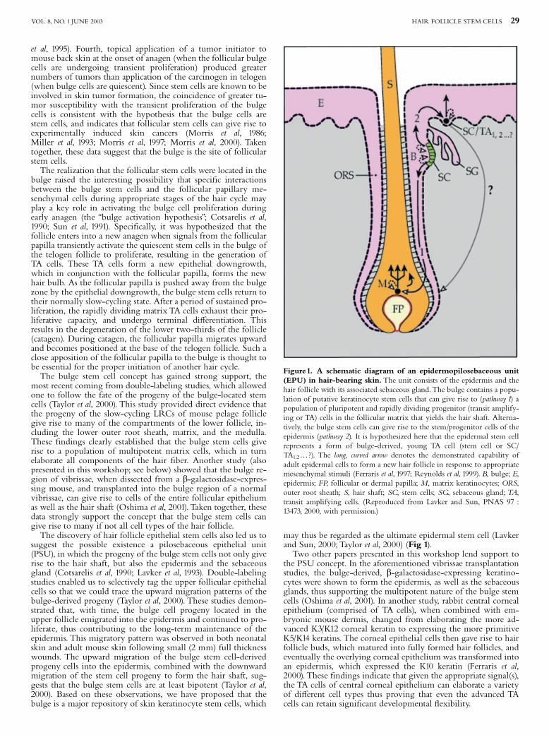
Figure1. A schematic diagram of an epidermopilosebaceous unit(EPU) in hair-bearing skin. The unit consists of the epidermis and thehair follicle with its associated sebaceous gland.The bulge contains a popu-lation of putative keratinocyte stem cells that can give rise to (pathway 1) apopulation of pluripotent and rapidly dividing progenitor (transit amplify-ing or TA) cells in the follicular matrix that yields the hair shaft. Alterna-tively, the bulge stem cells can give rise to the stem/progenitor cells of theepidermis (pathway 2). It is hypothesized here that the epidermal stem cellrepresents a form of bulge-derived, young TA cell (stem cell or SC/TA1,2y?). The long, curved arrow denotes the demonstrated capability ofadult epidermal cells to form a new hair follicle in response to appropriatemesenchymal stimuli (Ferraris et al, 1997; Reynolds et al, 1999). B, bulge; E,epidermis; FP, follicular or dermal papilla; M, matrix keratinocytes; ORS,outer root sheath; S, hair shaft; SC, stem cells; SG, sebaceous gland;TA,transit amplifying cells. (Reproduced from Lavker and Sun, PNAS 97 :13473, 2000, with permission.)
HAIR FOLLICLE STEM CELLS 29VOL. 8, NO. 1 JUNE 2003

In conclusion, available data strongly support the idea that fol-licular epithelial stem cells reside in the bulge, and that such stemcells can give rise to not only all the components of the hair shaft,but also the epidermis and the sebaceous gland.What is needed isa better understanding of the signals that cause the normallyquiescent follicular stem cells to divide and to give rise to youngmultipotent TA cells; of the signals that direct the TA cells to be-come a follicular matrix keratinocyte, a sebocyte, or an epidermalkeratinocyte; and of the degree of plasticity that exists within thekeratinocyte stem cell and transit amplifying cell compartments.
This work was supported by National Institutes of Health GrantsEY06769 (R.M.L.), and DK39753, DK52206, DK57269 (T.-T.S.)
MULTIPOTENT STEM CELL AND HAIR FOLLICLEMORPHOGENESIS
Location of follicular stem cells The hair follicle is anexcellent system to investigate the fate of epithelial stem cells. Itis postulated that stem cells have an unlimited self-renewalcapacity, but that they don’t proliferate at a high rate undernormal conditions. Labeling experiments and clonal analysisgive complementary information on the location and behaviorof stem cells and their progeny, making them very reliablemethods to investigate stem cells. The rapid proliferationobserved in the lower portion of the anagen whisker follicles ofthe rat resulted in the idea that within the matrix, the stem cellsgive rise to their progeny, transient amplifying cells (Ibrahim andWright, 1982; Oshima et al, 2001). This implied that the cellslocated in the lowest part of the hair bulb (the matrixkeratinocytes), were the stem cells (Reynolds and Jahoda, 1991;Hardy, 1992). However several lines of evidence indicate that
stem cells actually reside in the upper part of the follicle.Oliver’s seminal experiments demonstrate that a new hair bulbcan regenerate from the upper portion of a vibrissa follicle afteramputation of the follicle’s lower third (Oliver, 1966). In thepelage follicle of the mouse, tritiated thymidine (3H-TdR)- orbromodeoxyuridine (BrdU)-labeling experiments have revealedthat slow cycling cells (label-retaining cells, LRCs) reside in thebulge located at the level of the insertion of the arrector pilimuscle (Cotsarelis et al, 1990; Taylor et al, 2000). The cells locatedin this region proliferate infrequently in contrast to those locatedin the hair matrix, which proliferate very actively (Lavker et al,1991). Clonal analysis has demonstrated that 95% of thekeratinocyte colony forming cells (K-CFCs) of rat vibrissafollicles are located in the bulge region, whereas the remaining5% are located in the hair bulb (Kobayashi et al, 1993). Theseprevious studies indicated that bulge region is the site offollicular stem cells and led us to question (1) Is there a £ux ofstem cells from the bulge region to the bulb? (2) Does the bulgeregion serve as a reservoir of stem cells to sustain hair growth?
Migration of transplanted bulge cells To investigate the fateof the bulge cells in the vibrissa follicle, we conductedchimerical experiments using wild type and Rosa 26 transgenicmice that constitutively express a LacZ reporter gene. LacZexpression is maintained ubiquitously in tissues, and the b-galactosidase (b-gal.) positive cells originated from Rosa 26 areclearly identi¢ed by X-gal staining. We amputated the bulgeregion of vibrissa follicles of wild-type adult mice and replacedthem with bulges obtained from Rosa 26 adult mice. Chimericfollicles were then transplanted under the kidney capsule ofathymic mice, and harvested at regular intervals (Fig 2-A).Initially, b-gal. expressing cells were observed solely in the
upper region of the follicles. During the ¢rst four weeks
Figure 2. Schematic representation of the LacZ chimeric experiments. (A) generation of a chimeric follicle. (B) implantation of a Rosa 26 bulgeregion onto the back of a wild type embryo (modi¢ed from ¢gures in Oshima et al, 2001)
30 LAVKER ETAL JID SYMPOSIUM PROCEEDINGS

following transplantation, these cells were constantly observedopposite and above the transplanted region, indicating thatbulge-derived cells ¢rst migrated laterally and upward into theupper regions of the follicle. With time, b-gal. expressing cellswere also observed below the transplant, and, by eight weeks,they were present in the hair bulb. During the ¢rst weeksfollowing transplantation, the migrating b-gal. expressing cellswere mostly in the basal layer of the outer root sheath (ORS)and less frequently in the more di¡erentiated suprabasal layers(e.g. the ORS companion cell layer). b-gal. positive cells wereidenti¢ed solely in the ORS basal layer, after they reached thesuprabulbar region (see Figs 2, 3 in Oshima et al, 2001). Theseresults unambiguously demonstrate that the cells migrate in thebasal layer of the ORS from the upper region of the adultvibrissa follicle to the hair bulb. The b-gal. positive cells wereoften evenly distributed along the follicle length, suggestingthat there was a continuous £ux of migrating cells from thebulge region to the hair bulb.
Generation of pilo-sebaceous units After six weeks, b-gal.expressing cells were observed closer to the proximal end of thefollicle. Once the cells had reached the tip of the hair bulb, theystarted moving inwards to form the matrix, and later the innerroot sheath (IRS) and the hair shaft (see Fig 3 in Oshima et al,2001). This demonstrates that the transplanted cells contribute to
all the epithelial lineages involved in the formation of a hairfollicle (i.e., the ORS, the IRS, and the hair shaft). This indicatesthat the bulge region contains multipotent cells whose migrationis required for proper whisker growth.Transplanted Rosa 26 cells also migrated to the upper region
and to the opposite side of the follicle. b-gal.-positive lobularglands whose ducts opened into the hair canal (i.e., sebaceousglands) were observed. After the fourth week of transplantation,sebaceous glands were constantly observed on either side of theupper follicle but never in the lower part. These resultsdemonstrate that the transplanted bulge cells have the capacityto generate the sebaceous gland lineage. Moreover, they indicatethat sebaceous gland morphogenesis requires signals present onlyin the upper portion of the follicle. Collectively, these resultsdemonstrate that the bulge region contains multipotent stemcells that can generate all the epithelial lineages of the vibrissaefollicle.
Generation of hairy skin Individual bulge regions wereobtained from adult Rosa 26 vibrissae follicles and implanted inutero onto the back skin of E14.5-E16.5 mouse embryos. At birth,the regions containing the implants were transferred onto theback of adult athymic mice (Fig 2-B). Within days, b-gal.expressing cells were widely distributed in the epidermis andformed a hair germ (see Fig 4 in Oshima et al, 2001). Afterseveral weeks, mature follicles and associated sebaceous glandswere formed. Multiple b-gal. expressing hair follicles wereobserved, and a hair shaft of signi¢cant length was present inmost of the b-gal. expressing follicles. These results indicate thatthe transplanted bulge cells were functional in pelage follicles. Incontrast, the cells of the follicular papilla were b-gal. negative,indicating that the papilla originated from the recipient. Theseresults demonstrate that the bulge region of an adult vibrissafollicle contains multipotent stem cells with the capacity torespond to morphogenetic signals of hairy skin and to generateall the epithelial structures of the hairy skin.
Multipotent stem cells and hairy skin morphogenesis Vibrissafollicles are excellent models to study the cellular and molecularfactors involved in stem cell behavior during morphogenesis andrenewal of a complex epithelial structure. Hair follicles, sebaceousglands and the epidermis can be generated from adult bulgeregions implanted in embryos demonstrating that multipotentstem cells reside in the bulge.In vibrissa follicles, the undi¡erentiated progeny of the
multipotent stem cells migrate to de¢ned places where theygenerate the committed progenitors, i.e., above the bulge regionfor sebaceous progenitors and in the hair bulb for matrixprogenitors. Thus, the bulge region of the follicle serves as areservoir of multipotent stem cells (Fig 3). This location maintainsthe stem cells and allows them to receive the signals that instructtheir progeny to di¡erentiate into various lineages. Collectively thepopulation of K-CFCs identi¢ed in the bulge region, thepopulation of LRCs and the population of the multipotent stemcells that we described here, are most likely related.Stem cells and/or their progeny migrate in the basal layer of
the ORS over the entire length of a mature follicle. The reasonwhy the cells need to migrate over is most likely related towhisker growth. In pelage follicles, the proliferation of stemcells located in the bulge, is thought to reconstitute the pool ofmatrix cells at the end of telogen. The in£uence of the follicularpapilla, which then physically approach the stem cells, appearsdeterminant. This mechanism is known as the bulge activationhypothesis (Cotsarelis et al, 1990; Sun et al, 1991; Lavker et al,1993). However in vibrissa follicles, the follicular papilla cellsnever make contact with the cells located in the bulge region,even though vibrissa follicles do shorten slightly during catagen.Consequently, stem cells or their progeny must migrate thelength of the follicle to receive the necessary signals from thefollicular papilla.
Figure 3. Stem cell fate in a vibrissal follicle. The multipotent stemcells are located in the upper-region of the vibrissal follicle. They (or theircommitted progenitors) migrate to the hair bulb to generate the hair line-age or to the upper part of the follicle to generate sebaceous glands andepidermis (modi¢ed from ¢gures in Oshima et al, 2001).
HAIR FOLLICLE STEM CELLS 31VOL. 8, NO. 1 JUNE 2003

HAIR FOLLICLE STEM CELLS IN HUMAN SKINDEVELOPMENT
More than 10 years has passed since hair follicle stem cells werereported to lie in the bulge region of hair follicles in rodents(Cotsarelis et al, 1990). More recently, evidence has been presentedthat the hair follicle bulge is also thought to contain stem cells forthe human interfollicular epithelium (Lavker et al, 1993; Tayloret al, 2000; Oshima et al, 2001). In the developing hair follicles ofhuman fetuses, the bulge, an important ‘‘niche’’ for stem cells, is aprominent protrusion consisting of undi¡erentiated cells. However,in the adult human hair follicle, the bulge is not very prominent,often appearing as just a subtle swelling. In this presentation, cellswithin the bulge zone of human fetal hair follicles are character-ized, and discussed in the context of epithelial stem cells.
Localization of hair follicle stem cells during human fetalhair follicle development The sequential localization ofputative hair follicle stem cells was based on the expressionpatterns of several putative marker molecules for the epidermalstem cells. High levels of a2b1 and a3b1 integrin expression andweak staining for E-cadherin, b-catenin and a-catenin arethought to be markers for human epidermal stem cells (Mole' sand Watt, 1997; Watt, 1998); we utilized these markers tocharacterize regions enriched in putative stem cells duringhuman fetal hair follicle development (Akiyama et al, 2000). At65^84 days estimated gestational age (EGA), b1 integrin-rich,E-cadherin- and a- and b-catenin-poor cells (possible stem cells)were localized to the entire hair germ. By 85^104 days EGA, theb1 integrin-rich, E-cadherin- and a- and b-catenin-poor cellswere localized to the outermost cells of the hair peg. In the laterstages of development (bulbous hair peg ^105^135 days EGA anddi¡erentiated lanugo hair follicle ^4135 days EGA), theseputative follicular stem cells were identi¢ed in the bulge and theoutermost layer of the outer root sheath. These observationsindicate that cells in the bulge are unique in that they areuniformly b1 integrin-rich, E-cadherin- and a- and b-catenin-poor. The ¢nding of a population of putative hair follicle stemcells provides important clues to understanding the process ofhuman hair follicle morphogenesis. In addition, the regulationof growth and di¡erentiation via the integrin, cadherin andcatenin families is thought to be important in hair folliclemorphogenesis. The present results suggest that cell adhesionmolecules including integrins, cadherins and catenins may bekey regulators of the growth of follicular epithelial stem cellsduring human fetal follicle development.
Morphology of stem cells in developing human fetal hairfollicles Morphological characteristics of the b1 integrin-rich,E-cadherin- and a- and b-catenin-poor cells within the bulgecell, were investigated in the developing human hair follicles.These putative stem cells in each stage of hair follicledevelopment showed similar, undi¡erentiated morphologicfeatures (Akiyama et al, 2000). The bulge cells containedabundant free ribosomes and glycogen particles, but almost nocytoplasmic organelles, ultrastructural features indicative of arelatively undi¡erentiated state (Akiyama et al, 1995) (Fig 4).Mitochondria were rarely observed in the bulge cells. Onlysmall bundles of intermediate ¢laments were seen in theperipheral cytoplasm of the bulge cells, whereas larger bundlesof keratin ¢laments typical of more di¡erentiated keratinocyteswere not observed. The morphological features of human fetalbulge cells are consistent with those of the adult mousefollicular bulge cells (Cotsarelis et al, 1990), and supports the ideathat relatively undi¡erentiated cells (a feature of stem cells) arelocalized to the human fetal hair follicle bulge.
Hair follicle stem cells and growth factors, keratins andbasement membrane zone components in human skindevelopment The expression patterns of keratins, growthfactor receptors, and cell adhesion molecules were investigated
in developing hair follicles, with emphasis on the bulgeregion.In the bulbous hair peg stage, the putative stem cells within the
bulge region were high in epidermal growth factor (EGF)receptor expression (Akiyama et al, 1996; Akiyama et al, 2000).The bulge cells also expressed platelet-derived growth factor(PDGF) A chain and PDGF B chain, and the follicular sheathexpressed both PDGF a and b receptors (Akiyama et al, 1996).The low-a⁄nity nerve growth factor (NGF) receptor (p75) wasexpressed in the lower portion of the bulge and in mesenchymalcells around the hair follicle (Akiyama et al, 1996). These ¢ndingssuggested that EGF and NGF may be involved in regulation ofthe bulge cells and that PDGFs may play a role in theinteraction between the bulge and associated mesenchymal tissueat the bulbous hair peg stage.The bulge cells at the bulbous hair peg stage express keratins 5
and 14 (basal cell keratins) and keratin 19, the simple epithelialkeratin (Akiyama et al, 1995). The expression of basementmembrane zone components including the hemidesmosomalmolecules showed continuous bright staining along the dermal-epidermal junction of the hair germ and the hair peg, althoughthe staining was weak or negative in the lower portion of thehair peg.1 In the bulbous hair peg and the di¡erentiated lanugohair follicle, the basement membrane zone components were ex-pressed from the interfollicular epidermis to the bulge, and alsoin the region between the matrix cells and the dermal papilla cells(Akiyama et al, 1995). These ¢ndings suggest that basementmembrane zone components are continuously expressed in thestem cell sites during hair follicle morphogenesis.The nature and behavior of the hair follicle stem cells in
developing human skin has not yet been fully understood.Knowledge of the localization and features of hair follicle stemcells in developing human hair follicles will contribute to abetter understanding of the mechanisms underlying human hairfollicle morphogenesis.
CORNEAL EPITHELIAL BASAL CELLS CAN ACTIVATEEPIDERMAL GENETIC PROGRAMS BY REVERTING
FIRST TO HAIR STEM CELLS
During the last 10 years, the question has arisen as to whetheradult epithelial stem cell populations of the interfollicular skin,
Figure 4. Bulge cells of bulbous hair peg show undi¡erentiated ul-trastructure. Electron microscopy reveals that bulge cells in the humanfetal hair follicle (125 days EGA) lack cytoplasmic organelles indicative ofdi¡erentiation, but have abundant glycogen particles and free ribosomes inthe cytoplasm. The bulge cells show only a relatively small amount of ker-atin intermediate ¢lament bundles. Bars, 1 mm.
1Akiyama M, Matsuo I, Shimizu H: Remodeling of desmosome (DS)and hemidesmosome (HD) during human hair follicle development.J Invest Dermatol 117: 429 2001 (abstr.)
32 LAVKER ETAL JID SYMPOSIUM PROCEEDINGS

hair follicle, and corneal epithelium are committed to the pro-duction of only one cell lineage, i.e., whether epidermal interfol-licular stem cells give rise only to the epidermis, hair stem cells tohair follicle, and corneal stem cells to corneal epithelium (Milleret al, 1993). Alternatively, as suggested by several recent works onvarious organs (for a review, see Fuchs and Segre, 2000), adultstem cells may be pluripotent and consequently have equivalentpotentialities. In the second hypothesis, epithelial stem cells maystill possess embryonic features and therefore their microenviron-ment (e.g., the ¢broblasts with which they are associated), mayplay a crucial role in their di¡erentiation. It is well known thatthe adult rat dermal papilla is able to induce hair follicle forma-tion when associated with adult epidermis from di¡erent sources(i.e., the plantar region (Reynolds and Jahoda, 1992) or the fore-skin (Ferraris et al, 1997)). Since the basal population contains bothstem and transient amplifying (TA) type cells dispersed through-out the epidermis (among others: Jensen et al, 1999), the questionarises as to which epithelial cells of the adult epidermis can re-spond to new dermal in£uences. The adult corneal epithelium isan ideal system to answer this question because the stem cells ofcorneal epithelium are located in the limbal epithelium, whereasthe TA cells reside in the corneal epithelium. Identi¢cation of thelimbal epithelium as the preferential site of corneal epithelialstem cells was based on the observations that limbal basal cellsdid not express the K3 keratin (a marker of advanced cornealepithelial di¡erentiation), and that slow cycling cells wereexclusively located in the basal layer of the limbal epithelium(Schermer et al, 1986; Cotsarelis et al, 1989). More recent dataobtained in mouse showed that a hierarchy of TA cells are dis-tributed from the periphery to the centrum of the cornealepithelium, which contains primarily late or mature TA cells(Lehrer et al, 1998).Pluristrati¢ed epithelia of cornea and skin display distinct pro-
grams of di¡erentiation: corneal keratinocytes express the keratinpair K3/K12, epidermal keratinocytes the keratin pair K1-2/K10(Sun et al, 1983). Moreover, the epidermis forms cutaneous appen-dages, which express their own set of keratins. Thus, the results ofrecombinants between a hair-inducing dermis and an adult cen-tral corneal epithelium may provide insights into the questions ofequivalence between epithelial stem cells of ectodermal origin,and of the plasticity within a cell lineage. Indeed, the concept ofstem cells does not exclude the possibility that there exists a hier-archy of TA cells that under a given condition may revert back toa stem cell phenotype.The capacity of the epithelial component of adult mammalian
central cornea, to follow an alternative di¡erentiation pathwaywas investigated by associating the corneal epithelium with anembryonic dermis from a hair-forming region (Ferraris et al,2000). In addition to having a characteristic keratin expression,the cornea has two other advantages as a source of adult epithe-lium for recombination-type experiments: (1) it is easy to manip-ulate; and (2) it has no appendages. In order to identifyunequivocally the origin of the di¡erentiated structures, bispeci-
¢c epithelial-mesenchymal recombinants were performed, invol-ving rabbit adult central corneal epithelium and embryonicdorsal or upper-lip mouse dermis. Rabbit nuclei, which displayhomogeneous chromatin, are easy to distinguish from mouse nu-clei which have bright spots of condensed chromatin whenHoechst stained (Cunha and Vanderslice, 1984). The results showthat adult central corneal cells are able to respond to speci¢c in-formation originating from embryonic hair-forming dermis.They give rise ¢rst to a new basal layer, which no longer ex-presses the corneal-type keratins, then to pilosebaceous units,and ¢nally to upper layers expressing epidermal-type keratins(Fig 5).We thus provide the ¢rst evidence that a distinct TA cellpopulation can be reprogrammed (Ferraris et al, 2000).
Figure 5. Formation of hair follicles and of an epidermis aftergrafting recombinants of adult rabbit central corneal epitheliumand 14.5-day embryonic mouse dorsal dermis under the kidneycapsule of athymic mice. (A) Time zero, The corneal epithelium (e)comprises 6^7 cell layers. Fluorescent labeling showing that all the corneallayers, including the basal layer, synthesize the K12 corneal-type keratin.The rabbit corneal epithelium (e) nuclei are uniformly stained, whereasthe mouse dermal (d) nuclei exhibit bright £uorescent intranuclear bodies.The basal layer (bl) of the epithelium is composed entirely of rabbit cells.(B) After 12 days, the pluristrati¢ed epithelium (e) comprises numerousstrata characterized by the presence of corneal-type keratin K12 (red). Notethat the basal layer (bl), as well as the forming hair follicles (h) are not la-beled. A few cells localized at the point of attachment between hair folliclesand epithelium already synthesize the epidermal-type keratin, K10 (green).(C) After 21 days, the cells expressing keratin K12 are shedding, while con-tinuous layers of keratin K10 synthesizing cells have formed.
HAIR FOLLICLE STEM CELLS 33VOL. 8, NO. 1 JUNE 2003

Our interspeci¢c recombination experiments showed clearlythat signals from embryonic mouse dermis can be recognized by^ and elicit transformation of adult rabbit corneal epithelium to ^epidermis, hair follicles, and glands. In addition, and perhapsmore importantly, the detailed chain of events provided greaterinsight into questions relating to stem cell lineages and cell repro-gramming. Most researchers working on epithelial stem cellsfollow the conventional model of stem cell activity, in whichthere is a progressive and irreversible transition from stem cellsto transient amplifying cells to a postmitotic di¡erentiatedphenotype. The rabbit corneal epithelial cells used for our recom-binations were a transient amplifying population: the observationthat K12 was expressed throughout the epithelium at the time ofgrafting (Fig 5A) was con¢rmatory evidence that the cornealepithelium we used was centrally derived and contained no stemcells.Within a few days after being recombined with trichogenicdermis, the corneal epithelium formed a less-di¡erentiated basallayer, taking on a phenotype equivalent to the limbal epithelium,or the basal layer of the epidermis, both of which harbor epithe-lial stem cells. Thus it appears that the ¢rst stage of the transfor-mation process may be the restoration of a more primitive orstem cell-like phenotype from amongst a TA cell population in asomewhat more advanced stage of di¡erentiation. Our results ap-pear to re£ect the ideas of Loe¥er and Potten (1997), and may besupportive evidence for their spiral model of stem cell and TAbehavior. As part of this model,TA cells, which have left the stemcell niche, are not irreversibly committed to a terminal di¡eren-tiation pathway, but are able to revert to being stem cells in theevent of the removal or destruction of stem cells. The observationthat, after a few days, the corneal epithelial basal cells were bothparticipating in hair follicle morphogenesis and generating supra-basal K12 expressing cells (Fig 5B), illustrates clearly that prolif-eration and di¡erentiation are not mutually exclusive. In ourexperiments it must be assumed that hair follicle stem cells werealso established during the process of follicle morphogenesis. In arecent paper, it was suggested that stem cell were localizedthroughout the epithelium of developing hair follicles in the em-bryo (Akiyama et al, 2000). This implies that all hair bud epithe-lial cells are potential stem cells, and that subsequently the stemcell niche becomes progressively limited to the upper part of theouter root sheath. The developing follicles induced in the adultcorneal epithelium can be considered similar to embryonic folli-cles, which contain large numbers of stem cells. In relation tothis, we show that the ¢rst signs of epidermal di¡erentiation(K10 expression) always appeared at the top of the developinghair follicles (Fig 5B). At 21 days the K12 (corneal speci¢c) kera-tin is present in the higher and shedding layers of the epithelium(Fig 5C), while the lower epithelium expresses the epidermalspeci¢c K10 keratin.In conclusion, adult central corneal TA epithelial cells retain
the ability to transform into an epidermis and to produce hairfollicles with associated sebaceous glands when recombined withembryonic mouse hair-forming dermis. Moreover, the formationof the new epidermis originates from the induced hair follicles,which con¢rms the hair follicle as the main repository of epider-mal stem cells (Rochat et al, 1994; Taylor et al, 2000). Our resultsprovide: (i) a clear indication that a distinct TA corneal epithelialcell population can be reprogrammed; (ii) that this populationdoes so by ¢rst reverting to a hair stem-like condition (Fig 6).Our ¢ndings also imply that integumental stem cells haveequivalent potentialities.
THE HYPOTHESIS OF HAIR FOLLICLEPREDETERMINATION
The structural complexity of the hair follicle (HF), its potentialimmortality, and rapid growth rate, together with ability to re-turn periodically to its incipient state, make the HF a uniquestructure among other mammalian organs. Despite insights intomolecular aspects of HF biology, mechanisms governing the HF
cycle remain unclear. Several pioneering studies have attemptedto de¢ne the cellular dynamics of the HF cycle. However, mosthave focused on limited aspects of HF biology, discrete HF struc-tures or specialized types of HFs, and thus do not provide a uni-¢ed model of HF function. In 1990, the bulge activationhypothesis was published (Cotsarelis et al, 1990) ^ the ¢rst andthe most widely accepted model of the cellular kinetics in the HF.Here, we present a hypothesis of hair follicle predetermination,
which includes two critical re¢nements of preexisting models:the dual origin of the cycling portion of the HF and the recruit-ment of HF stem cells during the previous hair cycle that prede-termines the potential of hair germ cells to produce HF of thenext generation. The hypothesis of HF predetermination is basedon several conclusions made from an extensive analysis of the lit-erature and our own experimental data.
I. There are two cell populations with proliferation potentialin the telogen HF: the secondary hair germ and the cells ofthe bulge region Previous reports have clearly indicated thehigh proliferative potential of the bulge cells and their directcontribution to HF regrowth (Lane et al, 1991; Taylor et al, 2000).The telogen HF also contains a population of cells at its base,called the ‘‘hair germ’’ (Dry, 1926), situated in close apposition tothe follicular papilla (FP) (Fig 7A). Kinetic studies on earlyanagen HFs reveal a high proliferative activity in both the hairgerm and in the bulge region (Silver et al, 1969; Tezuka et al, 1991;Wilson et al, 1994). Furthermore, no migration of highlypersistent label-retaining cells (LRC) from the bulge into thehair germ during the initial anagen stages has been observed(Morris and Potten, 1999). Thus hair germ and bulge cellpopulations possess an intrinsic ability to proliferate and areboth directly involved in early anagen.
II. The hair germ cells, but not the stem cells in the bulgeregion, may play the primary role in anagen induction Studies
Figure 6. Interpretative schema. Corneal epithelium TA cells ¢rstrevert to hair-stem cells, before giving rise to hair follicles, andsubsequently epidermal cells.
34 LAVKER ETAL JID SYMPOSIUM PROCEEDINGS

of proliferative activity in early anagen HFs revealed that in anagen I,hair germ cells are actively proliferating, whereas mitotic activity inthe bulge region appears no earlier than anagen II and reachessigni¢cant levels only in anagen III (Silver et al, 1969; Tezuka et al,1991; Morris and Potten, 1999).The hair germ is ideally situated to participate in the direct FP-
epithelial crosstalk (Fig 7A) that is believed to play a pivotal role inanagen induction (Cotsarelis et al, 1990; Sun et al, 1991; Pauset al, 1999), while the bulge region is somewhat distant from the FPin telogen HFs. Furthermore, in anagen I, both the hair germ and theFP exhibit prominent up-regulation of bamacan, a putative organizerof mesenchymal^epithelial interaction (Couchman and du Cros,1995), thus further suggesting the hair germ as an immediate targetof FP inductive signaling and as one source of initial anagentransformations (Fig 7A,B).
III. Hair germ cells and cells of the bulge region contributeto di¡erent layers of the anagen HF Despite a belief that alllayers of the HF are products of the hair matrix germinative cells(e.g., Orwin, 1989), recent studies using a hair reconstitution assaydemonstrated that regeneration of IRS/hair shaft and ORSinvolves di¡erent progenitor cells (Kamimura et al, 1997). Thesame results were obtained using in vivo transduction of mouseskin with b-gal-encoding retroviral vectors (Ghazizadeh andTaichman, 2001). These data, together with the presence of twoproliferative units in the early anagen HF (Tezuka et al, 1991;Commo et al, 2000), suggest that cells in the hair germ and inthe bulge region may contribute to the development of di¡erentHF compartments. Speci¢cally, FP-associated hair germ cells maygive rise to the ascending HF portion (IRS and hair shaft) whilebulge cells may produce the ORS (Fig 7).Evidence in support of such a model are as follows: (1)
expression patterns of the presumptive stem cell markers in thebulge area and in the ORS are identical, whereas the matrix hasa di¡erent expression pro¢le (Lane et al, 1991; Akiyama et al, 1996;Commo et al, 2000); (2) bulge-derived tumors in human skin donot produce hair shaft- or IRS-like structures (Lever andSchaumburg-Lever, 1990), but instead keratinize along atrichilemmal pathway speci¢c for the ORS (Pinkus, 1969); (4)the cultured lower hair bulb microdissected from rat vibrissa isable to reconstitute all ascending HF layers, but not the ORS(Jahoda CAB; unpublished); (5) in hairless mouse skin, wherethe bulge cells are physically separated from FP, the ORS-likebulge outgrowths do not form any structures resembling IRSor hair shaft, while FP-associated epithelial cells produce HFslacking the ORS (Panteleyev et al, 1999b).Recently, it was proposed that presumptive stem cells located
in the bulge region undergo two distinct pathways of migration/
specialization: ‘‘the bulge-epidermal’’ (upward) and ‘‘the bulge-hair’’ (downward) (Taylor et al, 2000). We prefer to desig-nate these pathways as ‘‘bulge-epidermal’’ and ‘‘bulge-ORS’’, thusemphasizing their similarity and regulation by parallelmechanisms along with their di¡erence to the ‘‘hair’’ type of cellspecialization.
IV. In late anagen, the bulge-derived clonogenic cellsmigrate to the lower ORS and form a speci¢c compactstructure on the periphery of the hair bulb During matureanagen, a movement of undi¡erentiated ORS cells in thedownward direction has been reported (Reynolds and Jahoda,1991). This ¢nding is in line with the recently shown capabilityof the bulge cells to have an active and prolonged migrationthrough the ORS of anagen HFs (Oshima et al, 2001).Recent studies have revealed a region of asymmetrical
(unilateral) expression of a number of genes in the hair bulb ofmouse HFs, suggesting the presence of a speci¢c cell populationthat di¡ers from the matrix cells and seems to be contiguouswith the lowermost extension of the ORS (Paus et al, 1997; Gatet al, 1998; Gambardella et al, 2000; McGowan and Coulombe,2000; Muller-Rover et al, 2000; Panteleyev et al, 2000b). Wehypothesize that this structure ^ ‘‘the lateral disc’’ ^ mayrepresent the ¢nal destination of bulge-derived cells with highclonogenic potential migrating downward, where they graduallyconcentrate during anagen (Fig 8A).Currently, there is no evidence indicating the existence of the
lateral disc in human HFs. However, the substantial thickness ofthe lower ORS (at the level of upper hair bulb) in human HFmight be su⁄cient to harbor a distinct cell population. Perhaps theK19-positive (a putative marker of clonogenic cells) population ofinactive cells in the lower ORS in human anagen HFs (Commoet al, 2000) represents the human analog of the lateral disc.
V. During catagen, lateral disc cells survive apoptosis andtransform into the hair germ under the in£uence of FP Itis generally believed that all epithelial cells of the lower hair bulbare eliminated during catagen by means of apoptosis (Seiberget al, 1995). Nevertheless, electron microscope studies show thatin early catagen both FP-associated keratinocytes and FP¢broblasts reinforce their connection to the basal lamina.The keratinocytes accomplish this by formation of numerousadditional hemidesmosomes, and FP ¢broblasts by a depositionof extensive intercellular ¢lamentous network (Sugiyama et al,1976). This change in papilla-epithelial junction might re£ectthat some FP-associated keratinocytes are apoptosis resistant.Our studies of hairless gene expression revealed the presence of
a small population of h mRNA-positive keratinocytes associated
Figure 7. Proposed cellular kinetics in the early anagen hf. (A) the activation of hair germ cells by a fp-derived signal (yellow arrow). (B) in anagen ii,the activity of the hair germ (yellow arrow) induces proliferation of the bulge cells (black arrows). (C) in anagen iiia, the downward growth of bulge-derivedcells (black arrows) results in the formation of the ors. (D) in anagen iiib, the upward proliferation of hair germ cells (white arrows) results in formation of theascending compartment of the anagen hf (hair shaft and irs).
HAIR FOLLICLE STEM CELLS 35VOL. 8, NO. 1 JUNE 2003

with FP in early catagen HFs. These cells persist over catagen andconcentrate at the bottom of the telogen HF (Panteleyev et al,2000a). Thus, we propose that some hair bulb cells survivecatagen-associated apoptosis and contribute to the formation ofthe hair germ.What is the origin of these FP-associated keratinocytes? We
believe that in the mouse follicles, these cells representapoptosis-resistant bulge-derived cells of the lateral disc(Panteleyev et al, 1999a). In the human HF, these apoptosis-resistant cells may originate from the lowermost ORS thatcomes into direct contact with the FP during the destruction ofthe hair bulb in early catagen (Fig 8A^C).Assuming the conventional model of stem cell activity, in
which there is an irreversible transition from stem cells totransient amplifying (TA) cells to di¡erentiated cells (Lajtha,1979), it is di⁄cult to envision that lateral disc cells retain theirundi¡erentiated features and high proliferative potential. So,how do lateral disc cells reacquire (or retain) their ‘‘stem cell-like’’ features and transform into a hair germ with the potentialto form a new hair? Recently, it was shown that a distinct TAcell population of rabbit corneal epithelium can reacquire somestem cell characteristics under the in£uence of adjacentmesenchyme (Ferraris et al, 2000). A similar mechanism mayoccur in FP^ epithelial interactions and we believe that theformation of intimate contacts between the FP and the lateraldisc cells (lower ORS in human HF) in catagen is adetermining event in their ¢nal transformation into the hairgerm.
VI. By telogen, the hair germ cells become selectivelyreceptive to FP signaling and committed to producingascending HF layers The proliferative activity of the HFbulge cells can be induced by many nonspeci¢c factors (Holecekand Ackerman, 1993) and, in culture, these cells have an extensiveproliferative capacity (Yang et al, 1993). The proliferative activityof the hair matrix cells (which we believe originate from hairgerm cell precursors), by contrast, is strictly dependent on the
associated mesenchyme, and these cells cannot proliferate inculture in the absence of FP ¢broblasts (Reynolds and Jahoda,1993).Given the key role of mesenchymal cells in the determination
of cell fates (Ferraris et al, 2000), we propose that the intimatecontact between lateral disc/hair germ cells with FP ¢broblastsduring catagen-telogen phases (Fig 8B^D) speci¢cally primethese cells with high receptivity to FP-derived morphogeneticsignals. Thus, the receptivity of the hair germ cells to FPsignaling and their commitment to produce the ascending layersof the HF may be predetermined by a priming phase of the hairgerm precursors during the previous hair cycle.
Conclusion In the hypothesis of HF predetermination, wehave sought to provide a uni¢ed view of the process of stem cellrecruitment during HF cycling. The data and ideas put forth heresuggest that HF stem cell recruitment is a complex, relativelyprotracted, multistage process that requires speci¢c interactionswith microenvironment.Despite the lines of evidence in favor of our hypothesis, it
remains speculative and raises as many questions as it answers.Nevertheless, our intention is not to present a ¢nal andindisputable model of HF cycle progression but rather to take afresh look at some of the most critical but poorly understoodaspects of HF biology. We hope that our hypothesis inspiresfuture studies of the cellular dynamics that underlie HFprogression through the periods of growth, regression andquiescence.
The authors are grateful to Andrey Panteleyev, Jr. and Dmitry Panteleyev for theirhelp with artwork. Stimulating discussions with Drs. Colin Jahoda and VladimirBotchkarev were invaluable to this work.This study was supported in part by grantsfrom National Alopecia Areata Foundation, NIH USPHS, and Skin Disease Re-search Center of the Department of Dermatology, Columbia University (KO1-AR02204, R03-AR47403, toAP; P30-44534, and RO1-47338 toAMC).
Figure 8. Proposed scheme of the lateral disc transformation into the hair germ. (A) During anagen, bulge-derived cells with clonogenic potentialmigrate downward (blue arrow) and form the lateral disc that resides inactively on the periphery of the hair bulb. Hair matrix cells (red) are actively prolif-erating. (B) In early catagen, owing to the diminution of the hair matrix, the lateral disc cells come into direct contact with FP. (C) In late catagen, lateraldisc cells travel upward along with the FP and gradually transform into the hair germ (change of blue color into red). (D) In the telogen, hair germ and bulgecells reside as two separate and functionally discrete structures. Owing to FP-dependent ‘‘priming’’during previous catagen, hair germ cells acquire selectivesensitivity to FP-derived signaling and the commitment to produce ascending layers of HF of new generation. 1These experiments were performed 14months after completion of labeling and the behavior of ‘‘younger’’ LRC (e.g., 8^10 weeks after labeling) has not been reported.
36 LAVKER ETAL JID SYMPOSIUM PROCEEDINGS

CONTRIBUTORS
The contributors of this workshop were: RM Lavker and T-TSun, Hair Follicle Stem Cells: An Overview; H Oshima and YBarrandon, Multipotent Stem Cell and Hair Follicle Morpho-genesis; M Akiyama, Hair Follicle Stem Cells in Human SkinDevelopment; C Ferraris, G Chevalier, B Favier, CAB Jahodaand D Dhouailly, Corneal Epithelium Basal Cells Can ActivateEpidermal Genetic Programs by Reverting First to Hair StemCells; AA Panteleyev, CAB Jahoda and AM Christiano,The Hy-pothesis of Hair Follicle Predetermination.
REFERENCES
Akiyama M, Dale BA, Sun T-T, Holbrook KA: Characterization of hair folliclebulge in human fetal skin; The human fetal bulge is a pool of undi¡erentiatedkeratinocytes. J Invest Dermatol 105:844^850, 1995
Akiyama M, Smith LT, Holbrook KA: Growth factor and growth factor receptorlocalization in the hair follicle bulge and associated tissue in human fetus.J Invest Dermatol 106:391^396, 1996
Akiyama M, Smith LT, Shimizu H: Changing patterns of localization of putativestem cells in developing human hair follicles. J Invest Dermatol 114:321^327,2000
Bickenbach JR: Identi¢cation and behavior of label-retaining cells in oral mucosaand skin. J Dent Res 60:1611^1620, 1981
Commo S, Gaillard O, Bernard BA: The human hair follicle contains two distinctK19 positive compartments in the outer root sheath: A unifying hypothesis forstem cell reservoir? Di¡erentiation 66:157^164, 2000
Cotsarelis G, Cheng SZ, Dong G, Sun T-T, Lavker RM: Existence of slow-cyclinglimbal epithelial basal cells that can be preferentially stimulated to proliferate:Implications on epithelial stem cells. Cell 57:201^209, 1989
Cotsarelis G, Sun T-T, Lavker RM: Label-retaining cells reside in the bulge area ofpilosebaceous unit: Implications for follicular stem cells, hair cycle, and skincarcinogenesis. Cell 61:1329^1337, 1990
Couchman JR, du Cros DL: Proteoglycans and associated proteins of the mam-malian hair follicle. J Invest Dermatol 104 (5 (Suppl.):40S^41S, 1995
Cunha GR,Vanderslice KD: Identi¢cation in histological sections of species originof cells from mouse, rat and human. StainTechnol 59:7^12, 1984
Dry FW:The coat of the mouse (Mus musculus). J Genet 16:288^340, 1926Ferraris C, Bernard BA, Dhouailly D: Adult epidermal keratinocytes are endowed
with pilosebaceous forming abilities. Int J Dev Biol 41:491^498, 1997Ferraris C, Chevalier G, Favier B, Jahoda CA, Dhouailly D: Adult corneal epithe-
lium basal cells possess the capacity to activate epidermal, pilosebaceous andsweat gland genetic programs in response to embryonic dermal stimuli. Devel-opment 127:5487^5495, 2000
Fuchs E, Segre JA: Stem cells. A new lease of life. Cell 100:143^155, 2000Gambardella L, Schneider-Maunoury S,Voiculescu O, Charnay P, BarrandonY: Pat-
tern of expression of the transcription factor krox-20 in mouse hair follicle.Mech Dev 96:215^218, 2000
Gat U, DasGupta R, Degenstein L, Fuchs E: De Novo hair follicle morphogenesisand hair tumors in mice expressing a truncated beta-catenin in skin. Cell95:605^614, 1998
Ghazizadeh S, Taichman LB: Multiple classes of stem cells in cutaneous epithelium:A lineage analysis of adult mouse skin. EMBO J 20:1215^1222, 2001
Hardy MH:The secret life of the hair follicle.Trends Genet 8:55^61, 1992Holecek BU, Ackerman AB: Bulge-activation hypothesis: Is it valid? Am J Dermato-
pathol 15:235^255, 1993Ibrahim L,Wright EA: A quantitative study of hair growth using mouse and rat vi-
brissal follicles. J Embryol Exp Morph 72:209^224, 1982Jensen UB, Lowell S,Watt FM: The spatial relationship between stem cells and their
progeny in the basal layer of human epidermis: A new view based on whole-mount labeling and lineage analysis. Development 126:2409^2418, 1999
Kamimura J, Lee D, Baden HP, Brissette J, Dotto GP: Primary mouse keratinocytecultures contain hair follicle progenitor cells with multiple di¡erentiation po-tential. J Invest Dermatol 109:534^540, 1997
Kobayashi K, Rochat A, Barrandon Y: Segregation of keratinocyte colony-formingcells in the bulge of the rat vibrissa. Proc Natl Acad Sci (USA) 90:7391^7395,1993
Lajtha LG: Stem cells concepts. Di¡erentiation 14:23^34, 1979Lane EB,Wilson CA, Hughes BR, Leigh IM: Stem cells in hair follicles. Cytoskele-
tal studies. Ann NYAcad Sci 642:197^213, 1991Lavker RM, Sun TT: Epidermal stem cells. Properties, markers, and location. Proc
Nat Acad Sci USA 97:13473^13475, 2000Lavker RM, Cotsarelis G,Wei Z-G, Sun TT: Stem cells of pelage, vibrissa, and eye-
lash follicles: The hair cycle and tumor formation. Ann N YAcad Sci 642:214^225, 1991
Lavker RM, Miller S, Wilson C, Cotsarelis G, Wei Z-G, Yang J-S, Sun TT: Hairfollicle stem cells. Their location, role in hair cycle, and involvement in skintumor formation. J Invest Dermatol 101:16S^26S, 1993
Lehrer MS, Sun TT, Lavker RM: Strategies of epithelial repair: Modulationof stem cell and transit amplifying cell proliferation. J Cell Sci 111:2867^2875,1998
LeverWF, Schaumburg-Lever G: Histopathology of the Skin, 7th edn. Philadelphia:Lippincott, 1990
Loe¥er M, Potten CS: Stem cells and cellular pedigrees ^ a conceptual introduction.In: Potten C (ed). Stem Cells. London: Academic Press, 1997; p 1^27
Loe¥er M, Potten CS,Wichmann HE: Epidermal cell proliferation. II. A compre-hensive mathematical model of cell proliferation and migration in the basallayer predicts some unusual properties of epidermal stem cells.Virchows Arch BCell Pathol Incl Mol Pathol 83:286^300, 1987
McGowan KM, Coulombe PA: Keratin 17 expression in the hard epithelial contextof the hair and nail, and its relevance for the pachyonychia congenita pheno-type. J Invest Dermatol 114:1101^1107, 2000
Miller SJ, Lavker RM, Sun TT: Keratinocyte stem cells of cornea, skin and hair fol-licle: Common and distinguishing features. Dev Biol 4:217^240, 1993
Miller SJ,Wei ZG,Wilson C, Dzubow L, Sun TT, Lavker RM: Mouse skin is parti-cularly susceptible to tumor initiation during early anagen of the hair cycle:Possible involvement of hair follicle stem cells. J Invest Dermatol 101:591^594,1993
Mole' s JP, Watt FM: The epidermal stem cell compartment. Variation in expressionlevels of E-cadherin and catenins within the basal layer of human epidermis.J Histochem Cytochem 45:867^874, 1997
Morris RJ, Coulter K, Tryson K, Steinberg SR: Evidence that cutaneous carcino-gen-initiated epithelial cells from mice are quiescent rather than actively cy-cling. Cancer Res 57:3436^3443, 1997
Morris RJ, Fischer SM, Slaga TJ: Evidence that a slowly cycling subpopulationof adult murine epidermal cells retains carcinogen. Cancer Res 46:3061^3066,1986
Morris RJ, Potten CS: Highly persistent label-retaining cells in the hair follicles ofmice and their fate following induction of anagen. J Invest Dermatol 112:470^475, 1999
Morris RJ, Tryson KA,Wu KQ: Evidence that the epidermal targets of carcinogenaction are found in the interfollicular epidermis or infundibulum as well as inthe hair follicles. Cancer Res 60:226^229, 2000
Muller-Rover S, Bulfone-Paus S, Handjiski B, et al: Intercellular adhesion molecule-1and hair follicle regression. J Histochem Cytochem 48:557^568, 2000
Oliver RF:Whisker growth after removal of the dermal papilla and lengths of fol-licle in the hooded rat. J Embryol Exp Morph 15:331^347, 1966
Orwin DFG:Variations in wool follicle morphology. In: Rodgers GE, Reis PJ,WardKA, Marshall RC (eds). The Biology of Wool and Hair. London: Chapman &Hall, 1989; pp 227^242
Oshima H, Rochat A, Kedzig C, Kobayashi K, Barrandon Y: Morphogenesis andrenewal of hair follicles from adult multipotent stem cells. Cell 104:233^245,2001
PanteleyevAA, Botchkareva NV, Sundberg JP, Christiano AM, Paus R: The role ofthe hairless (hr) gene in the regulation of hair follicle catagen transformation.AmJ Pathol 155:159^171, 1999a
Panteleyev AA, Paus R, Christiano AM: Patterns of hairless (hr) gene expression inmouse hair follicle morphogenesis and cycling. Am J Pathol 157:1071^1079,2000a
Panteleyev AA, Paus R, Sundberg JP, Christiano AM: The ‘second wave’ of hairgrowth in hairless mouse skin: A model to study mechanisms of anagen hairfollicle induction. J Invest Dermatol 112:631, 1999b
Panteleyev AA, Tadin M, Paus R, Christiano AM: Hair cycle-dependent expressionpatterns of the transcription factor AP-2a in mouse skin. J Invest Dermatol114:795, 2000b
Paus R, Foitzik K, Welker P, Bulfone-Paus S, Eichmuller S: Transforming growthfactor-beta receptor type I and type II expression during murine hair follicledevelopment and cycling. J Invest Dermatol 109:518^526, 1997
Paus R, Muller-Rover S, Botchkarev VA: Chronobiology of the hair follicle: hunt-ing the ‘hair cycle clock’. J Invest Dermatol Symp ProcThe 4:338^345, 1999
Pinkus H: ‘Sebaceous cysts’ are trichilemmal cysts. Arch Dermatol 99:544^555, 1969Reynolds AJ, Jahoda CAB: Cultured dermal papilla cells induce follicle formation
and hair growth by transdi¡erentiation of an adult epidermis. Development115:587^593, 1992
Reynolds AJ, Jahoda CAB: Hair ¢bre progenitor cells Developmental status and in-teractive potential. Sem Dev Biol 4:241^250, 1993
Reynolds AJ, Jahoda CAB: Hair follicle stem cells? A distinct germinative epidermalcell population is activated in vitro by the presence of hair dermal papilla cells.J Cell Sci 99:373^385, 1991
Reynolds AJ, Lawrence C, Cserhalmi-Friedman PB, Christiano AM, Jahoda CAB:Trans-gender induction of hair follicles. Nature 402:33^34, 1999
Rochat A, Kobayashi K, BarrandonY: Location of stem cells of human hair folliclesby clonal analysis. Cell 76:1063^1073, 1994
Schermer A, Galvin S, Sun TT: Di¡erentiation-related expression of a major 64Kcorneal keratin in vivo and in culture suggests limbal location of cornealepithelial stem cells. J Cell Biol 103:49^62, 1986
Seiberg M, Marthinuss J, Stenn KS: Changes in expression of apoptosis-associatedgenes in skin mark early catagen. J Invest Dermatol 104:78^82, 1995
Silver AF, Chase HB, Arsenault CT: Early anagen initiated by plucking comparedwith early spontaneous anagen. In: Montagna W, Dobson RL (eds). Adv BiolSkin 9. NewYork: Pergamon Press, 1969; p 265^28
HAIR FOLLICLE STEM CELLS 37VOL. 8, NO. 1 JUNE 2003

Sugiyama S, Takahashi M, Kamimura M: The ultrastructure of the hair follicles inearly and late catagen, with special reference to the alteration of the junctionalstructure between the dermal papilla and epithelial component. J Ultrastruct Res54:359^373, 1976
Sun TT, Cotsarelis G, Lavker RM: Hair follicular stem cells: The bulge-activationhypothesis. J Invest Dermatol 98:77s^78s, 1991
Sun TT, Eichner R, Nelson WG, Tseng SCG, Weiss RA, Jarvinen M, Woodcock-Mitchell J: Keratin classes. Molecular markers for di¡erent types of epithelialdi¡erentiation. J Invest Dermatol 81:109s^115s, 1983
Taylor G, Lehrer MS, Jensen PJ, SunT-T, Lavker RM: Involvement of follicular stemcells in forming not only the follicle but also the epidermis. Cell 102:451^461,2000
Tezuka M, Ito M, Ito K,TazawaT, SatoY: Investigation of germenative cells in gen-erating and renewal anagen hair apparatus in mice using anti-bromodeoxyur-idine monoclonal antibody. J Dermatol Sci 2:434^443, 1991
Watt FM: Epidermal stem cells. Markers, patterning and the control of stem cell fate.PhilosTrans R Soc Lond B Biol Sci 353:831^837, 1998
Wilson C, Cotsarelis G,Wei ZG, et al: Cells within the bulge region of mouse hairfollicle transiently proliferate during early anagen: Heterogeneity and func-tional di¡erences of various hair cycles. Di¡erentiation 55:127^136, 1994
Yang JS, Lavker RM, Sun TT: Upper human hair follicle contains a subpopulationof keratinocytes with superior in vitro proliferative potential. J Invest Dermatol101:652^659, 1993
38 LAVKER ETAL JID SYMPOSIUM PROCEEDINGS