Embed Size (px)
Citation preview

Handbook of Pediatric Neuro-Ophthalmology
Handbook of PediatricNeuro-Ophthalmology
Edited by
Kenneth W. Wright, MDDirector, Wright Foundation for Pediatric Ophthalmology Director, Pediatric Ophthalmology, Cedars-Sinai MedicalCenter, Clinical Professor of Ophthalmology, University ofSouthern California—Keck School of Medicine, Los Angeles,California
Peter H. Spiegel, MDFocus On You, Inc., Palm Desert, CaliforniaInland Eye Clinic, Murrieta, CaliforniaChildren’s Eye Institute, Upland, California
Lisa S. Thompson, MDAttending Physician, Stroger Hospital of Cook County,Chicago, Illinois
IllustratorsTimothy C. Hengst, CMISusan Gilbert, CMIFaith Cogswell
Kenneth W. Wright, MD Peter H. Spiegel, MDDirector, Wright Foundation for Focus On You, Inc.
Pediatric Ophthalmology Palm Desert, CADirector, Pediatric Ophthalmology, Inland Eye Clinic
Cedars-Sinai Medical Center, Murrieta, CAClinical Professor of Children’s Eye
Ophthalmology, University of Institute Southern California—Keck School Upland, CAof Medicine USA
Los Angeles, CAUSA
Lisa S. Thompson, MDAttending PhysicianStroger Hospital of Cook CountyChicago, ILUSA
Library of Congress Control Number: 2005932933
ISBN 10: 0-387-27929-6 e-ISBN 0-387-27930-XISBN 13: 978-0387-27929-9
Printed on acid-free paper.
© 2006 Springer Science+Business Media, Inc.Reprinted from Wright and Spiegel: Pediatric Ophthalmology andStrabismus, second edition, copyright 2003 Springer Science+BusinessMedia.
All rights reserved. This work may not be translated or copied in wholeor in part without the written permission of the publisher (SpringerScience+Business Media, Inc., 233 Spring Street, New York, NY 10013,USA), except for brief excerpts in connection with reviews or scholarlyanalysis. Use in connection with any form of information storage andretrieval, electronic adaptation, computer software, or by similar ordissimilar methodology now known or hereafter developed is forbidden. The use in this publication of trade names, trademarks, service marks,and similar terms, even if they are not identified as such, is not to betaken as an expression of opinion as to whether or not they are subjectto proprietary rights. While the advice and information in this book are believed to be true andaccurate at the date of going to press, neither the authors nor the editorsnor the publisher can accept any legal responsibility for any errors oromissions that may be made. The publisher makes no warranty, expressor implied, with respect to the material contained herein.
Printed in the United States of America. (BS/EVB)
9 8 7 6 5 4 3 2 1
springer.com
Preface
Pediatric ophthalmology is a broad field encompassing manydiverse topics including embryology, chromosomal abnormali-ties, neurology, craniofacial abnormalities, systemic diseases,retina disease, and strabismus. This variety makes pediatric oph-thalmology interesting, and intellectually stimulating, but atthe time somewhat daunting. The handbook series is designedto give the practitioner an easy to understand, succinct yetdetailed reference on various subjects related to pediatric ophthalmology.
The Handbook of Pediatric Neuro-Ophthalmology is a prac-tical resource on the diagnosis and management of neurologicaldisorders with ocular manifestations. A broad range of topics are covered including the pediatric neuro-ophthalmology exa-mination, ocular motility disorders, optic nerve anomalies, nystagmus, and neuro-degenerative disease. Children with neurological disorders often have debilitating disease withoutviable treatment options. In these cases even the seasonedphysician can feel uncomfortable when speaking with thefamily. A beautifully sensitive chapter is included “Breaking theNews” that gives practical points to help the physician com-municate with the family.
Chapters in the handbook are reader friendly. They areorganized with clear sub-headings that allow the reader toquickly find the area of interest. Excellent color photographs anddiagrams illustrate the clinical points and help with diseaserecognition. Extensive use of tables and information boxes sim-plify and summarize complex topics. Each chapter is fully ref-erenced to provide evidence-based practice guidelines andfurther in-depth reading.
Another important use of the Handbook of Pediatric Neuro-Ophthalmology is patient and family education. Parents arerightfully concerned about the effects of neurological disease on
v

their child’s eyes. Information including diagrams and photo-graphs from the handbook about the eye manifestations of neu-rological disease can be shared with the families. This importantinformation is often lacking in general texts on ophthalmologyand pediatrics.
I hope you will find this book, Handbook of Pediatric Neuro-Ophthalmology, to be an invaluable adjunct to your pedi-atric practice.
Kenneth W. Wright, MD
vi preface
Contents
Preface . . . . . . . . . . . . . . . . . . . . . . . . . . . . . . . . . . . . vContributors . . . . . . . . . . . . . . . . . . . . . . . . . . . . . . . . ix
1 Embryology . . . . . . . . . . . . . . . . . . . . . . . . . . . . . 1Cynthia S. Cook, Kathleen K. Sulik, and Kenneth W. Wright
2 Pediatric Neuro-Ophthalmology Examination . . . . 62Edward G. Buckley
3 The Pediatric Low-Vision Patient . . . . . . . . . . . . . 85Anne Frances Walonker
4 Breaking the News: The Role of the Physician . . . 92Nancy Chernus-Mansfield
5 Ocular Motility Disorders . . . . . . . . . . . . . . . . . . . 107Mitra Maybodi, Richard W. Hertle, and Brian N. Bachynski
6 Congenital Optic Nerve Abnormalities . . . . . . . . 204Paul H. Phillips and Michael C. Brodsky
7 Cortical Visual Impairment . . . . . . . . . . . . . . . . . 247Susan M. Carden and William V. Good
8 Brain Lesions with Ophthalmologic Manifestations . . . . . . . . . . . . . . 255Michael X. Repka
9 Nystagmus and Ocular Oscillations in Infancy and Childhood . . . . . . . . . . . . . . . . . . . . . . . . . . . 289Richard W. Hertle
vii

10 Neurodegenerative Conditions of Ophthalmic Importance . . . . . . . . . . . . . . . . . . . . 324Mark S. Borchert and Sarah Ying
11 Neurocranial Defects with Neuro-Ophthalmic Significance . . . . . . . . . . . . . . 371Ronald M. Minzter and Edward G. Buckley
12 Management of Common Pediatric Neuro-Ophthalmology Problems . . . . . . . . . . . . . 400James W. McManaway III and Dean J. Bonsall
Index . . . . . . . . . . . . . . . . . . . . . . . . . . . . . . . . . . . . . 435
viii contents
Contributors
Brian N. Bachynski, MD
Dean J. Bonsall, MD
Mark S. Borchert, MD
Michael C. Brodsky, MD
Edward G. Buckley, MD
Susan M. Carden, MBBS, FRACO, FRACS
Nancy Chernus–Mansfield
Cynthia S. Cook, DVM, PhD
William V. Good, MD
Richard W. Hertle, MD, FACS
Mitra Maybodi, MD
James W. McManaway III, MD
Ronald M. Minzter, MD
Paul H. Phillips, MD
Michael X. Repka, MD
ix

Kathleen K. Sulik, PhD
Anne Frances Walonker, CO
Kenneth W. Wright, MD
Sarah Ying, MD
x contributors
EmbryologyCynthia S. Cook, Kathleen K. Sulik, and
Kenneth W. Wright
DIFFERENTIATION OF GERM LAYERS AND EMBRYOGENESIS
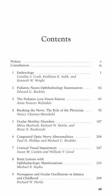
After fertilization of the ovum within the uterine tube, cellularmitosis results in formation of a ball of 12 to 16 cells, themorula. A fluid-filled cavity within this embryonic cell massforms, resulting in a transformation into a blastocyst that beginsto penetrate the uterine mucosa on approximately the sixth daypostfertilization. The cells of the blastocyst continue to dividewith the cells of the future embryo proper (embryoblast) accu-mulating at one pole. The cells of the primitive embryoblast dif-ferentiate into two layers, the epiblast and the hypoblast. Thesetwo cellular layers bridge the central cavity of the blastocyst,thus dividing the blastocyst into the amniotic cavity and theyolk sac (Fig. 1-1).
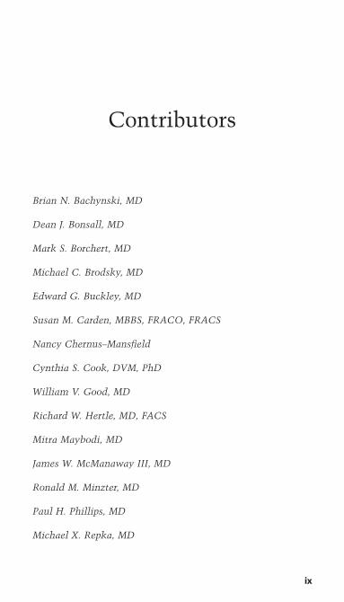
During the third week of gestation, the two-layered embry-oblast transforms into a trilaminar embryo as central epiblastcells invaginate between the epiblast and hypoblast layers.Invagination of central epiblast cells creates a longitudinalgroove through the midline of the caudal half of the epiblast, theprimitive streak. This invagination of epiblast cells is termedgastrulation (Fig. 1-2A,B). Invaginating epiblast cells differenti-ate to form the mesodermal germ layer, which spreads out tofill the space between the epiblast and hypoblast. Gastrulationproceeds in a cranial to caudal progression and continuesthrough the fourth week of human gestation. These invaginat-ing epiblast cells displace the hypoblast cells to form the endo-derm. The epiblast cells therefore give rise to all three definitivegerm layers: ectoderm, mesoderm, and endoderm (Fig. 1-2C).
1
1
2 handbook of pediatric neuro-ophthalmology
Maternalsinusoid
Amnioblast
Amnioticcavity
Epiblast
Blam
inarem
bryoHypoblast
Endoderm
Extraembryoniccoelom
ExocoelomicmembraneExtraembryonic
somatopleuric mesoderm
End
omet
rial s
trom
a
FIGURE 1-1. Drawing of a human blastocyst (12 days gestation) that haspenetrated the maternal endometrium. An embryoblast has formed thatconsists of two cell layers: the epiblast above and the hypoblast below.
Amniotic cavity
Primitive node Yolk sac
Yolk sac
Epiblast
AmnionPrimitivestreak
Primitivestreak
A
B
C
Hypoblast Invaginatingepiblast cells
Ectoderm
Intraembryonicmesoderm
Endoderm
FIGURE 1-2A–C. (A) Drawing of a 17-day-old embryo in gastrulation stage(dorsal view) with the amnion removed. (B) Cross section of a 17-day-oldembryo through the primitive streak. The primitive streak representsinvagination of epiblast cells between the epiblast and hypoblast layers.Note the epiblast cells filling the middle area to form the mesodermallayer. (C) Cross section of the embryo at the end of the third week showsthe three definitive germ layers: ectoderm, mesoderm, and endoderm.
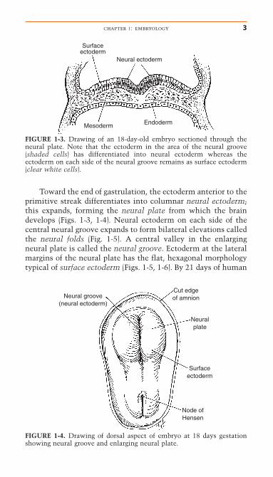
Toward the end of gastrulation, the ectoderm anterior to theprimitive streak differentiates into columnar neural ectoderm;this expands, forming the neural plate from which the braindevelops (Figs. 1-3, 1-4). Neural ectoderm on each side of thecentral neural groove expands to form bilateral elevations calledthe neural folds (Fig. 1-5). A central valley in the enlargingneural plate is called the neural groove. Ectoderm at the lateralmargins of the neural plate has the flat, hexagonal morphologytypical of surface ectoderm (Figs. 1-5, 1-6). By 21 days of human
chapter 1: embryology 3
Surfaceectoderm
Neural ectoderm
Mesoderm Endoderm
FIGURE 1-3. Drawing of an 18-day-old embryo sectioned through theneural plate. Note that the ectoderm in the area of the neural groove(shaded cells) has differentiated into neural ectoderm whereas the ectoderm on each side of the neural groove remains as surface ectoderm(clear white cells).
Neural groove(neural ectoderm)
Cut edgeof amnion
Surfaceectoderm
Node ofHensen
Neuralplate
FIGURE 1-4. Drawing of dorsal aspect of embryo at 18 days gestationshowing neural groove and enlarging neural plate.
Neural folds
Neuralectoderm
Surfaceectoderm
Neuralgroove
Somite
Node ofHensen
Primitivestreak
FIGURE 1-5. Dorsal view of ahuman embryo at 20 days gestation. The neural plate trans-forms into two neural folds oneach side of the neural groove.The neural groove in the middleof the embryo is shaded torepresent neural ectoderm; theunshaded surface of the embryois surface ectoderm.
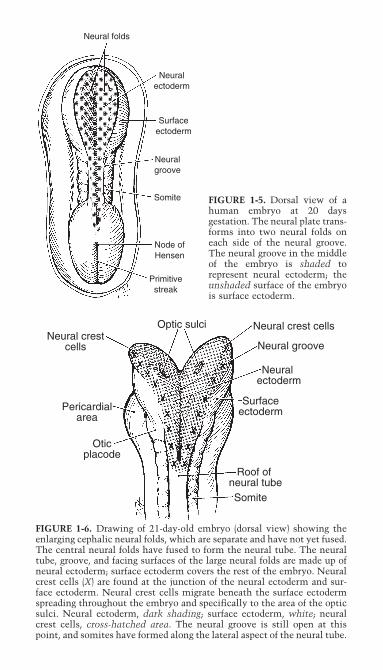
Optic sulci Neural crest cells
Neural groove
Neuralectoderm
Surfaceectoderm
Roof of neural tubeSomite
Neural crestcells
Pericardialarea
Oticplacode
FIGURE 1-6. Drawing of 21-day-old embryo (dorsal view) showing theenlarging cephalic neural folds, which are separate and have not yet fused.The central neural folds have fused to form the neural tube. The neuraltube, groove, and facing surfaces of the large neural folds are made up ofneural ectoderm; surface ectoderm covers the rest of the embryo. Neuralcrest cells (X) are found at the junction of the neural ectoderm and sur-face ectoderm. Neural crest cells migrate beneath the surface ectodermspreading throughout the embryo and specifically to the area of the opticsulci. Neural ectoderm, dark shading; surface ectoderm, white; neuralcrest cells, cross-hatched area. The neural groove is still open at thispoint, and somites have formed along the lateral aspect of the neural tube.
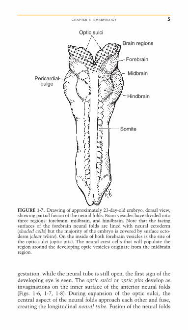
gestation, while the neural tube is still open, the first sign of thedeveloping eye is seen. The optic sulci or optic pits develop asinvaginations on the inner surface of the anterior neural folds(Figs. 1-6, 1-7, 1-8). During expansion of the optic sulci, thecentral aspect of the neural folds approach each other and fuse,creating the longitudinal neural tube. Fusion of the neural folds
chapter 1: embryology 5
Brain regions
Forebrain
Midbrain
Hindbrain
Somite
Pericardialbulge
Optic sulci
FIGURE 1-7. Drawing of approximately 23-day-old embryo, dorsal view,showing partial fusion of the neural folds. Brain vesicles have divided intothree regions: forebrain, midbrain, and hindbrain. Note that the facingsurfaces of the forebrain neural folds are lined with neural ectoderm(shaded cells) but the majority of the embryo is covered by surface ecto-derm (clear white). On the inside of both forebrain vesicles is the site ofthe optic sulci (optic pits). The neural crest cells that will populate theregion around the developing optic vesicles originate from the midbrainregion.
is initiated in the region of the future neck and proceeds alongthe midline in both caudal and cranial directions. Followingclosure of the neural tube, the neural ectoderm and optic sulciare internalized, and the embryo is then covered by surface ectoderm (Fig. 1-7).
Neural Crest Cell DevelopmentAs the neural folds elevate and approach each other, a special-ized population of mesenchymal cells, the neural crest cells,emigrate from the junction of the neural and surface ectoderm(see Fig. 1-6). Progenitor cells in the neural folds are multipo-tent, with potential to form multiple ectodermal derivatives,including epidermal, neural crest, and neural tube cells. These
6 handbook of pediatric neuro-ophthalmology
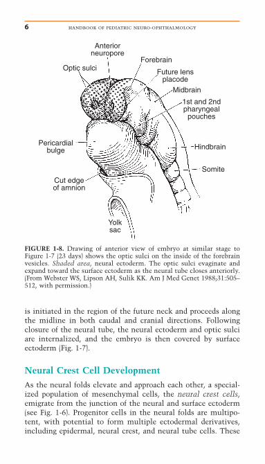
Anteriorneuropore
1st and 2ndpharyngeal
pouches
Hindbrain
Somite
Pericardialbulge
Optic sulci
Midbrain
Future lensplacode
Forebrain
Cut edgeof amnion
Yolksac
FIGURE 1-8. Drawing of anterior view of embryo at similar stage toFigure 1-7 (23 days) shows the optic sulci on the inside of the forebrainvesicles. Shaded area, neural ectoderm. The optic sulci evaginate andexpand toward the surface ectoderm as the neural tube closes anteriorly.(From Webster WS, Lipson AH, Sulik KK. Am J Med Genet 1988;31:505–512, with permission.)
cells are induced by interactions between the neural plate andepidermis. The competence of the neural plate to respond toinductive interactions changes as a function of embryonic age.92
These stellate cells migrate peripherally beneath the surfaceectoderm to spread throughout the embryo and surround thearea of the developing optic sulci. Neural crest cells play animportant role in eye development, as they are the precursors(anlage) to major structures, including cornea stroma, irisstroma, ciliary muscle, choroid, sclera, and orbital cartilage andbone (Table 1-1).55,64 The patterns of neural crest emergence andemigration correlate with the segmental disposition of the devel-oping brain.72 Migration and differentiation of the neural crestcells are influenced by the hyaluronic acid-rich extracellularmatrix and the optic vesicle basement membrane.17 This acel-lular matrix is secreted by a surface epithelium as well as thecrest cells and forms a space through which the crest cellsmigrate. Fibronectin secreted by the noncrest cells forms thelimits of this mesenchymal migration.65 Interactions betweenthe migrating neural crest and the associated mesoderm appearto be essential for normal crest differentiation.76,77
chapter 1: embryology 7
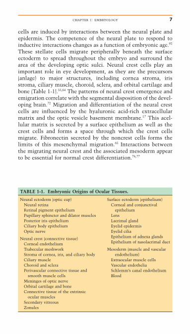
TABLE 1-1. Embryonic Origins of Ocular Tissues.
Neural ectoderm (optic cup)Neural retinaRetinal pigment epitheliumPupillary sphincter and dilator musclesPosterior iris epitheliumCiliary body epitheliumOptic nerve
Neural crest (connective tissue)Corneal endotheliumTrabecular meshworkStroma of cornea, iris, and ciliary bodyCiliary muscleChoroid and scleraPerivascular connective tissue and
smooth muscle cellsMeninges of optic nerveOrbital cartilage and boneConnective tissue of the extrinsic
ocular musclesSecondary vitreousZonules
Surface ectoderm (epithelium)Corneal and conjunctival
epitheliumLensLacrimal glandEyelid epidermisEyelid ciliaEpithelium of adnexa glandsEpithelium of nasolacrimal duct
Mesoderm (muscle and vascular endothelium)
Extraocular muscle cellsVascular endotheliaSchlemm’s canal endotheliumBlood
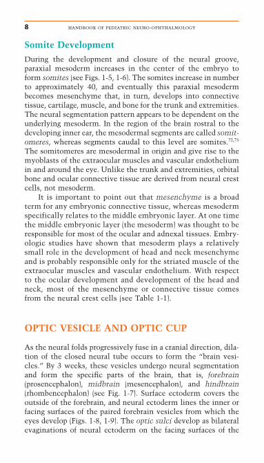
Somite DevelopmentDuring the development and closure of the neural groove, paraxial mesoderm increases in the center of the embryo to form somites (see Figs. 1-5, 1-6). The somites increase in numberto approximately 40, and eventually this paraxial mesodermbecomes mesenchyme that, in turn, develops into connectivetissue, cartilage, muscle, and bone for the trunk and extremities.The neural segmentation pattern appears to be dependent on theunderlying mesoderm. In the region of the brain rostral to thedeveloping inner ear, the mesodermal segments are called somit-omeres, whereas segments caudal to this level are somites.72,75
The somitomeres are mesodermal in origin and give rise to themyoblasts of the extraocular muscles and vascular endotheliumin and around the eye. Unlike the trunk and extremities, orbitalbone and ocular connective tissue are derived from neural crestcells, not mesoderm.
It is important to point out that mesenchyme is a broadterm for any embryonic connective tissue, whereas mesodermspecifically relates to the middle embryonic layer. At one timethe middle embryonic layer (the mesoderm) was thought to beresponsible for most of the ocular and adnexal tissues. Embry-ologic studies have shown that mesoderm plays a relativelysmall role in the development of head and neck mesenchymeand is probably responsible only for the striated muscle of theextraocular muscles and vascular endothelium. With respectto the ocular development and development of the head andneck, most of the mesenchyme or connective tissue comesfrom the neural crest cells (see Table 1-1).
OPTIC VESICLE AND OPTIC CUP
As the neural folds progressively fuse in a cranial direction, dila-tion of the closed neural tube occurs to form the “brain vesi-cles.” By 3 weeks, these vesicles undergo neural segmentationand form the specific parts of the brain, that is, forebrain(prosencephalon), midbrain (mesencephalon), and hindbrain(rhombencephalon) (see Fig. 1-7). Surface ectoderm covers theoutside of the forebrain, and neural ectoderm lines the inner orfacing surfaces of the paired forebrain vesicles from which theeyes develop (Figs. 1-8, 1-9). The optic sulci develop as bilateralevaginations of neural ectoderm on the facing surfaces of the
8 handbook of pediatric neuro-ophthalmology
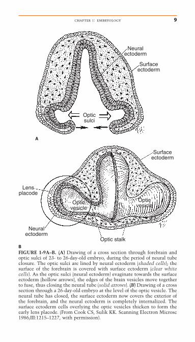
chapter 1: embryology 9
Surfaceectoderm
Opticsulci
Neuralectoderm
A
Surfaceectoderm
Neuralectoderm
Optic stalk
Opticvesicle
Opticvesicle
Lensplacode
BFIGURE 1-9A–B. (A) Drawing of a cross section through forebrain andoptic sulci of 23- to 26-day-old embryo, during the period of neural tubeclosure. The optic sulci are lined by neural ectoderm (shaded cells); thesurface of the forebrain is covered with surface ectoderm (clear whitecells). As the optic sulci (neural ectoderm) evaginate towards the surfaceectoderm (hollow arrows), the edges of the brain vesicles move togetherto fuse, thus closing the neural tube (solid arrows). (B) Drawing of a crosssection through a 26-day-old embryo at the level of the optic vesicle. Theneural tube has closed, the surface ectoderm now covers the exterior ofthe forebrain, and the neural ectoderm is completely internalized. Thesurface ectoderm cells overlying the optic vesicles thicken to form theearly lens placode. (From Cook CS, Sulik KK. Scanning Electron Microsc1986;III:1215–1227, with permission).
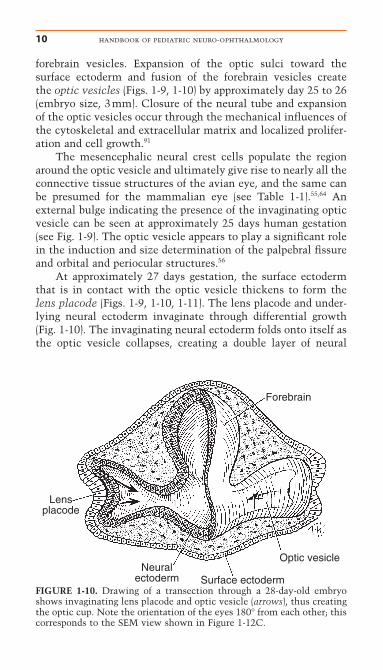
10 handbook of pediatric neuro-ophthalmology
Forebrain
Neuralectoderm Surface ectoderm
Optic vesicle
Lensplacode
FIGURE 1-10. Drawing of a transection through a 28-day-old embryoshows invaginating lens placode and optic vesicle (arrows), thus creatingthe optic cup. Note the orientation of the eyes 180° from each other; thiscorresponds to the SEM view shown in Figure 1-12C.
forebrain vesicles. Expansion of the optic sulci toward thesurface ectoderm and fusion of the forebrain vesicles create the optic vesicles (Figs. 1-9, 1-10) by approximately day 25 to 26(embryo size, 3mm). Closure of the neural tube and expansionof the optic vesicles occur through the mechanical influences ofthe cytoskeletal and extracellular matrix and localized prolifer-ation and cell growth.91
The mesencephalic neural crest cells populate the regionaround the optic vesicle and ultimately give rise to nearly all theconnective tissue structures of the avian eye, and the same canbe presumed for the mammalian eye (see Table 1-1).55,64 Anexternal bulge indicating the presence of the invaginating opticvesicle can be seen at approximately 25 days human gestation(see Fig. 1-9). The optic vesicle appears to play a significant rolein the induction and size determination of the palpebral fissureand orbital and periocular structures.56
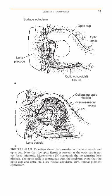
At approximately 27 days gestation, the surface ectodermthat is in contact with the optic vesicle thickens to form thelens placode (Figs. 1-9, 1-10, 1-11). The lens placode and under-lying neural ectoderm invaginate through differential growth(Fig. 1-10). The invaginating neural ectoderm folds onto itself asthe optic vesicle collapses, creating a double layer of neural
chapter 1: embryology 11
Surface ectoderm
Optic cup
Optic (choroidal)fissure
Lensplacode
Opticstalk
A
Collapsing opticvesicle
Neurosensoryretina
RPE
Lens vesicleB
FIGURE 1-11A,B. Drawings show the formation of the lens vesicle andoptic cup. Note that the optic fissure is present as the optic cup is notyet fused inferiorly. Mesenchyme (M) surrounds the invaginating lensplacode. The optic stalk is continuous with the forebrain. Note that theoptic cup and optic stalk are neural ectoderm. RPE, retinal pigmentepithelium.
12 handbook of pediatric neuro-ophthalmology
ectoderm, the optic cup (Fig. 1-11). The optic cup will eventuallydifferentiate into neurosensory retina (inner layer) and retinalpigment epithelium (RPE) (outer layer) (Fig. 1-11). Local apicalcontraction112 and physiological cell death91 have been identifiedduring invagination of the lens placode and formation of theoptic cup. In the mouse embryo, Msx2, a homeobox-containingtranscription factor, is expressed only in the cells of the opticcup that are destined to become neural retina. In vitro Msx2 hasbeen shown to suppress RPE differentiation and may be involvedin the initial patterning of the optic cup.48 Abnormal differenti-ation of the outer layer of the optic cup to form aberrant neuralretina has been demonstrated in several mutant mousestrains.21,26,109 The area of future retinal differentiation demon-strates the greatest concentration of vimentin (a cytoskeletalprotein) in the optic cup.53 Regionally, within the optic cup,spatial orientation is predicted by expression of the transcriptionfactor, vax2, which defines the ventral region (area of the opticfissure).10 The PAX6 gene has been demonstrated within cells ofneural ectodermal origin (optic cup and, later, in the ciliary bodyand retina), surface ectoderm (lens), and neural crest (cornea).74
The widespread distribution of this gene supports its involve-ment in many stages of ocular morphogenesis.
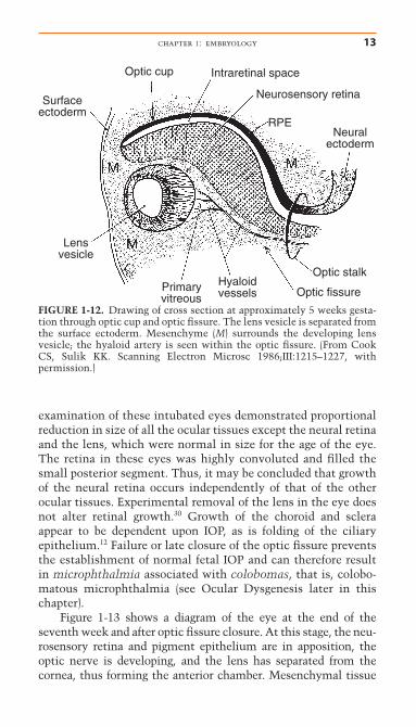
The Optic FissureInvagination of the optic cup occurs in an eccentric manner withformation of a seam, the optic fissure, inferiorly (Figs. 1-11, 1-12). The optic fissure is also known as the embryonic fissure orchoroidal fissure. Mesenchymal tissue (of primarily neural crestorigin) surrounds and is within the optic fissure and optic cup,and at 5 weeks the hyaloid artery develops from mesenchyme inthe optic fissure. This artery courses from the optic stalk (pre-cursor to the optic nerve) through the optic fissure to the devel-oping lens (Fig. 1-12). The lens vesicle separates from the surfaceectoderm at approximately 6 weeks, the same time as closure ofthe optic fissure. Closure of the optic cup occurs initially at theequator with progression anteriorly and posteriorly.
Once the fissure has closed, secretion of primitive aqueousfluid by the primitive ciliary epithelium establishes intraocularpressure (IOP), which contributes to expansion of the opticcup.15,29 Experimental studies have shown that placement of acapillary tube into the vitreous cavity of a chick eye reduces the IOP and markedly slows growth of the eye.29 Histological
examination of these intubated eyes demonstrated proportionalreduction in size of all the ocular tissues except the neural retinaand the lens, which were normal in size for the age of the eye.The retina in these eyes was highly convoluted and filled thesmall posterior segment. Thus, it may be concluded that growthof the neural retina occurs independently of that of the otherocular tissues. Experimental removal of the lens in the eye doesnot alter retinal growth.30 Growth of the choroid and scleraappear to be dependent upon IOP, as is folding of the ciliaryepithelium.12 Failure or late closure of the optic fissure preventsthe establishment of normal fetal IOP and can therefore resultin microphthalmia associated with colobomas, that is, colobo-matous microphthalmia (see Ocular Dysgenesis later in thischapter).
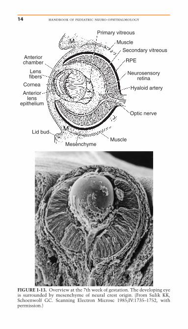
Figure 1-13 shows a diagram of the eye at the end of theseventh week and after optic fissure closure. At this stage, the neu-rosensory retina and pigment epithelium are in apposition, theoptic nerve is developing, and the lens has separated from thecornea, thus forming the anterior chamber. Mesenchymal tissue
chapter 1: embryology 13
Neuralectoderm
Hyaloidvessels Optic fissurePrimary
vitreous
Lensvesicle
Optic stalk
Optic cup
Surfaceectoderm
Neurosensory retina
RPE
Intraretinal space
FIGURE 1-12. Drawing of cross section at approximately 5 weeks gesta-tion through optic cup and optic fissure. The lens vesicle is separated fromthe surface ectoderm. Mesenchyme (M) surrounds the developing lensvesicle; the hyaloid artery is seen within the optic fissure. (From CookCS, Sulik KK. Scanning Electron Microsc 1986;III:1215–1227, with permission.)
14 handbook of pediatric neuro-ophthalmology
Muscle
Lid bud
Cornea
Lensfibers
Anteriorchamber
Primary vitreous
Muscle
Mesenchyme
Secondary vitreous
RPE
Neurosensoryretina
Anteriorlens
epithelium
Hyaloid artery
Optic nerve
FIGURE 1-13. Overview at the 7th week of gestation. The developing eyeis surrounded by mesenchyme of neural crest origin. (From Sulik KK,Schoenwolf GC. Scanning Electron Microsc 1985;IV:1735–1752, withpermission.)
chapter 1: embryology 15
(neural crest cell origin) around the primitive retina develops into the choroid and sclera. Peripheral to the developing globe arelinear accumulations of myoblasts (mesodermal origin) that areanlagen of the extraocular muscles. The eyelids are small budsabove and below the developing eye. The hyaloid vasculaturecourses from the primitive optic nerve to the posterior lenscapsule.
LENS
Thickening of the lens placode can be seen on gestational day 27in the human (see Fig. 1-10). Before its contact with the opticvesicle, the surface ectoderm must become competent to respondto lens inducers. It then receives inductive signals from the ante-rior neural plate, so that it gains a “lens-forming bias” specifiedfor lens formation. Complete lens differentiation requires bothinductive signals from the optic vesicle and an inhibitory signalfrom head neural crest to suppress any residual lens-forming biasin head ectoderm adjacent to the lens.38 In the chick, a tight extra-cellular matrix-mediated adhesion between the optic vesicle andthe surface ectoderm has been described.47,57,69 This anchoring ofthe mitotically active surface ectoderm results in cell crowding,cell elongation, and formation of the thickened placode.119
Adhesion between the optic vesicle and the lens placode isthought to ensure alignment of the lens in the visual axis.15
Although adhesion between the optic vesicle and surface ecto-derm exists, electron microscopic studies have demonstratedthat there is no direct cell contact.22,49,108 The basement mem-branes of the optic vesicle and the surface ectoderm remain separate and intact throughout the contact period. Experimentalstudies have demonstrated a requirement for functional PAX6gene in both the optic vesicle and surface ectoderm to mediatelens placode induction.23 The BMP4 gene, which is present onlyin the optic vesicle, is also required for lens induction.35
The lens placode invaginates forming the hollow lensvesicle (Figs. 1-11, 1-12). The size of the lens vesicle is deter-mined by the area of contact of the optic vesicle and the surfaceectoderm. Lens vesicle detachment from the surface ectodermoccurs on day 33 (7–9 mm) and is the initial event leading to theformation of the chambers of the eye. This process of separationis accompanied by active migration of epithelial cells out of thekeratolenticular stalk or junction,37 cellular necrosis, and base-
ment membrane breakdown.36 Although apoptosis (programmedcell death) is a normal feature of lens vesicle separation, exces-sive and persistent cell death is associated with aphakia in thelap mouse mutant.8
Induction of a small lens vesicle that fails to undergo normalseparation from the surface ectoderm is one of the characteris-tics of teratogen-induced anterior segment malformationsdescribed in animal models.24,28,81,102 In the mouse mutant (dyl),this failure of lens vesicle separation is caused by a mutation inthe FoxE3 gene that promotes survival and proliferation whilepreventing differentiation of the lens epithelium.18 AP-2 tran-scription factors also influence lens vesicle separation as well ascausing mis-expression of PAX6 and MIP26 genes.109 Anteriorlenticonus, anterior capsular cataracts, and anterior segmentdysgenesis with keratolenticular adhesions (Peters’ anomaly)may result from faulty keratolenticular separation. Further dis-cussion of anterior segment dysgenesis follows. Arrest of lensdevelopment at the lens stalk stage results in aphakia in mutantmice (ak mutation). In addition to aphakia, affected eyes exhibitabsence of a pupil and abnormalities in the iris, ciliary body, andvitreous.40,41
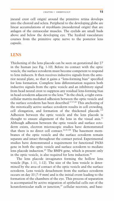
The hollow lens vesicle consists of a single layer of epithelial cells with cell apices directed toward the center of thesphere. Following detachment from the surface ectoderm, thelens vesicle is surrounded by a basal lamina, the future lenscapsule. Abnormalities in this basement membrane may resultin involution of the lens vesicle, resulting in later aphakia.8 Atapproximately 37 days gestation, primary lens fibers form fromelongation of the posterior lens epithelium of the lens vesicle(Fig. 1-14).51 The retinal anlage promotes primary lens fiber for-mation in the adjacent lens epithelial cells. Experimental in vivorotation of the lens vesicle in the chick eye by 180° results inelongation of the lens epithelial cells nearest the presumptiveretina, regardless of the orientation of the transplanted lens.31
Thus, the retina develops independently from the lens althoughthe lens appears to rely upon the retina for cytodifferentiation.In the mouse, the Prox1 and Maf genes have been demonstratedto mediate lens fiber elongation.88,110 As these posterior epithe-lial cells lengthen to fill the lumen of the lens vesicle, they losetheir nucleus and most organelles.14 Upregulation of lens-specific proteins, CP49 and CP95, is demonstrated after closureof the lens stalk.51 The primitive lens filled with primary lensfibers is the embryonic lens nucleus. After the epithelial cells
16 handbook of pediatric neuro-ophthalmology
chapter 1: embryology 17
Surfaceectoderm
Elongating primarylens fibers
Cuboidalepithelium(future lensepithelium)
Hyaloid artery/primary vitreous
RPE
Neurosensoryretina
FIGURE 1-14. Formation of the embryonic lens nucleus and primary lensfibers at approximately 7 weeks. Note that mesenchyme (M) of neural crestorigin surrounds the optic cup. The posterior lens epithelial cells (locatednearest the developing retina, R) elongate, forming the primary lens fibers(L). The anterior epithelium remains cuboidal and becomes the anteriorepithelium in the adult. The optic fissure is now closed.
18 handbook of pediatric neuro-ophthalmology
Anteriorlens epithelium
Embryonalnucleus
Anterior"Y" suture
Posterior"Y" suture
Secondarylens fibers
Fetal nucleus
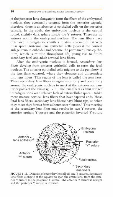
FIGURE 1-15. Diagram of secondary lens fibers and Y sutures. Secondarylens fibers elongate at the equator to span the entire lens, from the ante-rior Y suture to the posterior Y suture. The anterior Y suture is uprightand the posterior Y suture is inverted.
of the posterior lens elongate to form the fibers of the embyronalnucleus, they eventually separate from the posterior capsule;therefore, there is an absence of epithelial cells on the posteriorcapsule. In the adult, the embryonic nucleus is the centralround, slightly dark sphere inside the Y sutures. There are nosutures within the embryonal nucleus. The lens fibers haveextensive interdigitations with a relative absence of extracel-lular space. Anterior lens epithelial cells (nearest the cornealanlage) remain cuboidal and become the permanent lens epithe-lium, which is mitotic throughout life, giving rise to future secondary fetal and adult cortical lens fibers.
After the embryonic nucleus is formed, secondary lensfibers develop from anterior epithelial cells to form the fetalnucleus. The anterior epithelial cells migrate to the periphery ofthe lens (lens equator), where they elongate and differentiateinto lens fibers. This region of the lens is called the lens bow.These secondary lens fibers elongate anteriorly and posteriorlyaround the embryonic nucleus to meet at the anterior and pos-terior poles of the lens (Fig. 1-15). The lens fibers exhibit surfaceinterdigitations with relative lack of extracellular space. Unlikemore mature cortical lens fibers that have tapered ends, thesefetal lens fibers (secondary lens fibers) have blunt tips, so whenthey meet they form a faint adherence or “suture.” This meetingof the secondary lens fiber ends results in two Y sutures, theanterior upright Y suture and the posterior inverted Y suture
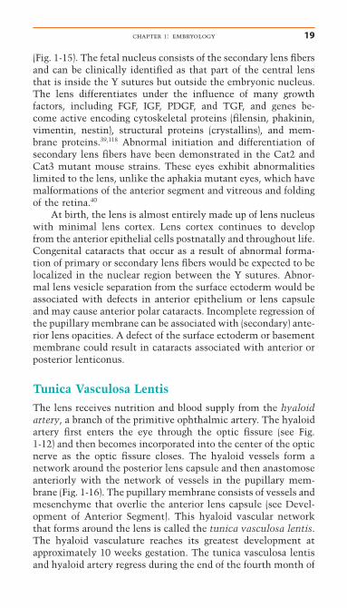
(Fig. 1-15). The fetal nucleus consists of the secondary lens fibersand can be clinically identified as that part of the central lensthat is inside the Y sutures but outside the embryonic nucleus.The lens differentiates under the influence of many growthfactors, including FGF, IGF, PDGF, and TGF, and genes be-come active encoding cytoskeletal proteins (filensin, phakinin,vimentin, nestin), structural proteins (crystallins), and mem-brane proteins.39,118 Abnormal initiation and differentiation ofsecondary lens fibers have been demonstrated in the Cat2 andCat3 mutant mouse strains. These eyes exhibit abnormalitieslimited to the lens, unlike the aphakia mutant eyes, which havemalformations of the anterior segment and vitreous and foldingof the retina.40
At birth, the lens is almost entirely made up of lens nucleuswith minimal lens cortex. Lens cortex continues to developfrom the anterior epithelial cells postnatally and throughout life.Congenital cataracts that occur as a result of abnormal forma-tion of primary or secondary lens fibers would be expected to belocalized in the nuclear region between the Y sutures. Abnor-mal lens vesicle separation from the surface ectoderm would beassociated with defects in anterior epithelium or lens capsuleand may cause anterior polar cataracts. Incomplete regression ofthe pupillary membrane can be associated with (secondary) ante-rior lens opacities. A defect of the surface ectoderm or basementmembrane could result in cataracts associated with anterior orposterior lenticonus.
Tunica Vasculosa LentisThe lens receives nutrition and blood supply from the hyaloidartery, a branch of the primitive ophthalmic artery. The hyaloidartery first enters the eye through the optic fissure (see Fig. 1-12) and then becomes incorporated into the center of the opticnerve as the optic fissure closes. The hyaloid vessels form anetwork around the posterior lens capsule and then anastomoseanteriorly with the network of vessels in the pupillary mem-brane (Fig. 1-16). The pupillary membrane consists of vessels andmesenchyme that overlie the anterior lens capsule (see Devel-opment of Anterior Segment). This hyaloid vascular networkthat forms around the lens is called the tunica vasculosa lentis.The hyaloid vasculature reaches its greatest development atapproximately 10 weeks gestation. The tunica vasculosa lentisand hyaloid artery regress during the end of the fourth month of
chapter 1: embryology 19
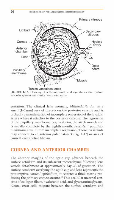
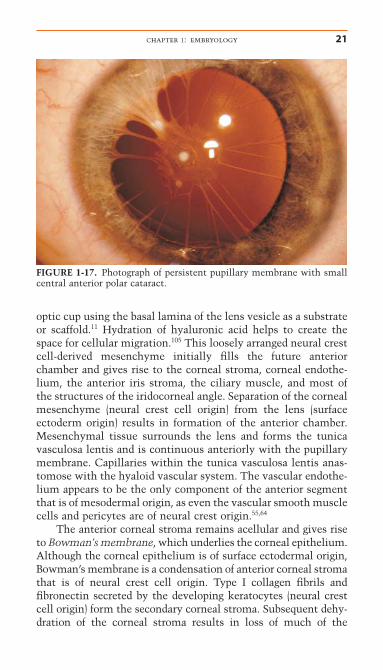
gestation. The clinical lens anomaly, Mittendorf’s dot, is a small (1–2mm) area of fibrosis on the posterior capsule and is probably a manifestation of incomplete regression of the hyaloidartery where it attaches to the posterior capsule. The regressionof the pupillary membrane begins during the sixth month andis usually complete by the eighth month. Persistent pupillarymembranes result from incomplete regression. These iris strandsmay connect to an anterior polar cataract (Fig. 1-17) or area ofcorneal endothelial fibrosis.
CORNEA AND ANTERIOR CHAMBER
The anterior margins of the optic cup advance beneath thesurface ectoderm and its subjacent mesenchyme following lensvesicle detachment at approximately day 33 of gestation. Thesurface ectoderm overlying the optic cup and lens represents thepresumptive corneal epithelium; it secretes a thick matrix pro-ducing the primary cornea stroma.43 This acellular material con-sists of collagen fibers, hyaluronic acid, and glycosaminoglycans.Neural crest cells migrate between the surface ectoderm and
20 handbook of pediatric neuro-ophthalmology
Lid bud
Primary vitreous
Anteriorchamber
Opticnerve
Tunica vasculosa lentis
Muscle
Hyaloidartery
Secondaryvitreous
Cornea
Lens
Pupillarymembrane
FIGURE 1-16. Drawing of a 2-month-old fetal eye shows the hyaloid vascular system and tunica vasculosa lentis.
optic cup using the basal lamina of the lens vesicle as a substrateor scaffold.11 Hydration of hyaluronic acid helps to create thespace for cellular migration.105 This loosely arranged neural crestcell-derived mesenchyme initially fills the future anteriorchamber and gives rise to the corneal stroma, corneal endothe-lium, the anterior iris stroma, the ciliary muscle, and most of the structures of the iridocorneal angle. Separation of the cornealmesenchyme (neural crest cell origin) from the lens (surface ectoderm origin) results in formation of the anterior chamber.Mesenchymal tissue surrounds the lens and forms the tunicavasculosa lentis and is continuous anteriorly with the pupillarymembrane. Capillaries within the tunica vasculosa lentis anas-tomose with the hyaloid vascular system. The vascular endothe-lium appears to be the only component of the anterior segmentthat is of mesodermal origin, as even the vascular smooth musclecells and pericytes are of neural crest origin.55,64
The anterior corneal stroma remains acellular and gives riseto Bowman’s membrane, which underlies the corneal epithelium.Although the corneal epithelium is of surface ectodermal origin,Bowman’s membrane is a condensation of anterior corneal stromathat is of neural crest cell origin. Type I collagen fibrils andfibronectin secreted by the developing keratocytes (neural crestcell origin) form the secondary corneal stroma. Subsequent dehy-dration of the corneal stroma results in loss of much of the
chapter 1: embryology 21
FIGURE 1-17. Photograph of persistent pupillary membrane with smallcentral anterior polar cataract.