Embed Size (px)
Citation preview

Handbook on Comprehensive Breast Cancer Care
for Healthcare Workers2021
Know about Breast Cancer: Prevent, Diagnose, Treat & Care
National Cancer Control Programme
Ministry of Health
II
ISBN: 978-624-5719-00-6
01st Print March 2021
The information contained in this report may be reproduced for non-commercial
purposes with attribution to the copyright holders.
© The National Cancer Control Programme, Ministry of Health
No. 555/5D, Elvitigala Mawatha,
Narahenpita, Colombo 05.
https://www.nccp.health.gov.lk/
Printed by
Ari Investments (Pvt) Ltd 19, St. Josephs’ Road, Nugegoda, Sri Lanka Tele: 285 2410 Fax: 282 2615 E-mail: [email protected]

III
Delivery Confirmation System
NCCP kindly requests from the end users of this Handbook to
“Confirm the Delivery”
Delivery can be confirmed directly
by accessing the
“e-Delivery Confirmation Form”
Simply scan QR code with your smart phoneor
follow the web link given below
QR code
Web link https://forms.gle/6n7b87jgc6WSrQEW9

IV
V
Special Acknowledgement
Dr. Sujatha Samarakoon - Public Health Specialist
Dr. Muzrif Munas - Consultant Community Physician
Dr. Buddhika Senananyake - Consultant Community Physician
Dr. Mekala Fernando - Senior Registrar in Community Medicine
Dr. Amila Suranga Malawige - Registrar in Community Medicine
Dr. Mangala Liyanage - Medical Officer
Dr. Jayantha Dissanayake - Medical Officer
Dr. Saddharma Weerakoon - Registrar in Community Medicine
Dr. Kalpanie Wijewardana - Medical Officer
Dr. Nimali Wijegunawardena - Senior Registrar in Community Medicine
Dr. Sumudu Hewage - Senior Registrar in Community Medicine

VI
VII
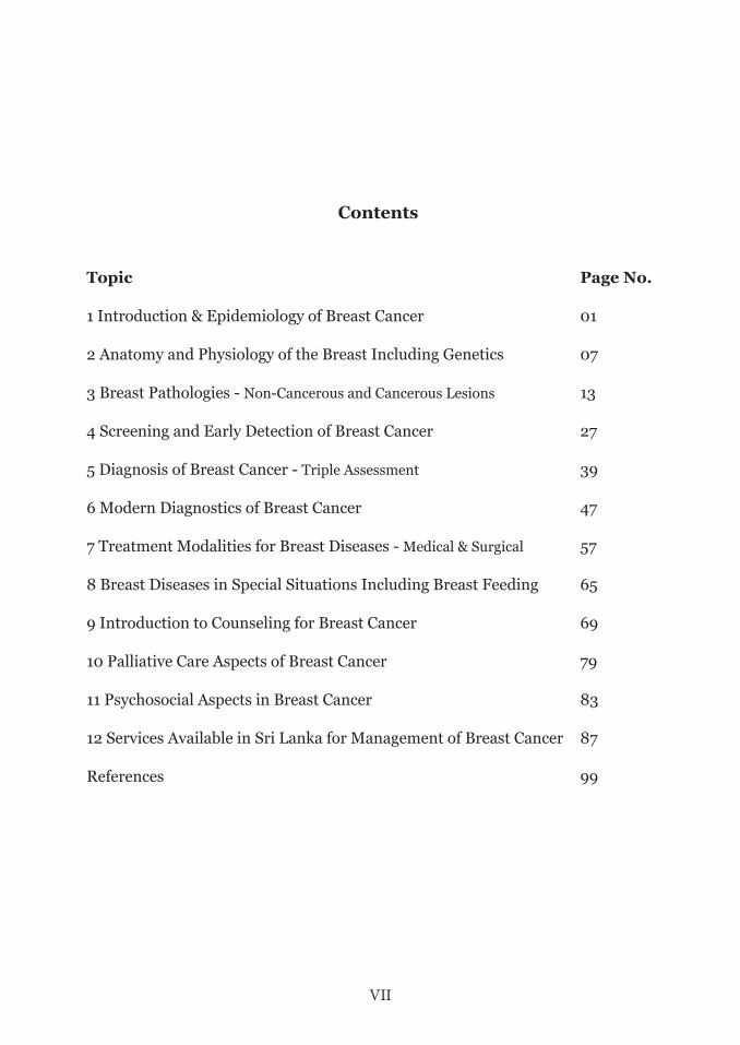
Contents
Topic Page No.
1 Introduction & Epidemiology of Breast Cancer 01
2 Anatomy and Physiology of the Breast Including Genetics 07
3 Breast Pathologies - Non-Cancerous and Cancerous Lesions 13
4 Screening and Early Detection of Breast Cancer 27
5 Diagnosis of Breast Cancer - Triple Assessment 39
6 Modern Diagnostics of Breast Cancer 47
7 Treatment Modalities for Breast Diseases - Medical & Surgical 57
8 Breast Diseases in Special Situations Including Breast Feeding 65
9 Introduction to Counseling for Breast Cancer 69
10 Palliative Care Aspects of Breast Cancer 79
11 Psychosocial Aspects in Breast Cancer 83
12 Services Available in Sri Lanka for Management of Breast Cancer 87
References 99

VIII
IX
The National Cancer Control Programme (NCCP) is the national focal point of the Ministry of Health responsible for planning, coordination, implementation, monitoring and evaluation of the cancer control and palliative care activities in Sri Lanka.
Breast cancer is the number one cancer among women in Sri Lanka and the incidence has been increasing since last twenty-five years.
With the aim of strengthening the early detection and fast track diagnosis of breast cancer, the Ministry of Health, Sri Lanka is currently working towards the establishment of breast clinics at tertiary care level island-wide. Enhancing the knowledge and skills of the human resources in the said breast clinics is thus an essential task to be achieved.
The handbook on breast cancer care for healthcare workers in Sri Lanka, developed and published by the National Cancer Control Programme consists of most of the subject matter with regard to breast cancer, and is aimed to provide adequate knowledge for the relevant healthcare staff.
It is my utmost pleasure to wish the National Cancer Control Programme for the publication of this worthy handbook and express my sincere gratitude to its contributors who may have been involved in various ways for the successful publication of this comprehensive book.
Dr. Asela GunawardenaDirector General of Health ServicesMinistry of Health
Message from the Director General of Health Services

X

XI
The National Cancer Control Programme (NCCP) was established in 1980 and it is the national focal point for prevention and control of cancer. NCCP plays a key role in planning, coordinating, implementing, monitoring and evaluating the cancer control activities in Sri Lanka. The Goal of the national cancer control strategy is to reduce the incidence of preventable cancers, detect early detectable cancers and provide continuum of care to all cancer patients in an equitable manner.
Breast cancer is the most common cancer among women in Sri Lanka. According to estimates, 24% of all newly diagnosed cancers among women are breast cancers. Around 3000-3500 women are diagnosed with breast cancer annually and there is an increasing incidence of breast cancer over the last 25 years. Breast cancer incidence is increasing with the age and comes to a peak at ages between 45 to 65 years.
Breast cancer is one of the cancers which has a good survival rate, if it is detected in the early stage. Key strategies for early detection of breast cancer is screening and early diagnosis. Screening for breast cancer is performed in women without any signs or symptoms of breast cancer to detect cancer as early as possible. By developing the “Handbook on Breast Cancer Care for Healthcare Workers in Sri Lanka” NCCP aims to increase the awareness, knowledge and skills on breast screening, diagnosis and treatment options among primary health care workers. Further, this book will serve as a guide on services, palliative care and counseling on breast cancer.
Overall, this handbook will provide a strong orientation on breast cancer, and is now our responsibility to rise to the challenge and combine the necessary individual and collective resources in our drive towards achieving targets of prevention and control of breast cancer. I would like to thank the World Health Organization and all authors, experts and professional colleges for their contribution in developing this comprehensive handbook on breast cancer care.
Dr. Champika WickramasingheMBBS, MSc, MD (Community Medicine)Deputy Director General-Non-Communicable DiseasesNon-Communicable Disease BureauMinistry of Health
Message from the Deputy Director General- Non-communicable Diseases

XII
XIII
Each year, the National Cancer Control Programme (NCCP) strives to strengthen the prevention early detection, diagnosis, treatment and care of breast cancers, which is the top most cancer detected among the Sri Lankan females.
The Technical Advisory Committee for Diagnosis and Treatment of Cancers has identified the need and initiated the implementation of breast clinics, extending services to all nine provinces of the country and it has been endorsed by the National Advisory Committee of Cancer Prevention and Care. As of now, seven out of nine provinces have established breast clinics.
With the aim of strengthening the services rendered in the said breast clinics, the Diagnostic and Treatment Unit of the NCCP was able to develop a comprehensive book on breast care with partnership of experts in the relevant fields. It gives me great pleasure to announce the first publication of the ‘handbook on “Comprehensive Breast Cancer Care for Healthcare Workers” in Sri Lanka’. The book focuses on noteworthy information on almost all aspects of breast cancer and would be of special interest to the healthcare personnel attached to these breast clinics.
The National Cancer Control Programme thank the authors and the commitment of those who contributed to complete this task with their continuous and enthusiastic efforts to make this creation a reality. The partnership from the World Health Organization and Dr. Sujatha Samarakoon, Public Health Specialist to the National Cancer Control Programme for this endeavor is gratefully acknowledged.
Dr. Janaki Vidanapathirana MBBS, MSc, MD (Community Medicine) Director, National Cancer Control Programme
Message from the Director
XIV
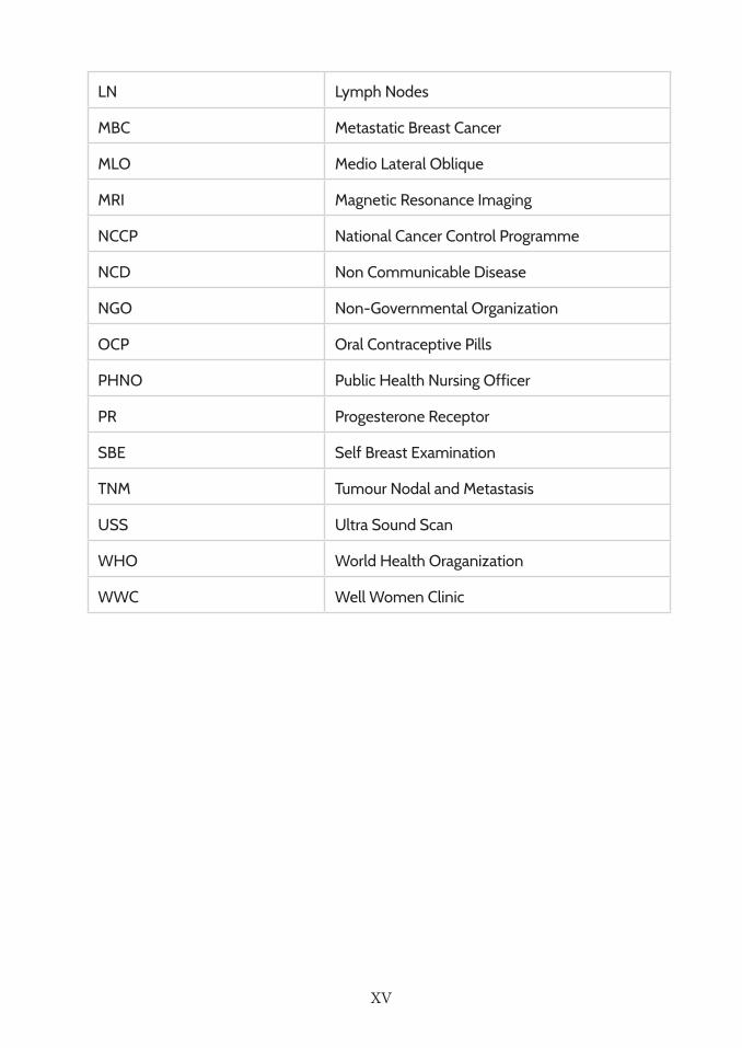
Abbreviations and Acronyms
AJCC American Joint Committee on Cancer
BCS Breast Conserving Surgery
BIRADS Breast Imaging- Reporting and Data System
BMI Body Mass Index
CBE Clinical Breast Examination
CC Cranio Caudal
CEDC Cancer Early Detection Center
CESM Contrast Enhanced Spectral Mammography
CPG Cancer Predisposition Gene
CT Computed Tomography
DBT Digital Breast Tomosynthesis
DCIS Ductal Carcinoma In Situ
EBC Early Breast Cancer
ER Estrogen Receptors
FNAC Fine Needle Aspiration Cytology
FSH Follicle Stimulating Hormone
G-CSF Granulocyte- Colony Stimulating Factor
GP General Practitioner
HER2 Human Epidermal growth factor Receptor 2
HLC Healthy Lifestyle Centers
HPL Human Placental Lactogen
HRT Hormone Replacement Therapy
LH Luteinizing Hormone
XV
LN Lymph Nodes
MBC Metastatic Breast Cancer
MLO Medio Lateral Oblique
MRI Magnetic Resonance Imaging
NCCP National Cancer Control Programme
NCD Non Communicable Disease
NGO Non-Governmental Organization
OCP Oral Contraceptive Pills
PHNO Public Health Nursing Officer
PR Progesterone Receptor
SBE Self Breast Examination
TNM Tumour Nodal and Metastasis
USS Ultra Sound Scan
WHO World Health Oraganization
WWC Well Women Clinic

XVI
Dr. Suraj Perera Consultant Community Physician, National Cancer Control Programme
Dr. Buddhika Senanayake Consultant Community Physician, National Cancer Control Programme
Chapter01
Introduction & Epidemiology of Breast Cancer
1
Chapter 1 – Introduction & Epidemiology of Breast Cancer
Introduction & Epidemiology of Breast Cancer
What is cancer?
Our body is made up of tiny building blocks called cells. Normal cells grow when the body needs them, and die when our body doesn’t need them any longer. This process is controlled by our body. Cancer starts when cells in the body change (mutate) and grow out of control. Cancer is made up of abnormal cells growing in such a manner, even though the body doesn’t need them. In most types of cancer, the abnormal cells grow to form a lump or mass called a tumor. A tumor can be cancerous or benign. A cancerous tumor is malignant, meaning it can grow and spread to other parts of the body. A benign tumor means the tumor can grow but will not spread. This could happen in any organ in the body, and we name the cancer according to the site of the origin. When the initial tumor occurs in the breast tissue, it is called a breast cancer.
Cancer burden
Cancer has become a leading cause of death worldwide, accounting for an estimated 19.3 million cases and 10 million deaths due to any form of cancer in 2020. Physical, emotional and financial well-being of individuals, families, communities and health systems have been seriously disturbed by the increasing trend of cancers. The World Health Organization (WHO) revealed that approximately 70% of deaths due to cancer, occur in low and middle-income countries.
Sri Lankan national cancer incidence data shows an upward trend of all forms of cancer and average of and 30,000 new cases each year. According to the national cancer registry, new cases of all cancers were 33,226 in 2019.
Incidence of Breast Cancer
Breast cancer is one of the most common types of cancer in women in the world as well as Sri Lanka. According to the GLOBOCAN 2020, female breast cancer has now surpassed lung cancer as the most commonly diagnosed cancer worldwide. Estimated 2.3 million new cases indicate that 1 in every 8 cancers diagnosed in 2020 was a breast cancer. The disease is the fifth-leading cause of cancer mortality worldwide, resulting 685,000 deaths in 2020. Among women, breast cancer accounts for 1 in 4 cancer cases and 1 in 6 cancer deaths. Regarding the sex difference, females are at a 100 times higher risk of getting a breast cancer compared to males.
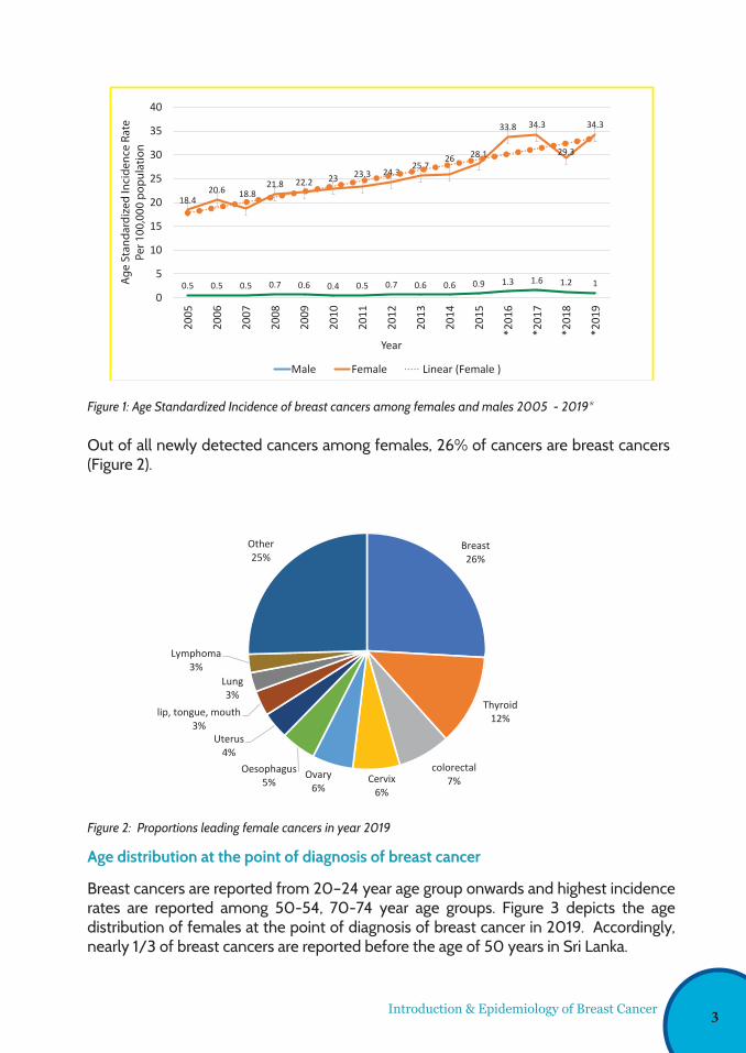
Breast cancer is the highest incident cancer reported in Sri Lanka and also the commonest cause of death due to cancers among females in Sri Lanka. In year 2019, 4594 breast cancers were reported among females while 119 breast cancers were reported among males. According to the National Cancer Registry Programme of Sri Lanka, the incidence of breast cancer is increasing over the years and the age standardized incidence rate among females is rising rapidly compared to males (Figure 1). The age standardized incidence rate was 18.4 /100,000 for the year 2005 to 24.3 /100,000 in the year 2014 and 34.4 in the year 2019 as shown in figure 1.
2
Introduction & Epidemiology of Breast Cancer
Figure 1: Age Standardized Incidence of breast cancers among females and males 2005 - 2019*
Figure 2: Proportions leading female cancers in year 2019
Out of all newly detected cancers among females, 26% of cancers are breast cancers (Figure 2).
Age distribution at the point of diagnosis of breast cancer
Breast cancers are reported from 20–24 year age group onwards and highest incidence rates are reported among 50-54, 70-74 year age groups. Figure 3 depicts the age distribution of females at the point of diagnosis of breast cancer in 2019. Accordingly, nearly 1/3 of breast cancers are reported before the age of 50 years in Sri Lanka.
Figure 2: Leading female cancers in year 2019
Age distribution at the point of diagnosis of breast cancer
Breast cancers are reported from 20–24-year age group onwards and highest incidence rates are
reported among 50-54, 70-74 year age groups. Figure 3 depicts the age distribution of females at
the point of diagnosis of breast cancer in 2019. Accordingly, nearly 1/3 of breast cancers are
reported before the age of 50 years in Sri Lanka.
Breast26%
Thyroid12%
colorectal7%Cervix
6%
Ovary6%
Oesophagus 5%
Uterus4%
lip, tongue, mouth3%
Lung3%
Lymphoma3%
Other25%
Leading cancers among females in Sri Lanka 2019
0.5 0.5 0.5 0.7 0.6 0.4 0.5 0.7 0.6 0.6 0.9 1.3 1.6 1.2 1
18.420.6 18.8
21.8 22.2 23 23.3 24.325.7
26 28.1
33.8 34.3
29.3
34.3
0
5
10
15
20
25
30
35
40
2005
2006
2007
2008
2009
2010
2011
2012
2013
2014
2015
*201
6
*201
7
*201
8
*201
9
Year
Male Female Linear (Female )
Breast Cancer ASR per 100,000 population 2005-2019
males (Figure 1). The age standardized incidence rate was 18.4 /100,000 for the year 2005 to
24.3 /100,000 in the year 2014 and 34.4 in the year 2019 as shown in figure 1.
Figure 1: Age Standardized Incidence of breast cancers among females and males 2005 - 2019*
Out of all newly detected cancers among females, 26% of cancers are breast cancers (Figure 2). Ag
e St
anda
rdiz
ed In
cide
nce
Rate
Per 1
00,0
00 p
opul
atio
n
3
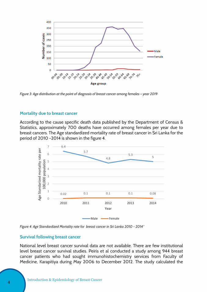
Figure 3: Age distribution at the point of diagnosis of breast cancer among females – year 2019
Figure 4: Age Standardized Mortality rate for breast cancer in Sri Lanka 2010 - 2014*
Mortality due to breast cancer
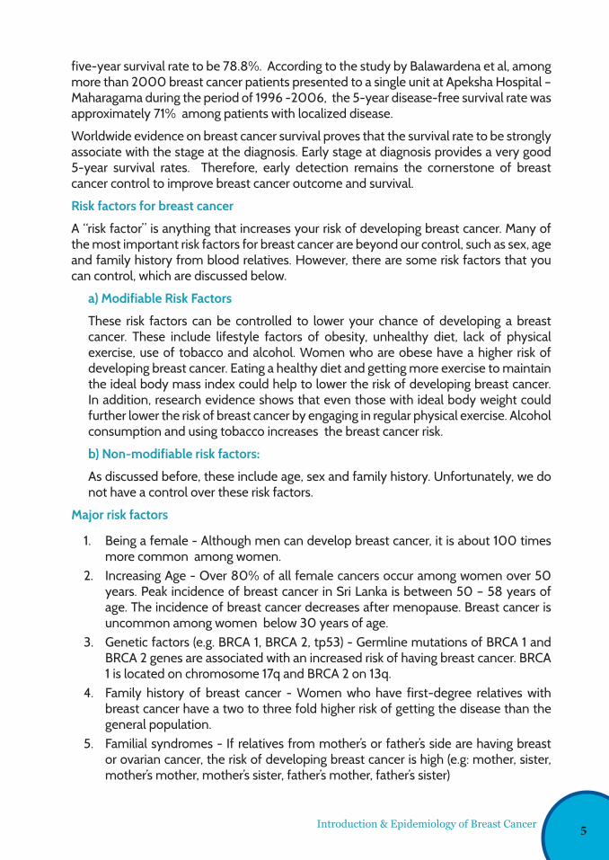
According to the cause specific death data published by the Department of Census & Statistics, approximately 700 deaths have occurred among females per year due to breast cancers. The Age standardized mortality rate of breast cancer in Sri Lanka for the period of 2010 -2014 is shown in the figure 4.
Survival following breast cancer
National level breast cancer survival data are not available. There are few institutional level breast cancer survival studies. Peiris et al conducted a study among 944 breast cancer patients who had sought immunohistochemistry services from Faculty of Medicine, Karapitiya during May 2006 to December 2012. The study calculated the
Introduction & Epidemiology of Breast Cancer
Figure 3: Age distribution at the point of diagnosis of breast cancer among females – year 2019
Mortality due to breast cancer
According to the cause specific death data published by the Department of Census & Statistics,
approximately 700 deaths have occurred among females per year due to breast cancers. The Age
standardized mortality rate of breast cancer in Sri Lanka for the period of 2010 -2014 is shown in
the figure 4.
Figure 4: Age Standardized Mortality rate for breast cancer in Sri Lanka 2010 - 2014*
6.45.7
4.85.3
5
0.02 0.1 0.1 0.1 0.080
1
2
3
4
5
6
7
2010 2011 2012 2013 2014
Age
Stan
dariz
ed m
orta
lity
rate
per
10
0,00
0 po
pula
tion
Year
Male Female
4
five-year survival rate to be 78.8%. According to the study by Balawardena et al, among more than 2000 breast cancer patients presented to a single unit at Apeksha Hospital – Maharagama during the period of 1996 -2006, the 5-year disease-free survival rate was approximately 71% among patients with localized disease.
Worldwide evidence on breast cancer survival proves that the survival rate to be strongly associate with the stage at the diagnosis. Early stage at diagnosis provides a very good 5-year survival rates. Therefore, early detection remains the cornerstone of breast cancer control to improve breast cancer outcome and survival.
Risk factors for breast cancer
A “risk factor” is anything that increases your risk of developing breast cancer. Many of the most important risk factors for breast cancer are beyond our control, such as sex, age and family history from blood relatives. However, there are some risk factors that you can control, which are discussed below.
a) Modifiable Risk Factors
These risk factors can be controlled to lower your chance of developing a breast cancer. These include lifestyle factors of obesity, unhealthy diet, lack of physical exercise, use of tobacco and alcohol. Women who are obese have a higher risk of developing breast cancer. Eating a healthy diet and getting more exercise to maintain the ideal body mass index could help to lower the risk of developing breast cancer. In addition, research evidence shows that even those with ideal body weight could further lower the risk of breast cancer by engaging in regular physical exercise. Alcohol consumption and using tobacco increases the breast cancer risk.
b) Non-modifiable risk factors:
As discussed before, these include age, sex and family history. Unfortunately, we do not have a control over these risk factors.
Major risk factors
1. Being a female - Although men can develop breast cancer, it is about 100 times more common among women.
2. Increasing Age - Over 80% of all female cancers occur among women over 50 years. Peak incidence of breast cancer in Sri Lanka is between 50 – 58 years of age. The incidence of breast cancer decreases after menopause. Breast cancer is uncommon among women below 30 years of age.
3. Genetic factors (e.g. BRCA 1, BRCA 2, tp53) - Germline mutations of BRCA 1 and BRCA 2 genes are associated with an increased risk of having breast cancer. BRCA 1 is located on chromosome 17q and BRCA 2 on 13q.
4. Family history of breast cancer - Women who have first-degree relatives with breast cancer have a two to three fold higher risk of getting the disease than the general population.
5. Familial syndromes - If relatives from mother’s or father’s side are having breast or ovarian cancer, the risk of developing breast cancer is high (e.g: mother, sister, mother’s mother, mother’s sister, father’s mother, father’s sister)
Introduction & Epidemiology of Breast Cancer 5
Minor risk factors:
1. Exposure to estrogen and progesterone hormones for longer periods during the lifetime is considered as a minor risk factor. Early Menarche (before age 12), late menopause (after age 55), nulliparity (never having children) or being older at first pregnancy and long term hormone replacement therapy are the examples for situations where a person is exposed to estrogen and/or progesterone for longer periods.
2. Exogenous oestrogen is considered as a risk factor of breast carcinoma. Large number of scientific researchers have been done on the effect of contraceptive agents on breast cancer and showed a slight risk. This should not be a reason to stop it. The risk is very small and is only significant in young women with a strong family history of breast cancers and in those who have been on the pill for more than 5 years. If the female has a family history, she could discuss other contraceptive options with her gynecologist.
3. Fibrocystic disease and epithelial hyperplasia are benign entities in the breast and they are known to associate with a minor risk of getting breast cancer.
4. Ionizing radiation is also considered as a minor risk of developing breast carcinoma. 5. There are studies that show evidence that breast augmentation surgery increase the
risk of breast cancer.
Table 1: Risk factors for breast cancer
Table 1: Risk factors for breast cancer
Non-Modifiable Factors
Major Factors
Being a female
Increasing age
Presence of BRCA 1 & BRACA 2 genes
Family history of blood relatives
Exposure to Radiation
Minor Factors
Nulliparity
Early Menarche
Late menopause
Modifiable Factors
Excess body weight
A high body mass index
Low physical activity
Sedentary lifestyles
Excessive alcohol consumption
Smoking
Unhealthy diets
Protective factors
Breastfeeding and regular physical exercises give protection from breast cancer. It must be noted here that breastfeeding provides protection against breast cancer with a 4.3% reduction per twelve months of breast feeding. Globally, breastfeeding can avert nearly 20 000 deaths due to breast cancer annually.
Introduction & Epidemiology of Breast Cancer6
Dr. Mekala Fernando Senior Registrar in Community Medicine, National Cancer Control Programme
Dr. Amila Suranga Malawige Registrar in Community Medicine, National Cancer Control Programme
Chapter02
Anatomy and Physiology of the Breast Including Genetics
7

Anatomy and Physiology of the Breast Including Genetics
Chapter 2 -Anatomy and Physiology of the Breast Including Genetics
Anatomy of Mammary Glands
The breasts are found at the anterior thoracic wall, anterior to the deep fascia and pectoral muscles; separated from them by the retromammary space. Each breast consists of mammary glands and surrounding connective tissue. The mammary glands are modified apocrine sweat glands. The gland is comprised of 15-20 secretory lobes which are separated by fibrous bands called the suspensory ligaments of the breast (Cooper’s ligaments). The secretory lobes contain numerous lobules comprised of the tubuloalveolar glands. The secretory ducts of the lobes, called the lactiferous ducts, converge and open onto nipple. Each lactiferous duct dilates into the lactiferous sinus before opening onto the nipple.
Lymphatic Drainage
Lymph from the breast lobules, nipple and areola areas collect into the subareolar lymphatic plexus. From here, around 75% of lymph (mostly from the lateral quadrants of the breast) drains into the pectoral lymph nodes, and then into the axillary lymph nodes. Whilst the remainder drains into the parasternal lymph nodes. The parasternal nodes drain into the bronchomediastinal trunks, which also drain the thoracic organs. Besides the axillary and parasternal nodes, some drainage of the breast
Figure 5: Cross section of the breast
Figure 6: Lymphatic drainage of the breast
3
Lymphatic Drainage
Lymph from the breast
lobules, nipple and areola
areas collect into the
subareolar lymphatic plexus.
From here, around 75% of
lymph (mostly from the lateral
quadrants of the breast)
drains into the pectoral lymph
nodes, and then into
the axillary lymph nodes. Whilst the
remainder drains into the parasternal lymph nodes. The parasternal nodes drain into
the bronchomediastinal trunks, which also drain the thoracic organs. Besides the
axillary and parasternal nodes, some drainage of the breast can occur via the
intercostal lymph nodes which are located around the heads and necks of the ribs. The
intercostal lymph nodes drain either into the thoracic lymph duct or the
bronchomediastinal lymph trunks.
It is important to understand the lymphatic system as breast carcinomas tend to
spread by travelling through the lymphatic vessels, creating metastatic deposits in
distant parts of the body.
Figure 2: Lymphatic drainage of the breast
2
Chapter 2 -Anatomy and physiology of the breast including genetics
Anatomy of Mammary Glands
The breasts are found at the anterior thoracic wall, anterior to the deep fascia and
pectoral muscles; separated from them by the retromammary space. Each breast
consists of mammary glands and surrounding connective tissue. The mammary
glands are modified apocrine sweat glands. The gland is comprised of 15-20
secretory lobes which are separated by fibrous bands called the suspensory ligaments
of the breast (Cooper’s ligaments). The secretory lobes contain numerous lobules
comprised of the tubuloalveolar glands. The secretory ducts of the lobes, called
the lactiferous ducts, converge and open onto nipple. Each lactiferous duct dilates into
the lactiferous sinus before opening onto the nipple.
Figure 1: Cross section of the breast
1. Cooper’s Ligaments
2. Breast Lobule
3. Extralobular Duct
4. Ductal Ampulla (Reservoir)
5. Main Duct
6. Nipple
7. Skin
8. Subcutaneous Fat
9. Mammary Layer Fatty Tissue
10. Retromammary Fat
11. Lymph nodes
12. Pectoralis Major muscle
13. Pectoralis Minor muscle
14. Rib
8
Anatomy and Physiology of the Breast Including Genetics
can occur via the intercostal lymph nodes which are located around the heads and necks of the ribs. The intercostal lymph nodes drain either into the thoracic lymph duct or the bronchomediastinal lymph trunks.
It is important to understand the lymphatic system as breast carcinomas tend to spread by travelling through the lymphatic vessels, creating metastatic deposits in distant parts of the body.
Blood Supply and Nerve Supply
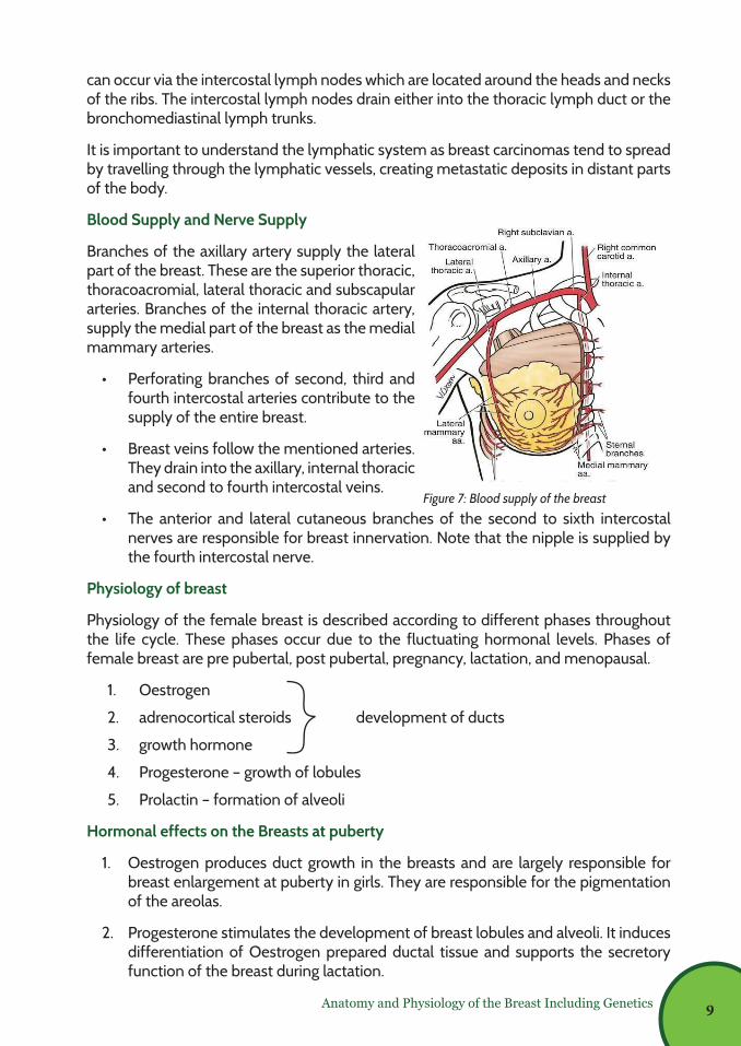
Branches of the axillary artery supply the lateral part of the breast. These are the superior thoracic, thoracoacromial, lateral thoracic and subscapular arteries. Branches of the internal thoracic artery, supply the medial part of the breast as the medial mammary arteries.
• Perforating branches of second, third and fourth intercostal arteries contribute to the supply of the entire breast.
• Breast veins follow the mentioned arteries. They drain into the axillary, internal thoracic and second to fourth intercostal veins.
• The anterior and lateral cutaneous branches of the second to sixth intercostal nerves are responsible for breast innervation. Note that the nipple is supplied by the fourth intercostal nerve.
Physiology of breast
Physiology of the female breast is described according to different phases throughout the life cycle. These phases occur due to the fluctuating hormonal levels. Phases of female breast are pre pubertal, post pubertal, pregnancy, lactation, and menopausal.
1. Oestrogen
2. adrenocortical steroids development of ducts
3. growth hormone
4. Progesterone – growth of lobules
5. Prolactin – formation of alveoli
Hormonal effects on the Breasts at puberty
1. Oestrogen produces duct growth in the breasts and are largely responsible for breast enlargement at puberty in girls. They are responsible for the pigmentation of the areolas.
2. Progesterone stimulates the development of breast lobules and alveoli. It induces differentiation of Oestrogen prepared ductal tissue and supports the secretory function of the breast during lactation.
4
Blood Supply and Nerve Supply
Branches of the axillary artery supply the lateral part of the breast. These are the superior thoracic, thoracoacromial, lateral thoracic and subscapular arteries. Branches of the internal thoracic artery, supply the medial part of the breast as the medial mammary arteries.
Perforating branches of second, third and fourth intercostal arteries contribute to the supply of the entire breast.
Breast veins follow the mentioned arteries. They drain into the axillary, internal thoracic and second to fourth intercostal veins.
The anterior and lateral cutaneous branches of the second to sixth intercostal nerves are responsible for breast innervation. Note that the nipple is supplied by the fourth intercostal nerve.
Physiology of breast
Physiology of the female breast is described according to different phases throughout
the life cycle. These phases occur due to the fluctuating hormonal levels. Phases of
female breast are pre pubertal, post pubertal, pregnancy, lactation, and menopausal.
Hormones which regulate the function of breast and their actions
1. Oestrogen
2. adrenocortical steroids development of ducts
3. growth hormone
4. Progesterone – growth of lobules
5. Prolactine – formation of alveoli
Figure 3: Blood supply of the breast Figure 7: Blood supply of the breast
9

Anatomy and Physiology of the Breast Including Genetics
Cyclical changes of breast during the menstrual cycle
The hormone Oestrogen is produced by the ovaries in the first half of the menstrual cycle. It stimulates the growth of milk ducts in the breasts. The increasing level of Oestrogen leads to ovulation halfway through the cycle. Next, the hormone progesterone takes over in the second half of the cycle. It stimulates the formation of the milk glands. During menstruation, many women also have changes in breast texture. Their breasts may feel very lumpy. This is because the glands in the breast are enlarging to get ready for a possible pregnancy. If pregnancy does not happen, the breasts go back to normal size. Once menstruation starts, the cycle begins again. Breast swelling, tenderness, and pain can occur during the 7 days preceding menstruation.
Breasts changes during pregnancy and Lactation
Breast changes are one of the earliest signs of pregnancy. This is a result of the hormone progesterone. In addition, the dark areas of skin around the nipples (the areolas) begin to swell. This is followed by the rapid swelling of the breasts themselves. Most pregnant women feel soreness down the sides of the breasts, and nipple tingling or soreness. This is because of the growth of the milk duct system and the formation of many more lobules. By the fifth or sixth month of pregnancy, the breasts are fully capable of producing milk. Many other hormones also play vital roles in milk production. These include follicle-stimulating hormone (FSH), luteinizing hormone (LH), prolactin, oxytocin, and human placental lactogen (HPL). Drop in oestrogen increases sensitivity to prolactin. This will stimulate milk production and suckling stimulates prolactin and oxytocin, stimulating milk ejection further.
Breast changes at menopause
By the time a woman reaches her late 40s and early 50s, perimenopause is starting or is well underway. At this time, the levels of oestrogen and progesterone begin to change. Oestrogen levels dramatically decrease. This leads to many of the symptoms commonly linked to menopause. Without oestrogen, the breast’s connective tissue becomes dehydrated and is no longer elastic. The breast tissue, which was prepared to produce milk, shrinks and loses shape. This leads to the “saggy” breasts associated with women of this age.
Figure 8: Hormonal changes during the menstrual cycle
5
Hormonal effects on the Breasts at puberty
1. Oestrogen produces duct growth in the breasts and are largely responsible for
breast enlargement at puberty in girls. They are responsible for the pigmentation
of the areolas.
2. Progesterone stimulates the development of breast lobules and alveoli. It induces
differentiation of Oestrogen prepared ductal tissue and supports the secretory
function of the breast during lactation.
Cyclical changes of breast during the menstrual cycle
Figure 4: Hormonal changes during the menstrual cycle
The hormone Oestrogen is produced by the ovaries in the first half of the menstrual
cycle. It stimulates the growth of milk ducts in the breasts. The increasing level of
Oestrogen leads to ovulation halfway through the cycle. Next, the hormone
progesterone takes over in the second half of the cycle. It stimulates the formation of
the milk glands. During menstruation, many women also have changes in breast
texture. Their breasts may feel very lumpy. This is because the glands in the breast are
enlarging to get ready for a possible pregnancy. If pregnancy does not happen, the
breasts go back to normal size. Once menstruation starts, the cycle begins again.
10
Anatomy and Physiology of the Breast Including Genetics
Common Breast Cancer Mutations
Researchers have identified several key gene changes linked to breast cancer. Some of these pose a high risk, while others seem to be less significant.
High-penetrance genes: BRCA 1, BRCA 2, T53, PTEN, CDH1
Moderate-penetrance genes: ATM, PALB2
High-penetrance genes
• BRCA 1 and BRCA 2
The BRCA 1 (breast cancer gene one) and BRCA 2 (breast cancer gene two) inherited gene mutations are the most common cause of hereditary breast cancer. Mutations in these genes account for up to 10% of all breast cancers. BRCA mutations also raise chances for ovarian cancer, pancreatic cancer, colon cancer and, in men, prostate cancer. Women with a BRCA 1 or BRCA 2 mutation have up to a 72% chance of breast cancer during their lifetime.
Tabel 2: Life time risk of developing breast cancer
Life time risk of developing breast cancer
BRCA 1 BRCA 2
Before the age of 50 years 50% 28%
Up to the age of 70 years 50-85% 50-85%
Male breast cancer 8.6% by age 65 years 15% by age 65 years 20% lifetime
• TP53
The TP53 gene helps to stop the growth of cells that have damaged DNA. An inherited TP53 mutation causes Li-Fraumeni syndrome, a disorder could cause breast cancer, leukemia, brain tumors, and cancers called sarcomas.
• PTEN
PTEN is a gene that helps control cell growth. An inherited change in PTEN can cause Cowden syndrome, a disorder that could lead to cancerous and noncancerous breast tumors and other growths. Women with a PTEN mutation have a lifetime breast cancer chance of between 25% and 50%, though some studies suggest the odds are even higher.
• CDH1
CDH1 makes a protein that helps cells bind together to create tissue. People with a faulty CDH1 gene are more likely to develop a rare type of stomach cancer. Women with this mutation also have a 39% to 52% lifetime chance of invasive lobular breast cancer (breast cancer that starts in the lobules of the breast, the glands that make milk).
11
Anatomy and Physiology of the Breast Including Genetics
Moderate-penetrance genes
• PALB 2
Normally, the PALB 2 gene makes a protein that works with the BRCA 2 gene protein to repair damaged DNA and stop tumor growth. But defects in the gene can lead to a higher likelihood of breast cancer. Some studies suggest that women with a PALB 2 mutation have a 14% chance of breast cancer by age 50 and a 35% chance by age 70.
• ATM
The ATM gene normally helps repair damaged DNA, but some people who inherit one bad copy of the gene are at high risk for breast cancer and pancreatic cancer. Research suggest the lifetime chance of breast cancer for those who carry an ATM mutation is between 33% and 38%. Those who have a type of mutation that affects a specific location on the ATM gene have a 69% lifetime chance.
Other genes
• PIK3CA
The PIK3CA gene gives instructions to make a protein that’s important for many cell functions. A PIK3CA mutation isn’t inherited (doesn’t pass down the family tree). Instead, it’s a mutation that develop during the life, called a sporadic mutation. PIK3CA gene mutations are found in about 30% to 40% of breast cancers.
• HER2
The HER2 gene makes a protein called HER2 (human epidermal growth factor receptor 2). This protein is found on the surface of all breast cells and that helps them grow. If the HER2 gene malfunctions and makes too many copies of itself, it tells cells to make too much HER2 protein. This causes the cells to grow out of control.
Like PIK3CA, HER2 is not an inherited gene mutation. Most breast cancers are HER2-negative. Research suggests about only 10% to 20% of cases are HER2-positive. This means there’s a change in the HER2 gene that makes breast cells grow and divide uncontrollably.
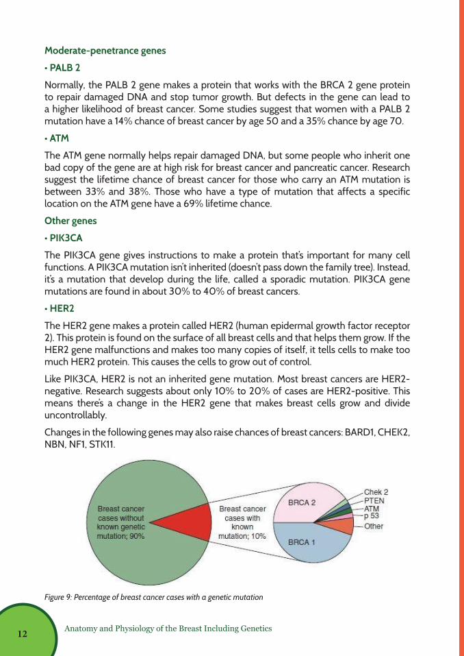
Changes in the following genes may also raise chances of breast cancers: BARD1, CHEK2, NBN, NF1, STK11.
Figure 9: Percentage of breast cancer cases with a genetic mutation
9
HER2
The HER2 gene makes a protein called HER2 (human epidermal growth factor receptor
2). This protein is found on the surface of all breast cells and that helps them grow. If
the HER2 gene malfunctions and makes too many copies of itself, it tells cells to make
too much HER2 protein. This causes the cells to grow out of control.
Like PIK3CA, HER2 is not an inherited gene mutation. Most breast cancers are HER2-
negative. Research suggests about only 10% to 20% of cases are HER2-positive. This
means there’s a change in the HER2 gene that makes breast cells grow and divide
uncontrollably.
Changes in the following genes may also raise chances of breast cancers: BARD1,
CHEK2, NBN, NF1, STK11.
Figure 5: Percentage of breast cancer cases with a genetic mutation
12
Professor Bimalka Senevirathne Consultant Pathologist, Department of Pathology, Faculty of Medical Sciences, University of Sri Jayewardenepura, Nugegoda
Chapter03
Breast Pathologies -Non-Cancerous and Cancerous Lesions
13
Breast Pathologies-Non-Cancerous and Cancerous Lesions
Chapter 3 - Breast pathologies-Non-Cancerous and Cancerous Lesions
Benign breast diseases
Benign breast disease represents a spectrum of disorders that come to clinical attention as palpable lesions found on physical examination or as imaging abnormalities. Following confirmation of a benign diagnosis, treatment in general is aimed at symptomatic relief and patient education. Some benign breast diseases, such as atypical hyperplasia, confer an increase in the patient’s future risk of developing breast cancer, which warrants counseling on follow up screening and risk reduction strategies.
Classification of benign breast diseases:
Histologically they can be divided into three groups which provide an idea regarding potential future cancer risk;
1) Non-proliferative disorders - no increased risk
2) Proliferative disorders without atypia - mild to moderate increase in risk
3) Atypical hyperplasia - substantial increase in risk (relative risk in the order of 4.1-5.3).
Clinically, classification of benign breast diseases by common presenting features may be more helpful;
a) Physiological swelling and tenderness
b) Nodularity
c) Breast pain (not usually associated with malignancy)
d) Palpable breast lumps
e) Nipple discharge including galactorrhoea
f) Breast infection and inflammation - usually associated with lactation
• Physiological swelling and tenderness
Puberty
Breast enlargement is sometimes unilateral initially. It is the first obvious sign of puberty in girls. Pubertal breast development is known as thelarche.
Cyclical mastalgia
The breasts are active organs that change throughout the menstrual cycle. Some degree of tenderness and nodularity in the premenstrual phase is so common that it may be considered as normal, affecting up to two thirds of all menstruating women. It rapidly resolves as menstruation starts. Conditions to exclude by history and examination are infection, pregnancy and malignancy.
14
Breast Pathologies-Non-Cancerous and Cancerous Lesions
Pregnancy – Discussed in Chapter 8.
• Nodularity
Fibrocystic change is the most common benign breast disorder, and most often it presents with pain and nodularity (lumpy breast). This usually affects women aged 20-50 years and appears to be hormonal in aetiology. Incidence of fibrocystic change decreases after menopause suggesting the role of oestrogen in its pathogenesis. Fibrocystic change could be associated with polycystic ovaries.
Clinical features
³ Patient presents with painful, tender swellings which are fibrous and have defined edges.
³ Usually bilateral and multifocal.
Gross features
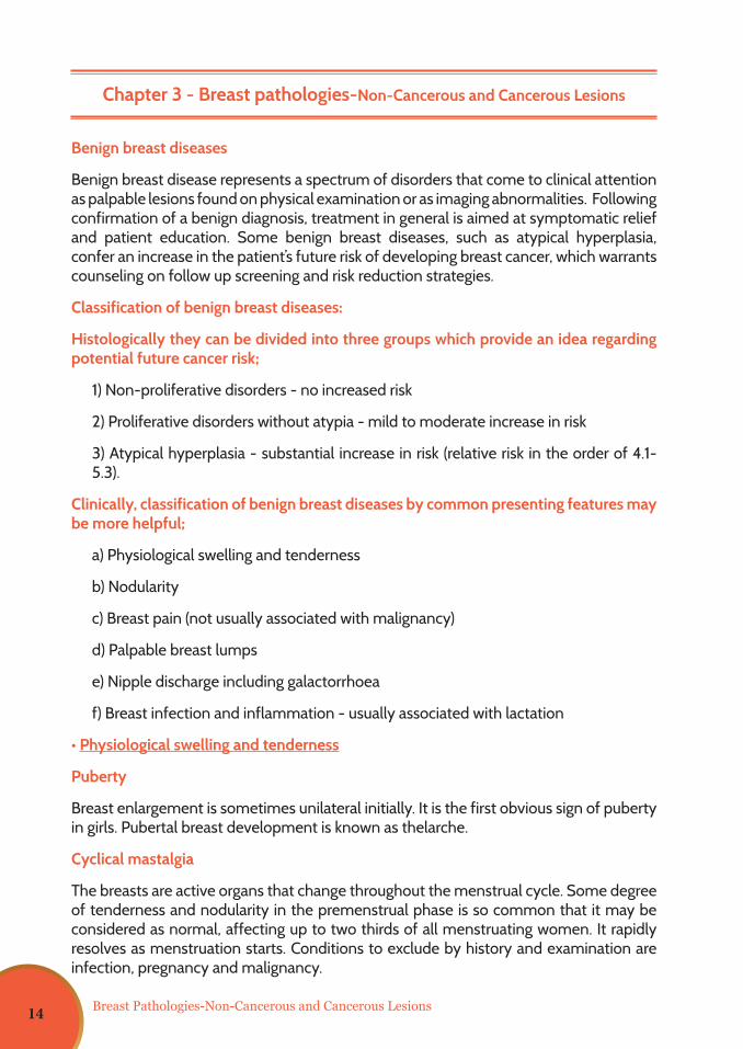
Gray, white, fibrous tissue with multiple cysts filled with semi translucent fluid giving them a blue colour (Blue domed cysts).
Microscopic features
Cystically dilated or ectatic ducts lined by metaplastic apocrine ductal cells having abundant eosinophilic granular cytoplasm.
It is important to identify the spectrum of histological changes since only a few subsets of changes have an increased risk of developing into breast cancer. The spectrum of histo-pathological changes is divided into two clinico-pathologically relevant groups;
1) Non proliferative fibrocystic change
2) Proliferative fibrocystic change with epithelial hyperplasia
Figure 10: Fibrocystic change
15
Presence of atypical hyperplasia is considered to be associated with increased risk of developing breast cancer.
• Palpable breast lumps
Many breast lumps are benign, especially in younger patients. Most benign lumps will be either cysts or fibroadenomas. Broadly speaking, a benign mass is usually three-dimensional, mobile and smooth, has regular borders and is solid or cystic in consistency. A malignant mass is usually firm in consistency, has irregular borders and may be fixed to the underlying skin or soft tissue. There may also be skin changes or nipple retraction. However, current guidelines recognize that it is not always possible to make an accurate diagnosis on the basis of clinical examination alone, and therefore all unexplained lumps should be referred for assessment in a specialist breast clinic.
³ Breast cysts
Cysts are most common between the ages of 35 and 50 years. They are palpable as discrete lumps and may be recurrent. They cannot be reliably distinguished from solid tumours on clinical examination. Aspiration is not usually recommended, and they may settle spontaneously. However, guidelines advise to refer all such cases to a breast clinic for imaging.
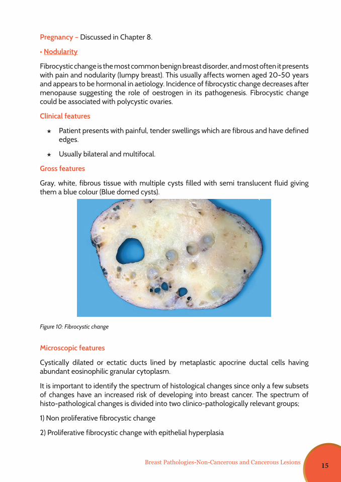
³ Fibroadenomas
These are the most common benign breast lesions which present in young women between the ages of 20-24 years.
Fibroadenomas arise in breast lobules and are composed of fibrous and epithelial tissue. They present as firm, non-tender, highly mobile, palpable lumps. Hormones seem to be involved in their aetiology, and hormone replacement therapy (HRT) increases the incidence.
Most fibroadenomas stop growing at 2-3cm.
The treatment of choice is surgical excision, but this may not be necessary if they are small. Diagnosis is confirmed by histo-pathology. It should be noted that complex and multiple fibroadenomas need follow up care as they are associated with an increased risk of breast cancer. All women presenting with unexplained breast lumps should be assessed at a specialist breast clinic and undergo triple assessment of examination, imaging (the first-line choice before the age of 40 years is ultrasound, whereas it’s the mammogram after the age of 40 years) and needle biopsy (not necessarily required for women under the age of 25 years).
Figure 11: Fibroadenoma
Breast Pathologies-Non-Cancerous and Cancerous Lesions16
³ Phylloides tumour
This is a rare tumour that tends to occur in women aged between 40 and 50 years. It can be difficult to distinguish from a fibroadenoma. It may be benign, borderline or malignant. A benign tumour may reappear after excision and may become malignant. Treatment is wide excision including some normal breast tissue. Follow-up is essential and the practice varies as there are no national guidelines.
³ Intraductal papilloma
This is a benign, warty lesion usually located just behind the areola.
• A small lump or a sticky, possibly blood-stained discharge may be noticed.
• Women aged in their 40s are more likely to have just one lesion, but younger women may have multiple lesions.
• Triple assessment is required in a specialist breast clinic, with examination, imaging and biopsy.
³ Atypical hyperplasia
This is a type of benign hyperplasia which can occur in the ducts or the lobules. However, it should be noted that:
• It may progress into a Lobular carcinoma in situ.
• In the presence of atypical hyperplasia, there is approximately a 29% risk of developing breast cancer over the next 25 years. Hence follow-up is essential.
• Risk is increased in the presence of a family history of breast cancer.
³ Sclerosing adenosis
This is a benign condition of sclerosis within the lobules.
• It may cause a lump, pain or be found on routine assessment.
• It can be very difficult to distinguish from malignancy, and biopsy is often advised.
• Once diagnosed, it does not need follow-up as it does not have malignant potential.
³ Fat necrosis
Fat necrosis is more likely to occur in larger, fatty breasts in overweight or obese women, but can occur in any woman and even occasionally in men. Breast fat necrosis has various etiologies and implications; therefore, a careful patient history is imperative to properly evaluate the patient. The most common etiology of fat necrosis is recent breast surgery; however, in non-operative patients, cancer or mechanical trauma to the breast tissue is often the culprit. Breast fat necrosis can be confused with malignancy on breast imaging (it can mimic malignancy on radiologic studies, as well as in the clinical presentation). Fat necrosis can be diagnosed clinically or radiographically in the majority
Breast Pathologies-Non-Cancerous and Cancerous Lesions 17
of cases, without the need for biopsy. In surgical patients who have recently undergone a breast surgical procedure such as breast reduction, reconstruction, implant removal or fat grafting after primary reconstruction, the most common presentation is the finding of a palpable mass or lump under the breast skin. Fat necrosis of breast should resolve or regress slowly with time.
³ Duct ectasia and periductal mastitis
Duct ectasia of the breast or mammary duct ectasia is a condition which occurs when a milk duct beneath the nipple widens, the duct walls thicken and the duct fills with fluid. This is the most common cause of greenish discharge. Mammary duct ectasia can mimic breast cancer. It is a disorder of peri- or post-menopausal age. Signs & symptoms of duct ectasia can include nipple retraction, inversion, pain and classic green-brown discharge. The duct widening is commonly believed to be a result of secretory stasis, including stagnant colostrum, which also causes periductal inflammation and fibrosis. However, because nonspecific duct widening is common, it might be also a coincidental finding in many processes.
• Infection
Infection (mastitis) may be associated with lactation or, more rarely, occur at other times.
With lactation - Breast ducts become blocked with engorged milk, and bacteria enter from cracks in the nipple.
• There may be engorgement of the breast and axillary lymphadenopathy.
• Cold compression and analgesia such as ibuprofen or paracetamol may give some relief.
• Encourage the woman to continue breast-feeding.
• An abscess may develop in the peripheral part of the breast tissue.
• A localised abscess will require incision and drainage, followed by antibiotics.
• Swabs should be sent for culture.
Without lactation - Spontaneous peripheral abscesses in non-lactating women are often associated with diabetes and immune compromise. Smoking and nipple rings can predispose women to non-lactational mastitis.
• Non-lactational mastitis produces peri-areolar abscesses, usually resulting from obstruction with cellular debris and lipid-laden material. Bacteria enter from the skin and produce periductal inflammation and abscess formation.
• There is a chronic recurrent course with noncyclical mastalgia, nipple discharge or retraction, peri-areolar abscess, subareolar mass or cellulitis of the overlying skin.
Inflammatory breast cancer causes pain, redness and induration of the skin, usually affecting the dependent portion of the breast. Symptoms progress very rapidly, and within a month the breast may have the peau d’orange appearance.
Breast Pathologies-Non-Cancerous and Cancerous Lesions18

Anyone in whom presumed mastitis does not resolve completely and who has residual breast change needs referral to exclude inflammatory or other types of breast cancer.
Breast cancer: types, spread & prognostic factors
Types of breast cancer:
Breast cancer is more common in the left breast than in the right breast, and rarely could be bilateral (<5%). Anatomically, the most common site is the upper, outer quadrant of the breast. There are several different histological types of breast cancer. It is important to identify the definitive type of breast cancer because each type has a different biological behaviour. Some types are highly aggressive and spread fast to distant sites.
Classification of breast cancer is based on the cell of origin. Most breast cancers begin in the epithelial cells of breast tissue and are known as carcinomas. Breast carcinomas are broadly divided in to invasive and in situ carcinoma groups. In situ refers to a carcinoma confined to the epithelium of the tissue, while invasive refers to carcinomas that have spread to the surrounding stroma and tissues. In situ carcinoma group also has several pathological types such as in situ ductal, in situ lobular and in situ papillary carcinoma, in which there are characteristic cyto-architectural features. Since in situ carcinoma hasn’t spread to surrounding tissues, it is associated with an excellent outcome in contrast to invasive carcinomas. Many breast cancers detected on screening mammograms are early cancers of in situ stage. Early breast cancers could be very small and may not be felt by the individual or by the doctor.
Invasive breast carcinomas
Invasive group of breast carcinoma also includes distinct pathological types. The classification of breast carcinoma has evolved over a long period of time, and as a result has had incorporated into it a wide range of criteria, such as cell type, architectural features and patterns of spread. The most common type of invasive breast carcinoma is ductal carcinoma. Ductal carcinoma arises from the epithelial cells of breast ducts that carry milk to the nipple. More than 80% of carcinomas are of ductal carcinoma type. There is another type of invasive breast carcinoma which is known as lobular carcinoma. Lobular carcinoma arises from the breast lobules / glands and occurs less frequently (approximately 5%) than ductal carcinoma. There are many more less common pathological types of invasive breast carcinomas such as medullary carcinoma, mucinous carcinoma, papillary carcinoma, tubular carcinoma, inflammatory carcinoma, adenoid cystic carcinoma, secretory carcinoma, etc. The type of breast cancer can only be identified and confirmed during the microscopic examination of tumour tissue, which is an integral part of the pathological assessment performed by a specialist.
Figure 12: Breast cancer
Breast Pathologies-Non-Cancerous and Cancerous Lesions 19
Sarcomas
There are breast cancers other than carcinomas. These cancers arise from non-epithelial cells of the breast. Tumours arising from stromal components are broadly classified as sarcomas. Primary sarcomas of the breast are very rare and include several distinct types such as angiosarcoma, fibrosarcoma, liposarcoma, leiomyosarcoma, rhabdomyosarcoma, etc. Malignant Phylloides tumour is another example in which the cancerous component is similar to a sarcoma.
Lymphomas are malignant tumours arising from lymphoid tissue of the mammary gland. Primary lymphomas of the breast are extremely uncommon (< 1%).
Paget’s disease of the breast is another malignant entity with distinct clinicopathological features. The disease was first described by Sir James Paget in 1874.
It usually presents as an eczematous lesion of the nipple. It is accompanied in nearly all instances by an underlying breast carcinoma of in situ ductal type with or without associated stromal invasion. Paget’s disease is diagnosed by biopsy examination of the suspected lesion. In addition, there are malignant tumours such as squamous cell carcinoma, basal cell carcinoma and skin appendageal tumours arising from the overlying skin of the breast.
Secondary tumours of the breast which have spread from a distant site are rare as opposed to primary cancers. These tumours typically present as superficial, well-defined multifocal masses. Malignant melanoma and carcinoma of lung, ovary, stomach and kidney are the most common sources. Metastasis from the contralateral breast carcinoma also should not be forgotten.
Grading and staging of breast cancer
Grade - Histological grade of the tumour is an important prognostic factor. Higher the histological grade, worse would be the outcome for the patient. Grading of breast carcinoma is done by histological examination of routinely stained sections of the tumour. Grading is done by determining the cyto-architectural features of the tumour, and the widely used method is the Nottingham grading system, which is a modification of the Bloom-Richardson system. In this system, the grade is obtained by adding up the scores for tubule formation, nuclear pleomorphism and mitotic count. Each of these variables are given a score from 1 to 3.
Nottingham modification of the Bloom-Richardson system
Tubule formation:1 point: Tubular formation in > 75% of the tumour2 points: Tubular formation in 10-75% of the tumour3 points: Tubular formation in < 10% of the tumour
Nuclear pleomorphism:1 point: Nuclei with minimal variation in size and shape2 points: Nuclei with moderate variation in size and shape3 points: Nuclei with marked variation in size and shape
Breast Pathologies-Non-Cancerous and Cancerous Lesions20
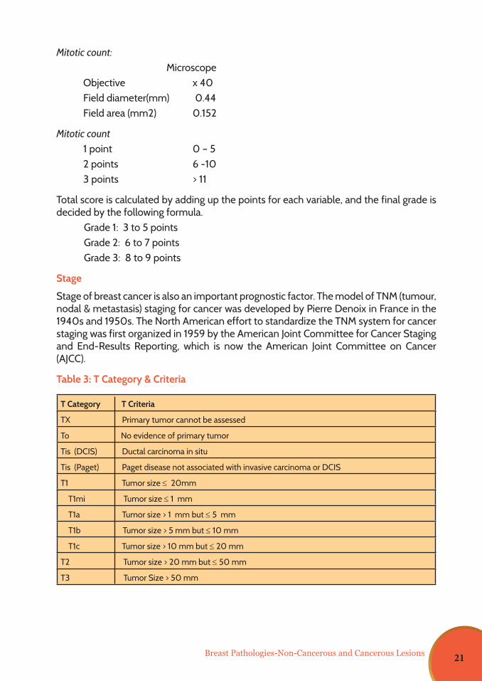
Mitotic count: Microscope
Objective x 40Field diameter(mm) 0.44Field area (mm2) 0.152
Mitotic count1 point 0 – 52 points 6 -103 points > 11
Total score is calculated by adding up the points for each variable, and the final grade is decided by the following formula.
Grade 1: 3 to 5 points Grade 2: 6 to 7 points Grade 3: 8 to 9 points
Stage
Stage of breast cancer is also an important prognostic factor. The model of TNM (tumour, nodal & metastasis) staging for cancer was developed by Pierre Denoix in France in the 1940s and 1950s. The North American effort to standardize the TNM system for cancer staging was first organized in 1959 by the American Joint Committee for Cancer Staging and End-Results Reporting, which is now the American Joint Committee on Cancer (AJCC).
Table 3: T Category & Criteria
T Category T Criteria
TX Primary tumor cannot be assessed
To No evidence of primary tumor
Tis (DCIS) Ductal carcinoma in situ
Tis (Paget) Paget disease not associated with invasive carcinoma or DCIS
T1 Tumor size ≤ 20mm
T1mi Tumor size ≤ 1 mm
T1a Tumor size > 1 mm but ≤ 5 mm
T1b Tumor size > 5 mm but ≤ 10 mm
T1c Tumor size > 10 mm but ≤ 20 mm
T2 Tumor size > 20 mm but ≤ 50 mm
T3 Tumor Size > 50 mm
Breast Pathologies-Non-Cancerous and Cancerous Lesions 21
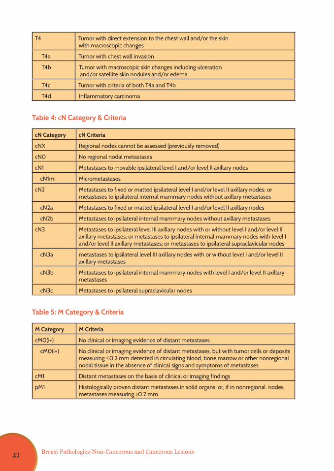
T4 Tumor with direct extension to the chest wall and/or the skin with macroscopic changes
T4a Tumor with chest wall invasion
T4b Tumor with macroscopic skin changes including ulceration and/or satellite skin nodules and/or edema
T4c Tumor with criteria of both T4a and T4b
T4d Inflammatory carcinoma
Table 4: cN Category & Criteria
cN Category cN Criteria
cNX Regional nodes cannot be assessed (previously removed)
cN0 No regional nodal metastases
cN1 Metastases to movable ipsilateral level I and/or level II axillary nodes
cN1mi Micrometastases
cN2 Metastases to fixed or matted ipsilateral level I and/or level II axillary nodes; or metastases to ipsilateral internal mammary nodes without axillary metastases
cN2a Metastases to fixed or matted ipsilateral level I and/or level II axillary nodes
cN2b Metastases to ipsilateral internal mammary nodes without axillary metastases
cN3 Metastases to ipsilateral level III axillary nodes with or without level I and/or level II axillary metastases; or metastases to ipsilateral internal mammary nodes with level I and/or level II axillary metastases; or metastases to ipsilateral supraclavicular nodes
cN3a metastases to ipsilateral level III axillary nodes with or without level I and/or level II axillary metastases
cN3b Metastases to ipsilateral internal mammary nodes with level I and/or level II axillary metastases
cN3c Metastases to ipsilateral supraclavicular nodes
Table 5: M Category & Criteria
M Category M Criteria
cM0(i+) No clinical or imaging evidence of distant metastases
cM0(i+) No clinical or imaging evidence of distant metastases, but with tumor cells or deposits measuring ≤0.2 mm detected in circulating blood, bone marrow or other nonregional nodal tissue in the absence of clinical signs and symptoms of metastases
cM1 Distant metastases on the basis of clinical or imaging findings
pM1 Histologically proven distant metastases in solid organs; or, if in nonregional nodes, metastases measuring >0.2 mm
Breast Pathologies-Non-Cancerous and Cancerous Lesions22
Previous systems for breast cancer staging have focused solely on the anatomic extent of disease; however, during the past decade, it has become recognized that biologic factors, such as tumor grade and hormone receptor expression, are as important as or more important than the anatomic extent of disease to determine prognosis and guide treatment decisions. Thus, the current eighth edition of the AJCC Cancer Staging Manual incorporates prognostic biomarkers to predict outcomes on an individualized basis.
Prognostic staging is preferred for patient care, but anatomic staging may be used in regions of the world where biomarker testing is not available. The integration of prognostic staging into patient management is of particular importance for breast cancer, because recent advances in therapies are based on an individual’s hormone receptor status and the findings from multigene panels which have revolutionized strategies for treatment.
The AJCC staging system for breast cancer applies to invasive carcinomas and ductal carcinoma in situ (DCIS), with or without microinvasion, and does not apply to breast sarcomas, phylloides tumors or breast lymphomas.
To determine a patient’s breast cancer stage, the National Comprehensive Cancer Network recommends the following workup: history and physical examination; blood tests; diagnostic bilateral mammography and US, as necessary; pathologic assessment review; and determination of hormone receptor status. Breast MRI is considered optional, with attention given to cases with mammographically occult tumours. Consideration of additional imaging studies, such as bone scintigraphy and CT, is directed by signs or symptoms.
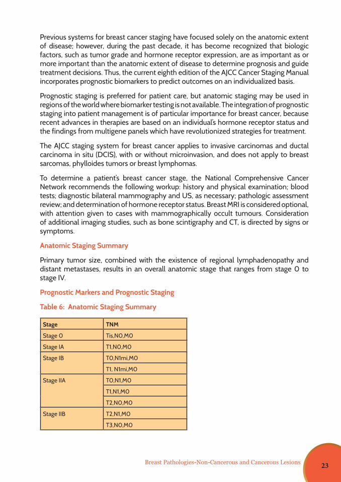
Anatomic Staging Summary
Primary tumor size, combined with the existence of regional lymphadenopathy and distant metastases, results in an overall anatomic stage that ranges from stage 0 to stage IV.
Prognostic Markers and Prognostic Staging
Table 6: Anatomic Staging Summary
Stage TNM
Stage 0 Tis,N0,M0
Stage IA T1,N0,M0
Stage IB T0,N1mi,M0
T1, N1mi,M0
Stage IIA T0,N1,M0
T1,N1,M0
T2,N0,M0
Stage IIB T2,N1,M0
T3,N0,M0
Breast Pathologies-Non-Cancerous and Cancerous Lesions 23
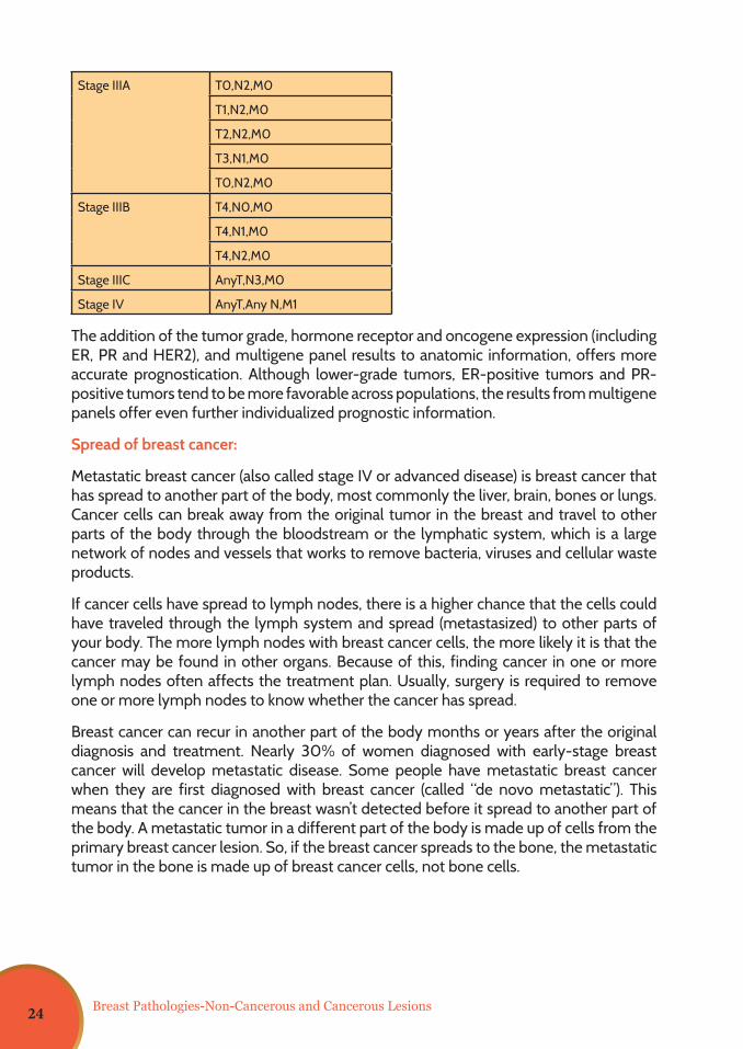
Stage IIIA T0,N2,M0
T1,N2,M0
T2,N2,M0
T3,N1,M0
T0,N2,M0
Stage IIIB T4,N0,M0
T4,N1,M0
T4,N2,M0
Stage IIIC AnyT,N3,M0
Stage IV AnyT,Any N,M1
The addition of the tumor grade, hormone receptor and oncogene expression (including ER, PR and HER2), and multigene panel results to anatomic information, offers more accurate prognostication. Although lower-grade tumors, ER-positive tumors and PR-positive tumors tend to be more favorable across populations, the results from multigene panels offer even further individualized prognostic information.
Spread of breast cancer:
Metastatic breast cancer (also called stage IV or advanced disease) is breast cancer that has spread to another part of the body, most commonly the liver, brain, bones or lungs. Cancer cells can break away from the original tumor in the breast and travel to other parts of the body through the bloodstream or the lymphatic system, which is a large network of nodes and vessels that works to remove bacteria, viruses and cellular waste products.
If cancer cells have spread to lymph nodes, there is a higher chance that the cells could have traveled through the lymph system and spread (metastasized) to other parts of your body. The more lymph nodes with breast cancer cells, the more likely it is that the cancer may be found in other organs. Because of this, finding cancer in one or more lymph nodes often affects the treatment plan. Usually, surgery is required to remove one or more lymph nodes to know whether the cancer has spread.
Breast cancer can recur in another part of the body months or years after the original diagnosis and treatment. Nearly 30% of women diagnosed with early-stage breast cancer will develop metastatic disease. Some people have metastatic breast cancer when they are first diagnosed with breast cancer (called “de novo metastatic”). This means that the cancer in the breast wasn’t detected before it spread to another part of the body. A metastatic tumor in a different part of the body is made up of cells from the primary breast cancer lesion. So, if the breast cancer spreads to the bone, the metastatic tumor in the bone is made up of breast cancer cells, not bone cells.
Breast Pathologies-Non-Cancerous and Cancerous Lesions24
While metastatic breast cancer may not go away completely, treatment may control it for a number of years. If one treatment stops working, there are other alternatives to try. The cancer can be active sometimes and then go into remission at other times. Many different treatments alone, in combination, or in sequence are often used. The symptoms of metastatic breast cancer can vary greatly depending on the location of the cancer. This section covers the symptoms of breast cancer that has spread to the bone, lung, brain and liver, and the tests used to diagnose metastatic breast cancer.
Bone Metastasis: The most common symptom of breast cancer that has spread to the bone is a sudden, noticeable new pain. Breast cancer can spread to any bone, but most often spreads to the ribs, spine, pelvis or the long bones in the arms and legs.
Lung Metastasis: When breast cancer moves into the lung, it often doesn’t cause symptoms. If a lung metastasis does cause symptoms, they may include pain or discomfort in the lung, shortness of breath, persistent cough and others.
Brain Metastasis: Symptoms of breast cancer that has spread to the brain can include headache, changes in speech or vision, memory problems and others.
Liver Metastasis: When breast cancer spreads to the liver, it often doesn’t cause symptoms. If a liver metastasis does cause symptoms, they may include pain or discomfort in the mid-section, fatigue and weakness, weight loss or poor appetite, fever, etc.
Prognostic factors of breast carcinoma
The prognosis of breast carcinoma is related to a large variety of clinical and pathological features. Important prognostic factors are listed below;
a. Age of the patient at the time of diagnosis – Women less than 50 years of age have a better prognosis when compared to older women and very young (<35 years) females.
b. Size – Larger the diameter of the primary tumour, worse the prognosis.
c. Site of the tumour – No correlation has been found with the quadrant location of the tumour.
d. Histological type – Certain histological types such as signet ring carcinoma and inflammatory carcinoma are known to have a bad prognosis.
e. Histological grade – Higher the grade, the prognosis becomes worse.
f. Stage of breast cancer – Prognosis worsens with advancing stage.
g. Presence or absence of invasion – In situ carcinoma has a much better prognosis than invasive malignancy.
h. Skin invasion - Breast carcinoma with skin involvement has an adverse outcome when compared with breast cancer without skin infiltration.
Breast Pathologies-Non-Cancerous and Cancerous Lesions 25
i. Pregnancy & lactation – Breast carcinoma presenting during pregnancy & lactation has a worse prognosis.
j. Paget’s disease- The presence or absence of Paget’s disease in invasive breast carcinoma has no prognostic relevance.
k. Oestrogen receptor status – Oestrogen receptor positive breast carcinoma has a better prognosis.
l. HER2/neu – HER2/neu positivity identifies a subset of patients with poor prognosis. HER2/neu positivity is an excellent predictor of response to Herceptin.
m. BRCA 1 & BRCA 2 mutations – Breast carcinoma associated with BRCA 1 & BRCA 2 mutations carries a poor prognosis.
n. p53 gene mutation – Accumulation of p53 due to genetic mutation is associated with a poor prognosis.
o. Lymphatic invasion and vascular emboli – Lymphatic invasion and the presence of vascular emboli correlates with a bad prognosis.
Breast Pathologies-Non-Cancerous and Cancerous Lesions26
Dr. Nayana De AlwisConsultant Community Physician, National Cancer Control Programme
Chapter04
Screening and Early Detectionof Breast Cancer
27
Screening and Early Detection of Breast Cancer
Chapter 4 - Screening and Early Detection of Breast Cancer
Breast cancer is one of the cancers which has a good survival rate if it is detected in the early stage. There are two strategies for early detection of breast cancer. These are early diagnosis and screening. If detected early, there are various treatment options available for breast cancer, which increase survival and improve the quality of life. Even though some lifestyle changes can reduce the risk, that alone cannot eliminate many breast cancers. Therefore, early detection is the cornerstone of prevention and control of breast cancer.
Advantage of early detection of breast cancer
• Less aggressive treatment
• Wide range of treatment options
• Better outcome
Available screening/early detection methods
Different methods of breast screening are recommended for women over 20 years of age who do not have any signs or symptoms of breast cancer.
1. Be breast aware
2. Self-Breast Examination
3. Clinical Breast Examination
4. Radio-imaging (Screening mammography and/or ultrasonography)
BE BREAST AWARE
What is Breast Awareness?
Breast awareness is a part of general body awareness. It is a process of getting to know about one’s own breasts and becoming familiar with their appearance. Learning how breasts feel at different times will help to know what is normal. One can become familiar with her breasts by looking and feeling (e.g: in the bath, shower, when dressing). Being breast aware and knowing what is normal will help to be aware of any changes from normal.
The Normal Breast
Before the menopause, normal breasts feel different at different times of the month. The milk-producing tissue in the breast becomes active in the days before a period starts. In some women, the breasts feel tender and lumpy at this time, especially near the armpits. After a hysterectomy, the breasts usually show the same monthly differences until the time when your periods would have stopped. After the menopause, activity in the milk-producing tissue stops. Normal breasts feel soft, less firm and not lumpy.
28
Screening and Early Detection of Breast Cancer
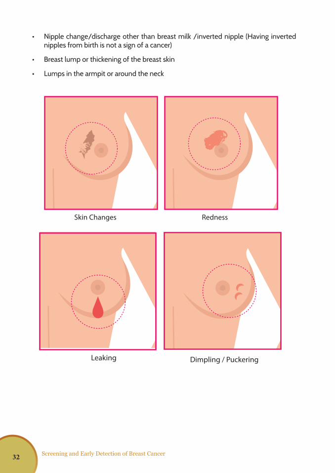
Changes to look out for
Appearance: Any change in the outline or shape of the breast, especially those caused by arm movements or by lifting the breasts or recent change in breast size. Any puckering or dimpling of the skin.
Feeling: Discomfort or pain in one breast that is different from normal, particularly if new and persistent.
Lumps: Any lumps, thickening or bumpy areas in one breast or armpit which seem to be different from the same part of the other breast and armpit. This is very important if new.
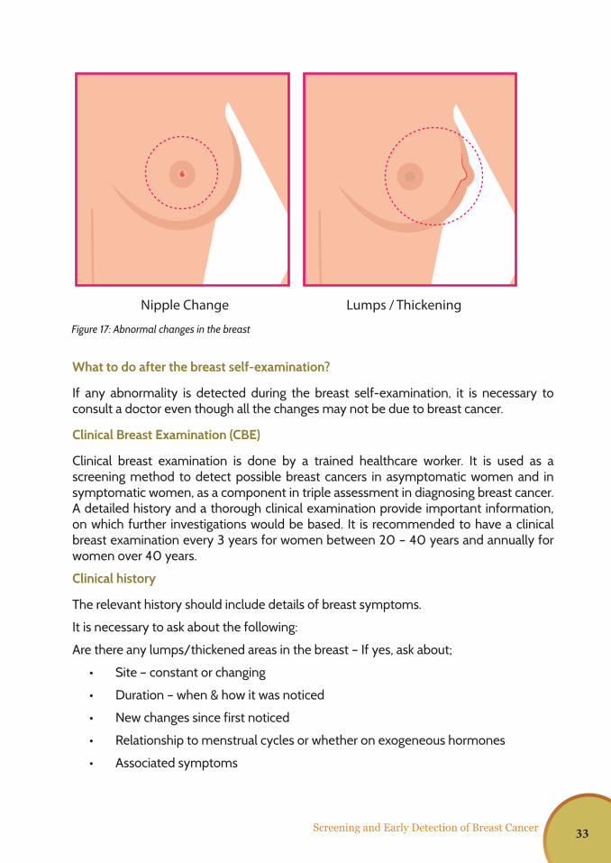
Nipple change: Nipple discharge, new and not milky. Bleeding or moist reddish areas which don’t heal easily. Any change in nipple position – pulled in or pointing differently. A nipple rash on or around the nipple.
Self-breast examination (SBE)
Self-breast examination is the inspection and palpation of the breasts on a monthly basis by the woman herself, and it is important for early detection of breast cancers.
It includes visual inspection and careful feeling of the breasts, the armpits and the areas around the collarbone (Clavicle), looking for lumps or abnormalities around the breast. It is recommended to do a self-breast examination once every month starting at the age of 20 years.
The following information should be provided to women >20 years when conducting health education activities.
When to carry out SBE ?
Self-breast examination should be conducted monthly. For women >20 years, it is recommended to conduct it one week after the start of menstruation, as during menstruation some women feel pain and lumpiness of breasts. In non-menstruating women, SBE should be done on a fixed date of each month.
Why is it important ?
If breast cancer is detected early, with early treatment it gives the best outcome. The practice of breast self-examination on a monthly basis is important for early detection of breast cancers.
Steps in Self breast examination
Place and postures to conduct SBE
A woman can use any place that suits her. It can be conducted in a lying down, sitting or a standing position or while bathing.
SBE has two components:1. Inspection2. Palpation
29

Screening and Early Detection of Breast Cancer
Inspection
Stand in front of the mirror exposing the chest up to the waist. Look at the breasts through the mirror while keeping the arms in positions shown in figure 13 (1. arms hanging by the side, 2. hands pressed on the waist, 3. arms lifted above the head)
The breasts should be inspected from the front and from the sides.
Pay particular attention to:
• Breast size, contour, shape, symmetry
• Skin changes such as erythema, dimpling, tethering or puckering, Peau d’ orange, eczematous skin changes, visible lumps
• Nipple – position, height, any inversion, retraction, erythema, eczema, nodules, ulceration and discharge
Palpate the breast using the palmar surface of the middle three fingers to identify thickened areas and or lumps.
On examining the right breast, raise the right arm over the head and palpate the right breast using the left hand. To palpate the right breast, keep the right palm beneath the head and palpate the breast using the left hand. Apply vice versa for the other.
Figure 14: Breast inspection in arms hanging by the side, hands pressed on the waist & arms lifted above the head positions
Figure 13: Inspection of the breast in standing position, sitting position & lying down position
FIGURE 13
FIGURE 14
FIGURE 21
Step 1 Step 2 Step 3
Standing Position Sitting Position Lying down Position
Arms hanging by the side Hands pressed on the waist Arms lifted above the head
30

Screening and Early Detection of Breast Cancer
Continue palpating the breast in a clockwise direction from outer circle of the breast towards the nipple using three pressure levels (superficial, intermediate and deep). Then examine the armpit and look for lumps. Check whether there is a nipple discharge by squeezing the areola using the thumb and the middle finger. Use the same technique to examine the other breast.
Palpation (in lying down position)
To palpate the right breast, keep the right palm beneath the head and palpate the breast using the left hand. Use the same technique to palpate the left breast.
Note changes mentioned below during inspection & palpation
• Skin changes of the breast
• Color changes of the breast
• Change in shape of the breast
• Orange peel / Peau d’orange appearance of breast
• Ulceration on the breast
• Late occurrence of breast asymmetry (usually both breasts are not of equal size. Therefore, a long- standing breast asymmetry is not a sign of a cancer)
Figure 15: Palpation of the breast
FIGURE 13
FIGURE 14
FIGURE 21
Step 1 Step 2 Step 3
FIGURE 22
FIGURE 17
FIGURE 18
Figure 16: Palpation of the breast in superficial, intermediate and deep pressure levels
Superficial Intermediate Deep
31
Screening and Early Detection of Breast Cancer
• Nipple change/discharge other than breast milk /inverted nipple (Having inverted nipples from birth is not a sign of a cancer)
• Breast lump or thickening of the breast skin
• Lumps in the armpit or around the neck
FIGURE 19
FIGURE 20
Skin Changes Redness Leaking
Nipple ChangeDimpling / Puckering Lumps / Thickening
FIGURE 19
FIGURE 20
Skin Changes Redness Leaking
Nipple ChangeDimpling / Puckering Lumps / Thickening
FIGURE 19
FIGURE 20
Skin Changes Redness Leaking
Nipple ChangeDimpling / Puckering Lumps / Thickening
32
Screening and Early Detection of Breast Cancer
What to do after the breast self-examination?
If any abnormality is detected during the breast self-examination, it is necessary to consult a doctor even though all the changes may not be due to breast cancer.
Clinical Breast Examination (CBE)
Clinical breast examination is done by a trained healthcare worker. It is used as a screening method to detect possible breast cancers in asymptomatic women and in symptomatic women, as a component in triple assessment in diagnosing breast cancer. A detailed history and a thorough clinical examination provide important information, on which further investigations would be based. It is recommended to have a clinical breast examination every 3 years for women between 20 – 40 years and annually for women over 40 years.
Clinical history
The relevant history should include details of breast symptoms.
It is necessary to ask about the following:
Are there any lumps/thickened areas in the breast – If yes, ask about;
• Site – constant or changing
• Duration – when & how it was noticed
• New changes since first noticed
• Relationship to menstrual cycles or whether on exogeneous hormones
• Associated symptoms
FIGURE 19
FIGURE 20
Skin Changes Redness Leaking
Nipple ChangeDimpling / Puckering Lumps / Thickening
Figure 17: Abnormal changes in the breast
33
Screening and Early Detection of Breast Cancer
Whether there is pain in the breast
• Site
• Characteristic of pain: constant or changing /unilateral or bilateral
• Duration
• Recent changes in pain such as intensity, frequency, site of pain
• Relationship to menstrual cycles or whether on exogeneous hormones
• Associated symptoms
Nipple discharge or any other nipple changes
• Duration – when and how they were first noted (spontaneous or not)
• Bilateral or unilateral
• From single duct or multi duct
• Risk factors – history should be taken on the risk factors
• Previous history of any pathological condition in either breast:
• Previous breast investigations:
o Most recent imaging if available (screening or diagnostic) - date and results
o Biopsy results – FNAC / Histology / Lumpectomy
Steps of clinical breast examination (CBE)
Clinical breast examination should be done in a covered room with good light. A female chaperone should be present if the examiner is a male. Before starting the examination, it is necessary to explain the procedure to the woman.
Inspection
Breasts should be inspected in each of the following positions 1. Arms relaxed at her sides 2. Hands placed on the hips and pushing inward (contraction of the Pectoralis Major muscle) 3. Arms raised over her head
The breasts should be inspected from the front and from each side. Pay particular attention to:
• Breast size, contour, shape, symmetry
• Skin changes such as erythema, dimpling, tethering or puckering, Peau d’ orange, eczematous skin changes, visible lumps
• Nipple – position, height, inversion, retraction, erythema, eczema, nodules ulceration and discharge
Palpation during CBE
The ability to identify breast cancers by palpation is influenced by the characteristics of the tumour, the surrounding breast tissue, the position of the lesion in the breast, proper
34
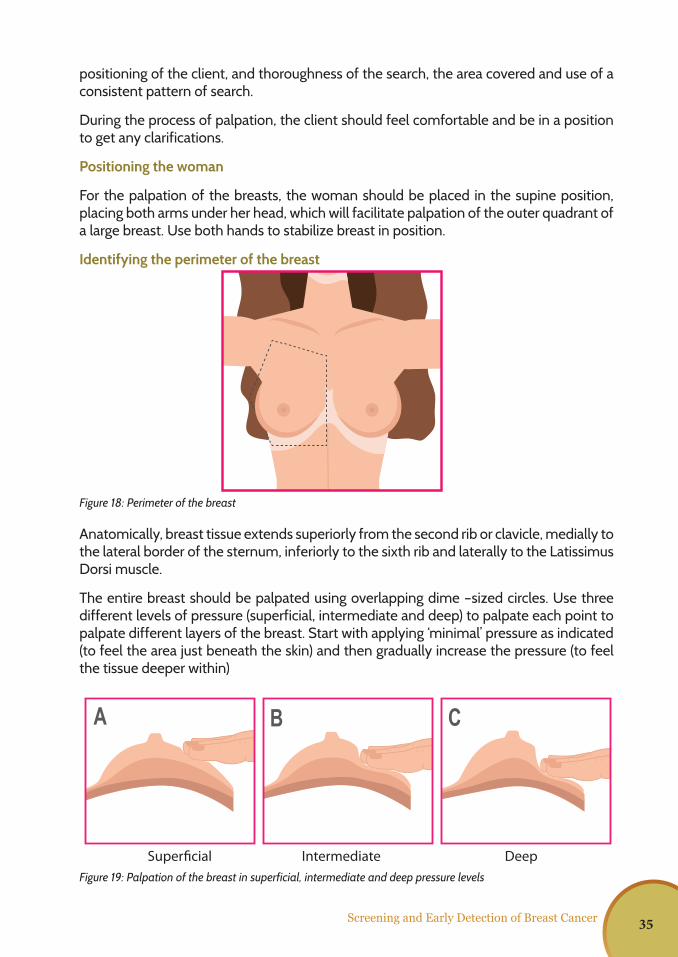
positioning of the client, and thoroughness of the search, the area covered and use of a consistent pattern of search.
During the process of palpation, the client should feel comfortable and be in a position to get any clarifications.
Positioning the woman
For the palpation of the breasts, the woman should be placed in the supine position, placing both arms under her head, which will facilitate palpation of the outer quadrant of a large breast. Use both hands to stabilize breast in position.
Identifying the perimeter of the breast
Anatomically, breast tissue extends superiorly from the second rib or clavicle, medially to the lateral border of the sternum, inferiorly to the sixth rib and laterally to the Latissimus Dorsi muscle.
The entire breast should be palpated using overlapping dime –sized circles. Use three different levels of pressure (superficial, intermediate and deep) to palpate each point to palpate different layers of the breast. Start with applying ‘minimal’ pressure as indicated (to feel the area just beneath the skin) and then gradually increase the pressure (to feel the tissue deeper within)
Screening and Early Detection of Breast Cancer
Figure 18: Perimeter of the breast
FIGURE 13
FIGURE 14
FIGURE 21
Step 1 Step 2 Step 3
FIGURE 22
FIGURE 17
FIGURE 18
Figure 19: Palpation of the breast in superficial, intermediate and deep pressure levels
Superficial Intermediate Deep
35
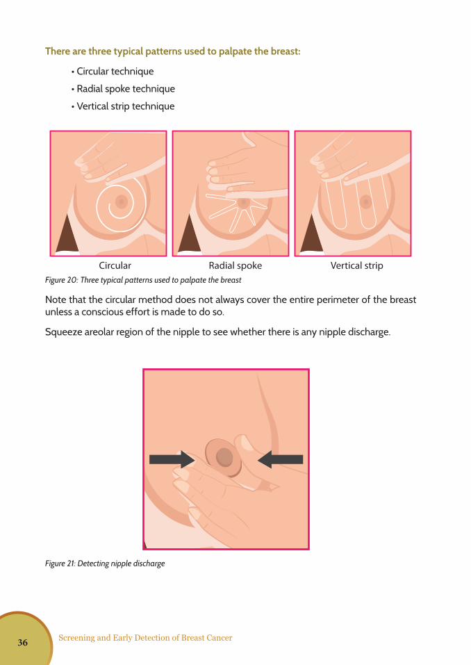
Screening and Early Detection of Breast Cancer
There are three typical patterns used to palpate the breast:
• Circular technique
• Radial spoke technique
• Vertical strip technique
Note that the circular method does not always cover the entire perimeter of the breast unless a conscious effort is made to do so.
Squeeze areolar region of the nipple to see whether there is any nipple discharge.
FIGURE 22
FIGURE 17
FIGURE 18Figure 20: Three typical patterns used to palpate the breast
FIGURE 22
FIGURE 17
FIGURE 18
Figure 21: Detecting nipple discharge
Circular Radial spoke Vertical strip
36
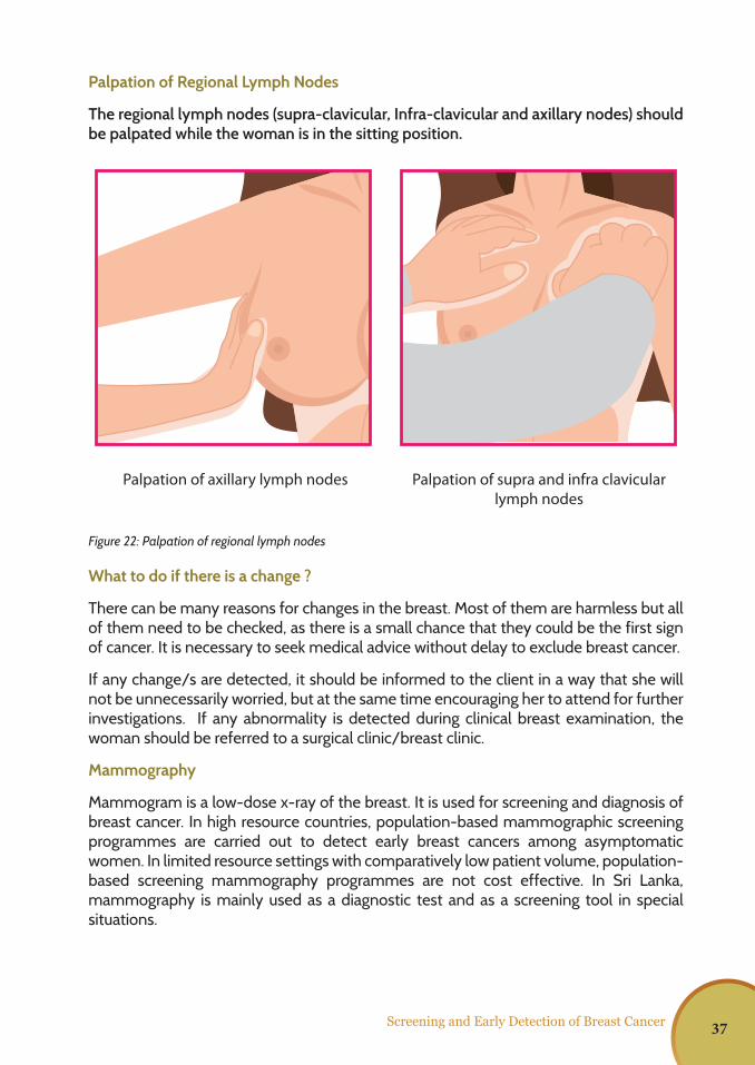
Screening and Early Detection of Breast Cancer
Palpation of Regional Lymph Nodes
The regional lymph nodes (supra-clavicular, Infra-clavicular and axillary nodes) should be palpated while the woman is in the sitting position.
What to do if there is a change ?
There can be many reasons for changes in the breast. Most of them are harmless but all of them need to be checked, as there is a small chance that they could be the first sign of cancer. It is necessary to seek medical advice without delay to exclude breast cancer.
If any change/s are detected, it should be informed to the client in a way that she will not be unnecessarily worried, but at the same time encouraging her to attend for further investigations. If any abnormality is detected during clinical breast examination, the woman should be referred to a surgical clinic/breast clinic.
Mammography
Mammogram is a low-dose x-ray of the breast. It is used for screening and diagnosis of breast cancer. In high resource countries, population-based mammographic screening programmes are carried out to detect early breast cancers among asymptomatic women. In limited resource settings with comparatively low patient volume, population-based screening mammography programmes are not cost effective. In Sri Lanka, mammography is mainly used as a diagnostic test and as a screening tool in special situations.
FIGURE 19
FIGURE 20
Skin Changes Redness Leaking
Nipple ChangeDimpling / Puckering Lumps / Thickening
Palpation of axillary lymph nodes Palpation of supra and infra clavicular lymph nodes
Figure 22: Palpation of regional lymph nodes
37

Screening and Early Detection of Breast Cancer 38
Dr. Rubecca Dissanayake Consultant Surgeon, Base Hospital, Balangoda
Chapter05
Diagnosis of Breast Cancer - Triple Assessment
39
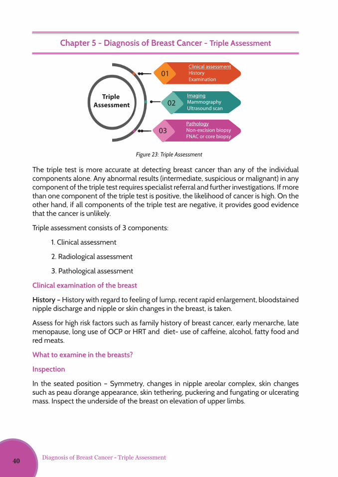
Clinical assessmentHistoryExamination
ImagingMammographyUltrasound scan
PathologyNon-excision biopsyFNAC or core biopsy
01
02
03
Triple Assessment
Diagnosis of Breast Cancer - Triple Assessment
Chapter 5 - Diagnosis of Breast Cancer - Triple Assessment
The triple test is more accurate at detecting breast cancer than any of the individual components alone. Any abnormal results (intermediate, suspicious or malignant) in any component of the triple test requires specialist referral and further investigations. If more than one component of the triple test is positive, the likelihood of cancer is high. On the other hand, if all components of the triple test are negative, it provides good evidence that the cancer is unlikely.
Triple assessment consists of 3 components:
1. Clinical assessment
2. Radiological assessment
3. Pathological assessment
Clinical examination of the breast
History – History with regard to feeling of lump, recent rapid enlargement, bloodstained nipple discharge and nipple or skin changes in the breast, is taken.
Assess for high risk factors such as family history of breast cancer, early menarche, late menopause, long use of OCP or HRT and diet- use of caffeine, alcohol, fatty food and red meats.
What to examine in the breasts?
Inspection
In the seated position – Symmetry, changes in nipple areolar complex, skin changes such as peau d’orange appearance, skin tethering, puckering and fungating or ulcerating mass. Inspect the underside of the breast on elevation of upper limbs.
Figure 23: Triple Assessment
40
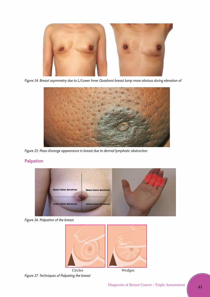
Diagnosis of Breast Cancer - Triple Assessment
3
Assess for high risk factors such as family history of breast cancer, early menarche, late
menopause, long use of OCP or HRT and diet- use of caffeine, alcohol, fatty food and
red meats.
What to examine in the breasts?
INSPECTION In the seated position – Symmetry, changes in nipple areolar complex, skin changes
such as peau d'orange appearance, skin tethering, puckering and fungating or
ulcerating mass. Inspect the underside of the breast on elevation of upper limbs.
Figure 1: Breast asymmetry due to L/LIQ breast lump more obvious during elevation of upper limbs
Figure 2: Peau d'orangeappearance in breast due to dermal lymphatic obstruction
Figure 24: Breast asymmetry due to L/Lower Inner Quadrant breast lump more obvious during elevation of
Figure 25: Peau d’orange appearance in breast due to dermal lymphatic obstruction
Figure 26: Palpation of the breast
Figure 27: Techniques of Palpating the breast
Palpation
4
Palpation
Palpation is performed in the lying down position –quadrant wise/ circular from
periphery to central, for lumps. Lumps should be examined for site, size, shape,
surface, consistency and skin or muscle attachment. or in any systematic order, with
the palmar surface of the fingers.
Complete the palpation in the seated up position to examine for any inframammary
lesions and to complete axillae examination.
Examine axillary lymph nodes (LN) – anterior, posterior, apical, central and lateral
groups. Look for margins, consistency and fixity. If indicated, examine supraclavicular
LNs
Figure 3:Palpation of the breast
Figure 4:
Circles Wedges
FIGURE 22
FIGURE 17
FIGURE 18
41

Palpation is performed in the lying down position – quadrant wise/ circular from periphery to central, for lumps. Lumps should be examined for site, size, shape, surface, consistency and skin or muscle attachment or in any systematic order, with the palmar surface of the fingers.
Complete the palpation in the seated up position to examine for any inframammary lesions and to complete axillae examination.
Examine axillary lymph nodes (LN) – anterior, posterior, apical, central and lateral groups. Look for margins, consistency and fixity. If indicated, examine supraclavicular LNs.
If the lump is obviously an advanced malignancy –examine for jaundice, hepatomegaly, lung signs and spinal tenderness.
Depending on the findings, we can categorize the clinical examination to 5 categories from P1-5.
P1 –No abnormal findings in the breast
P2- Benign findings
P3- Probably benign findings
P4- Findings are suspicious of malignancy
P5- Findings are of definite malignancy
Radiological assessment of the breast
Mammogram and Ultra-Sound (US) are the commonest investigations used in breast lumps. All females below 40 years coming with a breast lump should undergo US of the breasts and axillae.
All females 40 years or above should undergo both bilateral mammograms including CC (cranio-caudal) view and MLO (Medio Lateral Oblique) view and US of the breasts and axillae.
In pregnancy, mammogram can be done with proper shielding.
Mammogram
Indications for Mammogram
1. Breast cancer screening
2. Assessment of patients (usually >40 years) with clinical symptoms
3. Image guidance for biopsy
4. Preoperative staging
5. Preoperative localization
6. Therapy monitoring and follow-up
Diagnosis of Breast Cancer - Triple Assessment42
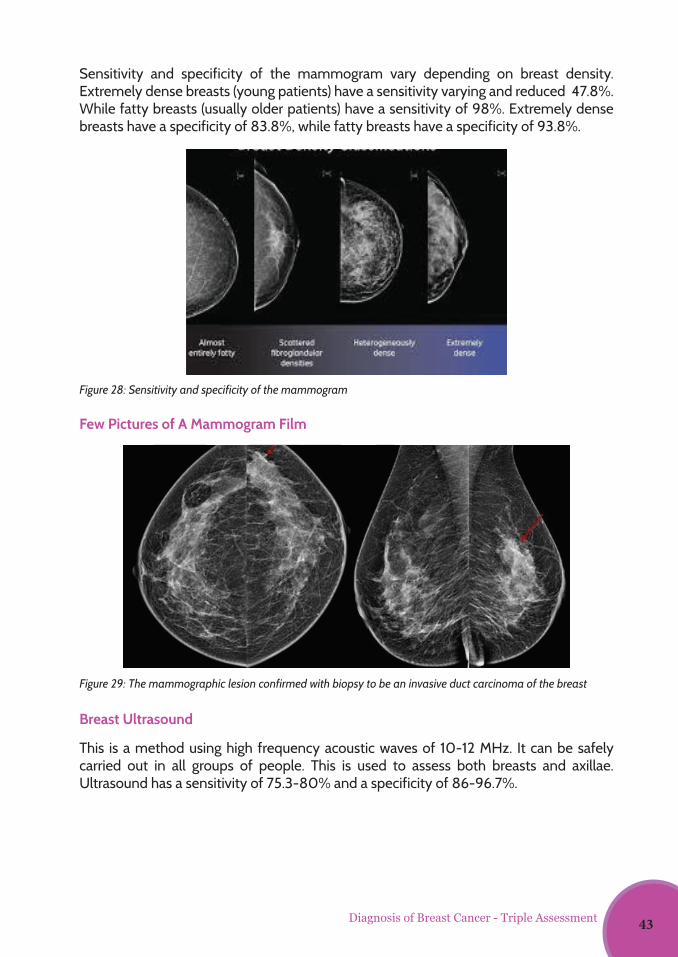
Sensitivity and specificity of the mammogram vary depending on breast density. Extremely dense breasts (young patients) have a sensitivity varying and reduced 47.8%. While fatty breasts (usually older patients) have a sensitivity of 98%. Extremely dense breasts have a specificity of 83.8%, while fatty breasts have a specificity of 93.8%.
Few Pictures of A Mammogram Film
Breast Ultrasound
This is a method using high frequency acoustic waves of 10-12 MHz. It can be safely carried out in all groups of people. This is used to assess both breasts and axillae.Ultrasound has a sensitivity of 75.3-80% and a specificity of 86-96.7%.
6
Sensitivity and specificity of the mammogram vary depending on breast density.
Extremely dense breasts (young patients) have a sensitivity varying and reduced 47.8%
while fatty breasts (usually older patients) have a sensitivity of 98%. Extremely dense
breasts have a specificity of 83.8%, while fatty breasts have a specificity of 93.8%.
Figure 5:
FEW PICTURES OF A MAMMOGRAM FILMS
Figure 6: The mammographic lesion confirmed with biopsy to be an invasive duct carcinoma of the breast
Figure 28: Sensitivity and specificity of the mammogram
Figure 29: The mammographic lesion confirmed with biopsy to be an invasive duct carcinoma of the breast
Diagnosis of Breast Cancer - Triple Assessment 43
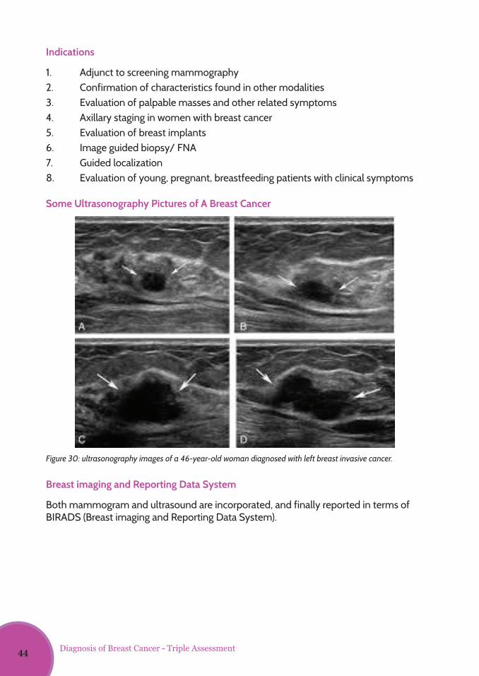
Indications
1. Adjunct to screening mammography2. Confirmation of characteristics found in other modalities3. Evaluation of palpable masses and other related symptoms4. Axillary staging in women with breast cancer5. Evaluation of breast implants6. Image guided biopsy/ FNA7. Guided localization8. Evaluation of young, pregnant, breastfeeding patients with clinical symptoms
Some Ultrasonography Pictures of A Breast Cancer
Breast imaging and Reporting Data System
Both mammogram and ultrasound are incorporated, and finally reported in terms of BIRADS (Breast imaging and Reporting Data System).
Figure 30: ultrasonography images of a 46-year-old woman diagnosed with left breast invasive cancer.
7
Breast Ultrasound This is a method using high frequency acoustic waves of 10-12 MHz. It can be safely
carried out in all groups of people. This is used to assess both breasts and axillae.
Ultrasound has a sensitivity of 75.3-80% and a specificity of 86-96.7%.
Indications
1. Adjunct to screening mammography 2. Confirmation of characteristics found in other modalities 3. Evaluation of palpable masses and other related symptoms 4. Axillary staging in women with breast cancer 5. Evaluation of breast implants 6. Image guided biopsy/ FNA 7. Guided localization 8. Evaluation of young, pregnant, breastfeeding patients with clinical
symptoms SOME ULTRASONOGRAPHY PICTURES OF A BREAST CANCER
Figure 7: ultrasonography images of a 46-year-old woman diagnosed with left breast invasive cancer.
Diagnosis of Breast Cancer - Triple Assessment44
BIRADS Final Assessment Categories
Category Management Likelihood of cancer
0Need additional imaging or prior examinations
Recall for additional imaging and/or await prior examinations
n/a
1 Negative Routine screening Essentially 0%
2 Benign Routine screening Essentially 0%
3 Probably Benign Short interval follow-up (6 month) or continued >0 % but ≤ 2%
4 Suspicious Tissue diagnosis
4a. low suspicion for malignancy (>2% to≤ 10%)
4b. moderate suspicion for malignancy (>10% to ≤ 50%)
4c. high suspicion for malignancy (>50% to <95%)
5 Highly suggestive of malignancy
Tissue diagnosis ≥95%
6 Known biopsy-proven
Surgical excision when clinically appropriate n/a
MRI breasts
Magnetic resonance imaging (MRI) is a non-radiating imaging modality with high sensitivity (75-99%). However, its specificity is between 78-97.5%. MRI is costlier and not readily available in most centers in the country. MRI is recognised as a screening tool either as a standalone or as an adjunct to mammography for screening of women at risk of breast cancer as determined by the Radiologist.
The following are recommended indications for breast MRI, which are supported by the published literature (NICE 2009, EUSOMA 2010):
Discordant conventional imaging - Conventional triple assessment includes mammography and breast ultrasound. If imaging does not correlate with clinical assessment or with pathology, MRI may act as an additional problem-solving tool in assessing tumour characteristics such as size, extent or multifocality.
Pre-operative staging MRI of both breasts - This is not routinely used for pre-operative staging, but in selected patients (e.g. when conventional imaging may underestimate the extent of disease, such as invasive lobular carcinoma, high grade DCIS or when there is significant discordance between the imaging and clinical findings), MRI may be useful in further assessing the tumour size, multicentricity and multifocality and to evaluate the contra-lateral breast for occult disease.
Occult primary tumour/malignant axillary nodes- The primary tumour may be occult on conventional imaging. Less than 1% of breast cancers present with involved axillary nodes but with normal conventional imaging.
Diagnosis of Breast Cancer - Triple Assessment
Table 7: Breast Imaging and Reporting Data System (BIRADS)
45
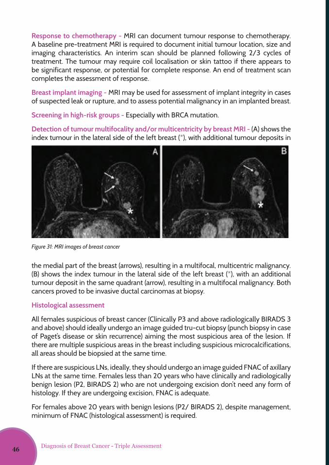
Response to chemotherapy - MRI can document tumour response to chemotherapy. A baseline pre-treatment MRI is required to document initial tumour location, size and imaging characteristics. An interim scan should be planned following 2/3 cycles of treatment. The tumour may require coil localisation or skin tattoo if there appears to be significant response, or potential for complete response. An end of treatment scan completes the assessment of response.
Breast implant imaging - MRI may be used for assessment of implant integrity in cases of suspected leak or rupture, and to assess potential malignancy in an implanted breast.
Screening in high-risk groups - Especially with BRCA mutation.
Detection of tumour multifocality and/or multicentricity by breast MRI - (A) shows the index tumour in the lateral side of the left breast (*), with additional tumour deposits in
the medial part of the breast (arrows), resulting in a multifocal, multicentric malignancy. (B) shows the index tumour in the lateral side of the left breast (*), with an additional tumour deposit in the same quadrant (arrow), resulting in a multifocal malignancy. Both cancers proved to be invasive ductal carcinomas at biopsy.
Histological assessment
All females suspicious of breast cancer (Clinically P3 and above radiologically BIRADS 3 and above) should ideally undergo an image guided tru-cut biopsy (punch biopsy in case of Paget’s disease or skin recurrence) aiming the most suspicious area of the lesion. If there are multiple suspicious areas in the breast including suspicious microcalcifications, all areas should be biopsied at the same time.
If there are suspicious LNs, ideally, they should undergo an image guided FNAC of axillary LNs at the same time. Females less than 20 years who have clinically and radiologically benign lesion (P2, BIRADS 2) who are not undergoing excision don’t need any form of histology. If they are undergoing excision, FNAC is adequate.
For females above 20 years with benign lesions (P2/ BIRADS 2), despite management, minimum of FNAC (histological assessment) is required.
Figure 31: MRI images of breast cancer
10
Breast implant imaging - MRI may be used for assessment of implant integrity in cases
of suspected leak or rupture, and to assess potential malignancy in an implanted
breast.
Screening in high-risk groups especially with BRCA mutation.
Figure 8: MRI images of breast cancer
Detection of tumour multifocality and/or multicentricity by breast MRI. - (A) shows
the index tumour in the lateral side of the left breast (*), with additional tumour
deposits in the medial part of the breast (arrows), resulting in a multifocal, multicentric
malignancy. (B) shows the index tumour in the lateral side of the left breast (*), with
an additional tumour deposit in the same quadrant (arrow), resulting in a multifocal
malignancy. Both cancers proved to be invasive ductal carcinomas at biopsy.
Histological assessment
All females suspicious of breast cancer (Clinically P3 and above radiologically BIRADS 3
and above) should ideally undergo an image guided tru-cut biopsy (punch biopsy in
case of Paget’s disease or skin recurrence) aiming the most suspicious area of the
lesion. If there are multiple suspicious areas in the breast including suspicious
microcalcifications, all areas should be biopsied at the same time.
10
Breast implant imaging - MRI may be used for assessment of implant integrity in cases
of suspected leak or rupture, and to assess potential malignancy in an implanted
breast.
Screening in high-risk groups especially with BRCA mutation.
Figure 8: MRI images of breast cancer
Detection of tumour multifocality and/or multicentricity by breast MRI. - (A) shows
the index tumour in the lateral side of the left breast (*), with additional tumour
deposits in the medial part of the breast (arrows), resulting in a multifocal, multicentric
malignancy. (B) shows the index tumour in the lateral side of the left breast (*), with
an additional tumour deposit in the same quadrant (arrow), resulting in a multifocal
malignancy. Both cancers proved to be invasive ductal carcinomas at biopsy.
Histological assessment
All females suspicious of breast cancer (Clinically P3 and above radiologically BIRADS 3
and above) should ideally undergo an image guided tru-cut biopsy (punch biopsy in
case of Paget’s disease or skin recurrence) aiming the most suspicious area of the
lesion. If there are multiple suspicious areas in the breast including suspicious
microcalcifications, all areas should be biopsied at the same time. Diagnosis of Breast Cancer - Triple Assessment46
Dr. Rupa Kannangara Consultant Radiologist, National Cancer Institute, Maharagama
Dr. Udari Liyanage Consultant Radiologist & Senior Lecturer, Faculty of Medicine University of Colombo
Dr. Rubecca Dissanayake Consultant Surgeon, Base Hospital, Balangoda
Chapter06
Modern Diagnostics of Breast Cancer
47
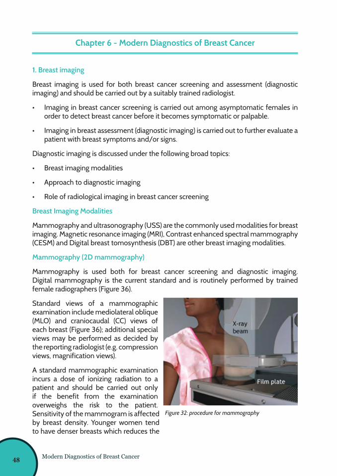
Modern Diagnostics of Breast Cancer
Chapter 6 - Modern Diagnostics of Breast Cancer
1. Breast imaging
Breast imaging is used for both breast cancer screening and assessment (diagnostic imaging) and should be carried out by a suitably trained radiologist.
• Imaging in breast cancer screening is carried out among asymptomatic females in order to detect breast cancer before it becomes symptomatic or palpable.
• Imaging in breast assessment (diagnostic imaging) is carried out to further evaluate a patient with breast symptoms and/or signs.
Diagnostic imaging is discussed under the following broad topics:
• Breast imaging modalities
• Approach to diagnostic imaging
• Role of radiological imaging in breast cancer screening
Breast Imaging Modalities
Mammography and ultrasonography (USS) are the commonly used modalities for breast imaging. Magnetic resonance imaging (MRI), Contrast enhanced spectral mammography (CESM) and Digital breast tomosynthesis (DBT) are other breast imaging modalities.
Mammography (2D mammography)
Mammography is used both for breast cancer screening and diagnostic imaging. Digital mammography is the current standard and is routinely performed by trained female radiographers (Figure 36).
Standard views of a mammographic examination include mediolateral oblique (MLO) and craniocaudal (CC) views of each breast (Figure 36); additional special views may be performed as decided by the reporting radiologist (e.g. compression views, magnification views).
A standard mammographic examination incurs a dose of ionizing radiation to a patient and should be carried out only if the benefit from the examination overweighs the risk to the patient. Sensitivity of the mammogram is affected by breast density. Younger women tend to have denser breasts which reduces the
Figure 32: procedure for mammography
48
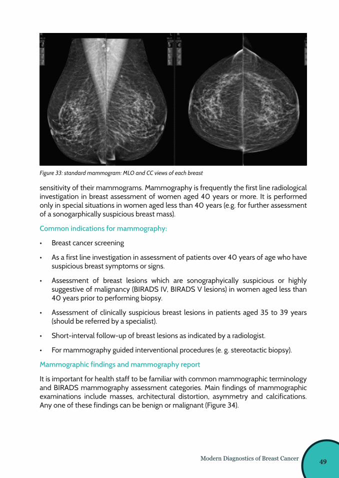
Modern Diagnostics of Breast Cancer
Figure 33: standard mammogram: MLO and CC views of each breast
sensitivity of their mammograms. Mammography is frequently the first line radiological investigation in breast assessment of women aged 40 years or more. It is performed only in special situations in women aged less than 40 years (e.g. for further assessment of a sonogarphically suspicious breast mass).
Common indications for mammography:
• Breast cancer screening
• As a first line investigation in assessment of patients over 40 years of age who have suspicious breast symptoms or signs.
• Assessment of breast lesions which are sonographyically suspicious or highly suggestive of malignancy (BIRADS IV, BIRADS V lesions) in women aged less than 40 years prior to performing biopsy.
• Assessment of clinically suspicious breast lesions in patients aged 35 to 39 years (should be referred by a specialist).
• Short-interval follow-up of breast lesions as indicated by a radiologist.
• For mammography guided interventional procedures (e. g. stereotactic biopsy).
Mammographic findings and mammography report
It is important for health staff to be familiar with common mammographic terminology and BIRADS mammography assessment categories. Main findings of mammographic examinations include masses, architectural distortion, asymmetry and calcifications. Any one of these findings can be benign or malignant (Figure 34).
49
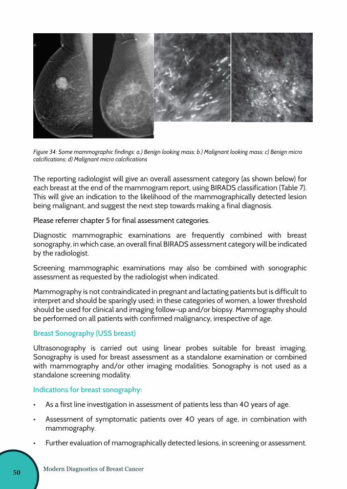
Figure 34: Some mammographic findings: a.) Benign looking mass; b.) Malignant looking mass; c) Benign micro calcifications; d) Malignant micro calcifications
The reporting radiologist will give an overall assessment category (as shown below) for each breast at the end of the mammogram report, using BIRADS classification (Table 7). This will give an indication to the likelihood of the mammographically detected lesion being malignant, and suggest the next step towards making a final diagnosis.
Please referrer chapter 5 for final assessment categories.
Diagnostic mammographic examinations are frequently combined with breast sonography, in which case, an overall final BIRADS assessment category will be indicated by the radiologist.
Screening mammographic examinations may also be combined with sonographic assessment as requested by the radiologist when indicated.
Mammography is not contraindicated in pregnant and lactating patients but is difficult to interpret and should be sparingly used; in these categories of women, a lower threshold should be used for clinical and imaging follow-up and/or biopsy. Mammography should be performed on all patients with confirmed malignancy, irrespective of age.
Breast Sonography (USS breast)
Ultrasonography is carried out using linear probes suitable for breast imaging. Sonography is used for breast assessment as a standalone examination or combined with mammography and/or other imaging modalities. Sonography is not used as a standalone screening modality.
Indications for breast sonography:
• As a first line investigation in assessment of patients less than 40 years of age.
• Assessment of symptomatic patients over 40 years of age, in combination with mammography.
• Further evaluation of mamographically detected lesions, in screening or assessment.
Modern Diagnostics of Breast Cancer50
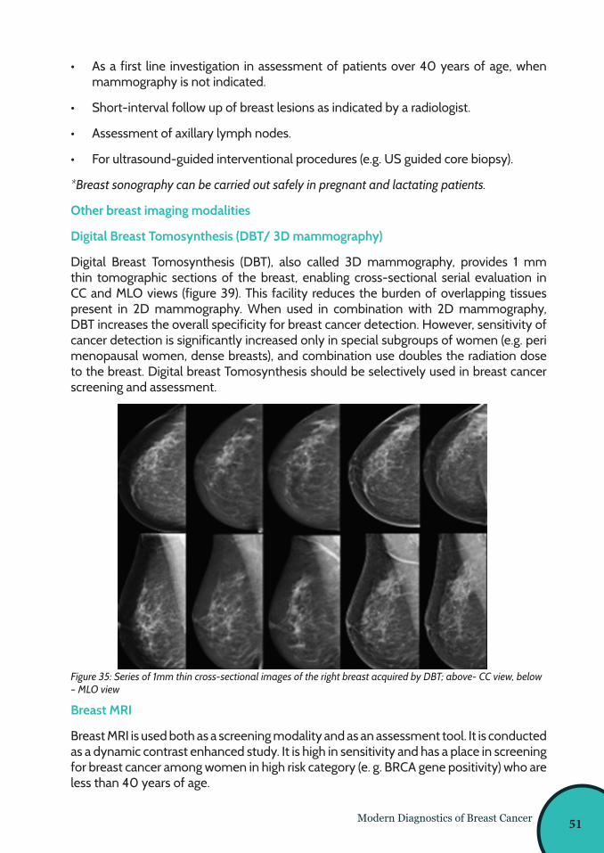
• As a first line investigation in assessment of patients over 40 years of age, when mammography is not indicated.
• Short-interval follow up of breast lesions as indicated by a radiologist.
• Assessment of axillary lymph nodes.
• For ultrasound-guided interventional procedures (e.g. US guided core biopsy).
*Breast sonography can be carried out safely in pregnant and lactating patients.
Other breast imaging modalities
Digital Breast Tomosynthesis (DBT/ 3D mammography)
Digital Breast Tomosynthesis (DBT), also called 3D mammography, provides 1 mm thin tomographic sections of the breast, enabling cross-sectional serial evaluation in CC and MLO views (figure 39). This facility reduces the burden of overlapping tissues present in 2D mammography. When used in combination with 2D mammography, DBT increases the overall specificity for breast cancer detection. However, sensitivity of cancer detection is significantly increased only in special subgroups of women (e.g. peri menopausal women, dense breasts), and combination use doubles the radiation dose to the breast. Digital breast Tomosynthesis should be selectively used in breast cancer screening and assessment.
Breast MRI
Breast MRI is used both as a screening modality and as an assessment tool. It is conducted as a dynamic contrast enhanced study. It is high in sensitivity and has a place in screening for breast cancer among women in high risk category (e. g. BRCA gene positivity) who are less than 40 years of age.
Figure 35: Series of 1mm thin cross-sectional images of the right breast acquired by DBT; above- CC view, below – MLO view
Modern Diagnostics of Breast Cancer 51
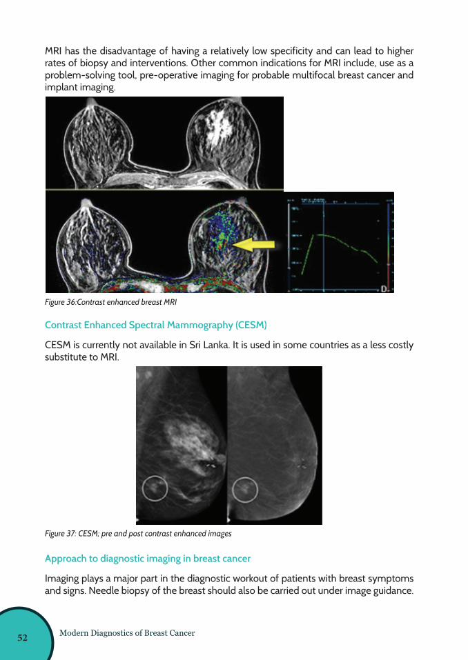
MRI has the disadvantage of having a relatively low specificity and can lead to higher rates of biopsy and interventions. Other common indications for MRI include, use as a problem-solving tool, pre-operative imaging for probable multifocal breast cancer and implant imaging.
Contrast Enhanced Spectral Mammography (CESM)
CESM is currently not available in Sri Lanka. It is used in some countries as a less costly substitute to MRI.
Approach to diagnostic imaging in breast cancer
Imaging plays a major part in the diagnostic workout of patients with breast symptoms and signs. Needle biopsy of the breast should also be carried out under image guidance.
Figure 36:Contrast enhanced breast MRI
Figure 37: CESM; pre and post contrast enhanced images
Modern Diagnostics of Breast Cancer52
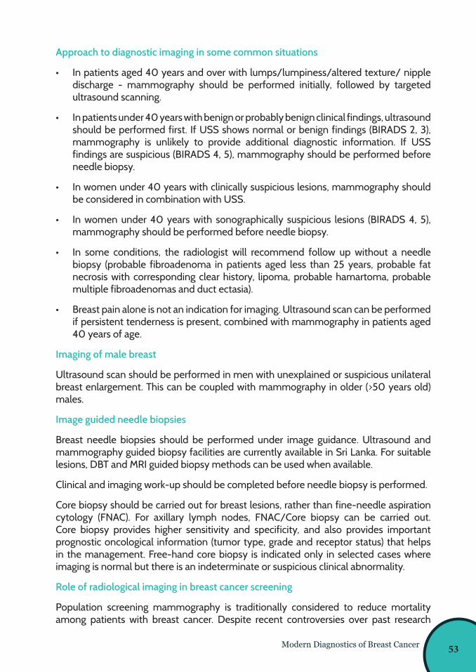
Modern Diagnostics of Breast Cancer
Approach to diagnostic imaging in some common situations
• In patients aged 40 years and over with lumps/lumpiness/altered texture/ nipple discharge - mammography should be performed initially, followed by targeted ultrasound scanning.
• In patients under 40 years with benign or probably benign clinical findings, ultrasound should be performed first. If USS shows normal or benign findings (BIRADS 2, 3), mammography is unlikely to provide additional diagnostic information. If USS findings are suspicious (BIRADS 4, 5), mammography should be performed before needle biopsy.
• In women under 40 years with clinically suspicious lesions, mammography should be considered in combination with USS.
• In women under 40 years with sonographically suspicious lesions (BIRADS 4, 5), mammography should be performed before needle biopsy.
• In some conditions, the radiologist will recommend follow up without a needle biopsy (probable fibroadenoma in patients aged less than 25 years, probable fat necrosis with corresponding clear history, lipoma, probable hamartoma, probable multiple fibroadenomas and duct ectasia).
• Breast pain alone is not an indication for imaging. Ultrasound scan can be performed if persistent tenderness is present, combined with mammography in patients aged 40 years of age.
Imaging of male breast
Ultrasound scan should be performed in men with unexplained or suspicious unilateral breast enlargement. This can be coupled with mammography in older (>50 years old) males.
Image guided needle biopsies
Breast needle biopsies should be performed under image guidance. Ultrasound and mammography guided biopsy facilities are currently available in Sri Lanka. For suitable lesions, DBT and MRI guided biopsy methods can be used when available.
Clinical and imaging work-up should be completed before needle biopsy is performed.
Core biopsy should be carried out for breast lesions, rather than fine-needle aspiration cytology (FNAC). For axillary lymph nodes, FNAC/Core biopsy can be carried out. Core biopsy provides higher sensitivity and specificity, and also provides important prognostic oncological information (tumor type, grade and receptor status) that helps in the management. Free-hand core biopsy is indicated only in selected cases where imaging is normal but there is an indeterminate or suspicious clinical abnormality.
Role of radiological imaging in breast cancer screening
Population screening mammography is traditionally considered to reduce mortality among patients with breast cancer. Despite recent controversies over past research
53
Modern Diagnostics of Breast Cancer
evidence, issues related to over-diagnosis and overtreatment, mammography continues to be the gold standard in breast cancer screening in most developed countries. However, this is not considered cost effective in low and middle income countries. Opportunistic screening mammography services are offered in Sri Lanka under selected circumstances.
2. Genetic testing
Advances in molecular genetics have led to the identification of numerous genes associated with inherited susceptibility to cancer including breast cancers. Inherited genetic alterations (germ line mutations) can be identified by testing DNA.
Cancer panel testing for germ line mutations in Cancer Predisposition Genes (CPGs) is now available in Sri Lanka. Testing is done on DNA extracted from venous blood and the test can identify genes associated with inherited predisposition to cancer, including genes associated with both common and rare cancers.
The steps involved in genetic testing:
• Test an affected family member FIRST after providing pre-test counseling and obtaining written informed consent to identify the mutation and confirm it.
• If a mutation is found, then other family members, including those who are not affected, can be tested for that mutation.
• Always provide post-test counseling.
Benefits of cancer gene panel testing:
• Clarify risks of hereditary breast cancer.
• Identify individuals who are at increased risk who could benefit from increased cancer surveillance or measures to decrease risk.
• Identify individuals who may not be at increased risk.
• Simultaneous analysis of multiple CPGs allows rapid diagnosis of specific hereditary breast cancer syndromes.
• Saves money and time than single gene testing for patients suspected of having multiple hereditary cancer syndromes.
• A greater likelihood of identifying a hereditary cause for the cancer(s) in patients and/or their families.
• Reduces false negative results.
• Offers treatment guidance once the mutation is identified.
• Identification of the mutation in the family allows other at-risk healthy family members to be screened with targeted genetic testing to determine their individual risk status.
• Allows for earlier intervention through targeted individualized cancer screening and prevention programs in individuals who are mutation carriers.
• Assists couples in reproductive decision making.
54
Modern Diagnostics of Breast Cancer
Limitations of cancer gene panel testing
The test may miss large structural genetic variations. These are usually very rare.
Who should undergo genetic testing?
Breast cancer, with at least one of the following
01. Diagnosed at age < 45 years
02. Diagnosed at age < 50 years with
• two breast primaries; or
• > 1 close relative with breast, pancreatic or prostate cancer
03. Diagnosed at age < 60 years with triple negative breast cancer
04. Family history of cancer:
• > 1 close relative with breast cancer diagnosed at age < 50 years
• > 1 close relative with ovarian cancer
• > 2 close relatives with breast, pancreatic or prostate cancer; or male breast
cancer relative
05. Male breast cancer
06. Ovarian cancer
07. Pancreatic cancer or prostate cancer with:
• > 1 close relative with ovarian cancer (any age) or breast cancer (age < 50
years); or
• > 2 close relatives with breast, pancreatic or prostate cancer
08. Unaffected individual (family history only):
• 1st-, 2nd-degree relative meeting any of the above criteria;
• 3rd-degree relative with breast/ovarian cancer and has > 2 relatives with
breast/ovarian cancer
Implications of a positive test result:
1. Clinical intervention can improve outcomes e.g. risk reduction mastectomy reduces risk of breast cancer and salpingo-oophorectomy reduces risk of ovarian and breast cancer (in premenopausal women).
2. Family members at risk can be offered testing and identified.3. Healthy lifestyles can be reinforced.
Implications of a negative test result:
Reassures the individual and their family members.
55
Modern Diagnostics of Breast Cancer
Management Guidelines
The options described below are available for managing the increased cancer risk in BRCA 1 and BRCA 2 mutation carriers:
For women:
• Breast awareness starting at age 18 years
• Clinical breast examination, every 6-12 months, starting at age 25 years
• Age 25-29 years, annual breast MRI screening with contrast or mammogram
• Age 30-75 years, annual mammogram and MRI with contrast
• Age >75 years, individual basis
• Consider options of risk-reducing mastectomy and/or salpingo-oophorectomy
• Consider trans-vaginal ultrasound starting at age 30-35 years or CA-125 screening, every 6 months
• Consider risk reduction agents
For men:
• Breast self-examination, starting at age 35 years
• Clinical breast examination, every 12 months, starting at age 35 years
• Prostate cancer screening, starting at age 45 years
• Pancreatic cancer and melanoma screening, based on family history
• Advise about options for prenatal diagnosis and assisted reproduction
56
Dr. Nurad Joseph Consultant Clinical Oncologist, DGH Chilaw
Dr. Sandani Wijerathne Consultant Onco-Surgeon, PGH Badulla
Chapter07
Treatment Modalities for Breast Diseases - Medical & Surgical
57
Treatment Modalities for Breast Diseases - Medical & Surgical
Chapter 7 - Treatment Modalities for Breast Diseases - Medical & Surgical
Treatment for breast cancer is essentially multimodal. Local disease is treated with surgery or radiotherapy or a combination of both. Systemic disease is treated with chemotherapy, endocrine therapy, targeted therapy or combinations. Selection of treatment modalities is based on tumour factors such as histology, staging of the disease and receptor status, and on patient factors such as age, co-morbidities, menopausal status and the patient’s wish.
The primary treatment of early breast cancer is surgery.
Surgery for breast cancer
Surgery for breast cancer can be done either with curative intention or palliative intention.
It includes surgery for the breast and for the axilla.
Surgery for the breast has a range of options, which include:
• Wide local excision i.e. - excision of the tumour with a margin of surrounding normal tissue
• Total mastectomy
Following wide local excision, the defect can be reconstructed with many options. These include simple closure, oncoplastic procedures and local rotational flaps/ pedicled flaps or free flaps.
Wide local excision should always be followed by whole breast irradiation, to reduce the risk of recurrence. The combination of wide local excision and whole breast irradiation is called breast conservation treatment. Nowadays, lots of patients are asking for breast reconstruction following total mastectomy. This is usually being done with either pedicled flaps or free flaps.
Breast conservation surgery (BCS)
Breast conservation surgery offers similar oncological outcomes as in total mastectomy, while giving an acceptable cosmetic appearance and lesser psychological morbidities. This can be usually offered for early tumours and relatively large tumours in larger breasts. It becomes useful in pregnancy where postoperative radiotherapy should be avoided, and in multifocal or multi-centric disease where a cosmetically acceptable outcome cannot be achieved. It is also relatively contraindicated in patients with previous radiotherapy to the chest wall, active connective tissue disorders and with tumours >5cm in size. There are certain situations that breast conservation carries a high risk of recurrence, as in high grade tumours, lobular carcinoma, extensive in situ component, lymphovascular invasion, triple negative tumours, etc.
58
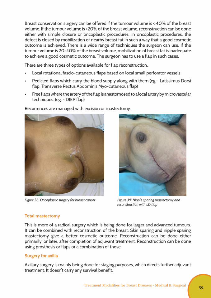
Treatment Modalities for Breast Diseases - Medical & Surgical
Breast conservation surgery can be offered if the tumour volume is < 40% of the breast volume. If the tumour volume is <20% of the breast volume, reconstruction can be done either with simple closure or oncoplastic procedures. In oncoplastic procedures, the defect is closed by mobilization of nearby breast fat in such a way that a good cosmetic outcome is achieved. There is a wide range of techniques the surgeon can use. If the tumour volume is 20-40% of the breast volume, mobilization of breast fat is inadequate to achieve a good cosmetic outcome. The surgeon has to use a flap in such cases.
There are three types of options available for flap reconstruction.
• Local rotational fascio-cutaneous flaps based on local small perforator vessels
• Pedicled flaps which carry the blood supply along with them (eg - Latissimus Dorsi flap, Transverse Rectus Abdominis Myo-cutaneous flap)
• Free flaps where the artery of the flap is anastomosed to a local artery by microvascular techniques. (eg. - DIEP flap)
Recurrences are managed with excision or mastectomy.
Total mastectomy
This is more of a radical surgery which is being done for larger and advanced tumours. It can be combined with reconstruction of the breast. Skin sparing and nipple sparing mastectomy give a better cosmetic outcome. Reconstruction can be done either primarily, or later, after completion of adjuvant treatment. Reconstruction can be done using prosthesis or flaps or a combination of those.
Surgery for axilla
Axillary surgery is mainly being done for staging purposes, which directs further adjuvant treatment. It doesn’t carry any survival benefit.
Figure 38: Oncoplastic surgery for breast cancer Figure 39: Nipple sparing mastectomy and reconstruction with LD flap
59
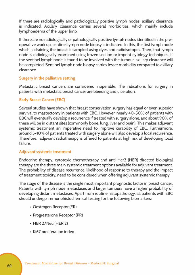
If there are radiologically and pathologically positive lymph nodes, axillary clearance is indicated. Axillary clearance carries several morbidities, which mainly include lymphoedema of the upper limb.
If there are no radiologically or pathologically positive lymph nodes identified in the pre-operative work up, sentinel lymph node biopsy is indicated. In this, the first lymph node which is draining the breast is sampled using dyes and radioisotopes. Then, that lymph node is radiologically examined using frozen section or imprint cytology techniques. If the sentinel lymph node is found to be involved with the tumour, axillary clearance will be completed. Sentinel lymph node biopsy carries lesser morbidity compared to axillary clearance.
Surgery in the palliative setting
Metastatic breast cancers are considered inoperable. The indications for surgery in patients with metastatic breast cancer are bleeding and ulceration.
Early Breast Cancer (EBC)
Several studies have shown that breast conservation surgery has equal or even superior survival to mastectomy in patients with EBC. However, nearly 40-50% of patients with EBC will eventually develop a recurrence if treated with surgery alone, and about 90% of these will be in distant sites (commonly bone, lung, liver and brain). This makes adjuvant systemic treatment an imperative need to improve curability of EBC. Furthermore, around 5-10% of patients treated with surgery alone will also develop a local recurrence. Therefore, adjuvant radiotherapy is offered to patients at high risk of developing local failure.
Adjuvant systemic treatment
Endocrine therapy, cytotoxic chemotherapy and anti-Her2 (HER) directed biological therapy are the three main systemic treatment options available for adjuvant treatment. The probability of disease recurrence, likelihood of response to therapy and the impact of treatment toxicity, need to be considered when offering adjuvant systemic therapy.
The stage of the disease is the single most important prognostic factor in breast cancer. Patients with lymph node metastases and larger tumours have a higher probability of developing distant metastases. Apart from routine histopathology, all patients with EBC should undergo immunohistochemical testing for the following biomarkers:
• Oestrogen Receptor (ER)
• Progresterone Receptor (PR)
• HER 2/Neu (HER 2)
• Ki67 proliferation index
Treatment Modalities for Breast Diseases - Medical & Surgical60

Adjuvant endocrine therapy
Adjuvant endocrine therapy is offered to all patients with ER+ and/or PR+ tumours, and it reduces the risk of disease recurrence by 50%. For postmenopausal patients, aromatase inhibitors (anastrazole, letrozole or exemestane) are the treatment of choice. Arthralgia and osteoporosis are common side effects, and these need to be monitored during treatment. In post-menopausal patients unsuitable for treatment with adjuvant aromatase inhibitors, the selective oestrogen receptor modulator tamoxifen could be considered as an alternative, even though it has a slightly inferior efficacy. Tamoxifen could increase the risk of thromboembolic events and endometrial cancer but decreases the risk of osteoporosis.
Early stage (less than 5cm in size and less than 4 involved lymph nodes: T1-2 N0-1) luminal A tumours have excellent outcomes with adjuvant endocrine therapy alone, and chemotherapy is not offered to these patients. The duration of adjuvant endocrine therapy was conventionally five years, but there is robust evidence to support extending this to 10 years, especially in patients with high-risk disease.
Anti- HER 2 /neu targeted therapy
The monoclonal antibody trastuzumab revolutionized the treatment of patients overexpressing the HER 2/Neu receptor. Compared to chemotherapy alone, the addition of trastuzumab reduces the risk of disease recurrence by more than 50%.
However, these drugs are prohibitively expensive, and trastuzumab is the only anti- HER 2 therapy which is currently available in the state health sector in Sri Lanka. Trastuzumab is sometimes delivered in combination with chemotherapy initially, and it is continued for 6-12 months. The main toxicity of trastuzumab is cardiac dysfunction, which is reversible if detected early. All patients on trastuzumab should have an echocardiogram performed every three months.
Cytotoxic chemotherapy
Chemotherapy is the oldest adjuvant treatment option in EBC. Unlike endocrine therapy and anti-HER 2 directed therapy, there are no robust predictive biomarkers for chemotherapy. Almost all patients with triple negative tumours and HER 2+ tumours are offered chemotherapy. As mentioned before, most patients with luminal A tumours can be spared chemotherapy, but patients with HER 2 negative luminal B tumours are commonly offered chemotherapy if the tumour size is more than 2 cm or if lymph node metastases are present.
Chemotherapy is most usually delivered in three weekly cycles. However, recent evidence has shown superior survival when the cycle duration is shortened to two weeks (dose dense chemotherapy). Alopecia, fatigue, nausea and neutropenia are the main side effects of chemotherapy, but contrary to popular belief, it is very well tolerated by most patients. Neutropenic sepsis is a potentially life-threatening complication of chemotherapy and requires immediate treatment with intravenous antibiotics. A high index of suspicion is needed, as fever may be masked due to the immunosuppression induced by chemotherapy. Colony stimulating factor (G-CSF) is sometimes given to reduce the incidence of neutropenic sepsis.
Treatment Modalities for Breast Diseases - Medical & Surgical 61
Neoadjuvant systemic therapy
Traditionally, adjuvant treatment was delivered after surgery. In certain instances, systemic therapy is delivered before surgery, with a view to downstaging the tumour to facilitate better surgery. It is routinely offered to patients with locally involved disease (skin and chest wall involvement). However, there is an emerging trend of delivering neoadjuvant systemic treatment for patients with HER 2+ and triple negative tumours (even if the tumours are operable), since the response is very good and may enable a superior cosmetic outcome with breast conservation surgery. If breast conservation surgery is to be performed following neoadjuvant therapy, clips are placed under ultrasound guidance before commencement of neoadjuvant therapy, to facilitate locating the tumour after treatment.
Bone protective agents
There is some evidence from recent phase III clinical trials that treatment with bisphosphonates reduces the incidence of bone metastasis and improves survival in post-menopausal patients with EBC. Based on the results of these trials, adjuvant zoledronic acid every six months is sometimes offered to postmenopausal patients.
Apart from hypocalcaemia, osteonecrosis of the jaw is a much-feared side effect of bisphosphonate therapy, and an assessment of dental and oral health is mandatory prior to treatment.
9
There is some evidence from recent phase III clinical trials that treatment with
bisphosphonates reduces the incidence of bone metastases and improves survival in
post-menopausal patients with EBC. Based on the results of these trials, adjuvant
zoledronic acid every six months is sometimes offered to postmenopausal patients.
Apart from hypocalcaemia, osteonecrosis of the jaw is a much-feared side effect of
bisphosphonate therapy, and an assessment of dental and oral health is mandatory
prior to treatment.
Figure 3: Example of use of the NHS predict tool to determine benefit of each adjuvant systemic treatment option
Adjuvant Radiotherapy
Radiotherapy is a local treatment option that acts by inflicting double strand DNA
damage in tumour cells. The older Cobalt-60 teletherapy units are now being replaced
with more modern linear accelerators which can deliver radiotherapy to the target
volumes with high precision.
Compared to systemic treatment its side effects are very minimal (mild skin erythema
and slight fatigue). Radiotherapy is very efficacious and reduces the risk of local
Figure 40: Example of use of the NHS predict tool to determine benefit of each adjuvant systemic treatment option
Treatment Modalities for Breast Diseases - Medical & Surgical62

10
recurrence by nearly 70%. Adjuvant whole breast radiotherapy is indicated in all
patients treated with breast conservation surgery. Following mastectomy, adjuvant
radiotherapy to the chest wall is delivered to patients with lymph node metastasis and
with tumours more than 5cm or with skin or chest wall infiltration. Sometimes, the
supraclavicular and internal mammary lymph nodes are included in the radiotherapy
treatment volume in high-risk patients.
Figure 4: A typical adjuvant whole breast radiotherapy plan
Metastatic Breast Cancer (MBC)
Nearly 14% of all patients with breast cancer in Sri Lanka have distant metastases at
presentation, while 20% of patients with EBC will also develop distant metastases after
adjuvant systemic treatment. Unfortunately, MBC is incurable. The intention of
treatment is focused on symptom relief and achieving maximal prolongation of
survival, while preserving quality of life. There is no evidence that removal of the
primary tumour is beneficial in MBC, and surgery is often not performed in patients
presenting with de novo metastatic disease. However, histological confirmation and
immunohistochemical staining as done in EBC, is mandatory in MBC, too. Radiotherapy
is highly effective in palliation of pain and pressure symptoms, and a durable response
can be achieved with a single dose of radiation.
Figure 41: A typical adjuvant whole breast radiotherapy plan
Adjuvant Radiotherapy
Radiotherapy is a local treatment option that acts by inflicting double strand DNA damage in tumour cells. The older Cobalt-60 teletherapy units are now being replaced with more modern linear accelerators which can deliver radiotherapy to the target volumes with high precision.
Compared to systemic treatment its side effects are very minimal (mild skin erythema and slight fatigue). Radiotherapy is very efficacious and reduces the risk of local recurrence by nearly 70%. Adjuvant whole breast radiotherapy is indicated in all patients treated with breast conservation surgery. Following mastectomy, adjuvant radiotherapy to the chest wall is delivered to patients with lymph node metastasis and with tumours more than 5cm or with skin or chest wall infiltration. Sometimes, the supraclavicular and internal mammary lymph nodes are included in the radiotherapy treatment volume in high-risk patients.
Metastatic Breast Cancer (MBC)
Nearly 14% of all patients with breast cancer in Sri Lanka have distant metastasis at presentation, while 20% of patients with EBC will also develop distant metastasis after adjuvant systemic treatment. Unfortunately, MBC is incurable. The intention of treatment is focused on symptom relief and achieving maximal prolongation of survival,
Treatment Modalities for Breast Diseases - Medical & Surgical 63
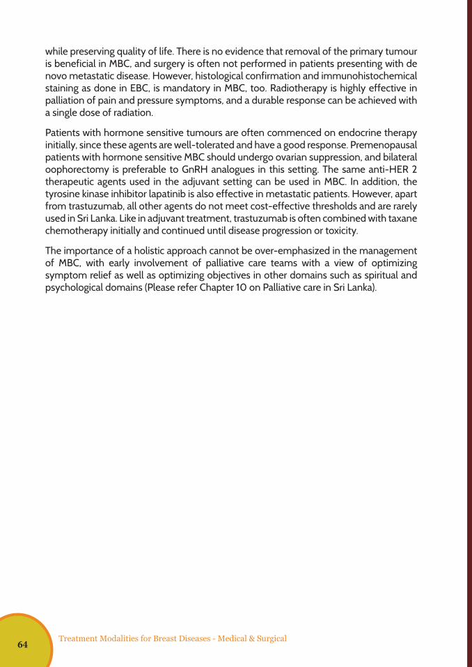
while preserving quality of life. There is no evidence that removal of the primary tumour is beneficial in MBC, and surgery is often not performed in patients presenting with de novo metastatic disease. However, histological confirmation and immunohistochemical staining as done in EBC, is mandatory in MBC, too. Radiotherapy is highly effective in palliation of pain and pressure symptoms, and a durable response can be achieved with a single dose of radiation.
Patients with hormone sensitive tumours are often commenced on endocrine therapy initially, since these agents are well-tolerated and have a good response. Premenopausal patients with hormone sensitive MBC should undergo ovarian suppression, and bilateral oophorectomy is preferable to GnRH analogues in this setting. The same anti-HER 2 therapeutic agents used in the adjuvant setting can be used in MBC. In addition, the tyrosine kinase inhibitor lapatinib is also effective in metastatic patients. However, apart from trastuzumab, all other agents do not meet cost-effective thresholds and are rarely used in Sri Lanka. Like in adjuvant treatment, trastuzumab is often combined with taxane chemotherapy initially and continued until disease progression or toxicity.
The importance of a holistic approach cannot be over-emphasized in the management of MBC, with early involvement of palliative care teams with a view of optimizing symptom relief as well as optimizing objectives in other domains such as spiritual and psychological domains (Please refer Chapter 10 on Palliative care in Sri Lanka).
Treatment Modalities for Breast Diseases - Medical & Surgical64
Dr. Ajith De Silva Consultant Surgeon, National Hospital, Colombo
Dr. Hiranya Jayawickrama Consultant Community Physician, Family Health Bureau
Dr. Nethmini Thenuwara Consultant Community Physician, Family Health Bureau
Dr. Nishani Lucas Consultant Neonatologist, Faculty of Medicine, University of Colombo
Dr. Kanchana Wijesinghe Consultant Surgeon, Faculty of Medical Sciences, University of Sri Jayawardenapura
Dr. A. Krishnamoorthy Senior Registrar in General Surgery National Hospital, Colombo
Dr. Nadun Danushka Registrar in General Surgery, National Hospital, Colombo
Chapter08
Breast Diseases in Special Situations Including Breast Feeding
65
Breast Diseases in Special Situations Including Breast Feeding
Chapter 8 - Special Situations Including Breast Feeding
Introduction
The management of breast cancer during pregnancy poses many challenges. Since pregnant patients are often excluded from clinical trials, treatment decisions are made based on first principles after extrapolation of data from non-pregnant patients. A full discussion in a multidisciplinary setting comprising surgeons, radiologists, pathologists, oncologists and obstetricians is mandatory. Patients presenting in the late third trimester (after 35-37 weeks) could have their treatment deferred until delivery.
Early Breast Cancer
Patients with early breast cancer are treated with curative intent and treatment should conform to protocols used in non-pregnant patients as far as possible. Mammography and ultrasonography may be safely performed with adequate precautions during pregnancy although interpretation may be difficult due to glandular changes in the breast during pregnancy. Exclusion of multifocal and multicentric disease, which is more prevalent in pregnant patients, could prove to be difficult. Consequently, although breast conservation surgery is still feasible in pregnant patients, challenges in excluding multifocal and multicentric disease may lead to most patients being offered modified radical mastectomy.
However multidisciplinary team will decide on the type of surgery (breast conversion or radical mastectomy) after considering several factors. It should be a team decision based on the trimester, stage, type of the cancer and the patient’s decision after providing the necessary information. If the multidisciplinary team decided to carry out a breast conservative surgery, then there is the question of giving postoperative radiotherapy before delivery, or delaying it until later. On the other hand, delaying radiotherapy in the management of breast cancer is likely to lead to an increased rate of local recurrence. Hence, the decision of administration of radiotherapy should be obtained after detailed discussion with patient, family members, multidisciplinary team considering the potential risk versus benefits.
In patients diagnosed later in the pregnancy, for example in the late second or third trimester, radiotherapy could well be postponed until after delivery without detriment to the maternal outcome, based on the recommendation of the multidisciplinary team.
Treatment modalities will be decided by a multidisciplinary team. Management depends on the stage of pregnancy and type and stage of cancer.
66
Breast Diseases in Special Situations Including Breast Feeding
Chemotherapy can be safely administered in the second and third trimester but tamoxifen and trastuzumab is contraindicated during pregnancy. Radiotherapy is also not generally offered during pregnancy although anecdotal evidence of its use without any adverse impact have been reported.
Metastatic Breast Cancer
Metastatic breast cancer is incurable and these patients are treated with palliative intent. As noted above, chemotherapy may be safely administered but radiotherapy, targeted treatment and hormonal therapy is contraindicated. Termination of pregnancy may be necessary in these patients, since rapid disease progression may compromise maternal safety.
Breast cancer and breastfeeding:
While breastfeeding is protective against breast cancer, it is contraindicated while undergoing radiotherapy and chemotherapy treatments for breast cancer. Breastfeeding is contraindicated while a mother is receiving radiotherapy as part of the treatment. However, breastfeeding can be resumed after stopping radiotherapy. Radiotherapy may affect the breastmilk production in the irradiated breast but will not affect the contralateral breast. Breastfeeding support would be beneficial to such mothers. Breastfeeding in a mother who is receiving chemotherapy would depend on the half-life of the drug. It takes approximately 5 half-lives for the clearance of a drug from the body. Therefore, it would be safe to resume breastfeeding / express breast milk to be given to the baby, after the duration of 5 half-lives after stopping chemotherapy. Chemotherapy may also affect breastmilk production and thereby these mothers would also benefit from skilled lactational support.
The management of breastfeeding in a breast cancer patient should be decided on an individual basis by a multidisciplinary team comprising of Surgeon, Oncologist and Neonatologist/Pediatrician.
67

Breast Diseases in Special Situations Including Breast Feeding
68
Dr. Janaki Vidanapathirana Director, Consultant Community Physician, National Cancer Control Programme
Chapter09
Introduction to Counseling for Breast Cancer
69
Introduction to Counseling for Breast Cancer
Chapter 9 - Introduction to Counseling for Breast Cancer
Introduction to Counseling
Counseling is a process of helping a person/people learn how to solve certain emotional, interpersonal & decisional problems. It is an interaction between a counselor and another person or persons, where the counselor offers the time, attention, and respect necessary to clarify ways of dealing with a problem. Counseling is an integral component in the management of an affected person. It’s also a tool for prevention of disease, promoting health and committing people to improve their lives with their own efforts. The action taken will be the person’s own decision, although it may be guided by the counselor.
Counseling should be:
• Client-centred: specific to the needs, issues & circumstances of each individual client
• An interactive, collaborative & respectful process
• Goal centred
• Developing autonomy & self- responsibility in clients
• Considerate of interpersonal situation, social / cultural context and readiness to change
• Asking questions, eliciting information, reviewing options & developing action plans
Counseling is not:
• Telling or directing
• Giving advice
• A conversation
• An interview
• A confession
• Praying
70
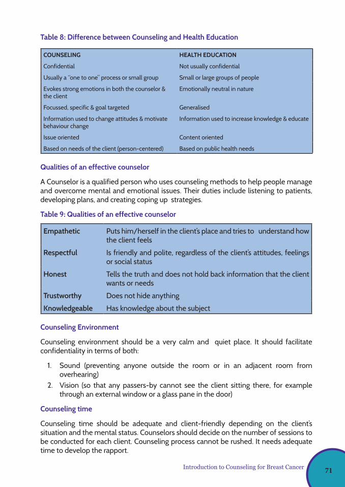
Introduction to Counseling for Breast Cancer
Table 8: Difference between Counseling and Health Education
COUNSELING HEALTH EDUCATION
Confidential Not usually confidential
Usually a “one to one” process or small group Small or large groups of people
Evokes strong emotions in both the counselor & the client
Emotionally neutral in nature
Focussed, specific & goal targeted Generalised
Information used to change attitudes & motivate behaviour change
Information used to increase knowledge & educate
Issue oriented Content oriented
Based on needs of the client (person-centered) Based on public health needs
Qualities of an effective counselor
A Counselor is a qualified person who uses counseling methods to help people manage and overcome mental and emotional issues. Their duties include listening to patients, developing plans, and creating coping up strategies.
Table 9: Qualities of an effective counselor
Empathetic Puts him/herself in the client’s place and tries to understand how the client feels
Respectful Is friendly and polite, regardless of the client’s attitudes, feelings or social status
Honest Tells the truth and does not hold back information that the client wants or needs
Trustworthy Does not hide anything
Knowledgeable Has knowledge about the subject
Counseling Environment
Counseling environment should be a very calm and quiet place. It should facilitate confidentiality in terms of both:
1. Sound (preventing anyone outside the room or in an adjacent room from overhearing)
2. Vision (so that any passers-by cannot see the client sitting there, for example through an external window or a glass pane in the door)
Counseling time
Counseling time should be adequate and client-friendly depending on the client’s situation and the mental status. Counselors should decide on the number of sessions to be conducted for each client. Counseling process cannot be rushed. It needs adequate time to develop the rapport.
71
Principles of Counseling
It is important to adhere to the following principles during the counseling process.
1. Principle of acceptance
Accept the client with his physical, psychological, social, economic and cultural conditions. Never make faces during the session and do not engage in stigmatizing and discriminating actions.
2. Principle of communication
Communication should be verbal as well as non-verbal and should be skillful. There should be eye contact with the client, and it should be maintained throughout the process. Always allow the client to talk first. It should be active listening without disturbing the client. Counselor can nod or say “Hmm”. The next step is paraphrasing, which is repeating what the client has said, and questions could be asked without disturbing the client. However, never ask questions in an authoritative voice. It should not be a client-victimizing form of question (eg: why didn’t you do that? - this is an incorrectly formulated question). The same thing can be asked as : “are there any obstacles or any other reasons to implement such activity, etc”. Acknowledge the client’s feelings, e.g. “I can see you feeling very sad”, I understand your feelings”, etc.
The following things are to be practiced when you communicate with the client:
- Use a tone of voice that shows interest, concern and friendliness.
- Ask one question at a time.
- Ask questions that lead the client to tell you about his/her problems.
- Avoid asking questions that can be answered by ‘yes’ or ’no’. Ask questions that encourage clients to say more.
- Avoid asking questions starting with “why”.
- Ask the same question in different ways if you think the client has not understood it.
- Avoid repeating questions that the client does not want to answer.
- Probe with both open and close ended questions.
Now and then repeat what you have heard. Then both you and the client know that you have understood each other. It is essential to give specific and factual information to the client. The counselor should never lie to the client. Repeat the main points of the discussion in similar but fewer words to ensure that you have understood the client correctly (paraphrase, reflect feelings, clarify and summarise). Do not engage in other tasks during the counseling sessions. Do not use mobile phones and do not watch television while having the counseling sessions.
Introduction to Counseling for Breast Cancer72
3. Principle of empathy
Instead of showing sympathy, put yourself in the client’s shoes and then give reflections accordingly (empathy is the ability to identify the feelings of a person). Never sympathize with the person.
4. Principle of non-judgement
Mental attitude: do not criticize or comment negatively regarding clients’ complaints. It is important to understand that clients are coming from different social backgrounds and with different attitudes.
5. Principle of confidentiality
Always keep the client’s name and the problems strictly secretive and assure the client about the same.
6. Principle of individuality
Treat each and every client as unique, and respect his / her problem, as well. Options and plans are different for each person even with the same problem.
7. Principle of non-emotional involvement
Do not get emotionally involved with the client and avoid getting carried away with his feelings. There should not be any personal connections with the clients. But need to ensure:
1. Listening keenly to the client is the main goal.
2. Identify the need of the client. e.g., parents need counseling for their children’s behaviour problems.
3. Help the client ventilate his emotions properly and help him to be aware of his own emotions and encourage him to be independent.
4. The main problem should be focused on, so that the sub-problems could be identified by the client himself.
5. Make the client accept his problem himself and help him to adjust with it till he/she gets over with it.
6. Focus on his strengths by studying the case, help in developing positive attitudes in him and ultimately help him to overcome his negativity.
Introduction to Counseling for Breast Cancer 73
Steps in the counseling process
Step 1: Greeting and Relationship Building
• A friendly, respectful greeting makes the client feel welcome, and establishes a good connection between the counselor and the client right from the start. Show them that they have your full attention by greeting them in a polite, friendly manner. It is also important to introduce yourself and offer a place to sit. You need to emphasize to clients that information about them and what they say will be confidential. It is essential to explain how you can help and explain what will happen during the visit. Ensure that the client is comfortable, and address the client by name with social conversation to reduce anxiety. At the same time, watch for non-verbal behaviour as signs of client’s emotional state.
• After greeting, the next step involves building a relationship, and focuses on engaging clients to explore issues that directly affect them. The first interview is important because the client is reading the verbal and nonverbal messages and makes inferences about the counselor and the counseling situation. Is the counselor able to empathize with the client? Does the client view the counselor as genuine?
• Invite the client to describe his or her reason for coming to talk.
• Allow the client time to respond.
• Indicate that you are interested in the person.
Step 2: Problem Assessment: While the counselor and the client are in the process of establishing a relationship, a second process is taking place, i.e. problem assessment. This step involves the collection and classification of information about the client’s life situation and reasons for seeking counseling. The counselors gather information to form a holistic view of their clients and the problems which they present, and this needs to be regularly assessed throughout the counseling process, especially in the early stages.
Step 3: Goal Setting: The counseling process must have a focus, and goals are the results or outcomes that a client wants to achieve at the end of counseling. This is where goals play an important role in giving direction. Goals should relate to the desired end or ends sought by the client. They should be defined in an explicit and measurable terms and should be feasible. Goals should also be within the range of the counselor’s knowledge and skills, and should be stated in positive terms that emphasize a positive change.
Step 4: Intervention: There are different points of view concerning what a good counselor should do with clients, depending on the theoretical positions that the counselor subscribes to. For example, the person-centered approach suggests that the counselor gets involved rather than intervenes by placing emphasis on the relationship. It should be a scientific approach. The behavioural approach attempts to initiate the intervention.
Step 5: Evaluation, follow-up, termination or referral: At the beginning of the counselling session, it is difficult to think of terminating the counseling process, as they are more
Introduction to Counseling for Breast Cancer74
concerned with beginning the counseling process. The aims towards terminating the counseling process will have to be done with the client knowing that it will have to end.
Counseling for breast cancer patients
Counseling helps breast cancer patients to respond to their mixed emotions about life’s challenges. Although counselors cannot always solve problems, they provide a safe place for breast cancer patients to talk about their concerns. Because counselors are separate from a person’s life, they provide a supportive, outside viewpoint and facilitates to overcome challenges. The main aim of counseling is to empower the patients with a mindfulness-based approach and confidence to make their own choices and decisions to explore ways to cope with their diagnosis, managing their emotional concerns and discuss ways to address and cope with any lifestyle changes. Breast cancer patients are provided with counseling at different stages and with different problems. It can be done by counselors, clinicians or other healthcare providers through different sessions. Depth of counseling sessions may be different from patient to patient. It should be tailormade to each and every patient. The number of sessions also depends on the patient’s understanding and coping strategies.
Some of the most common psychosocial concerns reported by women with breast cancer, which need to be addressed by counseling:
• Breaking bad news
• Fear of recurrence
• Physical symptoms such as fatigue, insomnia or pain
• Body image disruption
• Sexual dysfunction
• Treatment-related anxieties
• Intrusive thoughts about illness/persistent anxiety
• Marital/partner communication
• Feelings of vulnerability
• Existential concerns regarding mortality
Factors to be considered in a counseling session to improve mental well-being and quality of life for people with metastatic breast cancer:
• Manage feelings and emotions of sadness or anxiousness.
• Identify symptoms that can be treated by the healthcare team.
• Improve communication with family members and other loved ones.
Introduction to Counseling for Breast Cancer 75
• Reduce feelings of isolation and help from supportive groups. Emotional support is useful for people who are comfortable expressing their feelings and fears in a group setting.
• Discuss fears about death and dying.
• Try to find out the needs and preferences of the patients, and implement as much as possible (property matters, weddings of children, loan settlement, family functions, etc.)
• If the patient wishes, try to encourage spiritual or religious activities.
• Connect with social support.
Breaking Bad News:
It is important to have specific skills (SPIKES) when you are breaking bad news, and it can be accomplished by completing six tasks.
Table 10: Six Steps of SPIKES:
S – Setting • Arrange for some privacy and ask the person to sit.• Involve significant others, make connection and establish
rapport with the patient.• Consider managing time constraints and interruptions.
P – Perception of condition/seriousness
• Find out the patient’s understanding about the medical condition or what she suspects.
• Listen to the patient’s level of comprehension and accept denial but do not confront at this stage.
I – Invitation from the patient to give information
• Ask the patient if she/he wishes to know the details of the medical condition and/or treatment.
• Accept the patient’s right not to know. • Offer to answer questions later by giving medical facts. • Use effective communication skills with information in
small chunks.• Consider educational level, socio-cultural background
and current emotional state. • Find out whether the patient has understood what you
said, and respond to the patient’s reactions as they occur. • Give any positive aspects first e.g: Cancer has not spread to
the lymph nodes, highly responsive to therapy, treatment available locally, etc.
• Give facts accurately about treatment options, prognosis, costs, etc.
Introduction to Counseling for Breast Cancer76
E - Explore emotions and sympathize
• Prepare to give an empathetic response: 1. Identify emotions expressed by the patient (sadness,
silence, shock, etc.) 2. Identify cause/source of emotion.3. Allow the patient to express his or her feelings, then
respond in a way that demonstrates that you have recog-nized connection between 1 and 2.
S – Strategy and summary
• Close the interview. • Ask whether they want to clarify something else.• Offer an agenda for the next meeting. eg: I will speak to
you again when we have the opinion of the cancer special-ist.
Healthcare providers need to support breast cancer patients in the following situations:
• Pre- and post-surgery counseling
• Radiation therapy counseling
• Chemotherapy counseling
• Any procedure (like Biopsy, FNAC, Pet Scan, Genetic Testing, etc) counseling
• Pain and palliative counseling
• Terminal counseling
• Bereavement and grief counseling
• Fear of cancer recurrence (Survivor) counseling
• Compliance and adherence counseling
• Group and individual counseling
• Counseling about fertility issues in cancer
• Family and couples counseling/therapy
• Tobacco/alcohol cessation counseling
• Preventive and lifestyle change related counseling
Introduction to Counseling for Breast Cancer 77
During the counseling sessions, it is important to provide information needed for the patient and the family about illness, adverse effects of treatment, hygienic and dietary regimen, lifestyle, alternative treatment, etc. Also, it needs to facilitate the patient’s communication with medical staff, to facilitate adherence to the prescribed treatment and facilitate patient communication with social services staff and community support groups to obtain the required facilities easily. At the end of a successful counselling process, expected results at patient level will improve in the areas of level of knowledge about the treatment, procedure and the condition. It will help to prepare the patient emotionally and increase the patient’s self-efficiency and increase the patient’s satisfaction related to medical services. Many people describe being in a state of shock after diagnosis, causing numbness, confusion and fear. Most people experience a range of intense emotions including shock, fear, confusion, stress, worry, anxiety, distress, sadness, anger, frustration, guilt or shame, loneliness and grief/loss. These feelings are all common and normal. Many people feel like they’re on an emotional rollercoaster. You may find that some emotions come and go, while others last longer. After the initial shock of diagnosis, most people begin to adjust, and these feelings reduce over time. However, it is normal for the patient’s distress to go up and down when different events occur, e.g: starting or finishing treatment, having follow-up tests or facing reminders about cancer. People commonly feel anxious at these times. While some people worry that they are not reacting in the ‘right’ way, it can help to accept the reactions as part of the process of adjusting to cancer.
Introduction to Counseling for Breast Cancer78
Dr. Irosha Nilaweera Consultant Community Physician, National Cancer Control Programme
Chapter10
Palliative Care Aspects of Breast Cancer
79
Palliative Care Aspects of Breast Cancer
What is palliative care?
The number of breast cancer survivors has increased with the improvement of early detection and availability of comprehensive breast care with evidence based cost effective treatment pathways. Therefore, their health needs must be addressed by survivorship care and rehabilitation services.
Advanced breast cancer is characterized by many physical, psychological and spiritual manifestations which can affect the quality of life (most related to the cancer and some to treatments). Patients with advanced breast cancer and their families have complex needs that have to be addressed in order to minimize severe distress and deterioration in the quality of life of patients and their family members.
Palliative care is an approach that improves the quality of life of patients (adults & children) and their families who are facing the problems associated with life-threatening illness, through the prevention and relief of suffering by means of early identification, impeccable assessment and treatment of pain and other problems, physical, psychosocial and spiritual. (WHO, 2016). It is a right of every person with life-threatening illness to receive appropriate palliative care and a responsibility of every health care professional to practice palliative care according to the need of the patient.
Holistic Approach in Palliative Care
Palliative care, is a specialized medical care focused on relief of physical symptoms including the pain, as well as psychological (eg: depression, anxiety and worries about body image and sexuality etc.) and spiritual problems of the patient with a life threatening illness such as breast cancer. The goal is to improve quality of life for both patient and the family. Palliative care is appropriate at any age, at any stage in the illness, and it can be provided along with curative treatment.
Model of Palliative Care
• Palliative care begins at the point of diagnosis, continues throughout the rest of the ‘Patient Journey’ and extends beyond death into bereavement care.
• Bereavement care is the care extended to the surviving family members of the deceased to help them in the mourning following the loss of their loved one.
Multidisciplinary Team in Palliative Care
Palliative care team (see below) works in partnership with cancer specialists for people living with breast cancer to experience relief of symptoms, better communication, psychological and spiritual care and to help them plan for the future. Once symptoms are controlled, patients can get back to daily activities.
Chapter 10 - Palliative Care Aspects of Breast Cancer
80

Palliative care team members
• Consultant (Team leader)
• Medical officers
• Nursing officers
• Consultant Psychiatrist/ counselor
• Pharmacist
• Social service worker
• Physiotherapist, speech therapist, occupational therapist and nutritionist, etc
Palliative Care Delivery Points
Hospital based
• 24 cancer treatment centers (specialist palliative care)
• Palliative Care Centre: TH Karapitiya (provides residential palliative care) please refer Fig 49 & 50 in chapter 12
• Primary health care institutions / OPD setting / Family practice setting
Community based
• Home based care- e.g. PHNOs attached to primary care institutions
• Hospices – 7 hospices managed by NGOs island wide (please refer table in chapter 12)
For details please refer Chapter 12: Services available in Sri Lanka for management of breast cancer (sections: palliative care services and Supportive services)
Care planning and coordination:
• Identify support and resources available: Develop and implement care plan based on patient’s need
• Provide care in the last weeks /days of the life
• Facilitate the availability and access to medications
• Identify the psychosocial / spiritual needs of professionals providing care
Palliative Care Aspects of Breast Cancer 81
Health care professional assessment and care plan
Based on assessment, a survivorship care plan for the patient has to be formulated addressing all aspects of care related to cancer.
The Plan should include:
• Medical condition of the patient and the goals of care
• Description of the involved family members and health care professionals
• Patient issues: physical, psychological, existential, social, communication, understanding
• Family issues: physical, psychological, existential, social, communication, understanding
• Professional care issues: staffing, training, resources, resource/need match, emotional coping
• Coping assessment: patient, family, health care professionals
• Contingency planning: anticipated contingencies, planned interventions
Care for a palliative care patient is a longitudinal commitment. Therefore, it needs repeated assessments at appropriate intervals. This will be determined by the rate of change in the patient’s clinical conditions or at points of major change in goals, care plan, or the patient’s conditions.
Key Facts – Palliative Care
• It is care for patients with life-threatening illness such as chronic non communicable diseases (cancer, cardio vascular disease, kidney diseases, respiratory diseases, neurological conditions, etc.) and their families
• It improves quality of life
• It relieves physical, psychosocial and spiritual suffering
• It can be given in hospitals, health centers, hospices and homes
• It can be done by many types of health / non-health professionals and volunteers
• It benefits health systems, by reducing unnecessary hospital admissions
Palliative Care Aspects of Breast Cancer82
Dr. S.P. Kandapola Arachchige Consultant Psychiatrist, National Cancer Institute, Maharagama
Dr. W.N. Sachintha Dilhani Senior Registrar in Community Medicine, National Cancer Control Programme
Chapter11
Psychosocial Aspects in Breast Cancer
83
Psychosocial Aspects in Breast Cancer
Chapter 11 - Psychosocial Aspects in Breast Cancer
The diagnosis of cancer is almost always a devastating news to the patient, family and loved ones. It is invariably associated with many negative emotions such as fear, anxiety and grief. There is lot of uncertainty around the word cancer that affect the psychological well-being of not only the patient, but everyone around. Hence, psychological, emotional, spiritual, social, and practical aspects that patients and their families have as a consequence of cancer and its treatment should also be given priority and addressed in the oncology care.
The degree of negative psychosocial effects related to breast cancer vary on a number of factors including,
• Socio- economic status of the patient
• Psychological make-up and coping abilities of the patient and the family
• Disease related factors such as stage of the cancer at the diagnosis
• Type of the surgery they undergo (mastectomy / lumpectomy)
• Other forms of treatment they are receiving
• Side effects of the treatment
• Availability of emotional and financial support
Attending to psychosocial needs of the cancer patients should be a prioritized integral component of quality cancer care. All health care providers should pay their attention to patient’s psycho-social well-being as well. Early detection of psycho-social distress, psycho-social care needs and diagnosis of psychiatric co-morbidities for early intervention is the best approach to provide comprehensive psycho-social care.
Figure 42 depicts various aspects of the psychological needs of breast cancer patients and others.
Role of the healthcare providers in addressing psychosocial aspects in breast cancer patients
Figure 42: Breast cancer patients’ and partners’ psychosocial needs –diagrammatical presentation
3
Figure 2: Physical needs and emotional needs accompanying illness and treatment
Women with Breast Cancer
Partner / family
Physical & treatment
related needs
Psychological / emotional
Needs
Need of Informational & professional
support
Spiritual Needs
Financial / Logistic
needs
Figure 1 Breast cancer patients’ and partners’ psychosocial needs –diagrammatical presentation 84
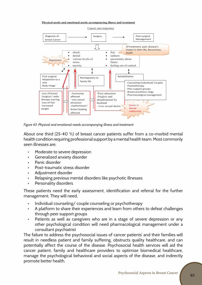
Psychosocial Aspects in Breast Cancer
Figure 43: Physical and emotional needs accompanying illness and treatment
About one third (25-40 %) of breast cancer patients suffer from a co-morbid mental health condition requiring professional support by a mental health team. Most commonly seen illnesses are;
• Moderate to severe depression• Generalized anxiety disorder• Panic disorder• Post-traumatic stress disorder• Adjustment disorder • Relapsing previous mental disorders like psychotic illnesses• Personality disorders
These patients need the early assessment, identification and referral for the further management. They will need,
• Individual counseling/ couple counseling or psychotherapy • A platform to share their experiences and learn from others to defeat challenges
through peer support groups • Patients as well as caregivers who are in a stage of severe depression or any
other psychological condition will need pharmacological management under a consultant psychiatrist
The failure to address the psychosocial issues of cancer patients’ and their families will result in needless patient and family suffering, obstructs quality healthcare, and can potentially affect the course of the disease. Psychosocial health services will aid the cancer patient, family and healthcare providers to optimize biomedical healthcare, manage the psychological behavioral and social aspects of the disease, and indirectly promote better health.
2
Physical needs and emotional needs accompanying illness and treatment
Cancer care trajectory
About a third (25-40 %) of cancer patients suffer from a co-morbid mental health conditions requiring professional support by a mental health team. Among them are;
Moderate to severe depression Generalized anxiety disorder Panic disorder Post traumatic stress disorder Adjustment disorder Relapsing previous mental disorders like psychotic illnesses Personality disorders
shock fear denial sadness various levels of
stress uncertainty about
future anxiety feeling out of control
Diagnosis of breast Cancer
Surgery Post-surgical Management
Of treatment, pain, disease’s impact in their life, Recurrence, Death
Post surgical-Adaptation to a new Body image
Reintegration to family life
Rehabilitation
-Loss of breast -Surgical / radio therapy scarring -Loss of Hair -Increased weight
- Femininity affected - Less sexual attraction -motherhood / breast feeding affected
-Poor attraction -Neglect and abandonment by husband - Low sexual desire
Depression
Issues in Sexual relationships
-Counseling (Individual/ Couple) -Psychotherapy -Peer support groups -Breast prosthesis /wigs -Pharmacological management
85
Provision of healthcare information
Information related to the disease, treatments, side effects, prognosis, psychosocial services etc. to the patient, family member or caregiver is a very important component in cancer care. This empower them to overcome any unpleasant emotions and adjust to the stressful experience. Similarly, health care providers should help patients /families in understanding and using the information provided. Thus, healthcare providers should be aware of the informational needs of the patients throughout the course of their care and they should be ready to allocate a time to discuss patient’s concern during their consultations. Readymade educational packages/ leaflets/ booklets related to breast cancer can be used as catalysts during those discussions.
Financial advice and/or assistance
With the unexpected diagnosis of breast cancer, the patient and the family may have to face numerous financial issues, including the costs during diagnostic, treatment and follow-up phases of the disease. Costs for transportation, reduced income due to employment issues with the diagnosis such as either termination of the employment or taking leave unlimitedly and out of pocket expenditure for treatments or investigations and inability of getting the assistance to replace lost income are the mainly emerging issues related to economic impact on families.
Healthcare providers could help the patients with financial burden by identifying and providing them information on available financial resources. They could even be referred to relevant officers/ agencies such as social service officers, support groups, Non- governmental organizations or others knowledgeable about available financial resources for further assistance.
Provision of material & logistic resources
Transportation facilities, lodging for patients and caregivers when they must travel long distances for outpatient therapy, child care, wigs and breast prostheses, and supplies for managing the side effects of cancer and its treatment (e.g., compression bandages or sleeves for lymphedema control) are some examples, where the breast cancer patients need the material / logistic support.
Though all these things could not be addressed directly by the health care providers, they can direct the patients to the available supportive care services. Hence, it is important that health care providers are aware of the available supportive care services locally, in and around their institution and nationally.
Spiritual needs
Enhancement of spiritual wellbeing has a great effect on quality of life of cancer patients and it has been proven in many studies. Many survivors find that life takes on new meaning after being diagnosed with cancer and will renew their commitment to spiritual practices. This will go through strong social support network, adaptive coping, lessened depression and better psychological function.
Addressing women’s spiritual concerns, with attention to cultural differences is much important in rendering holistic patient-centered care.
Available supportive care services for the breast cancer patients are elaborated in chapter 12.
Psychosocial Aspects in Breast Cancer86
Dr. Muzrif Munas Consultant Community Physician, National Cancer Control Programme
Dr. Sashiprabha Nawaratne Senior Registrar in Community Medicine, National Cancer Control Programme
Chapter12
Services Available in Sri Lanka for Management of Breast Cancer
87
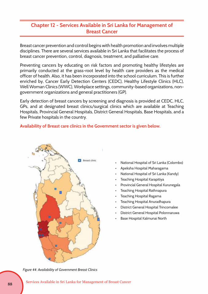
Services Available in Sri Lanka for Management of Breast Cancer
Chapter 12 - Services Available in Sri Lanka for Management of Breast Cancer
Breast cancer prevention and control begins with health promotion and involves multiple disciplines. There are several services available in Sri Lanka that facilitates the process of breast cancer prevention, control, diagnosis, treatment, and palliative care.
Preventing cancers by educating on risk factors and promoting healthy lifestyles are primarily conducted at the grass-root level by health care providers as the medical officer of health. Also, it has been incorporated into the school curriculum. This is further enriched by, Cancer Early Detection Centers (CEDC), Healthy Lifestyle Clinics (HLC), Well Woman Clinics (WWC), Workplace settings, community-based organizations, non-government organizations and general practitioners (GP).
Early detection of breast cancers by screening and diagnosis is provided at CEDC, HLC, GPs, and at designated breast clinics/surgical clinics which are available at Teaching Hospitals, Provincial General Hospitals, District General Hospitals, Base Hospitals, and a few Private hospitals in the country.
Availability of Breast care clinics in the Government sector is given below.
• National Hospital of Sri Lanka (Colombo) • Apeksha Hospital Maharagama• National Hospital of Sri Lanka (Kandy)• Teaching Hospital Karapitiya• Provincial General Hospital Kurunegala• Teaching Hospital Rathnapura• Teaching Hospital Ragama• Teaching Hospital Anuradhapura• District General Hospital Trincomalee• District General Hospital Polonnaruwa• Base Hospital Kalmunai North
3
Availability of Breast care clinics in the Government sector is given below.
Figure 1: Availability of Government Breast Clinics
WWC /HLC and other service providers have the opportunity to refer patients directly
to breast clinics. In the absence of a dedicated breast clinic in the area, women with
breast symptoms can be referred to a surgical clinic at the nearest government
hospital.
National Hospital of Sri Lanka (Colombo)
Apeksha Hospital Maharagama National Hospital of Sri Lanka
(Kandy) Teaching Hospital Karapitiya Provincial General Hospital
Kurunegala Teaching Hospital Rathnapura Teaching Hospital Ragama Teaching Hospital Anuradhapura District General Hospital
Trincomalee District General Hospital
Polonnaruwa Base Hospital Kalmunai North
Figure 44: Availability of Government Breast Clinics
88
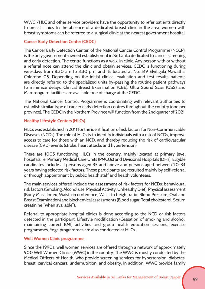
Services Available in Sri Lanka for Management of Breast Cancer
WWC /HLC and other service providers have the opportunity to refer patients directly to breast clinics. In the absence of a dedicated breast clinic in the area, women with breast symptoms can be referred to a surgical clinic at the nearest government hospital.
Cancer Early Detection Center (CEDC)
The Cancer Early Detection Center, of the National Cancer Control Programme (NCCP), is the only government-owned establishment in Sri Lanka dedicated to cancer screening and early detection. The centre functions as a walk-in clinic. Any person with or without a referral note can attend the clinic and obtain services. CEDC is functioning during weekdays from 8.30 am to 3.30 pm, and it’s located at No. 519 Elvitigala Mawatha, Colombo 05. Depending on the initial clinical evaluation and test results patients are directly referred to the specialized units by-passing the routine patient pathways to minimize delays. Clinical Breast Examination (CBE), Ultra Sound Scan (USS) and Mammogram facilities are available free of charge at the CEDC.
The National Cancer Control Programme is coordinating with relevant authorities to establish similar type of cancer early detection centres throughout the country (one per province). The CEDC in the Northern Province will function from the 2nd quarter of 2021.
Healthy Lifestyle Centers (HLCs)
HLCs was established in 2011 for the identification of risk factors for Non-Communicable Diseases (NCDs). The role of HLCs is to identify individuals with a risk of NCDs, improve access to care for those with an NCD, and thereby reducing the risk of cardiovascular disease (CVD) events (stroke, heart attacks and hypertension).
There are 1005 functioning HLCs in the country, mainly located at primary level hospitals i.e. Primary Medical Care Units (PMCUs) and Divisional Hospitals (DHs). Eligible candidates include all persons aged 35 and above and persons aged between 20-34 years having selected risk factors. These participants are recruited mainly by self-referral or through appointment by public health staff and health volunteers.
The main services offered include the assessment of risk factors for NCDs: behavioural risk factors (Smoking, Alcohol use, Physical Activity, Unhealthy Diet), Physical assessment (Body Mass Index, Waist circumference, Waist to height ratio, Blood Pressure, Oral and Breast Examination) and biochemical assessments (Blood sugar, Total cholesterol, Serum creatinine “when available”).
Referral to appropriate hospital clinics is done according to the NCD or risk factors detected in the participant. Lifestyle modification (Cessation of smoking and alcohol, maintaining correct BMI) activities and group health education sessions, exercise programmes, Yoga programmes are also conducted at HLCs.
Well Women Clinic programme
Since the 1990s, well women services are offered through a network of approximately 900 Well Women Clinics (WWC) in the country. The WWC is mostly conducted by the Medical Officers of Health, who provide screening services for hypertension, diabetes, breast, cervical cancers, undernutrition, and obesity. In addition, WWC provide family
89

planning services and health education to its participants.
The main target population for WWC are, women aged 35 years and 45 years. However, women outside the target population are also welcome at the WWCs.
In the implementation of the WWC programme, the Family Health Bureau work closely with the National Cancer Control Programme, the College of Pathologists of Sri Lanka and the Sri Lanka College of Obstetricians and Gynecology.
Mammography and ultrasound scan investigations
Mammography and ultrasound scan investigations are used in breast cancer screening and act as a component of triple assessment in diagnosing breast cancer.
Figure 45: availability of mammogram facilities in Sri Lanka
Referral pathway for women with or without breast symptomsWell Women Clinics (WWC) Healthy Lifestyle Centers
(HLC) OPDs of small hospitalsCancer Early Detection Center(CEDC)
Women with breast symptoms, abnormal findings on clinical breast examination or abnormal
mammography findings
Surgical clinics/ Breast clinics at TH/PGH/DGH/ BH / Private Hospitals
Triple assessmentHistology and Clinical Examination/ mammography/
Ultrasound scan / FNAC/ Biopsy
Abnormal findings in triple assessment
Advice to conduct self breast examination monthly If between 20-40 years advice to
come for clinical breast examination in three years
Further management based on the findings of the triple assessment
If triple assessment findings are negative
Normal clinical breast examination findings or normal mammography findings in
asymptomatic women
Services Available in Sri Lanka for Management of Breast Cancer
Figure 46: Referral pathway for women with or without breast symptoms
6
Mammography and ultrasound scan investigations are used in breast cancer screening
and act as a component of triple assessment in diagnosing breast cancer.
Figure 2: availability of mammogram facilities in Sri Lanka
90
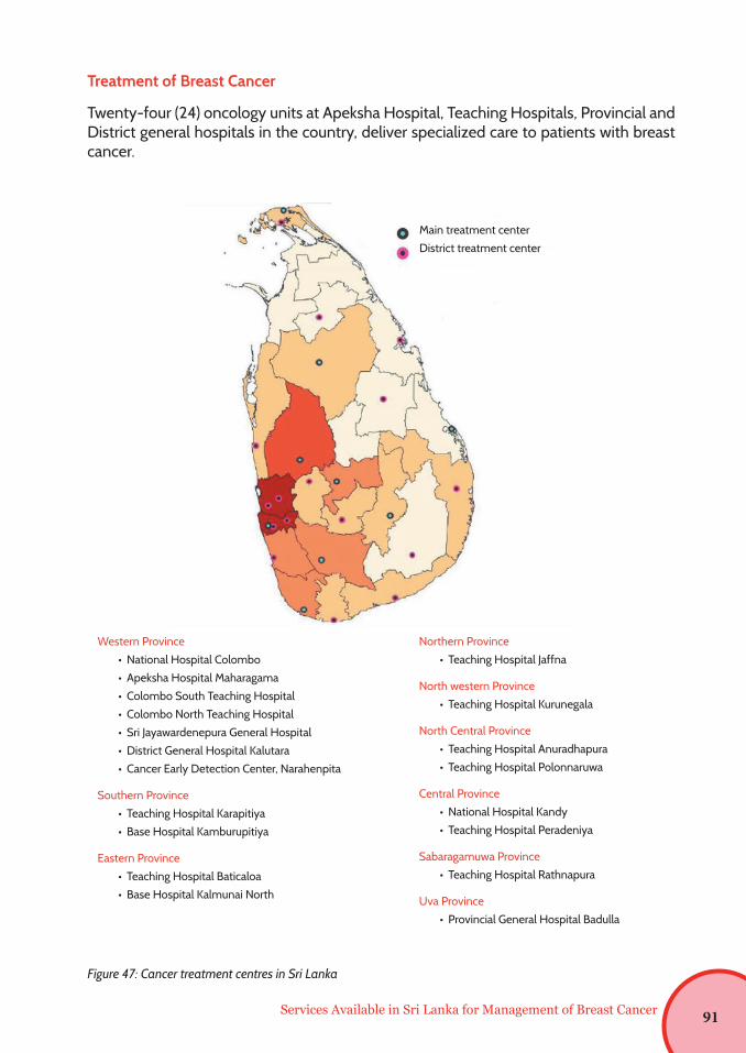
Figure 47: Cancer treatment centres in Sri Lanka
Treatment of Breast Cancer
Twenty-four (24) oncology units at Apeksha Hospital, Teaching Hospitals, Provincial and District general hospitals in the country, deliver specialized care to patients with breast cancer.
Services Available in Sri Lanka for Management of Breast Cancer
Main treatment center
District treatment center
Western Province
• National Hospital Colombo
• Apeksha Hospital Maharagama
• Colombo South Teaching Hospital
• Colombo North Teaching Hospital
• Sri Jayawardenepura General Hospital
• District General Hospital Kalutara
• Cancer Early Detection Center, Narahenpita
Southern Province
• Teaching Hospital Karapitiya
• Base Hospital Kamburupitiya
Eastern Province
• Teaching Hospital Baticaloa
• Base Hospital Kalmunai North
Northern Province
• Teaching Hospital Jaffna
North western Province
• Teaching Hospital Kurunegala
North Central Province
• Teaching Hospital Anuradhapura
• Teaching Hospital Polonnaruwa
Central Province
• National Hospital Kandy
• Teaching Hospital Peradeniya
Sabaragamuwa Province
• Teaching Hospital Rathnapura
Uva Province
• Provincial General Hospital Badulla
91
The main treatment modalities for breast cancer are:
• Surgery
• Chemotherapy
• Radiotherapy
• Hormone therapy
Breast cancer diagnosed at routine screening may be at an early stage of disease, but breast cancer diagnosed when symptoms are present may be at a later stage and therefore require a different treatment.
Surgery
There are many surgical interventions for breast cancer (refer chapter 8). Breast conservation surgeries, total mastectomies or wide local incision of tumors of breast cancer is conducted in general surgical units in the country as well as onco- surgical units at cancer treatment centers.
Chemotherapy
Chemotherapy involves using anti-cancer (cytotoxic) medicine to kill cancer cells. The choice of medicine and the combination for chemotherapy will depend on the type of breast cancer and how far it has spread. Several different medicines are used in chemotherapy, and two to three drugs are often given at once.
Chemotherapy is usually given as an
• In-ward patient or
• Out-patient treatment, which means without a hospital stay overnight.
In Sri Lanka, chemotherapy is offered in the government oncology units free of charge. Dedicated In-ward treatment facilities for cancer patients is available in 12 hospitals island wide.
• Apeksha Hospital, Maharagama
• National Hospital, Kandy
• Teaching Hospital, Karapitiya
• Teaching Hospital, Jaffna/BH- Thellipalai*
• District General Hospital, Badulla
• Teaching Hospital, Batticaloa
• Teaching Hospital, Rathnapura
• Teaching Hospital, Kurunegala
• Teaching Hospital, Anuradhapura
Services Available in Sri Lanka for Management of Breast Cancer92
• District General Hospital, Nuwaraeliya
• District General Hospital, Pollonnaruwa
• District General Hospital, Hambatota
Further, there are eight treatment centers which provides only daycare facilities.
• District General Hospital, Chilaw
• District General Hospital, Vavuniya
• District General Hospital, Matara
• District General Hospital, Hambantota
• District General Hospital, Monaragala
• Kotalawala University Hospital
• Colombo North Teaching Hospital (CNTH) - Ragama
• District General Hospital, Gampaha
Therefore, there are 13 hospitals with both daycare and in-ward facilities while, oral cytotoxic drugs are available at all 24 cancer treatment centers in the country.
Radiotherapy
Radiotherapy is very efficacious and reduces the risk of local recurrence of breast cancer. Medical linear accelerators (LINAC) and cobalt-60 machines are both mature technologies for external beam radiotherapy.
Cobalt treatment services are available at the following hospitals in the country;
• Apeksha Hospital, Maharagama
• National Hospital, Kandy
• Teaching Hospital, Karapitiya
• Teaching Hospital, Jaffna/BH- Thellipalai
• Teaching Hospital, Anuradhapura
• Provincial General Hospital, Badulla
Services Available in Sri Lanka for Management of Breast Cancer 93
Services Available in Sri Lanka for Management of Breast Cancer
While, linear accelerator facilities are available at ;
• Apeksha Hospital, Maharagama
• Teaching Hospital, Karapitiya
• Teaching Hospital, Batticaloa
• Teaching Hospital, Jaffna/BH- Thellipalai
Hormone Therapy
Hormone therapy is recommended for women with tumors that are hormone receptor positive. Hormone receptors can be identified using immunohistochemistry or by molecular testing.
Immunohistochemistry Services are available at;• Apeksha Hospital, Maharagama• National Hospital, Kandy• BH- Tellipalei • TH- Anuradhapura• TH-Karapitiya
Moreover, Molecular testing facilities are available at Apeksha Hospital, Maharagama and TH-Karapitiya. Furthermore, there are nine centers of excellence for cancer in each province. Summary of services available at these nine centers of excellence are described in the following table.
94
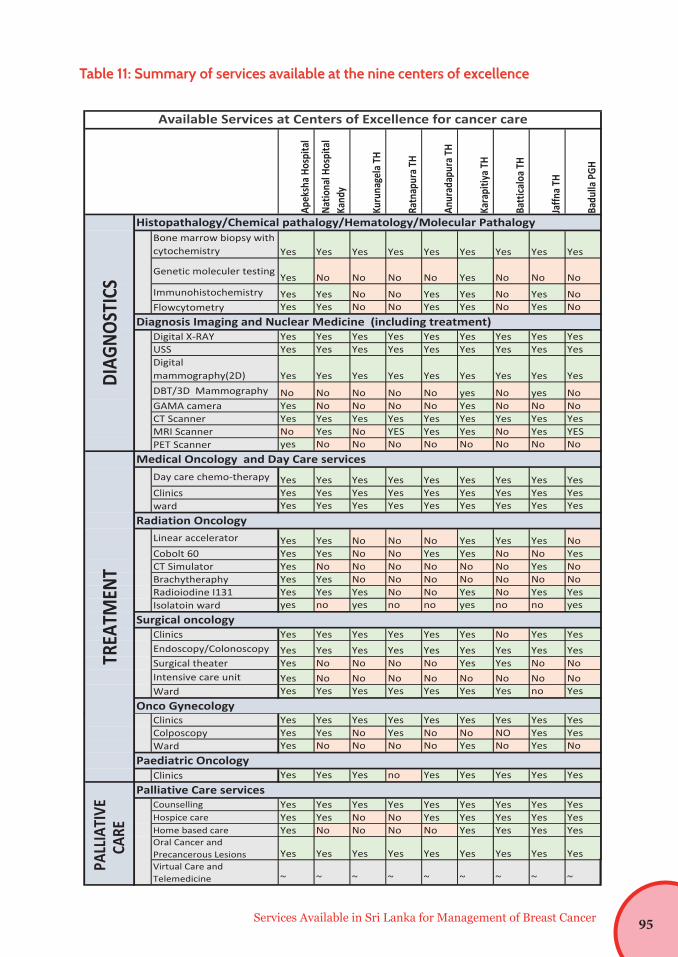
Table 11: Summary of services available at the nine centers of excellence
Services Available in Sri Lanka for Management of Breast Cancer
Apek
sha H
ospi
tal
Natio
nal H
ospi
tal
Kand
y
Kuru
nage
la TH
Ratn
apur
a TH
Anur
adap
ura T
H
Kara
pitiy
a TH
Batti
calo
a TH
Jaffn
a TH
Badu
lla P
GH
Bone marrow biopsy with cytochemistry Yes Yes Yes Yes Yes Yes Yes Yes Yes
Genetic moleculer testing Yes No No No No Yes No No NoImmunohistochemistry Yes Yes No No Yes Yes No Yes NoFlowcytometry Yes Yes No No Yes Yes No Yes No
Digital X-RAY Yes Yes Yes Yes Yes Yes Yes Yes YesUSS Yes Yes Yes Yes Yes Yes Yes Yes YesDigital mammography(2D) Yes Yes Yes Yes Yes Yes Yes Yes YesDBT/3D Mammography No No No No No yes No yes NoGAMA camera Yes No No No No Yes No No NoCT Scanner Yes Yes Yes Yes Yes Yes Yes Yes YesMRI Scanner No Yes No YES Yes Yes No Yes YESPET Scanner yes No No No No No No No No
Day care chemo-therapy Yes Yes Yes Yes Yes Yes Yes Yes YesClinics Yes Yes Yes Yes Yes Yes Yes Yes Yesward Yes Yes Yes Yes Yes Yes Yes Yes Yes
Linear accelerator Yes Yes No No No Yes Yes Yes NoCobolt 60 Yes Yes No No Yes Yes No No YesCT Simulator Yes No No No No No No Yes NoBrachytheraphy Yes Yes No No No No No No NoRadioiodine I131 Yes Yes Yes No No Yes No Yes YesIsolatoin ward yes no yes no no yes no no yes
Clinics Yes Yes Yes Yes Yes Yes No Yes YesEndoscopy/Colonoscopy Yes Yes Yes Yes Yes Yes Yes Yes YesSurgical theater Yes No No No No Yes Yes No NoIntensive care unit Yes No No No No No No No NoWard Yes Yes Yes Yes Yes Yes Yes no Yes
DIAG
NOST
ICS
Histopathalogy/Chemical pathalogy/Hematology/Molecular Pathalogy
Diagnosis Imaging and Nuclear Medicine (including treatment)
Available Services at Centers of Excellence for cancer care
Surgical oncology
Onco Gynecology
TREA
TMEN
T
Medical Oncology and Day Care services
Radiation Oncology
Clinics Yes Yes Yes Yes Yes Yes Yes Yes YesColposcopy Yes Yes No Yes No No NO Yes YesWard Yes No No No No Yes No Yes No
Clinics Yes Yes Yes no Yes Yes Yes Yes Yes
Counselling Yes Yes Yes Yes Yes Yes Yes Yes YesHospice care Yes Yes No No Yes Yes Yes Yes YesHome based care Yes No No No No Yes Yes Yes YesOral Cancer and Precancerous Lesions Yes Yes Yes Yes Yes Yes Yes Yes YesVirtual Care and Telemedicine ~ ~ ~ ~ ~ ~ ~ ~ ~PA
LLIA
TIVE
CA
RE
Palliative Care services
TREA
TMEN
T
Paediatric Oncology
95
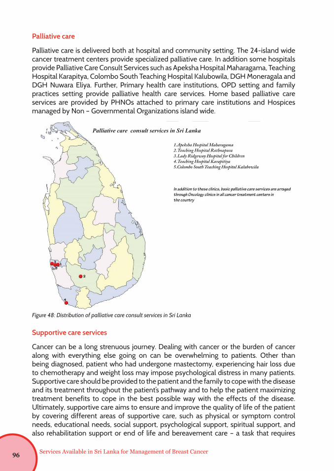
Services Available in Sri Lanka for Management of Breast Cancer
Palliative care
Palliative care is delivered both at hospital and community setting. The 24-island wide cancer treatment centers provide specialized palliative care. In addition some hospitals provide Palliative Care Consult Services such as Apeksha Hospital Maharagama, Teaching Hospital Karapitya, Colombo South Teaching Hospital Kalubowila, DGH Moneragala and DGH Nuwara Eliya. Further, Primary health care institutions, OPD setting and family practices setting provide palliative health care services. Home based palliative care services are provided by PHNOs attached to primary care institutions and Hospices managed by Non – Governmental Organizations island wide.
Supportive care services
Cancer can be a long strenuous journey. Dealing with cancer or the burden of cancer along with everything else going on can be overwhelming to patients. Other than being diagnosed, patient who had undergone mastectomy, experiencing hair loss due to chemotherapy and weight loss may impose psychological distress in many patients. Supportive care should be provided to the patient and the family to cope with the disease and its treatment throughout the patient’s pathway and to help the patient maximizing treatment benefits to cope in the best possible way with the effects of the disease. Ultimately, supportive care aims to ensure and improve the quality of life of the patient by covering different areas of supportive care, such as physical or symptom control needs, educational needs, social support, psychological support, spiritual support, and also rehabilitation support or end of life and bereavement care – a task that requires
15
Palliative care
Palliative care is delivered both at hospital and community setting. The 24-island wide
cancer treatment centers (specialist palliative care), Apeksha Hospital, Teaching
Hospital Ratnapura, Lady Ridgeway Hospital for Children, Teaching Hospital Karapitya,
Colombo South Teaching Hospital Kalubowila, (Palliative care consult services) and
Primary health care institutions / OPD setting / Family practice setting provide
palliative health care at hospital setting. Similarly, community based palliative care
services are provided by PHNOs attached to primary care institutions and Hospices
managed by Non – Governmental Organizations island wide.
Figure 4: Distribution of palliative care consult services in Sri Lanka
Palliative care consult services in Sri Lanka
Figure 48: Distribution of palliative care consult services in Sri Lanka
96
Services Available in Sri Lanka for Management of Breast Cancer
multidisciplinary cooperation and coordination. The supportive care needs are not the same for every breast cancer patient and differ according to the stage of the disease (early, advanced or metastatic breast cancer), the age of the patient (e.g. young women with childbearing potential or aged women), the individual social and cultural context, or simply according to individual perceptions. Moreover, needs may also change during the course of disease. Most important, however, is that they must be handled timely and appropriately.
Psychological support
For patients being identified as being at risk of anxiety and depression, psychological support is more important, and appropriate referral mechanisms and access to psychosocial experts are needed to ensure appropriate psychosocial care. Therefore, it is necessary to assess the patient and refer to a counseling center based on the need. There may be a necessity to refer family members or caregivers also for counseling (refer chapter 11). The psychological support will be available free of charge at all the cancer treatment centers and other Teaching Hospitals, Provincial/ District general hospitals and Base hospitals around the country.
Pain management
Treatment-related pain is often acute problem in breast cancer patients, and symptom management therefore is of the highest priority. National and international guidelines have been put in place to provide best possible pain management for breast cancer patients.
Social support
Equally important for the patient is the social support aspect of cancer care which involves obtaining financial assistance from the government. Cancer patients can seek financial assistance for cancer treatment and surgery from the President’s fund after submitting the necessary documents according to their website. Moreover, further financial, and social assistance is provided by the Department of Social Services after evaluation by a social service officer attached to the divisional secretariat office.
In addition to financial assistance, Government officers diagnosed with cancer are privileged to take leave under the establishment code. In sub section 1:3 Chapter XXIII of the Establishments Code, Special leave with full pay can be granted for a period not exceeding 06 months. An additional period of leave not exceeding 03 months can be granted with full pay if the Medical Board recommends at the end of the special leave of 06 months.
In addition, there are Non-Governmental Organizations (NGO) around the country carrying out services, providing various supportive services, including counseling, home based care, provision of wigs and breast prosthesis for cancer patients to uplift their quality of life and the lives of their loved ones. Among them the following Non-Governmental Organizations (NGO) works closely with the National Cancer Control Programme and cancer treatment centers of Ministry of Health, to provide psychosocial support for cancer patients.
97
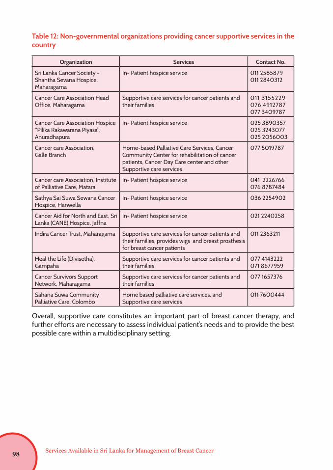
Services Available in Sri Lanka for Management of Breast Cancer
Table 12: Non-governmental organizations providing cancer supportive services in the country
Organization Services Contact No.
Sri Lanka Cancer Society - Shantha Sevana Hospice, Maharagama
In- Patient hospice service 011 2585879 011 2840312
Cancer Care Association Head Office, Maharagama
Supportive care services for cancer patients and their families
011 3155229 076 4912787 077 3409787
Cancer Care Association Hospice “Pilika Rakawarana Piyasa”, Anuradhapura
In- Patient hospice service 025 3890357 025 3243077 025 2056003
Cancer care Association, Galle Branch
Home-based Palliative Care Services, Cancer Community Center for rehabilitation of cancer patients, Cancer Day Care center and other Supportive care services
077 5019787
Cancer care Association, Institute of Palliative Care, Matara
In- Patient hospice service 041 2226766 076 8787484
Sathya Sai Suwa Sewana Cancer Hospice, Hanwella
In- Patient hospice service 036 2254902
Cancer Aid for North and East, Sri Lanka (CANE) Hospice, Jaffna
In- Patient hospice service 021 2240258
Indira Cancer Trust, Maharagama Supportive care services for cancer patients and their families, provides wigs and breast prosthesis for breast cancer patients
011 2363211
Heal the Life (Divisetha), Gampaha
Supportive care services for cancer patients and their families
077 4143222 071 8677959
Cancer Survivors Support Network, Maharagama
Supportive care services for cancer patients and their families
077 1657376
Sahana Suwa Community Palliative Care, Colombo
Home based palliative care services. and Supportive care services
011 7600444
Overall, supportive care constitutes an important part of breast cancer therapy, and further efforts are necessary to assess individual patient’s needs and to provide the best possible care within a multidisciplinary setting.
98
Services Available in Sri Lanka for Management of Breast Cancer
References
1. Antypas C, Sandilos P, Kouvaris J, et al. Fetal dose evaluation during breast cancer radiotherapy. Int J Radiat Oncol Biol Phys 1998;40(4):995–9
2. Aviles A, Neri N. Hematological malignancies and pregnancy: a final report of 84 children who received chemotherapy in utero. Clin Lymphoma 2001;2(3):173–7.
3. Aviles A, Neri N, Nambo MJ. Long-term evaluation of cardiac function in children who received anthracyclines during pregnancy. Ann Oncol 2006;17(2):286–8.
4. Azim Jr HA, Peccatori FA, Liptrott SJ, et al. Breast cancer and pregnancy: how safe is trastuzumab? Nat Rev Clin Oncol 2009;6(6):367–70.
5. Bader AA, Schlembach D, Tamussino KF, et al. Anhydramnios associated with administration of trastuzumab and paclitaxel for metastatic breast cancer during pregnancy. Lancet Oncol 2007;8(1):79–81.
6. Balawardena J, Skandarajah T.,Rathnayake W., Joseph N., ‘Breast Cancer Survival in Sri Lanka’, JCO Global Oncol 6:589-599
7. Beale JM, Tuohy J, McDowell SJ. Herceptin (trastuzumab) therapy in a twin pregnancy with associated oligohydramnios. Am J Obstet Gynecol 2009;201(1):e13–4.
8. Berveiller P, Mir O, Sauvanet E, et al. Ectopic pregnancy in a breast cancer patient receiving trastuzumab. Reprod Toxicol 2008;25(2):286–8.
9. Buckman, R. Breaking bad news: the S-P-I-K-E-S strategy. Community Oncology 2005; 2: 183-142
10. Cohen Y, Tatcher M, Robinson E. Radiotherapy in pregnancy. A case report with estimation of the dose to the fetus. Radiol Clin Biol 1973;42(1):34–9.
99
References
11. Dale DC, Cottle TE, Fier CJ, et al. Severe chronic neutropenia: treatment and follow-up of patients in the Severe Chronic Neutropenia International Registry. Am J Hematol 2003;72(2):82–93.
12. Department of Census & Statistics http://www.statistics.gov.lk/Duncan PG, Pope WD, Cohen MM, et al. Fetal risk of anesthesia and surgery during pregnancy. Anesthesiology 1986;64(6):790–4
13. Fanale MA, Uyei AR, Theriault RL, et al. Treatment of metastatic breast cancer with trastuzumab and vinorelbine during pregnancy. Clin Breast Cancer 2005;6(4):354–6.
14. Gilbert FJ, Tucker L, Gillan MGet al. Accuracy of Digital Breast Tomosynthesis for Depicting Breast Cancer Subgroups in a UK Retrospective Reading Study (TOMMY Trial). Radiology. 2015 Dec;277(3):697-706. doi: 10.1148/radiol.2015142566. Epub 2015 Jul 15. PMID: 26176654.
15. Guidance on screening and symptomatic breast imaging- The Royal College of Radiologists. Published 2019. https://www.rcr.ac.uk/publication/guidance-screening-and-symptomatic-breast-imaging-fourth-edition
16. Goldhirsch A, Ingle JN, Gelber RD, et al. Thresholds for therapies: highlights of the St Gallen International Expert Consensus on the primary therapy of early breast cancer 2009. Ann Oncol 2009;20(8):1319–29
17. Hoskin PJ and Ostler P. Clinical Oncology: Basic Principles and Practice. 2020. CRC Press. Chapter 8: Breast Cancer. pp 84-110.
18. Isaacs RJ, Hunter W, Clark K. Tamoxifen as systemic treatment of advanced breast cancer during pregnancy – case report and literature review. Gynecol Oncol 2001;80(3):405–8
19. Kouvaris JR, Antypas CE, Sandilos PH, et al. Postoperative tailored radiotherapy for locally advanced breast carcinoma during pregnancy: a therapeutic dilemma. Am J Obstet Gynecol 2000;183(2):498–9. 41.
100
References
20. Ni Mhuireachtaigh R, O’Gorman DA. Anesthesia in pregnant patients for nonobstetric surgery. J Clin Anesth 2006;18(1):60–6
21. Lawrence, R. A. and Lawrence, R. M. (2016) ‘Medical complications of the mother’ in Breastfeeding: A guide for the medical profession. 8th edn. USA: Elsevier, pp. 592 – 594.
22. Leal, S.C. Stuart, S.R. Carvalho, H de A. (2013) ‘Breast irradiation and lactation: a review’. Expert Rev Anticancer Ther. 13(2), pp.159-164. doi:10.1586/era.12.178.
23. Liyanage UA, 2016. Evolving role of radiological imaging in early detection of breast cancer: beyond 2D mammography. Sri Lanka Journal of Surgery, 34(1), pp.11–14. DOI: http://doi.org/10.4038/sljs.v34i1.8226
24. Liyanage, U.A. and Samaranayake, U.M.J.E., 2019. Evidence based mammography screening: is evidence changing? Sri Lanka Journal of Surgery, 37(2), pp.12–16. DOI: http://doi.org/10.4038/sljs.v37i2.8625
25. Luis SA, Christie DR, Kaminski A, et al. Pregnancy and radiotherapy: management options for minimising risk, case series and comprehensive literature review. J Med Imaging Radiat Oncol 2009;53(6):559–68.
26. Mandelson MT. Breast Density as a Predictor of Mammographic Detection: Comparison of Interval- and Screen-Detected Cancers. J Natl Cancer Inst . 2000 Jul 5;92(13):1081–7. http://dx.doi.org/10.1093/jnci/92.13.1081
27. Meyer-Wittkopf M, Barth H, Emons G, et al. Fetal cardiac effects of doxorubicin therapy for carcinoma of the breast during pregnancy: case report and review of the literature. Ultrasound Obstet Gynecol 2001;18(1):62–6.
28. Mir O, Berveiller P, Ropert S, et al. Emerging therapeutic options for breast cancer chemotherapy during pregnancy. Ann Oncol 2008;19(4):607–13.
29. National Control Programme: National Cancer Registry https://www.nccp.health.gov.lk/en
101
References
30. National Institute of Health and Care Excellence. NICE clinical guidelines (CG 164): Familial breast cancer
31. National Control Programme: Pathology based cancer incidence data 2017
32. Pant S, Landon MB, Blumenfeld M, et al. Treatment of breast cancer with trastuzumab during pregnancy. J Clin Oncol 2008;26(9):1567–9.
33. Peiris H.H., Mudduwa L.K.B., Thalagala N.I., Jayatilaka K.A.P.W. ‘Do Breast Cancer Risk Factors Affect the Survival of Breast Cancer Patients in Southern Sri Lanka?’, Asian Pac J Cancer Prev, 18 (1), 69-79
34. Sanson BJ, Lensing AW, Prins MH, et al. Safety of lowmolecular-weight heparin in pregnancy: a systematic review. Thromb Haemost 1999;81(5):668–72.
35. Sekar R, Stone PR. Trastuzumab use for metastatic breast cancer in pregnancy. Obstet Gynecol 2007;110(2 Pt 2):507–10.
36. Shrim A, Garcia-Bournissen F, Maxwell C, et al. Favorable pregnancy outcome following trastuzumab (herceptin) use during pregnancy – case report and updated literature review. Reprod Toxicol 2007;23(4):611–3.
37. Van der Giessen PH. Measurement of the peripheral dose for the tangential breast treatment technique with Co-60 gamma radiation and high energy X-rays. Radiother Oncol 1997;42(3):257–64. 37. Ngu SL, Duval P, Collins C. Foetal radiation dose in radiotherapy for breast cancer. Australas Radiol 1992;36(4):321–2.
38. Van Calsteren K, Verbesselt R, Ottevanger P, et al. Pharmacokinetics of chemotherapeutic agents in pregnancy: a preclinical and clinical study. Acta Obstet Gynecol Scand 2010;89:1338–45.
39. Van Calsteren K, Berteloot P, Hanssens M, et al. In utero exposure to chemotherapy: effect on cardiac and neurologic outcome. J Clin Oncol 2006;24(12):e16–7.
40. Victora, C. G. Bhal, R. Barros, A. J. D. Franca, G. V. A. Hortan, S. Krasevec, J. Murch, S. Sankar, M. J. Walker, N. Rollins, N. J. (2016) ‘Breastfeeding in the 21st century: epidemiology, mechanisms and lifelong effect’ The Lancet, 387 (January 30), pp. 475 -490.
102
References
41. Warraich Q, Smith N. Herceptin therapy in pregnancy: continuation of pregnancy in the presence of anhydramnios. J Obstet Gynaecol 2009;29(2):147–8.
42. Watson WJ. Herceptin (trastuzumab) therapy during pregnancy: association with reversible anhydramnios. Obstet Gynecol 2005;105(3):642–3. 62.
43. Waterston AM, Graham J. Effect of adjuvant trastuzumab on pregnancy. J Clin Oncol 2006;24(2):321–2.
44. Weber-Schoendorfer C, Schaefer C. Trastuzumab exposure during pregnancy. Reprod Toxicol 2008;25:390–1.
45. Witzel ID, Muller V, Harps E, et al. Trastuzumab in pregnancy associated with poor fetal outcome. Ann Oncol 2008;19(1):191–2.
103

104