Embed Size (px)
Citation preview
Handling and Nursing Reptiles
(What’s Normal & What’s Not)
Michael Cannon BVSc, MACVSc, Grad Dip Ed
Cannon & Ball Veterinary Clinic
461 Crown Street 2750
West Wollongong, NSW, 2500
Robert Johnson BVSc MACVSc IVAS
South Penrith Veterinary Clinic
South Penrith NSW Australia
Introduction
The examination procedure should not begin as the client walks into the consulting room, but with
the client’s first contact with the Veterinary Hospital – in most cases this will be with a telephone
call. A well-trained Veterinary Nurse can give this procedure a good foundation by giving clear
instructions to the client before they come. This will help make the examination more fruitful.
Client Instructions Prior to Attending
If it is cold or windy, take the necessary steps to protect the reptile.
Closely examine the cage floor prior to coming in. Bring in the cage floor covering or a sample
of any vomitus, diarrhoea etc.
Bring any medications etc. that have been used.
Make sure that the reptile is accompanied by someone who knows the current history. If they
are unable to attend have a telephone contact available, or provide a detailed written history.
You may find it useful to place the following list near the telephone for you or your nurse to discuss
with a client seeking advice about an ill reptile. The list includes clinical signs, easily recognisable
by the concerned reptile owner, which may suggest significant problems for the reptile:
General Signs
Weight loss
Dehydration
Abnormal odour
Seeking areas of higher temperature (behavioural fever)
Changes to skin or mucous membranes
Reduction in appetite or complete cessation of eating
Inability to adequately swallow or manipulate food within the mouth
Swelling around head, mouth or neck
Vomiting/regurgitation or rejection of food
Inactivity
A break in reptile’s routine – abnormal behaviour
Visible lumps or masses – anywhere on the body
Bleeding – always should be treated as an emergency situation
Abnormal skin shedding (dysecdysis)
Eyes
Unilateral or bilateral discharges
Changes in clarity or colour
Closing of one or both eyes –partial (squinting) or complete
Swelling around one or both eyes
Nostrils/Nares
Unilateral or bilateral discharges
Plugging of one or both nostrils
Bubbles appearing at nares
Respiratory
Open-mouthed breathing when at rest (very serious)
Audible respiratory noises (Sneezing, wheezing, gasping etc.)
Forks of tongue not separating (snakes & varanids)
Musculoskeletal/Neurological
Balance problems
Inability to coil on branches or logs
Limping or lack of full weight-bearing on one limb
Swollen foot/feet and or joint/joints
Scats
Change in quality and/or quantity of components of scats
Handling & Transport
Many different containers are used to transport reptiles including bags, pillow slips, boxes and
bins. It should be noted that any container used to transport a reptile should be leak proof, crush
proof and secure. Special conditions apply for the transport of venomous species. Confidence,
knowledge and assistance are required when handling reptiles. Depending on the order and
species various precautions need to be taken.
Chelonia
Glands either end of the bridge (between plastron and carapace) discharge a pungent fluid.
Side-necked turtles – All except one Australian freshwater species are side-necked. The
necks are usually difficult to straighten and examine.
Restrain by holding the caudal or lateral edge of the carapace. Avoid the mouth in Emydura
spp (short-necked).
Lizards
Tails may drop off when handling some lizards (small skinks and geckoes).
Some may bite, e.g. Blue-tongued lizards, bearded dragons and eastern water dragons.
Some may bite, scratch and flick their tail, e.g. varanids (monitors).
Snakes
Pythons of all sizes may bite and constrict
Venomous snakes must be accurately identified before the physical examination and only
handled if the handler is experienced and known by the veterinarian to be competent. There is
no need for any veterinarian to “head” or catch a venomous snake. Never put your hand in a
“snake bag” without knowing EXACTLY what is in it!
1. History
The time spent collecting the history should allow the reptile to settle down from the excitement of
transport. Use this time to perform a distant examination of the patient to look for subtle signs of
disease.
The basic questions that should be asked are:
If the reptile was acquired recently, from what source? Wild caught or captive bred?
Reptiles that have recently been through a transfer may have been stressed or exposed to a host
of infectious agents and environmental conditions. Some sources are know by reputation to be
either good or bad.
What is the animal’s age? Often this is unknown. If it was hatched recently ha it undergone its
first slough (ecdysis).
How long has the reptile been in its present environment? Is the animal settling in to a new
environment or is it well established. New arrivals are more likely to suffer from stress and
acute problems such as infectious diseases. Established animals are more likely to suffer from
husbandry problems, dietary problems or chronic diseases such as neoplasia or parasites.
Any recent changes to the environment? This may indicate sources of stress or toxins.
The size and type of cage? Pay particular attention to the nature of the floor surface or
substrate. What are the climbing and hiding facilities provided? Is the reptile normally kept in
an indoor enclosure or an outdoors aviary? If it is in an aviary ask for a description noting the
size, floor type, design, aspect it faces and any preventative medicine program currently in use?
The frequency of cage cleaning and the cleaning methods used? This is a means of
determining the client’s approach to hygiene and some potential sources of toxins.
The range of temperature and humidity in the cage? Is there a temperature gradient in the
cage? Is there a basking spot and what is the temperature where the reptile is able to bask?
Water supply? Determine the source and quality of the water and the type of water container
used. How often is the water changed? Are there bathing facilities? What is the size and how
often it is used?
Light sources? Type of light provided (incandescent, fluorescent). What is the availability of
ultraviolet light or exposure to direct sunlight? What is the normal photoperiod provided? Many
reptiles housed indoors are subject to internal lighting as people are in the room. This can result
in them being exposed to very long light intervals – much longer than the natural interval.
Describe any cage mates? Pay attention to size, gender, species and any interactions. What is
their health status? Any new introductions to or departures from the enclosure – when was the
most recent? Any other reptiles acquired recently in the collection? Also question about
quarantine procedure used.
What are the characteristics of the diet? Nutritional disease is extremely common. Many
reptiles are suffering from malnutrition (especially obesity).
- Type of food items provided
- Quantity offered
- Frequency of feeding
- Method of presentation
- Any supplements provided
- When was the last meal
Describe the recent scats (faeces/urates/urine) passed? What was its relation to feeding
(normally should be passed within 4-5 days of a feed)?
Describe the most recent skin shedding (ecdysis)? When did it occur? Was it normal?
Describe normal handling? How often is it done? Describe the techniques used? Is the reptile
a pet that is handled regularly?
Have there been any changes to the reptile’s behaviour? Behavioural changes are often the
first subtle signs that are noticeable. These are often overlooked. Has the reptile been less
active or sitting in an unexpected position? Does the reptile respond differently to the owner’s
presence or approach? Are there periods of excessive inactivity or hiding?
What are the current signs of disease that are causing concern & how long have they been
evident? Describe these signs of this patient or of any other reptiles on the premises.
What treatments or medications have been used? What was the response to these? Check
the dose, frequency etc. Always ask the client to describe how the drug was used to rule out any
errors in administration.
2. Examination of Environment
Often this is not available as the animals come in a cloth bag or small travel cage. Much of the
information regarding the environment will be gathered during the history taking or a visit to their
home.
While the history is being gathered you can also quickly visually assess the animal, its current
environment or any scats that have been passed, to glean any further information that may be
available. Take note of such details as cleanliness of the water dish or transport enclosure. Use this
observation to make an assessment of the general level of hygiene and sanitation.
3.Distant Examination
Spend a little time assessing the reptile’s demeanour (alert and responsive). Is it normally active?
Are there any obvious physical abnormalities? Assess respiratory rate and depth. Make a subjective
assessment of the reptile’s weight by assessing the level of fat deposition at the base of the tail or at
the latero-dorsal body wall.
Make an assessment of general appearance and behaviour1.
Chelonians1: Bright, alert and responsive. Swim evenly balanced in the water. When first
caught, they pull their head and limbs beneath the carapace and strongly hold them in when you
try to remove them. When walking, they support all the weight on their legs. The plastron bears
no weight and does not touch the ground.
Lizards1: They stand high and strongly on all four limbs. The body and limb muscles are well
rounded and firm. There is no excessive folding or creasing of the skin. They run away quickly
when you attempt to catch them.
Snakes1: The body is flat on the substrate with the weight borne by the ventral scales. They
respond to you approaching them by moving their head and flicking their tongue. If they feel
threatened they will form a coil. If not they will begin to explore the surroundings.
Crocodilians1: They have a very sedentary life, rarely moving unless threatened or at feeding
time. Resent handling and restraint. Will thrash their head and neck, trying to bite, while
lashing their tail in your direction.
4. Physical Examination
Collect all the equipment you will require before moving onto the physical examination. This
allows you to be as efficient as possible with your examination and causes less stress for the reptile.
The reptile should be given a complete physical examination. I prefer to start at the head and move
distally. You should develop your approach to the examination as a habit and carry out the
examination the same way each time so that nothing is overlooked. Pay particular emphasis to any
suggestive signs detected during the history and distant examination, so that they may be explored
in more detail.
Begin by examining the head, eyes, ears and nares (pay particular attention to the nares as dried
discharge may occlude on or both- this is the equivalent of a “runny” nose). The head should be
symmetrical.
Examine the skin creases around the eyes ventral neck and labial pits as this is often a location
to detect mites. Avoid handling snakes and lizards if the animal is in ecdysis.
Assess the hydration status. Signs of dehydration are: decreased turgor – skin tents when pulled
from the animal; skin is standing in multiple folds; abnormal ecdysis; in snakes and some
lizards, spectacle is shrunken and opaque; sunken eyes in lizards, chelonians and crocodilians.
Open the mouth and examine the tongue, teeth, gums, other oral structures and pharynx – note
any sour or abnormal odours. Is there excessive mucous or are cheesy plaques or petechiae
present? Examine the gum margins carefully for any sign of necrosis or ulceration.
Carefully palpate the entire body, beginning at the neck and working down the body. Gently
palpate the ventral coelomic cavity between the ribs. Feel for organs and any firm masses or
fluid accumulation. Take care if the animal has been fed recently that you do not place pressure
on the ingesta as it is passing through the intestine. An animal that has eaten recently may
regurgitate if the stress of handling is too great.
Locate the cardiac impulse and auscultate the heart and surrounding lungs. Auscultate the chest
(dorsal and ventral) and the abdomen. Heart rate is extremely variable and may be difficult to
count. The sounds of inspiration are louder and shorter in duration than expiration. Abnormal
respiratory wheezes or whistling are signs of severe pathology. Most unrestrained reptile s will
have 1-2 respirations per minute. Healthy animals breathe with the mouth closed. Rate and
depth of respiration will vary with the ambient temperarture.
The body wall muscle mass should have firm muscle tone The lower coelomic cavity contents
are often easily palpable. If the abdomen is enlarged palpation must be extremely gentle. The
reptile should not demonstrate pain with normal, gentle abdominal palpation.
Examine the cloaca and vent for swellings, encrustations or soiling (indicative of loose
droppings). If the tail is gently flexed dorsally, the vent will open slightly and allow inspection
of the cloacal mucosa or insertion of a swab or sexing probe.
Most reptiles will pass faeces that is formed. Urates are often passed at the same time.
Normally they precede the stool. Urates are normally chalky paste that is pure white to yellow
in colour. Terrestrial reptiles will also pass a small amount of clear liquid urine.
Biliverdinuria (green discoloration of the urates) is abnormal and is usually associated with
liver disease, anorexia or haemolytic anaemia. The reptiles have biliverdin rather than bilirubin.
Any abnormal faeces should be examined, by performing a faecal flotation and fresh warm,
saline smear.
Examine the skin. Look for any missing scales or scars. Assess any lumps or swellings present.
Pull out each limb individually. Palpate the bones from shoulder or hip to distal end of each
digit, paying particular attention to the joints. Examine each joint for full range of motion or
any swelling. A weak limb may be indicative of abdominal tumours, fractures or neurological
disease. Unilateral lameness is more common than bilateral. Assess the length of the claws.
Overgrown claws may be associated with poor substrate. Inspect the plantar aspect of each foot.
This whole procedure should take less time to perform than it takes to read.
Once the examination is finished immediately place the reptile back into its cage and assess its
tolerance of the procedure. Most reptiles will return to normal behaviour. If the reptile looks
obviously stressed or is breathing heavily, it is safe to assume it is ill.
Hints for Specific Reptile Groups
The technique for physical examination of reptiles is similar in principle to that used for most
other pets. Animals must be adequately restrained but not distressed. Like most other animals,
reptiles resent oral examination.
CHELONIA
Chelonia are generally easy to handle. A healthy turtle can be surprisingly mobile.
Demeanour varies from species to species. Of the commonly occurring species the common
eastern long-necked turtle is quite docile. Short-necked turtles (Emydura spp) are usually
more mobile and liable to bite.
Turtles should be weighed and measured (straight carapace length [SCL]). A data base can be
developed and used to gauge body condition4.
Carapace and plastron injuries and lesions are common. A central groove in the carapace
deepens with age.
Skin lesions may be an indication of more serious disease.
Oral examination may be revealing in an otherwise healthy animal. Look for crusts,
ulceration and rostral abrasion. Abscesses occur frequently, especially around the eyes and
ears.
Oedema of the neck and limbs with accompanying petechial haemorrhages may indicate
septicaemia.
SQUAMATA
i.Lizards
The attitude of lizards varies greatly with species, age and state of health. The larger skinks
such as blue-tongued lizards (Tiliqua helonian) and shingle backs (Tiliqua rugosa rugosa)
are usually placid and used to handling. Bearded dragons, except for the eastern species
(Pogona barbata) are also quite docile. Eastern water dragons (Physignathus leseurii) are
flighty and can be aggressive as adults. Young lizards of this species may be “hypnotised” by
lying them on their backs and gently stroking their abdomens. This is similar to the condition
of “tonic immobility” reported in many animals, e.g. chickens.
Skin tenting, as in mammals, may indicate dehydration.
Dysecdysis (difficulty with sloughing) can cause constriction or strangulation of digits and
the tail tip in some species.
Mites occur frequently, especially in blue-tongued lizards.
Abnormalities in muscle tone and posture, fitting and muscle fasciculation may indicate
metabolic bone disease (MBD). Lizards may also exhibit hindlimb and tail paresis. Eastern
water dragons are more commonly affected and sometimes present as a “neurological”
problem. A rubbery mandible occurs in the more chronic form of MBD.
Examination of the mouth is difficult in many lizard species. Guitar plectra are useful for
prying open mouths in skinks and agamids. Nematode infestation of the pharynx occurs
commonly in bearded dragons. Stomatitis presents as swollen and malodorous gingivae.
Abdominal swelling may occur for a variety of reasons; a full stomach, eggs, tumours and
foreign bodies. Agamids, especially smaller ones, may gorge themselves on invertebrates and
consequently have a very swollen stomach. Tumours are not common. Eggs may be palpated
in the agamidae and easily differentiated from other swellings. Monitor lizards are scavengers
and may ingest bone scraps that lead to intestinal or gastric obstruction.
ii. Snakes
Restraining the head of a python will usually cause it to struggle, whereas if it is handled
gently by supporting the body only it will tend to relax. Snakes will excrete voluminous
faeces and urates if distressed. Australian pythons, except for the scrub python (Morelia
amethistina) and water python (Liasis fuscus), are usually easy to handle. Venomous snakes
should be expertly restrained for the physical examination. It is recommended that only
experienced reptile veterinarians examine and treat venomous species.
Initially the snake should be weighed and its movements closely observed, watching for any
unusual coiling, twisting motions or weakness of the distal body and decreased constriction.
Flaccidity or any neurological sign in a python, especially Morelia spp may indicate
Inclusion Body Disease (IBD).
Snakes with respiratory disease may “mouth breathe”. Gentle and patient palpation is then
required for a closer examination. Inspection of the head and mouth should be left as a final
procedure as the snake will frequently become distressed when its mouth is opened.
The snake is a conveniently linear animal and charts have been made to facilitate organ
location2. The heartbeat (approximately 25% of the snout/vent length [SVL]) can be palpated
in debilitated or anaesthetised pythons. It is difficult to feel in a healthy, well muscled snake.
Auscultation using a stethoscope is unrewarding.
The skin will vary in texture and smoothness depending on the state of hydration, ecdysis and
external parasitism. Unlike lizards and turtles, healthy snakes shed their skin in one piece.
Dysecdysis may be associated with external parasitism or inappropriate humidity.
Common Problems revealed at Physical Examination
i. Lumps in snakes
Lumps occur frequently in snakes for many reasons. Abscesses occur in most parts of the body.
Swelling 25% along the snout/vent length in Morelia spp. May indicate cardiac enlargement. The
author has also seen a case of a fatty tumour in the precardial fat pad causing a similar swelling.
Granulomata caused by ascarid infestation are common in the stomach and proximal small
intestine. They have a higher incidence in wild caught snakes and those fed live or “wild” prey
items. Hypertrophic gastritis due to cryptosporidiosis is mainly seen in elapids and presents as a
mid-body swelling. Sparganosis is common in wild caught snakes especially elapids and
colubrids (frog eaters). Lesions usually appear as small raised lumps in the subcutis. They occur
more commonly in the distal body. Tumours occur but are not common. Prey items and eggs can
be detected by patient palpation and good history taking.
ii. Cloacal region
Cloacal infection may occur after mating. The cloaca is usually swollen, inflamed and covered in
a crusty discharge. Abscessation of the hemipene sulcus and associated gland may be noted.
iii. Mouth
Oral examination is carried out by holding the head in the right hand (for those who are left
handed) and gently pulling down on the ventral skin using the thumb and middle finger to expose
the gingivae. A probe may be introduced by the left hand and used to lever the mouth open in
order to inspect the glottis and pharynx. Cotton buds should not be used as they catch on the
small, delicate teeth.
Stomatitis may be graded as follows:
1. early – petechiation of the gingivae and ptyalism
2. mild – swollen and malodorous gingivae, occasional pockets of pus
3. severe – abscessation and exposure of underlying bone
Check for purulent or cheesy material on the glottis. This may indicate respiratory disease.
iv. Head and eyes
Inspection of the head and eyes may reveal the presence of mites. They often hide in the
periorbital region and the heat sensing pits of pythons. Normally the conjunctivae are not
visible. Hypertrophy of the conjunctival tissue is usually a sign of Ophionyssus natricis
infestation.
Retained spectacle (post ecdysis) and subspectacular abscess occur commonly.
v. Ecdysis1
All reptiles have a periodic sloughing of the superficial, keratinised epithelium. This is termed
“ecdysis” and an abnormal slough is called “dysecdysis”.
1. Crocodilians: These slough small, pieces continually. As the old scales are worn away, they are
replaced.
2. Snakes: These usually slough the skin in one piece, including the spectacles covering each eye.
The skin takes on a dull sheen 1-2 weeks prior to ecdysis. The spectacles become a milky blue
colour (similar to that seen with keratitis in a mammal) and the snake has poor vision. Many
become more flighty or aggressive because of their vision deficit. Most snakes will reuse food
at this time. After 4-7 days, the spectacles clear and the slough begins 4-7 days later. The
slough begins with the snake rubbing its nose and chin on a rough surface. This dislodges the
skin from around the mouth and lips. The snake then crawls out of its sloughed skin, turning it
inside out. Snakes that lack a rough surface in their enclosure may have difficulty shedding and
will become quite distressed. Dysecdysis is characterised by the skin coming off in pieces and
shredding or single scales being shed.
3. Lizards: These have periodic sheds of large sections of the skin in pieces over several days.
They also require a rough surface (log or rock) to rub against to assist in dislodging the skin
pieces.
4. Chelonians: The skin covering the head, neck and limbs is shed in a similar fashion to lizards.
The upper epithelium of the scutes of the shell are shed 1-2 at a time and then replaced.
Frequency of ecdysis varies with food availability, species, age, growth rate and ambient
temperature in its environment. When the reptiles are eating regularly (Spring, Summer and early
Autumn) they grow more quickly and will slough more often – as frequently as once a month.
The skin after a slough, has a shiny, lustrous appearance. This becomes duller as it shows signs of
wear and tear.
5. Body Weight
This is an extremely useful tool in health assessment. The reptile should be placed upon a set of
scales (either triple beam balance or small electronic scales) and an accurate measurement taken. I
find it useful to place them in a cloth bag during this procedure. This minimises struggling and
panic. Objective weight measurement can be used as a regular monitor to assess the reptile and
should be recorded on the reptile’s file or card. As an in-hospital tool, objective measurement is an
excellent prognostic indicator for a reptile receiving treatment. Weight loss is always a sign of a
poor prognosis.
As well it is useful to make a subjective assessment of the reptile’s general condition after palpating
the body wall muscles. With time and experience this can be used to determine the optimum weight
for an individual reptile. This is a useful skill for clients to develop as a general health assessment
tool.
6. Snout: Cloaca Measurement (Body Length)
A measuring tape should be placed along the ventral aspect of the snake. Determine the distance
from the snout to the cloaca. Also determine to distance to any abnormal structures or masses.
This is a useful means of locating normal structures and identifying amy abnormal masses or
structures you may encounter.
Diagnostic Approach to Abnormal Internal masses in Snakes
Snakes often develop anorexia. On physical examination you may discover an internal mass.
This may be difficult to identify as you are uncertain if it is normal or abnormal.
Common causes: tumours; intestinal impaction; retained eggs or foeti; abscesses or
granulomas.
Snakes organs are elongated and overlap each other as they are distributed along the
coelomic cavity.
The position of most organs is constant within each species as a proportion of the total body
length. It is quite variable between species. McCracken1,2
studied several species of snakes to
make a table to allow more accurate determination of normal organ positioning. This is a
useful method of determining the source or organ involved in a problem.
The initial aim is to determine if the mass in intra or extra-intestinal.
History1
How long has the mass been present?
Any changes in the size of the mass since detected? Impactions should not increase.
Any other health problems been detected recently?
Date of last meal and defaecation? Faeces should pass within 1 week of feeding.
What is the ambient temperature and water availability in its enclosure? Inadequate ambient
temperature, humidity and drinking water, following feeding may cause constipation.
Physical Examination: Are there any other problems present? Is there excess fluid r gas in the
GIT.
Measurement: determine snout-vent length and compare it to snout-mass length (measure to
anterior aspect of the mass). Determine the ratio of snout-mass:snout-vent. Compare this to
the table developed.
Palpation: Assess hardness, texture, discreteness and ability to move. If it will advance
caudally, it is likely to be in the GIT or oviduct.
Radiology: Plain radiographs followed by Contrast studies if indicated.
Initially the mass should be treated as obstipation unless proven otherwise. Deliver paraffin
oil via a stomach tube or per rectum depending on the location of the mass. Allow the snake
to have a soak in warm water for several hours. Try gentle heloni to move the mass
caudally.
If there is no movement within 3 days, perform double contrast radiography. Use 5ml/kg
barium Sulphate and 45ml/kg air via a stomach tube. If the mass is demonstrated in the GIT
but does not move after 2-3 days more conservative therapy, perform an exploratory
coeliotomy.
If the mass is demonstrated to be outside the GIT, try the following steps:
Fine needle aspirate
Check for cryptosporidiosis
Perform Blood tests to assess the renal system (particularly Uric Acid)
Endoscopy, via the oesophagus if mass is anterior to the pylorus.
Perform an exploratory coeliotomy.
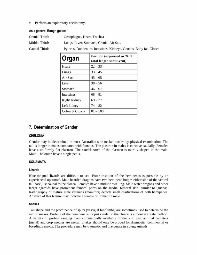
As a general Rough guide:
Cranial Third: Oesophagus, Heart, Trachea
Middle Third: Lungs, Liver, Stomach, Cranial Air Sac.
Caudal Third: Pylorus, Duodenum, Intestines, Kidneys, Gonads, Body fat, Cloaca
Organ Position (expressed as % of
total length snout-vent)
Heart 22 – 33
Lungs 33 – 45
Air Sac 45 – 65
Liver 38 – 56
Stomach 46 – 67
Intestines 68 – 81
Right Kidney 69 – 77
Left kidney 74 – 82
Colon & Cloaca 81 – 100
7. Determination of Gender
CHELONIA
Gender may be determined in most Australian side-necked turtles by physical examination. The
tail is longer in males compared with females. The plastron in males is concave caudally. Females
have a uniformly flat plastron. The caudal notch of the plastron is more v-shaped in the male.
Male helonian have a single penis.
SQUAMATA
Lizards
Blue-tongued lizards are difficult to sex. Exteriorisation of the hemipenes is possible by an
experienced operator6. Male bearded dragons have two hemipene bulges either side of the ventral
tail base just caudal to the cloaca. Females have a midline swelling. Male water dragons and other
larger agamids have prominent femoral pores on the medial femoral skin, similar to iguanas.
Radiography of mature male varanids (monitors) detects small ossifications of both hemipenes.
Absence of this feature may indicate a female or immature male.
Snakes
Tail shape and the prominence of spurs (vestigial hindlimbs) are sometimes used to determine the
sex of snakes. Probing of the hemipene sulci just caudal to the cloaca is a more accurate method.
A variety of probes, ranging from commercially available products to nasolacrimal catheters
(metal) and crop needles are useful. Snakes should only be probed for diagnostic, commercial or
breeding reasons. The procedure may be traumatic and inaccurate in young animals.
Probing Method
Two handlers are required for larger snakes. One person holds the front end and the other person
holds the distal end “belly up” with one hand about 10 cms cranial to the cloaca. Gently introduce
the sterilised (alcohol will do) and lubricated probe into one of the hemipene sulci which run
down either side of the tail. Males – probe – 10-12 subcaudal scales. Females – probe – 4
subcaudal scales. These lengths are only estimates. Probing depths may vary for different species
eg Morelia amethistina, the female has deep pockets, 6-8 scales.
8. Basic Diagnostic Procedures
The focus for this section is on procedures rather than different diagnostic tests and laboratory
analysis (i.e. histopathology, haematology, biochemistry).
8.1 Blood collection (and intravenous injection sites)
Chelonia
In most side-necked turtles the left jugular vein is used. The subcarapacial method7 has also been
described but is not used commonly.
Squamata
The ventral caudal tail vein is used for bleeding and intravenous injection in most lizards (skinks,
dragons, monitors). This route is also commonly used in snakes. Blood collection by cardiac
puncture and the palatine veins (large pythons) has also been described,8,9
but is not
recommended by the author in conscious animals. The heart is often difficult to locate in snakes.
In most cases a one millilitre tuberculin syringe and 25 gauge needle are adequate for
venipuncture. A three millilitre syringe and 23 gauge needle may be used in pythons larger than 3
kg and large monitors.
8.2 Cloacal wash
A small amount of warm saline may be introduced into the cloaca to obtain a diagnostic sample.
Protozoa and the ova of other endoparasites may be detected by direct faecal analysis.
8.3 Tracheal wash
A transtracheal aspirate is useful for microbiological and cytological analysis in cases of
respiratory disease. The glottis in turtles, snakes and monitors is conveniently situated in the
rostral part of the mouth. The technique is identical to that used in small mammal medicine.
8.4 Radiology
Radiology is a useful diagnostic aid. For example, it is essential for gender determination in
varanidae, assessing gravid turtles and MBD in lizards. The latter may be graded over the course
of treatment using a step wedge device (PT-11 Penetrometer [Eseco-Speedmaster, Oklahoma]).
Fractures of the carapace, plastron and limbs in chelonia and limb fractures in lizards are also
routinely radiographed. Fine detail mammography film is useful in small reptiles.
8.5 Other
Ultrasound, computerised tomography and MRI are used in the USA,UK and Europe as
diagnostic tools to varying degrees. Their usefulness appears limited in everyday reptile practice.
8.6 Endoscopy
Endoscopy is commonly used in reptile practice in the USA and UK, especially in iguanas and
increasingly in chelonia. Previously endoscopy was used mainly as a diagnostic tool to examine
and biopsy internal organs but recently it has been used for local, targeted medical therapy and
minimally invasive surgical procedures.10
In Australia where snakes are more popular, there
appears to be less demand for endoscopy. Palpation, transcutaneous biopsy and coeliotomy are
more commonly used for diagnostic purposes.
Summary
Physical examination and diagnosis in reptiles can be challenging, physically and intellectually.
First principles from mammalian veterinary practice are easily adapted to the class Reptilia.
Extensive knowledge of the normal animal is essential in order to recognise the abnormal.
References
McCracken, H.E. (1994). Husbandry and Diseases of Captive Reptiles. Proceedings Wildlife
Refresher Course. Post Graduate Foundation in Veterinary Science, University of Sydney. Pp.
461-545.
McCracken, H.E.. (1999). Organ Location in Snakes for Diagnostic and Surgical Evaluation. In
Zoo and Wild Animal Medicine: Current Therapy 4. ed Fowler, M.E. & Miller, R.W. WB
Saunders. Pp. 243-248.
Reiss, A. (1999). Current Therapy in Reptile Medicine. Proceedings Wildlife in Australia:
Healthcare & Management. Post Graduate Foundation in veterinary Science, University of
Sydney. Pp. 97-118.
Divers S, 1996. Basic reptile husbandry, history taking and clinical examination. In Practice,
February 1996. The Veterinary Record, pp 51-65.
McCracken H, 1994. Husbandry and diseases of captive reptiles. In: Wildlife, Proceedings 233,
pp 461-546. Post Graduate Committee in Veterinary Science, University of Sydney.
Shea G, Senior lecturer in Veterinary Anatomy, University of Sydney, personal communication.
Hernandez-Divers SM, Hernandez-Divers SJ and Wyneken J, 2002. Angiographic, anatomic and
clinical technique descriptions of a subcarapacial venipuncture site for chelonians. Journal of
Herpetological Medicine and Surgery, 12(2): pp 32-37.
Rosskopf WJ, Woerpel RW, Fudge A, Pitts BJ and Whittaker D, 1982. A practical method of
performing venipuncture in snakes. Exotic Practice, Vet Med Small Anim Clin, 77(5): pp 820-
821.
Raiti P, 2002. Snakes. In: Meredith A and Redrobe S (Eds.), BSAVA Manual of Exotic Pets,
Fourth Edition, pp 241-256. BSAVA, Gloucester.
Hernandez-Divers SJ and Hanley CS, 2003. Advances in reptile endoscopy and development of
minimally invasive surgical techniques in the green iguana (Iguana iguana). In: Proceedings,
Association of Reptilian and Amphibian Veterinarians, Tenth Annual Conference, Minneapolis,
p31.

Caring for Captive Reptiles Michael Cannon BVSc, MACVSc, Grad Dip Ed Cert BA
Cannon & Ball Veterinary Clinic
461 Crown Street 2750
West Wollongong, NSW, 2500
Robert Johnson BVSc MACVSc IVAS
South Penrith Veterinary Clinic
South Penrith NSW Australia
1. Introduction The most common cause of disease in captive reptiles is incorrect or poor husbandry. A sound
knowledge of reptile husbandry is required by the veterinarian in order to diagnose and treat
diseases appropriately. Environmental and dietary needs must be considered carefully. Specific
requirements of some species of reptiles will be mentioned. The emphasis in this lecture will be
on husbandry and not therapeutics.
2. Husbandry The eight H’s of husbandry:
Heat
Hide
Humidity
Health
Hygiene
Healthy appetite
Habitat
Handling
2.1 Heat (and light)
2.1.1 Heat
Reptiles are ectothermic vertebrates that regulate body temperature by behavioural and
physiological processes1. Reptiles should be housed at temperatures similar to field conditions,
providing temperature variation within the enclosure that allows the animal to choose its thermal
environment (thermoregulate)..2
A thermogradient or “mosaic” is achieved by having sufficient room to place heat sources
strategically within the enclosure. Two main types of heating are used by herpetologists, radiant
(lamps, ceramic globes) and convective (heat mats, tape).
“Hot rocks” are not recommended as heat sources. Snakes will tend to bask on them for extended
periods occasionally sustaining burns. This occurs more often in snakes that have recently
undergone ecdysis (sloughing). It has been shown that large reptiles rely primarily on radiant heat
sources for thermoregulation whereas smaller species tend to depend on convective sources.3
Terrestrial or ground dwelling snakes such as Childrens pythons (Antaresia childreni) prefer
subfloor heating. Ambient room temperature should be stable and not place undue stress on the
thermogradient in the vivarium. Mistakes are commonly made when enclosures are kept in rooms
subject to temperature extremes.
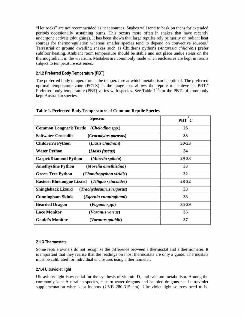
2.1.2 Preferred Body Temperature (PBT)
The preferred body temperature is the temperature at which metabolism is optimal. The preferred
optimal temperature zone (POTZ) is the range that allows the reptile to achieve its PBT.4
Preferred body temperature (PBT) varies with species. See Table 15,6
for the PBTs of commonly
kept Australian species.
Table 1. Preferred Body Temperature of Common Reptile Species
SSppeecciieess PPBBTT
oo
CC
CCoommmmoonn LLoonnggnneecckk TTuurrttllee ((CChheellooddiinnaa sspppp..)) 2266
SSaallttwwaatteerr CCrrooccooddiillee ((CCrrooccooddyylluuss ppoorroossuuss)) 3333
CChhiillddrreenn’’ss PPyytthhoonn ((LLiiaassiiss cchhiillddrreennii)) 3300--3333
WWaatteerr PPyytthhoonn ((LLiiaassiiss ffuussccuuss)) 3344
CCaarrppeett//DDiiaammoonndd PPyytthhoonn ((MMoorreelliiaa ssppiilloottaa)) 2299--3333
AAmmeetthhyyssttiinnee PPyytthhoonn ((MMoorreelliiaa aammeetthhiissttiinnaa)) 3333
GGrreeeenn TTrreeee PPyytthhoonn ((CChhoonnddrrooppyytthhoonn vviirriiddiiss)) 3322
EEaasstteerrnn BBlluueettoonngguuee LLiizzaarrdd ((TTiilliiqquuaa sscciinnccooiiddeess)) 2288--3322
SShhiinngglleebbaacckk LLiizzaarrdd ((TTrraacchhyyddoossaauurruuss rruuggoossuuss)) 3333
CCuunnnniinngghhaamm SSkkiinnkk ((EEggeerrnniiaa ccuunnnniinngghhaammii)) 3333
BBeeaarrddeedd DDrraaggoonn ((PPooggoonnaa sspppp..)) 3355--3399
LLaaccee MMoonniittoorr ((VVaarraannuuss vvaarriiuuss)) 3355
GGoouulldd’’ss MMoonniittoorr ((VVaarraannuuss ggoouullddiiii)) 3377
2.1.3 Thermostats
Some reptile owners do not recognise the difference between a thermostat and a thermometer. It
is important that they realise that the readings on most thermostats are only a guide. Thermostats
must be calibrated for individual enclosures using a thermometer.
2.1.4 Ultraviolet light
Ultraviolet light is essential for the synthesis of vitamin D3 and calcium metabolism. Among the
commonly kept Australian species, eastern water dragons and bearded dragons need ultraviolet
supplementation when kept indoors (UVB 280-315 nm). Ultraviolet light sources need to be
replaced according to manufacturer’s instructions. There is some discussion as to whether the
diamond python (Morelia spilota spilota) requires UVB supplementation in captivity.
2.2 Hide
All captive reptiles need somewhere to hide. Items such as toilet rolls or small cardboard boxes
are ideal for hatchling snakes and the smaller terrestrial varieties. When soiled, simply replace
them. Porcelain hides and inverted flower pots are also popular. These structures must be
waterproof and easy to disinfect. Certain species are more secretive compared with others e.g.
Antaresia spp (Childrens pythons).
Large vivaria should be furnished with several hides in a variety of positions in order to facilitate
thermoregulation. Hides can be used as an aid to handling. This is especially relevant to more
aggressive or venomous species. The entrance to a favourite shelter may be blocked securely and
then used to transport the reptile.
2.3 Humidity
Humidity requirements vary with species (40-80%). For example, the environment of a green
python (Morelia viridis) needs to be much more humid than that of the inland bearded dragon
(Pogona vitticeps). All snakes need a large water bowl for bathing and drinking. The humidity of
a vivarium can be controlled by altering the size of the surface are of the water bowl (a larger
bowl or tip the bowl on its size to change the surface area exposed. Always place it at the cooler
end of the exhibit, except in cases where a rapid increase in humidity is required. In these cases a
heat lamp or mat may be placed under the water container to aid evaporation. All vivaria, dry and
humid, should be adequately ventilated.
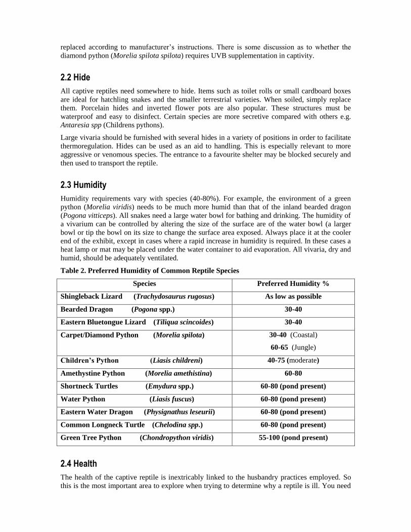
Table 2. Preferred Humidity of Common Reptile Species
Species Preferred Humidity %
Shingleback Lizard (Trachydosaurus rugosus) As low as possible
Bearded Dragon (Pogona spp.) 30-40
Eastern Bluetongue Lizard (Tiliqua scincoides) 30-40
Carpet/Diamond Python (Morelia spilota) 30-40 (Coastal)
60-65 (Jungle)
Children’s Python (Liasis childreni) 40-75 (moderate)
Amethystine Python (Morelia amethistina) 60-80
Shortneck Turtles (Emydura spp.) 60-80 (pond present)
Water Python (Liasis fuscus) 60-80 (pond present)
Eastern Water Dragon (Physignathus leseurii) 60-80 (pond present)
Common Longneck Turtle (Chelodina spp.) 60-80 (pond present)
Green Tree Python (Chondropython viridis) 55-100 (pond present)
2.4 Health
The health of the captive reptile is inextricably linked to the husbandry practices employed. So
this is the most important area to explore when trying to determine why a reptile is ill. You need
to discuss all aspects of the reptile’s husbandry to assist in preventing the ill-health from
recurring. Client’s may find this unnecessary as they feel they are providing “best care” for their
pet, but your role is to explain the importance of this information.
2.5 Hygiene
Good hygiene is dependent upon the use of an appropriate substrate and good disinfection and
cleaning practices.
2.5.1 Substrate
Substrates vary according to the special needs of a species. Newspaper or butchers paper is
suitable for arboreal species such as diamond and carpet pythons. Terrestrial species such as
Childrens pythons and blue-tongued lizards do best on pelleted newspaper “kitty litter” products.
Hatchling and small pythons should not be fed in containers with pelleted newspaper substrate.
Pellets may be inadvertently swallowed, causing intestinal obstruction. The feeding area should
be a separate container lined with paper. Bearded dragons thrive on fine sand. Gravid females
need a suitable substrate for digging and oviposition. Artificial turf, bark chips and dirt are
unsuitable and provide good media for bacterial and fungal growth.
2.5.2 Water quality
The commonest cause of disease in captive turtles is poor water quality. Water should be tested
daily using a standard aquarium kit, measuring pH, ammonia levels, nitrites and nitrates. Minor
skin ailments will heal if the turtle is removed from the water for a short period. It is preferable to
feed turtles in a separate tank. This will avoid contamination of the water with food particles.
2.6 Healthy appetite
Dietary needs vary greatly in Australian herpetofauna, depending on species, size and state of
maturity. Blue-tongued lizards start off life as insectivores and molluscivores and gradually
become more omnivorus in their feeding habits. Bearded dragons similarly begin as insectivores
and as they mature develop a taste for vegetables and some fruits. All Australian snakes are
carnivorous. Python hatchlings will eat small pinkie mice. As they grow food items should be
larger (pinkie-fuzzy-weaner mice-adult mice-rats) and feeding intervals further apart. Hatchlings
are generally fed every 4-5 days, while an adult python may be fed every 1-2 weeks. Small
lizards need to eat daily or at least every second day. Adult lizards are usually fed every 2-3 days.
Turtles will normally only eat in water. Some may be trained to accept food out of water. Usually
reptiles are fed no more than 20% of their body weight at a time. All mammalian prey items must
be prefrozen (preferably 4 weeks at least) for animal welfare reasons and to limit parasitism.
Never feed snakes together in the same vivarium. Assisted feeding may be necessary at times.
Smaller snakes can be fed pinkies with gentle pressure, patience and lubrication. Larger snakes
may be tube fed with canine or feline invalid diets such as Hills a/d. Long crop needles or a
variety of sizes of stomach tubes are used. Turtles are more difficult to “assist feed” due to the
length of their necks and a tendency to regurgitate. Lizards may be fed food items by hand if
necessary. Guitar plectra are useful for opening the mouths of turtles and some lizards.
2.7 Habitat - Huge or small?
Novice reptile keepers frequently make the mistake of transferring a hatchling or small snake to a
large vivarium (often one that a proud and “serpent- deprived as a youngster” Dad has built).
Snakes should always be housed so that they can stretch to their full body length but very large
enclosures may make it difficult for a small reptile to thermoregulate. Recommended vivarium
sizes4,5
are included in Table 2. Small plastic pet containers are sufficient for hatchling pythons.
Subfloor heating is usually provided by heatmats or tape. According to some authors the size of
the cage may not be as important as how it is furnished.7
2.8 Handling
Reptiles, especially snakes and small lizards should not be overhandled. Snakes should not be
handled for at least 3 days after eating due to the risk of regurgitation. Hands should be washed
before touching reptiles to limit the spread of disease and to be rid of any mammalian scent. A
snake will strike instinctively if it can smell mammals. Frequently reptiles are brought to the
clinic draped around the arm of their owner and not in a container. This can be stressful for the
snake and non reptile owning clients in the waiting room. Such a practice is to be actively
discouraged.
3. Common conditions associated with poor husbandry
3.1 Anorexia
Anorexia is a sign and not a disease. Reasons for anorexia may be physiological or medical.
8 It is
imperative that a thorough history is taken paying particular attention to diet and heating of the
vivarium. Physical examination and further diagnostics will help to differentiate between a
primary environmental problem and a medical one.
3.2 Stomatitis
Stomatitis is probably the most commonly seen condition in captive pythons. The snakes are
usually inappetant. In early stages it can present as petechiation and ptyalism. More severe forms
of the disease involve gingival swelling, abscessation and exposure of underlying bone. Reptiles
will not eat when affected. All orders are affected. Aetiology is poor or inappropriate husbandry,
especially suboptimal vivarium temperatures, or in the case of chelonia, poor water quality.
Severe cases in snakes will need surgical debridement as well as antibiotic therapy.
3.3 Mites
The snake mite, Ophionyssus natricis, has a very short life-cycle. Snakes infested with mites may
exhibit dysecdysis (difficulty sloughing skin) and spend long periods soaking in their water
bowls. Mite infestation is directly related to poor hygiene and quarantine practices.
3.4 Internal parasitism
Ascarid infestation (Ophidascaris moreliae, Polydelphis anoura) is common in pythons,
especially those sourced from the wild or fed live prey items of dubious provenance. Cestodes,
Strongyloides spp. Pentastomids, oxyurids, Capillaria spp. and Rhabdias spp. also occur.
Bearded dragons are frequently affected by ascarid infestation. Worms may be seen in the
pharynx of affected lizards. Symptoms of endoparasitism vary according to the organ systems
affected.
3.5 Skin infections – blisters – ventral necrotic dermatitis
Skin infections are usually caused by excessive humidity, inappropriate substrate, suboptimal
temperatures or poor hygiene (or all of the above). Early stages of the infection may appear as
localised lesions or blistering of single scales. As the disease progresses the skin may slough in
patches and become greasy and malodorous.
3.6 Abscess
Abscess formation occurs in any reptile but is more common in animals kept in poor conditions
where vivarium temperatures are suboptimal, substrate is not ideal and hygiene is poor.
Abscesses may form after haematogenous spread of pathogens or due to a more direct cause such
as nematode migration or wound infection. Pythons and turtles seem to be more prone to this
condition than other reptiles.
3.7 Metabolic bone disease (MBD)
Metabolic bone disease can be a confusing condition to diagnose, especially in small lizards.
Affected reptiles often twitch or have hindlimb and tail paresis and are misdiagnosed as
neurological problems. Usually there is a history of being fed calcium deficient diets such as
crickets that are not “gutloaded” and mealworms (fast food for reptiles – very high fat content).
Ultraviolet light is essential for the production of vitamin D3 in the skin. Eastern water dragons
and other agamids are prone to MBD. The author has also seen a severe case of MBD in a
common eastern blue-tongue fed a diet of mince and lettuce. Snakes are not usually affected due
to their habit of eating whole prey items.
3.8 Respiratory disease
Respiratory disease of various aetiologies occurs in all orders of the class Reptilia. Pythons
affected usually display mouth breathing, upper respiratory stridor and anorexia. Turtles and
lizards are also commonly affected. Juvenile blue-tongues frequently show upper respiratory
disease. Epiphora, blepharitis, sneezing and a decreased appetite are the usual signs. Cases often
respond to increased vivarium temperature without antibiotic therapy. Ophidian paramyxovirus
(OPMV) is an important cause of respiratory disease in captive reptiles in the UK and USA.
OPMV has recently been reported in snakes in Australia.
3.9 Obesity
Certain species are more prone to obesity and associated problems. Bearded dragons (P. vitticeps
and P. henrylawsonii) will overeat and “fatten up” especially around the neck and tail base.
Black-headed pythons (Aspidites melanocephalus) and womas (Aspidites ramsayi), two species
that are mainly reptile eaters in the wild, are at risk of hepatic lipidosis in captivity when fed a
fatty diet of rodents and day-old chicks. Prey quantities and frequency of feeding need to be
carefully monitored in these species. The author has also seen this condition in jungle carpet
pythons (Morelia spilota variegata).
3.10 Dystocia
Dystocia occurs more frequently in pythons and chelonia. The author has also seen dystocic
frilled lizards (Chlamydosaurus kingii), inland bearded dragons (Pogona vitticeps) and lace
monitors (Varanus varius). Dystocia may occur as a sequelum to injury, debilitation or disease in
reptiles. Lack of appropriate substrate for oviposition may also lead to the condition. Injured
female turtles should always be radiographed to determine if they are gravid and observed closely
for difficulties with egg-laying. Pythons are frequently affected. Occasionally a python will lay
most of her eggs except for the last one or two.9
The major cause of dystocia in pythons appears
to be of the non-obstructive type. Many factors may be involved, including the lack of a proper
nesting site, suboptimal vivarium temperature, dehydration and poor physical condition (poor
muscle tone, obesity).
3.11 Dispositon-related voluntary hypothermia10
Disposition-related voluntary hypothermia describes a condition mainly seen in captive lizards
and snakes. Reptiles may sometimes choose the coolest part of the vivarium instead of the
warmest and remain there, seemingly unable to thermoregulate. This type of hypothermia appears
to be an environmentally-induced shut-down rather than a disease-induced shut-down. There may
be a history of stress-related behaviour or poorly furnished enclosures. In the experience of the
author this problem occurs most commonly when snakes are placed in vivaria that are too large.
4. Welfare Issues
4.1 Live feeding
There is no need to feed live sentient animals (amphibian, reptile, avian, rodent) prey items to
reptiles. Herpetoculturalists should be educated by veterinarians as to the animal welfare and
health issues involved in this practice.
4.1.1 Rat bites
Rat bite wounds are unfortunately seen too often in captive pythons. It is the sign not only of a
careless, non-caring keeper, but also a lazy one. Rats may inflict deep tissue damage at multiple
sites over a very short period of time. Often the snake is sick or hypothermic (see 3.10). Bites
usually occur when the owner puts a rat in the vivarium and then leaves both snake and rodent to
their own devices. The solution is simple; feed dead, prefrozen prey.
4.1.2 Frozen prey items
Prey items should be prefrozen for at least one month in order to limit the risk of parasitism, both
internal and external. Thawing out is usually done by placing the prey in lukewarm water,
towelling it dry and either holding it in the hand or gently blow-drying it to warm it up.
4.2 Hygiene, disinfection and quarantine
Too often reptile owners will add a new snake to their collection without paying attention to
quarantine procedures. A period of three to six months should be recommended, depending on
species and disease risk. Clients should be educated as to proper hygiene and disinfection
practices. The health status and welfare of collections can be ruined by a hasty or nonexistent
quarantine period and substandard disinfection.
4.3 Rostral abrasions
Rostral abrasions occur frequently in agamids (dragon lizards) kept outdoors. It is especially
common in eastern water dragons kept in aviary style enclosures. Distressed animals rub their
noses on the wire as they exhibit “escape” behaviour. A solid opaque barrier should be placed
around the enclosure at ground level to discourage this behaviour. Ensure that cage furnishings
and shelter is also adequate.
4.4 Eastern water dragons
Eastern water dragons (Physignathus lesurii) are overrepresented in cases of metabolic bone
disease. These lizards are an attractive and popular pet for the novice reptile keeper.
It is distressing for the animal, owner and veterinarian to see small lizards (often less than 10
grams in body weight) suffering from MBD. It is timely that these reptiles were “reclassified” and
only available to experienced keepers.
Summary
A sound knowledge of reptile husbandry is essential in order to diagnose and treat captive
reptiles. Reading and consulting with herpetologists and reptile veterinarians will enable you to
build on your experience.
References
1. Cowles RB, Bogert CM, 1944. A preliminary study of the thermal requirements of desert
reptiles. Bulletin of the American Museum of Natural History, 83: pp 256-296.
2. Guillette Jr LJ, Cree A and Rooney A, 1995. Biology of stress: interactions with reproduction,
immunology and intermediary metabolism. In: Warwick C, Frye FL and Murphy JB (Eds.),
Health and Welfare of Captive Reptiles, pp 32-81. Chapman and Hall, London.
3. Arena PC, Warwick C, 1995. Miscellaneous factors affecting health and welfare. In: Warwick
C, Frye FL and Murphy JB (Eds.), Health and Welfare of Captive Reptiles, pp 262- 283.
Chapman and Hall, London.
4. Divers S, 1996. Basic reptile husbandry, history taking and clinical examination. In Practice,
February 1996. The Veterinary Record, pp 51-65.
5. McCracken H, 1994. Husbandry and diseases of captive reptiles. In: Wildlife. Post Graduate
Committee in Veterinary Science, pp 461-546. University of Sydney, Sydney.
6. Boylan T, 1994. Reptile housing for rehabilitation. In: Wildlife Rehabilitation – Reptiles.
Course Notes. Taronga Zoo, Sydney.
7. Burghardt GM and Layne GL, 1995. Effects of ontogenic processes and rearing conditions. In:
Warwick C, Frye FL and Murphy JB (Eds.), Health and Welfare of Captive Reptiles, pp 165-185.
Chapman and Hall, London.
8. Mader DR, 1997. Approach to the anorexic reptile. In: Proceedings of the 21st Waltham/OSU
Symposium, 1997.
9. DeNardo D, 1996. Dystocias. In: Mader DR, Reptile Medicine and Surgery, pp 370-374.
Saunders, Philadelphia, PA.
10. Warwick C, 1995. Psychological and behavioural principles and problems. In: Warwick C,
Frye FL and Murphy JB (Eds.), Health and Welfare of Captive Reptiles, pp 205-235. Chapman
and Hall, London.