Embed Size (px)
Citation preview
Handout 5 Carbohydrate, Fat, and Protein Digestion
1
ANSC 619 PHYSIOLOGICAL CHEMISTRY OF LIVESTOCK SPECIES Digestion and Absorption of Carbohydrates, Fats, and Proteins
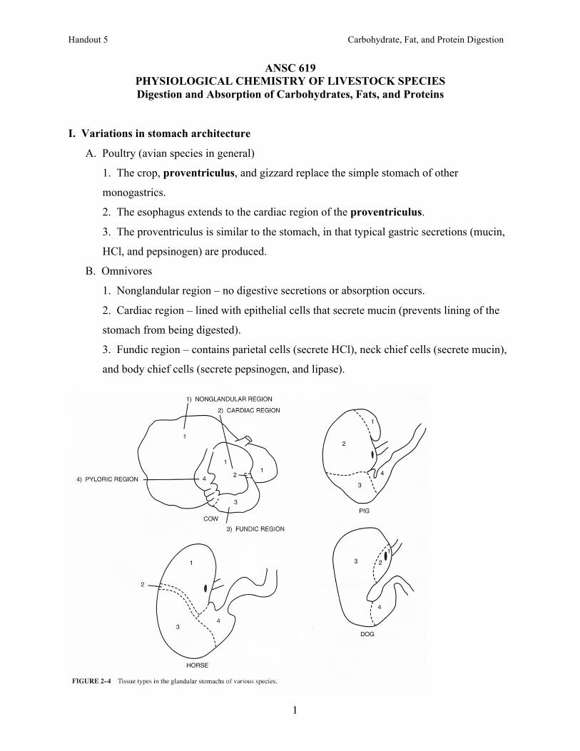
I. Variations in stomach architecture
A. Poultry (avian species in general)
1. The crop, proventriculus, and gizzard replace the simple stomach of other
monogastrics.
2. The esophagus extends to the cardiac region of the proventriculus.
3. The proventriculus is similar to the stomach, in that typical gastric secretions (mucin,
HCl, and pepsinogen) are produced.
B. Omnivores
1. Nonglandular region – no digestive secretions or absorption occurs.
2. Cardiac region – lined with epithelial cells that secrete mucin (prevents lining of the
stomach from being digested).
3. Fundic region – contains parietal cells (secrete HCl), neck chief cells (secrete mucin),
and body chief cells (secrete pepsinogen, and lipase).
Handout 5 Carbohydrate, Fat, and Protein Digestion
2
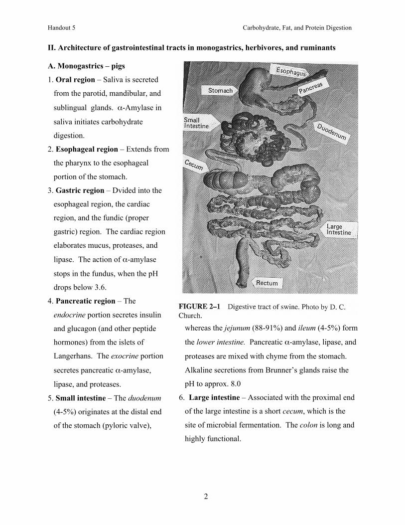
II. Architecture of gastrointestinal tracts in monogastrics, herbivores, and ruminants A. Monogastrics – pigs
1. Oral region – Saliva is secreted
from the parotid, mandibular, and
sublingual glands. α-Amylase in
saliva initiates carbohydrate
digestion.
2. Esophageal region – Extends from
the pharynx to the esophageal
portion of the stomach.
3. Gastric region – Dvided into the
esophageal region, the cardiac
region, and the fundic (proper
gastric) region. The cardiac region
elaborates mucus, proteases, and
lipase. The action of α-amylase
stops in the fundus, when the pH
drops below 3.6.
4. Pancreatic region – The
endocrine portion secretes insulin
and glucagon (and other peptide
hormones) from the islets of
Langerhans. The exocrine portion
secretes pancreatic α-amylase,
lipase, and proteases.
5. Small intestine – The duodenum
(4-5%) originates at the distal end
of the stomach (pyloric valve),
whereas the jejunum (88-91%) and ileum (4-5%) form
the lower intestine. Pancreatic α-amylase, lipase, and
proteases are mixed with chyme from the stomach.
Alkaline secretions from Brunner’s glands raise the
pH to approx. 8.0
6. Large intestine – Associated with the proximal end
of the large intestine is a short cecum, which is the
site of microbial fermentation. The colon is long and
highly functional.
Handout 5 Carbohydrate, Fat, and Protein Digestion
3
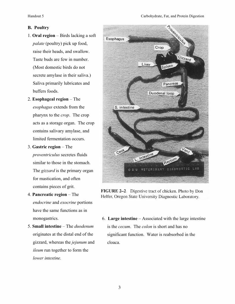
B. Poultry
1. Oral region – Birds lacking a soft
palate (poultry) pick up food,
raise their heads, and swallow.
Taste buds are few in number.
(Most domestic birds do not
secrete amylase in their saliva.)
Saliva primarily lubricates and
buffers foods.
2. Esophageal region – The
esophagus extends from the
pharynx to the crop. The crop
acts as a storage organ. The crop
contains salivary amylase, and
limited fermentation occurs.
3. Gastric region – The
proventriculus secretes fluids
similar to those in the stomach.
The gizzard is the primary organ
for mastication, and often
contains pieces of grit.
4. Pancreatic region – The
endocrine and exocrine portions
have the same functions as in
monogastrics.
5. Small intestine – The duodenum
originates at the distal end of the
gizzard, whereas the jejunum and
ileum run together to form the
lower intestine.
6. Large intestine – Associated with the large intestine
is the cecum. The colon is short and has no
significant function. Water is reabsorbed in the
cloaca.
Handout 5 Carbohydrate, Fat, and Protein Digestion
4
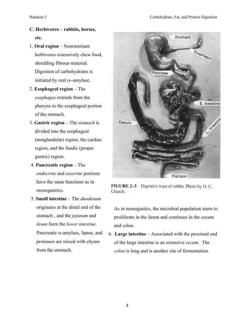
C. Herbivores – rabbits, horses,
etc.
1. Oral region – Nonruminant
herbivores extensively chew food,
shredding fibrous material.
Digestion of carbohydrates is
initiated by oral α−amylase.
2. Esophageal region – The
esophagus extends from the
pharynx to the esophageal portion
of the stomach.
3. Gastric region – The stomach is
divided into the esophageal
(nonglandular) region, the cardiac
region, and the fundic (proper
gastric) region.
4. Pancreatic region – The
endocrine and exocrine portions
have the same functions as in
monogastrics.
5. Small intestine – The duodenum
originates at the distal end of the
stomach , and the jejunum and
ileum form the lower intestine.
Pancreatic α-amylase, lipase, and
proteases are mixed with chyme
from the stomach.
As in monogastics, the microbial population starts to
proliferate in the ileum and continues in the cecum
and colon.
6. Large intestine – Associated with the proximal end
of the large intestine is an extensive cecum. The
colon is long and is another site of fermentation.
Handout 5 Carbohydrate, Fat, and Protein Digestion
5
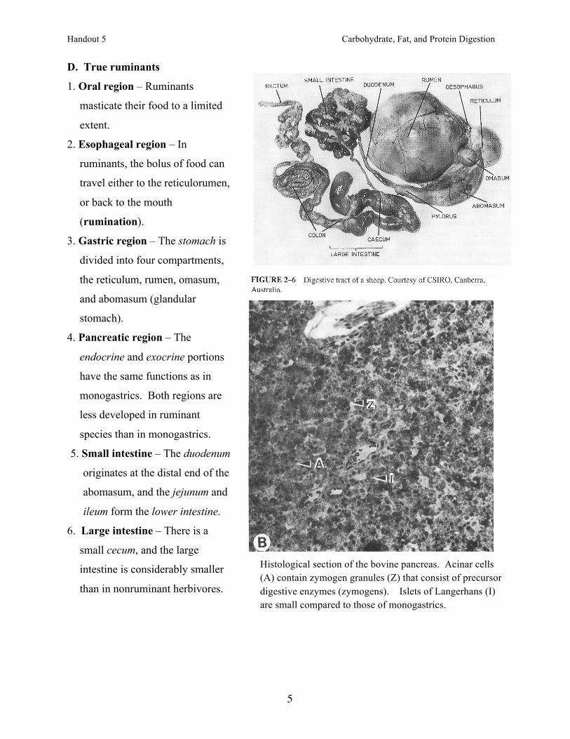
D. True ruminants
1. Oral region – Ruminants
masticate their food to a limited
extent.
2. Esophageal region – In
ruminants, the bolus of food can
travel either to the reticulorumen,
or back to the mouth
(rumination).
3. Gastric region – The stomach is
divided into four compartments,
the reticulum, rumen, omasum,
and abomasum (glandular
stomach).
4. Pancreatic region – The
endocrine and exocrine portions
have the same functions as in
monogastrics. Both regions are
less developed in ruminant
species than in monogastrics.
5. Small intestine – The duodenum
originates at the distal end of the
abomasum, and the jejunum and
ileum form the lower intestine.
6. Large intestine – There is a
small cecum, and the large
intestine is considerably smaller
than in nonruminant herbivores.
Histological section of the bovine pancreas. Acinar cells (A) contain zymogen granules (Z) that consist of precursor digestive enzymes (zymogens). Islets of Langerhans (I) are small compared to those of monogastrics.
Handout 5 Carbohydrate, Fat, and Protein Digestion
6
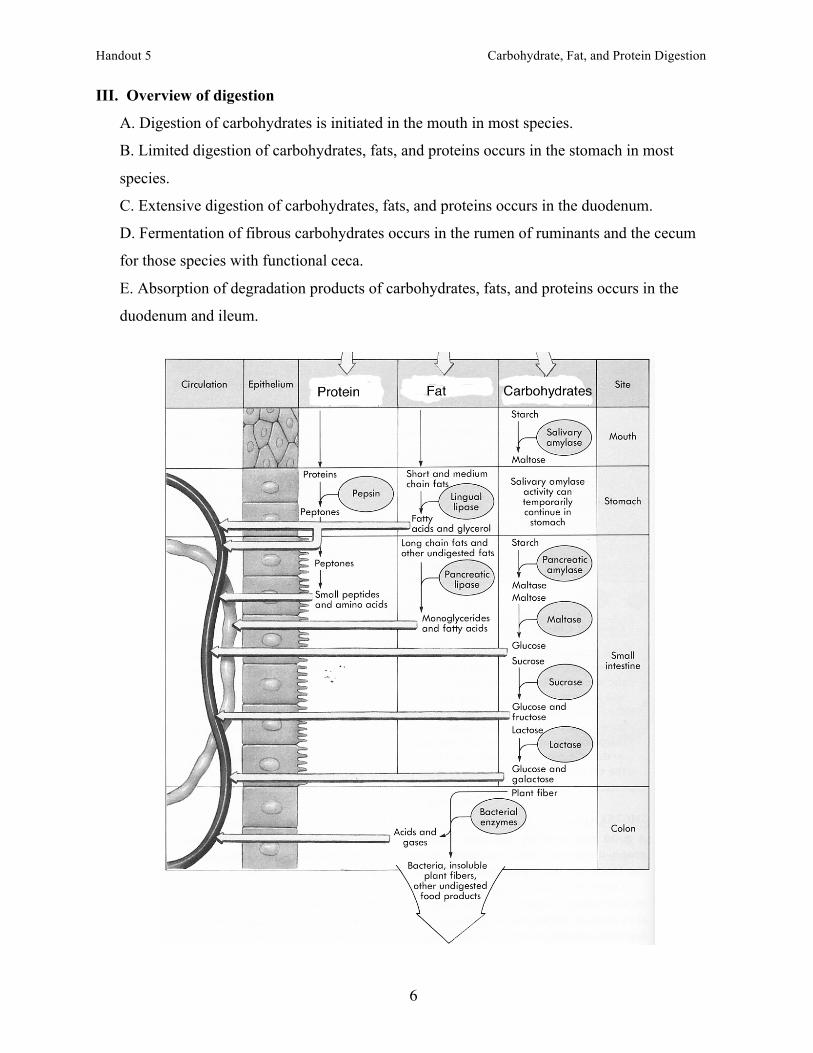
III. Overview of digestion
A. Digestion of carbohydrates is initiated in the mouth in most species.
B. Limited digestion of carbohydrates, fats, and proteins occurs in the stomach in most
species.
C. Extensive digestion of carbohydrates, fats, and proteins occurs in the duodenum.
D. Fermentation of fibrous carbohydrates occurs in the rumen of ruminants and the cecum
for those species with functional ceca.
E. Absorption of degradation products of carbohydrates, fats, and proteins occurs in the
duodenum and ileum.
Handout 5 Carbohydrate, Fat, and Protein Digestion
7
IV. Digestion of carbohydrates: overview
A. Oral digestion
1. Ingested starch is hydrolyzed by α-amylase.
2. pH optimum is 6.7.
B. Stomach
1. Salivary amylase digestion continues.
2. Some fermentation of lactose to lactic acid can occur.
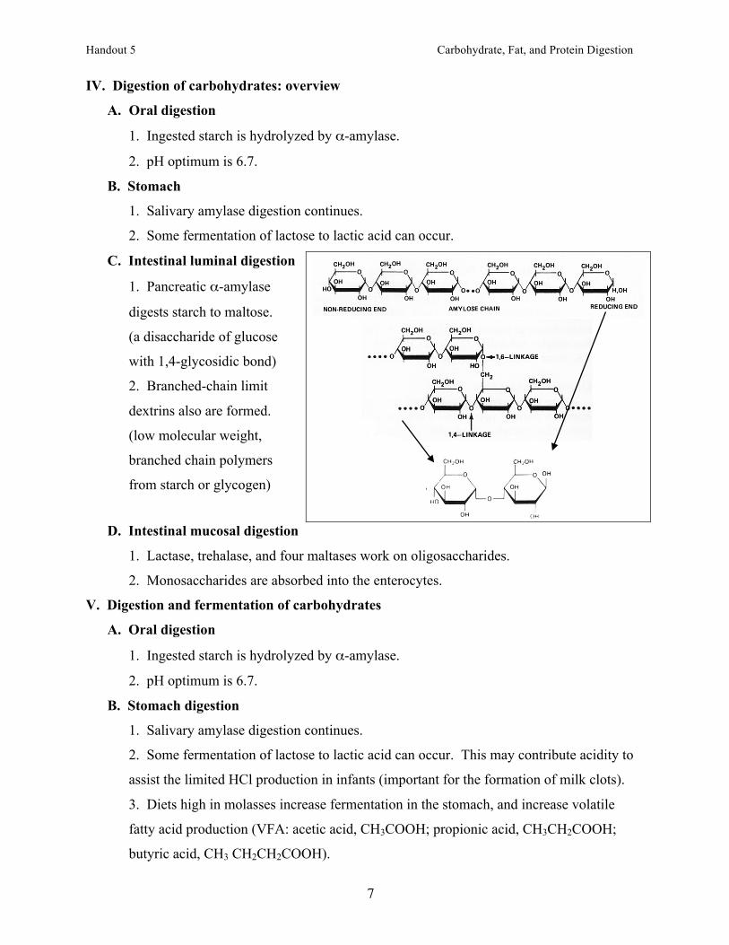
C. Intestinal luminal digestion
1. Pancreatic α-amylase
digests starch to maltose.
(a disaccharide of glucose
with 1,4-glycosidic bond)
2. Branched-chain limit
dextrins also are formed.
(low molecular weight,
branched chain polymers
from starch or glycogen)
D. Intestinal mucosal digestion
1. Lactase, trehalase, and four maltases work on oligosaccharides.
2. Monosaccharides are absorbed into the enterocytes.
V. Digestion and fermentation of carbohydrates
A. Oral digestion
1. Ingested starch is hydrolyzed by α-amylase.
2. pH optimum is 6.7.
B. Stomach digestion
1. Salivary amylase digestion continues.
2. Some fermentation of lactose to lactic acid can occur. This may contribute acidity to
assist the limited HCl production in infants (important for the formation of milk clots).
3. Diets high in molasses increase fermentation in the stomach, and increase volatile
fatty acid production (VFA: acetic acid, CH3COOH; propionic acid, CH3CH2COOH;
butyric acid, CH3 CH2CH2COOH).
Handout 5 Carbohydrate, Fat, and Protein Digestion
8
C. Intestinal luminal digestion
1. Pancreatic α-amylase digests starch to maltose, branched-chain limit dextrins, and
traces of glucose.
2. These products of digestion migrate to the mucosal surface of the duodenum
following a concentration gradient.
D. Intestinal mucosal digestion
1. Lactase, trehalase, and four maltases (including sucrase) work on oligosaccharides.
2. Monosaccharides are absorbed into the enterocytes.
3. Absorption takes place in the duodenum and jejunum.
E. Microbial activity in the small intestine
1. Gut microorganisms can digest nonstarch structural carbohydrates.
2. This is associated with a substantial amount of VFA production.
F. Large intestinal digestion and fermentation
1. Microorganisms in the colon and cecum produce cellulases, hemicellulases, and
pectinsases.
2. Primary products are VFA and methane.
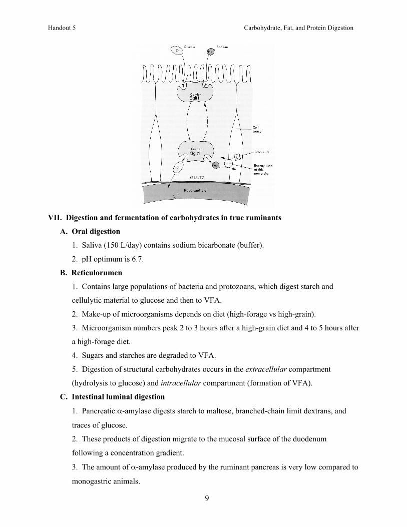
VI. Absorption of monosaccharides
A. Glucose and galactose
1. Both are transported into the enterocyte by a Na-dependent glucose transporter
(Sglt1).
2. Both then are released into the blood by a facilitated sugar transporter (GLUT2).
B. Fructose
1. Fructose is absorbed first by a Na-independent brush border fructose transporter
(GLUT5).
2. Fructose is released into the blood by GLUT2.
Handout 5 Carbohydrate, Fat, and Protein Digestion
9
VII. Digestion and fermentation of carbohydrates in true ruminants
A. Oral digestion
1. Saliva (150 L/day) contains sodium bicarbonate (buffer).
2. pH optimum is 6.7.
B. Reticulorumen
1. Contains large populations of bacteria and protozoans, which digest starch and
cellulytic material to glucose and then to VFA.
2. Make-up of microorganisms depends on diet (high-forage vs high-grain).
3. Microorganism numbers peak 2 to 3 hours after a high-grain diet and 4 to 5 hours after
a high-forage diet.
4. Sugars and starches are degraded to VFA.
5. Digestion of structural carbohydrates occurs in the extracellular compartment
(hydrolysis to glucose) and intracellular compartment (formation of VFA).
C. Intestinal luminal digestion
1. Pancreatic α-amylase digests starch to maltose, branched-chain limit dextrans, and
traces of glucose.
2. These products of digestion migrate to the mucosal surface of the duodenum
following a concentration gradient.
3. The amount of α-amylase produced by the ruminant pancreas is very low compared to
monogastric animals.
Handout 5 Carbohydrate, Fat, and Protein Digestion
10
D. Intestinal mucosal digestion
1. Lactase, trehalase, and four maltases (including sucrase) work on oligosaccharides.
2. Monosaccharides are absorbed into the enterocytes.
3. Absorption takes place in the duodenum and jejunum.
E. Microbial activity in the small intestine
1. Gut microorganisms can digest nonstarch structural carbohydrates.
2. This is associated with a small amount of VFA production in ruminants.
F. Large intestinal digestion and fermentation
1. Microorganisms in the colon and cecum produce cellulases, hemicellulases, and
pectinsases.
2. Primary products are VFA and methane. This is limited in ruminants.
VIII. Digestion of fats
A. Initial digestion of dietary fats
1. Saliva of rats (not humans) contains lingual lipase, which digests milk fats.
a. Primary products – 2,3-diacylglycerols
b. pH optimum – 4.5 - 5.4, so lingual lipase is active in the stomach
c. Does not require bile salts for activity.
d. Lingual lipase probably does not exist in human infants.
2. Lipase from human breast milk, bile salt-stimulated lipase, is taken up by human
infants and activated by bile salts in the small intestine.
B. Digestion of fat in the stomach
1. Stomach causes physical reduction in fat particle size.
2. Gastric lipase
a. Responsible for up to 25% of TAG hydrolysis in adults and infants.
b. pH optimum around 4.0.
C. Digestion of fat in the small intestine
1. Secretion of cholecystokinin from the intestinal mucosal cells (stimulated by fat in the
intestine) causes:
2. Gall bladder contraction.
a. The gall bladder contains 40 to 70 mL.
b. The gall bladder releases approximately 700 mL/d (extensive recirculation).
3. Secretion of pancreatic digestive enzymes (approximately 1,200 mL/d).
Handout 5 Carbohydrate, Fat, and Protein Digestion
11
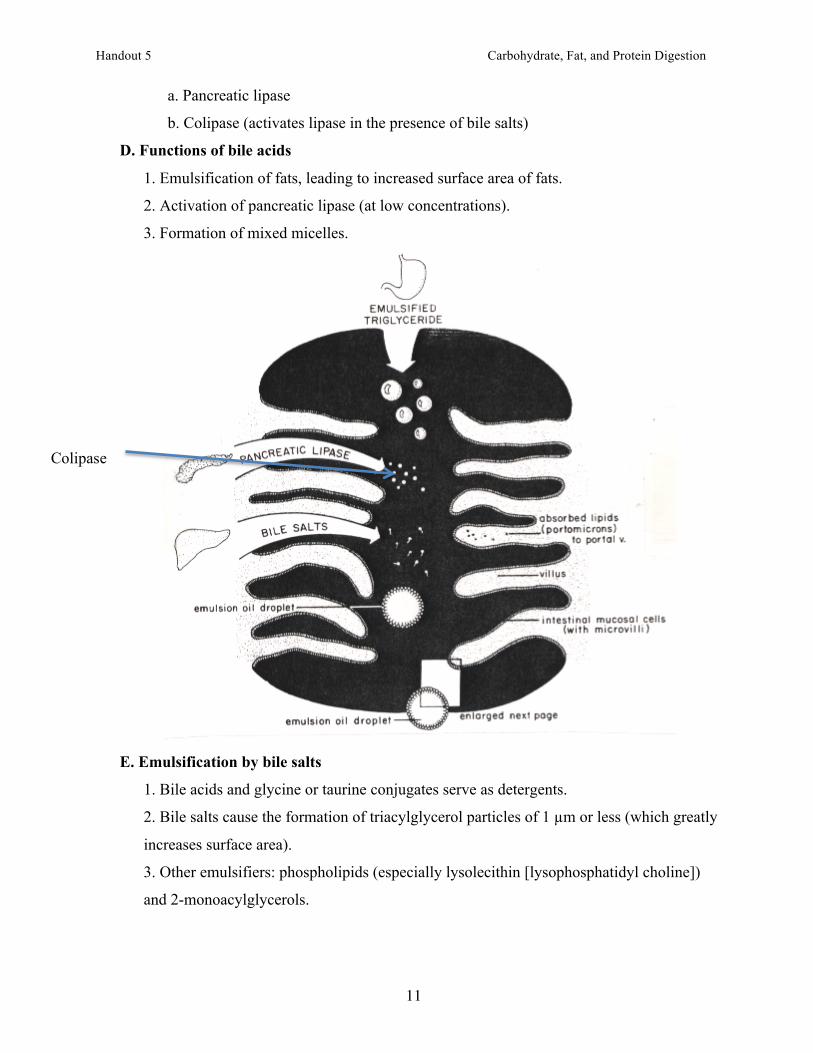
a. Pancreatic lipase
b. Colipase (activates lipase in the presence of bile salts)
D. Functions of bile acids
1. Emulsification of fats, leading to increased surface area of fats.
2. Activation of pancreatic lipase (at low concentrations).
3. Formation of mixed micelles.
E. Emulsification by bile salts
1. Bile acids and glycine or taurine conjugates serve as detergents.
2. Bile salts cause the formation of triacylglycerol particles of 1 µm or less (which greatly
increases surface area).
3. Other emulsifiers: phospholipids (especially lysolecithin [lysophosphatidyl choline])
and 2-monoacylglycerols.
Colipase
Handout 5 Carbohydrate, Fat, and Protein Digestion
12
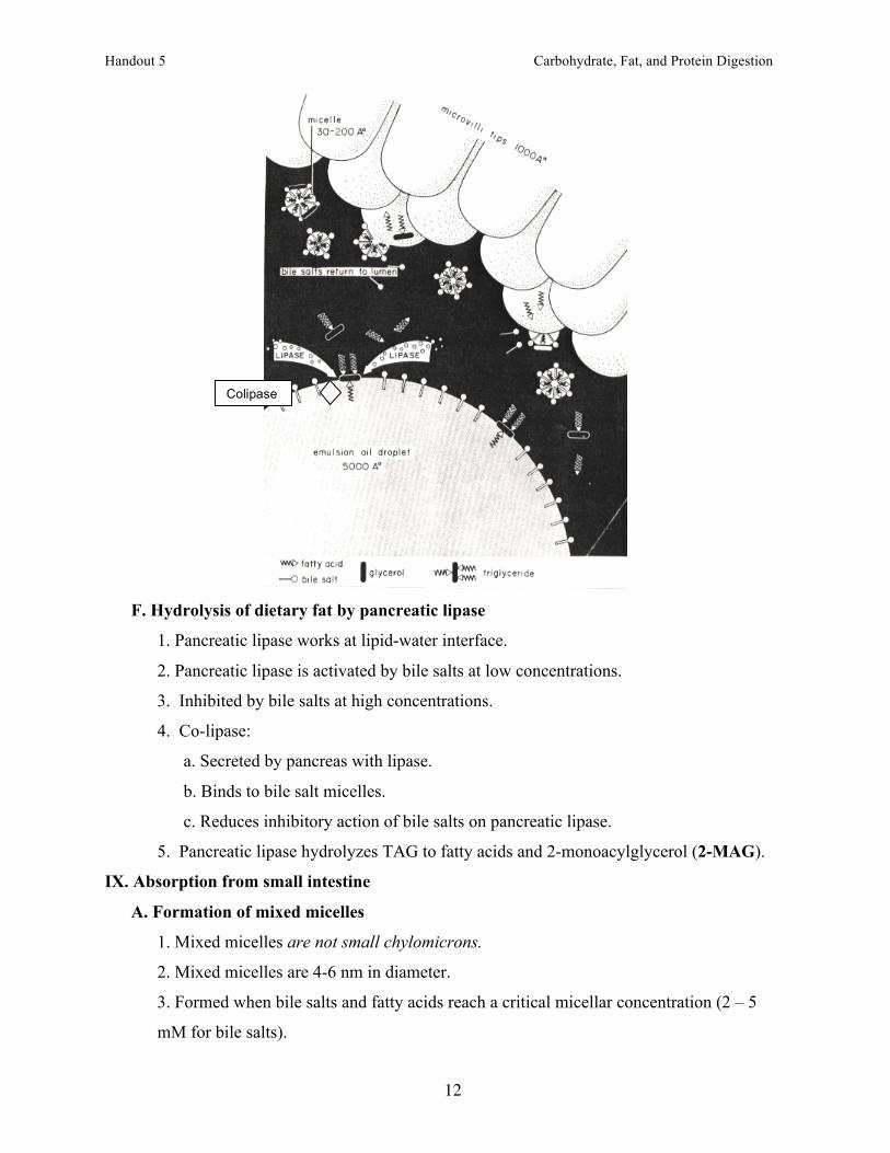
F. Hydrolysis of dietary fat by pancreatic lipase
1. Pancreatic lipase works at lipid-water interface.
2. Pancreatic lipase is activated by bile salts at low concentrations.
3. Inhibited by bile salts at high concentrations.
4. Co-lipase:
a. Secreted by pancreas with lipase.
b. Binds to bile salt micelles.
c. Reduces inhibitory action of bile salts on pancreatic lipase.
5. Pancreatic lipase hydrolyzes TAG to fatty acids and 2-monoacylglycerol (2-MAG).
IX. Absorption from small intestine
A. Formation of mixed micelles
1. Mixed micelles are not small chylomicrons.
2. Mixed micelles are 4-6 nm in diameter.
3. Formed when bile salts and fatty acids reach a critical micellar concentration (2 – 5
mM for bile salts).
Colipase
Handout 5 Carbohydrate, Fat, and Protein Digestion
13
4. Mixed micelles incorporate 2-MAGs, lysolecithin, cholesterol, and long-chain fatty
acids.
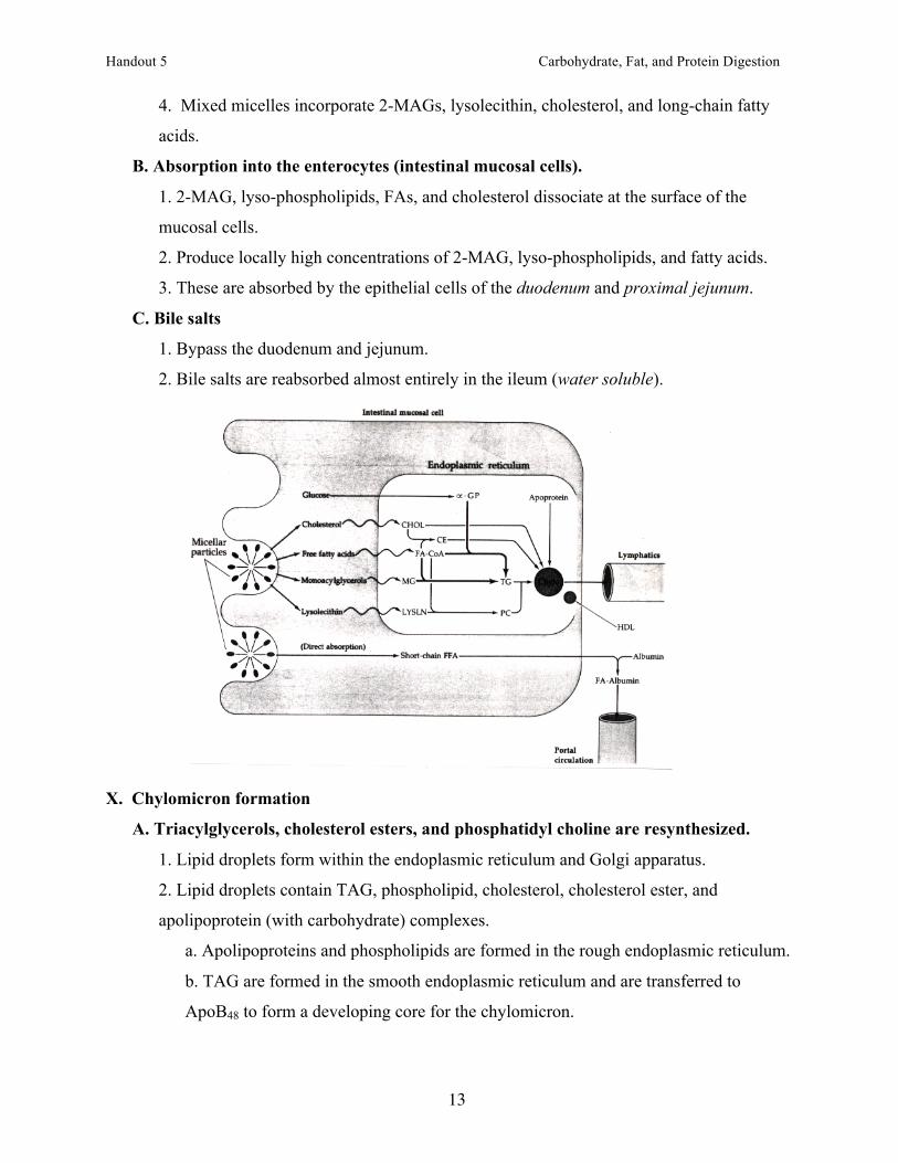
B. Absorption into the enterocytes (intestinal mucosal cells).
1. 2-MAG, lyso-phospholipids, FAs, and cholesterol dissociate at the surface of the
mucosal cells.
2. Produce locally high concentrations of 2-MAG, lyso-phospholipids, and fatty acids.
3. These are absorbed by the epithelial cells of the duodenum and proximal jejunum.
C. Bile salts
1. Bypass the duodenum and jejunum.
2. Bile salts are reabsorbed almost entirely in the ileum (water soluble).
X. Chylomicron formation
A. Triacylglycerols, cholesterol esters, and phosphatidyl choline are resynthesized.
1. Lipid droplets form within the endoplasmic reticulum and Golgi apparatus.
2. Lipid droplets contain TAG, phospholipid, cholesterol, cholesterol ester, and
apolipoprotein (with carbohydrate) complexes.
a. Apolipoproteins and phospholipids are formed in the rough endoplasmic reticulum.
b. TAG are formed in the smooth endoplasmic reticulum and are transferred to
ApoB48 to form a developing core for the chylomicron.
Handout 5 Carbohydrate, Fat, and Protein Digestion
14
c. After accumulating TAG and CE, Golgi vesicles form and carbohydrate moieties
are added to the apolipoproteins.
2. Golgi vesicles fuse with the cell membrane and are extruded into the lacteals.
3. Chylomicrons are transported via the lymphatics to the subclavean vein.
B. Composition
70 – 90% TAG
4 – 8% phospholipid
3% cholesterol
4% cholesterol ester
2% protein (apolipoprotein B)
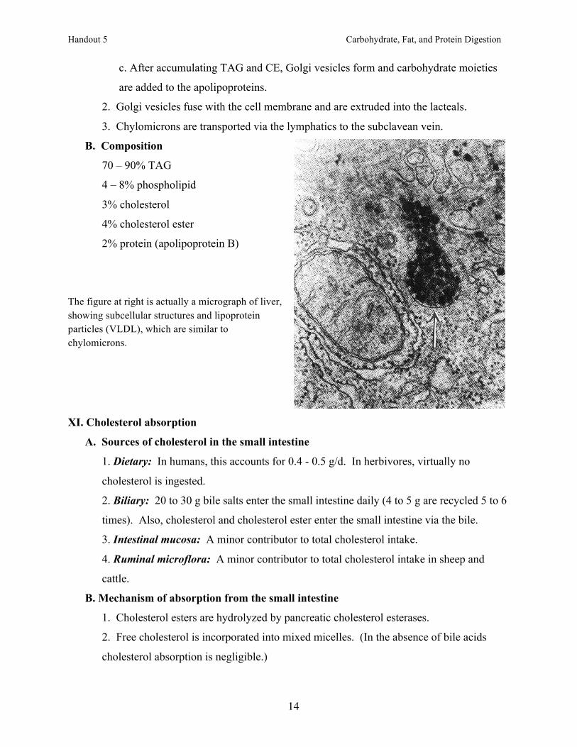
The figure at right is actually a micrograph of liver, showing subcellular structures and lipoprotein particles (VLDL), which are similar to chylomicrons.
XI. Cholesterol absorption
A. Sources of cholesterol in the small intestine
1. Dietary: In humans, this accounts for 0.4 - 0.5 g/d. In herbivores, virtually no
cholesterol is ingested.
2. Biliary: 20 to 30 g bile salts enter the small intestine daily (4 to 5 g are recycled 5 to 6
times). Also, cholesterol and cholesterol ester enter the small intestine via the bile.
3. Intestinal mucosa: A minor contributor to total cholesterol intake.
4. Ruminal microflora: A minor contributor to total cholesterol intake in sheep and
cattle.
B. Mechanism of absorption from the small intestine
1. Cholesterol esters are hydrolyzed by pancreatic cholesterol esterases.
2. Free cholesterol is incorporated into mixed micelles. (In the absence of bile acids
cholesterol absorption is negligible.)
Handout 5 Carbohydrate, Fat, and Protein Digestion
15
3. Free cholesterol is absorbed into the intestinal mucosal cells and re-esterified to form
cholesterol esters.
C. Sources of loss of cholesterol from the dietary tract
1. Bile salts
a. 0.8 g/d bile salts are lost in the feces.
b. The remainder (98 - 99%) is taken up in the ileum (enterohepatic circulation).
2. Cholesterol: cholesterol is poorly absorbed.
a. Approximately 0.4 g/d lost in the feces
b. Only 30 to 60% dietary plus biliary cholesterol absorbed.
c. Increased cholesterol intake results in a greater amount of
cholesterol absorbed, but a lesser percentage absorbed.
3. Plant sterols
a. 200 to 300 mg/d are ingested.
b. Plant sterols are absorbed only in trace amounts.
c. In large amounts, plant sterols inhibit cholesterol absorption (mechanism
unknown).
4. 0.2 to 0.4 g/d of cholesterol is lost as sloughed skin.
XII. Fat digestion in ruminants
A. Bacterial lipases
1. Bacteria (Propionibacterium avidum, P. acnes, Anaerovibrio lipolyticus, and
Butyrivibrio fibrisolvens) synthesize and secrete (express) lipases.
2. Extracellular lipases expressed by bacteria hydrolyze mono-, di-, and triacylglycerols
to free fatty acids and glycerol.
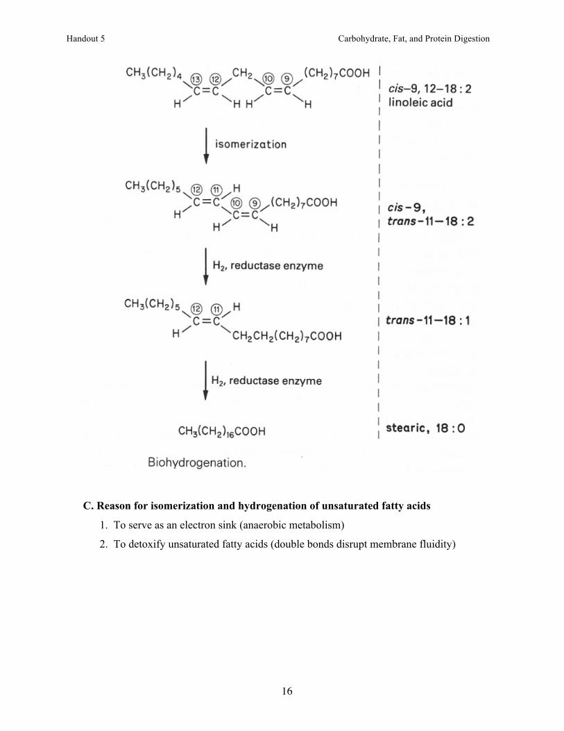
B. Isomerization and hydrogenation of unsaturated fatty acids
1. The double bond toward the methyl carbon is isomerized to a trans-double bond.
2. The double bond nearest the #1 carbon is reduced (hydrogenated).
3. The trans-double bond is reduced, usually producing stearic acid (18:0).
4. Each reaction is carried out by a different microorganism.
Handout 5 Carbohydrate, Fat, and Protein Digestion
16
C. Reason for isomerization and hydrogenation of unsaturated fatty acids
1. To serve as an electron sink (anaerobic metabolism)
2. To detoxify unsaturated fatty acids (double bonds disrupt membrane fluidity)
Handout 5 Carbohydrate, Fat, and Protein Digestion
17
XIII. Phases of protein digestion
A. Gastric
1. Secretion of hydrochloric acid (denatures proteins)
2. Activation of pepsin from pepsinogen by HCl; cleaves most peptide bonds.
B. Pancreatic
1. Secretion of secretagogues from mucosal endoncrine cells
a. Cholecystokinin (CCK): stimulates pancreatic digestive enzyme secretion into the
small intestine from pancreatic acinar cells; stimulates contraction of the gall bladder;
stimulates release of brush border enzymes (e.g., enteropeptidase).
b. Secretin: stimulates secretion of bicarbonate and water from pancreas.
2. Partial digestion of proteins by pancreatic enzymes
a. Trypsinogen à trypsin (catalyzed by enteropeptidase); cleaves peptide bonds
adjacent to dibasic amino acids.
b. Procarboxkypeptidases à carboxkypeptidases (catalyzed by trypsin); cleaves C-
terminal amino acids.
c. Chymotrypsinogen à chymotrypsin (catalyzed by trypsin); cleaves peptide bonds
adjacent to aromatic amino acids.
C. Intestinal
1. Digestion of protein fragments by:
a. Brush border peptidases
b. Peptidases in the cytoplasm of the mucosal cells
2. Hydrolysis of peptides to free amino acids, dipeptides and tripeptides
3. Absorption/transport of small peptides (66% of total) and amino acids
4. Hydrolysis of small peptides to amino acids within mucosal cells
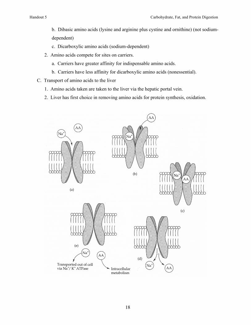
XIV. Transport of amino acids into mucosal cells (proximal small intestine)
A. Diffusion
1. Diffusion of amino acids is limited
2. Most amino acids have specific transport systems to ensure complete uptake.
B. Transport systems
1. Carrier types are specific for amino acids.
a. Leucine and other neutral amino acids (not sodium-dependent)
b. Threonine, alanine, other neutral amino acids (sodium-dependent)
Handout 5 Carbohydrate, Fat, and Protein Digestion
18
b. Dibasic amino acids (lysine and arginine plus cystine and ornithine) (not sodium-
dependent)
c. Dicarboxylic amino acids (sodium-dependent)
2. Amino acids compete for sites on carriers.
a. Carriers have greater affinity for indispensable amino acids.
b. Carriers have less affinity for dicarboxylic amino acids (nonessential).
C. Transport of amino acids to the liver
1. Amino acids taken are taken to the liver via the hepatic portal vein.
2. Liver has first choice in removing amino acids for protein synthesis, oxidation.