Embed Size (px)
Citation preview
Seediscussions,stats,andauthorprofilesforthispublicationat:https://www.researchgate.net/publication/228115821
HantavirusinfectionsinEuropeandtheirimpactonpublichealth
ArticleinReviewsinMedicalVirology·January2013
DOI:10.1002/rmv.1722·Source:PubMed
CITATIONS
93
READS
231
6authors,including:
AnttiVaheri
UniversityofHelsinki
905PUBLICATIONS34,035CITATIONS
SEEPROFILE
LiinaVoutilainen
UniversityofHelsinki
41PUBLICATIONS546CITATIONS
SEEPROFILE
TarjaSironen
UniversityofHelsinki
58PUBLICATIONS784CITATIONS
SEEPROFILE
OlliVapalahti
UniversityofHelsinki
303PUBLICATIONS7,785CITATIONS
SEEPROFILE
Availablefrom:LiinaVoutilainen
Retrievedon:25September2016
Rev. Med. Virol.Published online in Wiley Online Library
(wileyonlinelibrary.com)DOI: 10.1002/rmv.1722
R E V I E W
Hantavirus infectioon public healthReviews in Medical Virology
*Corresponding authoInstitute, POB 21, 00E-mail: antti.vaheri@h
Abbreviations usedCRP, C-reactive prothantavirus cardiopulwith renal syndromnephropathia epidemivirus; SEOV, Seoul v
Copyright © 201
ns in Europe and their impact
Antti Vaheri1,2*, Heikki Henttonen3, Liina Voutilainen1,3,Jukka Mustonen4,5, Tarja Sironen1 and Olli Vapalahti1,2,61Department of Virology, Haartman Institute, and Research Programs Unit, Infection Biology, University ofHelsinki, Helsinki, Finland2Department of Virology and Immunology, HUSLAB, Helsinki University Central Hospital, Helsinki,Finland3Finnish Forest Research Institute, Vantaa, Finland4School of Medicine, University of Tampere, Tampere, Finland5Department of Internal Medicine, Tampere University Hospital, Tampere, Finland6Department of Veterinary Biosciences, Faculty of Veterinary Medicine, University of Helsinki, Helsinki,Finland
SUMMARYHantaviruses (genusHantavirus, family Bunyaviridae) are enveloped tri-segmented negative-stranded RNAviruses eachcarried by a specific rodent or insectivore host species. Several different hantaviruses known to infect humans circulatein Europe. The most common is Puumala (PUUV) carried by the bank vole; another two important, genetically closelyrelated ones are Dobrava–Belgrade (DOBV) and Saaremaa viruses (SAAV) carried by Apodemus mice (species namesfollow the International Committee on Taxonomy of Viruses nomenclature). Of the two hantaviral diseases, hemor-rhagic fever with renal syndrome (HFRS) and hantaviral cardiopulmonary syndrome, the European viruses cause onlyHFRS: DOBV with often severe symptoms and a high case fatality rate, and PUUVand SAAV more often mild disease.More than 10,000 HFRS cases are diagnosed annually in Europe and in increasing numbers. Whether this is because ofincreasing recognition by the medical community or due to environmental factors such as climate change, or both, isnot known. Nevertheless, in large areas of Europe, the population has a considerable seroprevalence but only relativelyfew HFRS cases are reported. Moreover, no epidemiological data are available frommany countries. We know now thatcardiac, pulmonary, ocular and hormonal disorders are, besides renal changes, common during the acute stage ofPUUVand DOBV infection. About 5% of hospitalized PUUVand 16%–48% of DOBV patients require dialysis and someprolonged intensive-care treatment. Although PUUV–HFRS has a low case fatality rate, complications and long-termhormonal, renal, and cardiovascular consequences commonly occur. No vaccine or specific therapy is in general usein Europe. We conclude that hantaviruses have a significant impact on public health in Europe. Copyright © 2012 JohnWiley & Sons, Ltd.
Received: 31 January 2012; Revised: 4 May 2012; Accepted: 8 May 2012
INTRODUCTIONHantaviruses (family Bunyaviridae, genusHantavirus)are tri-segmented negative-stranded envelopedRNA
r: Antti Vaheri, Department of Virology,Haartman014 University of Helsinki, Helsinki, Finland.elsinki.fi
ein; DOBV, Dobrava–Belgrade virus; HCPS,monary syndrome; HFRS, hemorrhagic fevere; IDO, indoleamine 2,3-dioxygenase; NE,ca; PUUV, Puumala virus; SAAV, Saaremaairus; TULV, Tula virus.
2 John Wiley & Sons, Ltd.
viruses carried by rodents and insectivores. Theycause two diseases, hemorrhagic fever with renalsyndrome (HFRS) in Eurasia and hantavirus cardio-pulmonary syndrome (HCPS) [1–3]. Humans getmainly infected from aerosolized rodent excretabut HCPS may be also transmitted from person-to-person and HFRS from blood transfusions [4,5].Several hantaviruses cause HFRS in Europe, anendemic zoonosis, diagnosed in more than 10,000individuals in Europe annually. The principal HFRS-inducing hantaviruses in Europe are Puumala(PUUV) carried byMyodes voles and two interrelatedviruses carried byApodemusmice, Dobrava–Belgrade
A. Vaheri et al.
virus (DOBV), and Saaremaa virus (SAAV). These arethe species listed by the International Committee onTaxonomy of Viruses, but the nomenclature of theEuropean Apodemus-derived hantaviruses has beenand still is, under debate and revision: in literatureDOBVvariants inApodemus flavicollis are also referredto as DOBV-Af, and variants in Apodemus ponticus asDOBV-Ap. Some strains recovered from Apodemusagrarius are described as a genotype DOBV-Aa. Seoulvirus (SEOV) is the causal virus for medium severeHFRS in Asia and in many cities worldwide buthas been detected only once with certainty as thecause of HFRS in Europe [6,7] Similarly, Tula virus(TULV) although common in Microtus voles inCentral and Eastern Europe, has been associatedwithHFRS in one patient [8]. No specific antiviral therapyor vaccine is in general use in Europe. Recently,several complications and long-term consequenceshave been associated with HFRS. In the following,we will evaluate the disease burden of hantavirusinfections and HFRS in Europe.
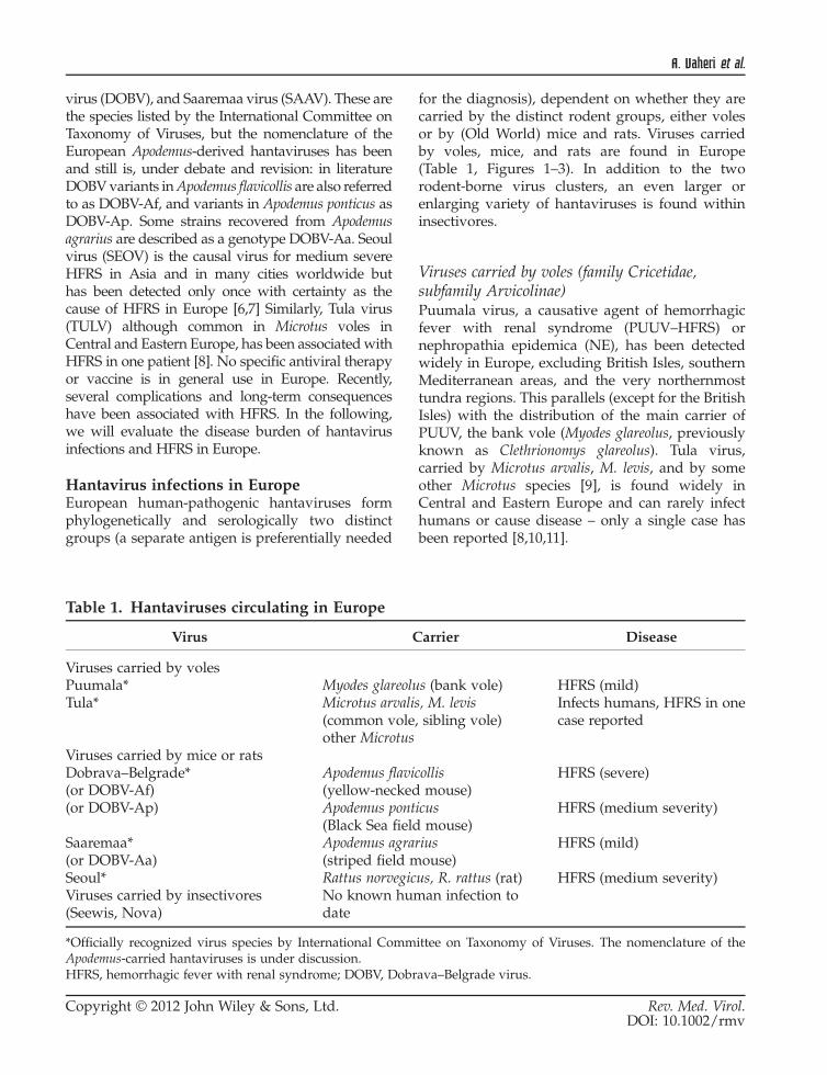
Hantavirus infections in EuropeEuropean human-pathogenic hantaviruses formphylogenetically and serologically two distinctgroups (a separate antigen is preferentially needed
Table 1. Hantaviruses circulating in Europe
Virus C
Viruses carried by volesPuumala* Myodes glareoluTula* Microtus arvali
(common voleother Microtus
Viruses carried by mice or ratsDobrava–Belgrade* Apodemus flavi(or DOBV-Af) (yellow-necked(or DOBV-Ap) Apodemus pont
(Black Sea fieldSaaremaa* Apodemus agra(or DOBV-Aa) (striped field mSeoul* Rattus norvegicViruses carried by insectivores(Seewis, Nova)
No known humdate
*Officially recognized virus species by International CommApodemus-carried hantaviruses is under discussion.HFRS, hemorrhagic fever with renal syndrome; DOBV, Dobr
Copyright © 2012 John Wiley & Sons, Ltd.
for the diagnosis), dependent on whether they arecarried by the distinct rodent groups, either volesor by (Old World) mice and rats. Viruses carriedby voles, mice, and rats are found in Europe(Table 1, Figures 1–3). In addition to the tworodent-borne virus clusters, an even larger orenlarging variety of hantaviruses is found withininsectivores.
Viruses carried by voles (family Cricetidae,subfamily Arvicolinae)Puumala virus, a causative agent of hemorrhagicfever with renal syndrome (PUUV–HFRS) ornephropathia epidemica (NE), has been detectedwidely in Europe, excluding British Isles, southernMediterranean areas, and the very northernmosttundra regions. This parallels (except for the BritishIsles) with the distribution of the main carrier ofPUUV, the bank vole (Myodes glareolus, previouslyknown as Clethrionomys glareolus). Tula virus,carried by Microtus arvalis, M. levis, and by someother Microtus species [9], is found widely inCentral and Eastern Europe and can rarely infecthumans or cause disease – only a single case hasbeen reported [8,10,11].
arrier Disease
s (bank vole) HFRS (mild)s, M. levis, sibling vole)
Infects humans, HFRS in onecase reported
collis HFRS (severe)mouse)
icus HFRS (medium severity)mouse)
rius HFRS (mild)ouse)us, R. rattus (rat) HFRS (medium severity)an infection to
ittee on Taxonomy of Viruses. The nomenclature of the
ava–Belgrade virus.
Rev. Med. Virol.DOI: 10.1002/rmv
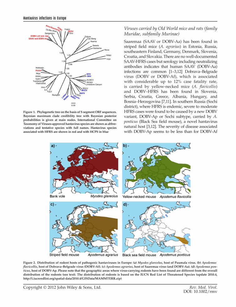
Figure 1. Phylogenetic tree on the basis of S segmentORF sequences.Bayesian maximum clade credibility tree with Bayesian posteriorprobabilities is given at main nodes. International Committee onTaxonomyofViruses-approvedhantavirus species are shownas abbre-viations and tentative species with full names. Hantavirus speciesassociated with HFRS are shown in red and with HCPS in blue
Figure 2. Distribution of rodent hosts of pathogenic hantaviruses in Eflavicollis, host of Dobrava–Belgrade virus (DOBV-Af). (c) Apodemus aticus, host of DOBV-Ap. Please note that the geographic areas where virdistribution of the rodents (see text). The distribution of rodents is bahttp://i.iucnredlist.org/spatial-data/2010.4/GISData/MAMMTERR.zip)
Hantavirus infections in Europe
Copyright © 2012 John Wiley & Sons, Ltd.
Viruses carried by Old World mice and rats (familyMuridae, subfamily Murinae)
Saaremaa (SAAV or DOBV-Aa) has been found instriped field mice (A. agrarius) in Estonia, Russia,southeastern Finland, Germany, Denmark, Slovenia,Croatia, and Slovakia. There are nowell-documentedSAAV-HFRS cases but serology including neutralizingantibodies indicates that human SAAV (DOBV-Aa)infections are common [1–3,12] Dobrava–Belgradevirus (DOBV or DOBV-Af), which is associatedwith considerable up to 12% case fatality rate,is carried by yellow-necked mice (A. flavicollis)and DOBV–HFRS has been found in Slovenia,Serbia, Croatia, Greece, Albania, Hungary, andBosnia–Herzegovina [7,11]. In southern Russia (Sochidistrict), where HFRS is endemic, severe to moderateHFRS cases were found to be caused by a newDOBVvariant, DOBV-Ap or Sochi subtype, carried by A.ponticus (Black Sea field mouse), a novel hantavirusnatural host [3,12]. The severity of disease associatedwith DOBV-Ap seems to be less than for DOBV-Af
urope: (a) Myodes glareolus, host of Puumala virus. (b) Apodemusgrarius, host of Saaremaa virus (and DOBV-Aa). (d) Apodemus pon-us-carrying rodents have been found are different from the overallsed on the IUCN Red List of Threatened Species (update 2010.4,
Rev. Med. Virol.DOI: 10.1002/rmv
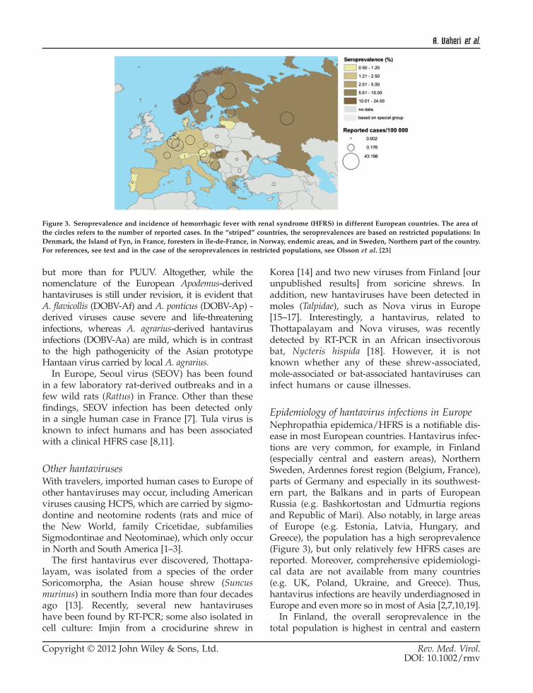
Figure 3. Seroprevalence and incidence of hemorrhagic fever with renal syndrome (HFRS) in different European countries. The area ofthe circles refers to the number of reported cases. In the “striped” countries, the seroprevalences are based on restricted populations: InDenmark, the Island of Fyn, in France, foresters in île-de-France, in Norway, endemic areas, and in Sweden, Northern part of the country.For references, see text and in the case of the seroprevalences in restricted populations, see Olsson et al. [23]
A. Vaheri et al.
but more than for PUUV. Altogether, while thenomenclature of the European Apodemus-derivedhantaviruses is still under revision, it is evident thatA. flavicollis (DOBV-Af) and A. ponticus (DOBV-Ap) -derived viruses cause severe and life-threateninginfections, whereas A. agrarius-derived hantavirusinfections (DOBV-Aa) are mild, which is in contrastto the high pathogenicity of the Asian prototypeHantaan virus carried by local A. agrarius.In Europe, Seoul virus (SEOV) has been found
in a few laboratory rat-derived outbreaks and in afew wild rats (Rattus) in France. Other than thesefindings, SEOV infection has been detected onlyin a single human case in France [7]. Tula virus isknown to infect humans and has been associatedwith a clinical HFRS case [8,11].
Other hantavirusesWith travelers, imported human cases to Europe ofother hantaviruses may occur, including Americanviruses causing HCPS, which are carried by sigmo-dontine and neotomine rodents (rats and mice ofthe New World, family Cricetidae, subfamiliesSigmodontinae and Neotominae), which only occurin North and South America [1–3].The first hantavirus ever discovered, Thottapa-
layam, was isolated from a species of the orderSoricomorpha, the Asian house shrew (Suncusmurinus) in southern India more than four decadesago [13]. Recently, several new hantaviruseshave been found by RT-PCR; some also isolated incell culture: Imjin from a crocidurine shrew in
Copyright © 2012 John Wiley & Sons, Ltd.
Korea [14] and two new viruses from Finland [ourunpublished results] from soricine shrews. Inaddition, new hantaviruses have been detected inmoles (Talpidae), such as Nova virus in Europe[15–17]. Interestingly, a hantavirus, related toThottapalayam and Nova viruses, was recentlydetected by RT-PCR in an African insectivorousbat, Nycteris hispida [18]. However, it is notknown whether any of these shrew-associated,mole-associated or bat-associated hantaviruses caninfect humans or cause illnesses.
Epidemiology of hantavirus infections in EuropeNephropathia epidemica/HFRS is a notifiable dis-ease in most European countries. Hantavirus infec-tions are very common, for example, in Finland(especially central and eastern areas), NorthernSweden, Ardennes forest region (Belgium, France),parts of Germany and especially in its southwest-ern part, the Balkans and in parts of EuropeanRussia (e.g. Bashkortostan and Udmurtia regionsand Republic of Mari). Also notably, in large areasof Europe (e.g. Estonia, Latvia, Hungary, andGreece), the population has a high seroprevalence(Figure 3), but only relatively few HFRS cases arereported. Moreover, comprehensive epidemiologi-cal data are not available from many countries(e.g. UK, Poland, Ukraine, and Greece). Thus,hantavirus infections are heavily underdiagnosed inEurope and even more so in most of Asia [2,7,10,19].
In Finland, the overall seroprevalence in thetotal population is highest in central and eastern
Rev. Med. Virol.DOI: 10.1002/rmv
Hantavirus infections in Europe
Finland; males contract NE at the mean age of40 years, females at 44 years. From the seropreva-lence (5% in Finland) and incidence (Figure 3), itmay be calculated that only 20%–30% of infectedhumans experience clinical problems severe enoughto seek medical attention leading to serological con-firmation [20–22].The epidemiological pattern has a particular
temporal cyclicity and can change geographically.In Northern Europe, there are 3- to 4-year cyclesof M. glareolus, and up to the late 1990s many partsof Finland were in non-synchronous phases of volecycles. More recently, the whole southern part ofthe country has been synchronous leading to asimultaneous epidemic peak in a large area insteadof smaller local non-synchromous peaks every year[23]. Consequently, in 1999, 2002, and 2005, Finlandhad about 2500 serologically diagnosed HFRScases, and in 2008, a record year, 3259 PUUV–HFRScases. Belgium had peak years in 2007 (298 cases)and 2008 (336 cases), Sweden in 2007 (2195 cases),and Germany in 2007 (1688 cases) and in 2010(>2000 cases) [2,7,10,24].
Environmental factors predicting HFRSepidemicsHemorrhagic fever with renal syndrome epidemicsis spatially associated with the natural habitats ofhantavirus carrier rodents. As the bank vole is aforest-dwelling species, the risk of PUUV infectionincreases with the proportion of forested land cover[23,25–27], the vicinity of forests [28], and greenbiomass [29]. Within boreal forested habitats, in-creased bank vole abundance and hence the abun-dance of PUUV-infected bank voles appears to beassociated with characteristics of old-growth moistforest [30]. A. flavicollis, carrier of DOBV, preferstemperate deciduous forests. A. agrarius, carrier ofSAAV/DOBV-Aa viruses on the other hand, isconnected to agricultural habitats [23].The human epidemiology of PUUV–HFRS fol-
lows the local rodent dynamics, that is, human casesoccur in the same rhythm as the rodent fluctuations[31,32]. In temperate Europe, HFRS follows mastyears of deciduous trees (beech and oak), which inturn tend to follow warm summers [32,33]. A heavycrop of beech mast and acorns induces an outbreakof M. glareolus and A. flavicollis [9,33–36]. Interest-ingly, although bank voles and climate-driven mastyears also occur on British Isles, no hantavirus
Copyright © 2012 John Wiley & Sons, Ltd.
infections have been reported in humans or rodents[37]. Human epidemics typically occur in summerof the rodent peak year. In the North, bank volesundergo 3- to 4-year population cycles and humanHFRS epidemics coincide with vole peaks in lateautumn and winter; there are annually two seasonalpeaks: a minor peak in August (urban people areinfected during their summer vacations in July) butamajor peak inNovember–February (after themajorbank vole density peak, and when rodents typicallyhave entered human dwellings) [6,20,23]. The largeoutbreak of PUUV infections in Northern Swedenin 2007 was preceded not only by an increase inrodent population but also by unusual weatherconditions: mild early winter with rain and meltingsnow followed by heavy frost and ice on the ground,which presumably forced rodents to human dwell-ings [38]. It is of particular interest here that eventhough rodent peaks superficially look similar intemperate and boreal Europe, the underlying causesare very different. In the north, it is primarily aquestion of top-down ecological processes (preda-tion) causing the cyclicity while in temperate zonebottom-up processes (masting) govern the outbreaksof forest rodents. It is also worth remembering thatit takes 1–2years for a rodent peak to develop,and therefore, current climatic conditions during anepidemic are not the primary cause even thoughthey may contribute to it.
Hantaviruses, despite being enveloped RNAviruses, are unexpectedly stable, >10 days at roomtemperature and >18 days at +4 �C and �20 �C[39,40]. The considerably colder conditions inNorthern Europe, particularly during the humanepidemic peak, could therefore contribute to thehigh disease burden. PUUV has also an impact onits carrier rodent: no visible disease but notablyimpaired winter survival [41].
Risk factors to catch hantavirus infectionsThe incidence of PUUV infection varies geographi-cally considerably between countries and withineach country. The male gender is a clear risk factorwith a male/female ratio of, for example, 1.67 inFinland and 1.52 in Sweden [24,42]. It is evidentthat rodent contact, “seeing rodents”, marks anincreased risk. The most important risk factorsinclude smoking and condition of the housing(whether there are holes allowing rodents to enter)and opening closed buildings/premises suggestingthat hantavirus infections occur mainly indoors
Rev. Med. Virol.DOI: 10.1002/rmv
A. Vaheri et al.
and by inhalation and are therefore affected bycondition of the respiratory tract [43–45]. Furtherrisk factors include use of rodent traps instead ofpoison in rodent control, and risk has been attrib-uted also to woodcutting and house warming withfirewood and spending time and working in theforest. Increased incidence or occupational risk isattributed also military activity and crises, farming,forestry, camping, and summer cottages [24,45,46].
Clinical pictureThe course of PUUV and DOBV infection is highlyvariable ranging from asymptomatic to lethaloutcome. The most common clinical findings arefever, headache, abdominal pains, backache, andnausea/vomiting [6]. Patients usually do not haveremarkable respiratory tract symptoms. Sloveniahas both DOBV-infected and PUUV-infected HFRSpatients that has made comparison of the clinicalpresentations possible [47]. Hemorrhagic complica-tions, pleural and abdominal effusion, shock andcase-fatality rate were found to be all more commoninDOBV-HFRS. Similarly, thrombocytopenia ismoresevere, and alanine aminotransferase as well asserum creatinine levels is higher in DOBV–HFRS.Ocular findings are very common (70%) in acute
PUUV–HFRS. A total of 87% had reduced visualacuity, 78% had myopic shift, 88% had decreasedintraocular pressure, and 88% thickening of thelens [48]. Thus, ocular findings combined withfever, headache, and thrombocytopenia may bepathognomonic. PUUV-related CNS symptomsseen in magnetic resonance imaging and electroen-cephalography and as signs of inflammation andPUUV-IgM in cerebrospinal fluid are common inacute PUUV–HFRS [49]. Lethal cases, in whichthe pituitary gland was invaded by PUUVresultingin local hemorrhages and necrosis, have beendescribed [50].The most typical laboratory findings in the
acute phase are leukocytosis, thrombocytopenia,increased serum C-reactive protein (CRP), and cre-atinine levels as well as proteinuria and hematuria.The clinical picture and laboratory findings arebasically similar in PUUV and DOBV-inducedHFRS, but in DOBV infection the findings arecommonly more severe [6,47]. Thus, the serumlevels of IL-10, IFN-g, TNF-a, and of procalcitoninwere higher in patients infected with DOBV thanPUUV [51,52].
Copyright © 2012 John Wiley & Sons, Ltd.
The clinical course of HFRS in Central-Europeanand Balkan DOBV infections varies from mild tomoderate to severe [53–55]. The severity of thedisease caused by DOBV-Aa resembles that ofHFRS caused by PUUV, and the severity of thedisease caused by DOBV-Ap infections is moreoften moderate to severe [12,56,57].
Typical renal histological finding in PUUV–HFRS is acute tubulointerstitial nephritis. An im-munocytochemical study indicated that TNF-a isstrongly expressed in the peritubular area of thekidneys [58]. The level of IL-6 in urine correlateswith the amount of proteinuria in acute PUUV–HFRS suggesting local production of this cytokinein the HFRS kidneys [59].
Severe clinical course of PUUV–HFRS is stronglyassociated with HLA-B8 and mild with HLA-B27[60–63]. In a recent study, Slovenian DOBV-infectedpatients had a significantly higher frequency ofHLA-B*35 than PUUV-infected patients [64].According to preliminary evidence, the same HLAhaplotype may be associated with a severe courseof Sin Nombre HCPS infection [65]. Interestingly,in M. glareolus, the DQA MHC class II gene andTNF-a polymorphism are associated with PUUVinfections [66–68]. Could rodent host geneticsexplain why only some European A. flavicollispopulations carry DOBV?
There is good evidence that complement activa-tion contributes to the pathogenesis of PUUV infec-tion [69,70]. Levels of the soluble terminal SC5b-9complex were higher, and C3 levels were lowerin the acute stage than during convalescence, espe-cially in patients with chest x-ray abnormalities.These changes had a significant correlationwith clinical and laboratory parameters reflectingdisease severity. These results suggest that comple-ment activation via the alternate pathway contri-butes to the pathogenesis of acute PUUV [70],and we have obtained further support for thesefindings from analysis of lethal PUUV–HFRS cases[Sironen et al., in manuscript]. It was concludedthat in lethal PUUV–HFRS, pulmonary involve-ment is critical—in addition to multiorgan failure(liver, pituitary gland) and that complement activa-tion leading to vascular leakage, especially in thelungs may contribute to the pathogenesis.
Acute-phase complicationsSeveral severe complications have been describedboth in PUUV-caused and DOBV-caused HFRS
Rev. Med. Virol.DOI: 10.1002/rmv
Hantavirus infections in Europe
(Table 2). Many of them are rare but can lead tointensive care treatment of the patients and over-all long hospital treatment or even a lethal out-come. Case fatality in PUUV–HFRS is very lowranging from 0.08% [24] to 0.4% [42]. In contrast,the case-fatality rate of DOBV infections hasbeen reported up to 12% for DOBV-Af [6,47],and about 6% for DOBV-Ap. For SAAV/DOBV-Aa, the case-fatality rate is 0.6%–0.9% [12,57].Fatal cases have been due to fluid imbalance aftershock, hemorrhages and necrosis in the pituitarygland, and encephalitis. Severe complicationsand deaths in PUUV–HFRS have been reportedonly in adult patients [101].Dialysis treatment is needed in 0%–6% of PUUV-
infection-induced acute renal failure [73,93,97]. InDOBV infection, the corresponding figures havebeen 16%–48% [47,97].A third of acute-stage PUUV–HFRS patients
have abnormal chest radiography findings butalmost all have lung parenchymal abnormalitieswhen studied by high-resolution computed tomog-raphy [88,90,104]. The most severe abnormality,pulmonary edema is a rare complication. These
Table 2. Severe complications of acutePuumala and Dobrava–Belgrade infection
NeurologicalMeningoencephalitis [71–74]Acute disseminated encephalomyelitis [75,76]Generalized seizure [77]Pituitary hemorrhage [49,50,78–81]Guillain–Barré syndrome [72,83]Urinary bladder paralysis [80]Epileptic seizures and hemiparesis due to focalencephalitis [84]CardiopulmonaryShock [6]Perimyocarditis [71,73,74,81,85,86]Pulmonary edema [54,87–92,102]HematologicalDisseminated intravascular coagulopathy [81,87,93–95]Multiple bleedings [47,82]OthersNeed for dialysis [47,73,93,96,97]Pancreatitis [98]Multiorgan failure [99,100]Lethal outcome [6]
Copyright © 2012 John Wiley & Sons, Ltd.
findings indicate that HFRS is a general diseaseand not so different from HCPS [92]. The mostsevere abnormality, pulmonary edema is a rarecomplication, but it has been observed in bothPUUV and DOBV infections [54,87,88,102]. Thesefindings indicate that HFRS is a general diseaseand not so different from HCPS [92,103].
About half of the PUUV and DOBV infectionpatients have abnormal cardiac findings [85,86].Most common are electrocardiographic changes,but also abnormal echography findings have beenobserved [86]. All these cardiac changes revertedto normal during the follow-up.
Long-term consequencesHormonal deficiencies are common during andafter PUUV infection. More than 50% of patientshad abnormalities of the gonadal and/or thyroidaxis during acute PUUV–HFRS [105]. Notably,17% had a chronic overt hormonal deficit 5 yearsafter acute PUUV–HFRS. In several cases, continu-ous hormone-replacement therapy (hydrocortisone,thyroxin, and testosterone) was required. Chronichormonal defects could not be predicted by the se-verity of acute PUUV–HFRS [105]. Hypopituitarismis the most common endocrinological complicationof HFRS (Table 3), but in our recent study, alsoseveral cases of primary hormonal defects (hypothy-roidism and testicular failure) were observed [105].
Acute tubulointerstitial nephritis caused byhantaviruses has a good prognosis. In most patients,a total recovery of the renal function is observed.Depressed renal tubular function, however, has beenreported in many studies (Table 3). This manifests
Table 3. Long-term consequences of Puumalaand Dobrava–Belgrade infection
NephrologicalDepressed tubular function [71,93,106–111]Glomerular hyperfiltration [108,110,111]Chronic glomerulonephritis [112,113]CardiovascularHypertension [71,108,110,111,114–117]EndocrinologicalHypopituitarism [49,50,81,82,98,105,118–122]Primary hypothyroidism [105]Testicular failure [105]
Rev. Med. Virol.DOI: 10.1002/rmv
A. Vaheri et al.
itself as increased tubular proteinuria several yearsafter acute PUUVor DOBV infection.In rare cases, glomerulonephritis may complicate
the convalescent phase of PUUV–HFRS [112,113].The clinical manifestation has been the nephroticsyndrome, and the renal histopathological findinghas usually been membranoproliferative glomeru-lonephritis. The long-term outcome has been favor-able in most patients.In our two independent series, PUUV–HFRS
patients had higher glomerular filtration rate, moreproteinuria and higher blood pressure than healthycontrols 5–6 years after acute disease [108,111].After 10 years of follow-up, the effect had largely,but not totally, disappeared. It seemed possiblethat PUUV–HFRS may predispose some patientsto the development of hypertension [123]. Hyper-tension, first observed for the ratborne Seoul virusin Baltimore [124], has been reported in severalother studies as a consequence of PUUV or DOBVinfection (Table 3).Finally, our unpublished work based on a large
serum bank shows that in Finland PUUV seroposi-tivity is associated with increased tendency tocardiovascular disease (myocardial infarction) inmen aged ≥50 years.
New markers for severe course of hantavirusinfectionThrombocytopenia is a known hallmark of hanta-viral disease, both HFRS and HCPS. Althoughthe mechanism of thrombocytopenia is obscure, itis now known to be associated with increasedthrombin formation and fibrinolysis [95]. It is alsoknown that the circulating adhesive plateletligands are altered in acute PUUV–HFRS. Fibrino-gen and von Willebrand factor antigen are mark-edly upregulated, and fibronectin is decreased[125]. These findings imply several rearrangedinteractions between platelets and their ligands.It is possible that the interaction of plateletswith endothelium could provide the mechanismof thrombocytopenia. [95,125].High plasma IL-6 levels are associated with
severe renal failure and thrombocytopenia inPUUV–HFRS and can be used as a marker of theseverity of the disease [126]. Interestingly, highplasma CRP may have a protective effect on renalfunction [126]. Pentraxins are a family of acute-phase proteins with a cyclic multimeric structure.
Copyright © 2012 John Wiley & Sons, Ltd.
They are related to the short pentraxins, CRP, andserum amyloid P. We recently found that highplasma pentraxin-3 levels associate with overallclinical severity of PUUV–HFRS so that pentraxin-3 may even be involved in the pathogenesis ofthrombocytopenia [127].
Indoleamine 2,3-dioxygenase (IDO) is an immu-nomodulatory enzyme produced by, for example,activated macrophages. IDO is involved in trypto-phan catabolism leading to tryptophan depletionand halted growth of microbes as well as inhibitionof T-cell responses. IDO is induced by IFN-g. Highserum IDO levels are associated with increaseddisease severity, especially renal impairment inPUUV infection [128].
At the acute stage, the degree of leukocytosis andof GATA-3 mRNA in urinary cells is a risk factorfor severe acute kidney injury in PUUV–HFRS[129]. GATA family transcription factors playmultiple vital roles in hematopoiesis in many celllineages, and in particular, T cells require GATA-3for execution of several developmental steps. Thus,it seems possible that the elevated GATA-3 mRNAin urinary sediment reflects kidney injury in distaltubular or collecting duct cells in PUUV-infectedkidneys.
Increased levels of Mac-2 binding protein (Mac-2BP; also known as tumor-associated antigen 90Kor galectin-3 binding protein) have been detectedin the circulation of patients with certain tumorsand patients with chronic virus infections (HIV-1,HBV, and HCV) in which the levels correlatewith the severity of the disease [for review onMac-2BP, see Hepojoki J, PhD thesis, availableat http://ethesis.helsinki.fi]. When purifying Tulahantavirus, we found that it copurifies and bindsto Mac-2BP. The results indicated that hantavirusbinds Mac-2BP. We found high Mac-2BP levels inacute-stage PUUV–HFRS and that the levels corre-late with disease severity and increased complementactivation [Hepojoki et al. submitted]. The physiolog-ical functions of Mac-2BP are linked with immunedefense against invading microbes and tumors, butthis is the first timeMac-2BP has been shown to binda microbe. The results suggest a role for Mac-2BP inthe recognition of an invading virus and activationof the innate-immune response.
DiagnosticsHemorrhagic fever with renal syndrome should besuspected if high fever is accompanied with head/
Rev. Med. Virol.DOI: 10.1002/rmv
Hantavirus infections in Europe
backache, thrombocytopenia, acute renal deficiency,and ocular findings. However, because the symp-toms are so variable, the diagnosis should beconfirmed serologically [22]. When the patientseeks medical attention ordinarily, both IgM andIgG antibodies are found in more than 95% of thecases, and on day 6 after onset of symptoms at thelatest. Acute HFRS is diagnosed by detection ofIgM antibodies, most commonly today by enzymeimmunoassay on the basis of recombinant nucleo-capsid protein. Many laboratories also use immuno-fluorescence on acetone-fixed virus-infected cellssince the early IgG antibodies are primarily targetedto the nucleocapsid protein, which gives a granularstaining pattern; during convalescence antibodiesto envelope proteins (Gn and Gc) also appear,which are seen as diffuse cytoplasmic staining. Com-mercially-available user-friendly immunochromato-graphic tests are available for detection of IgMantibodies within 15min. Although the hantaviralantibodies show antigenic cross-reactions, two testsshould be used in many areas of Eurasia, one detect-ing antibodies to the vole-borne Puumala virusand another one to a mouse/rat borne virus suchas Hantaan, Seoul, and Dobrava–Belgrade viruses.As serology is usually diagnostic in the beginning,
RT-PCRmay not be needed for diagnosis. The extentof viremia (and of viral RNA) varies in HFRS andHCPS and depends largely on the hantavirus type.In general, viral RNA is readily detected or a highviral load is found in severe hantavirus infections(caused, for example, by Hantaan, Dobrava, SinNombre, or Andes viruses. Both classical and real-time RT-PCR methods have been developed thataccurately provide the diagnosis from blood, serum,urine, cerebrospinal fluid, or saliva even before IgMantibodies [130–134]. Notably, in the case of HFRS,no evidence exists that hantaviruses are transmittedfrom person-to-person, for example, by kissing.Similar to Sin Nombre HCPS, a high DOBV viralRNA load may be associated with severe HFRS[133,135]. The different hantavirus infections canbe sero/genotyped by neutralization/RT-PCR-sequencing tests.In many endemic areas in Europe, diagnostics
are available and used. However, there are regionswhere the medical community does not recognizeHFRS, and diagnostics are not used [6]. Forexample, in France, HFRS is recognized in theArdennes and northeastern regions but not aroundOrleans, 100 km south of Paris, where a large
Copyright © 2012 John Wiley & Sons, Ltd.
proportion of bank voles carry PUUV [Noël Tordo,personal communication]. In short, if there are nodiagnostics, there is no HFRS. Moreover, becausethe incubation time is quite long and variable,2–6weeks, it may sometimes be difficult to knowwhere the infection was caught.
PreventionVaccines have been used in the Republic of Koreaand in China for a number of years but not outsideAsia. Hantavax, derived from formalin-inactivatedHantaan virus infected suckling mouse brain hasbeen widely used, but frequent booster doses areneeded to develop neutralizing antibodies andprotective immunity [136]. Jay Hooper andConnie Schmaljohn at the United States ArmyMedical Research Institute for Infectious Diseaseshave developed promising DNAvaccines encodingseparately Hantaan and Puumala virus glycopro-teins that induce neutralizing antibodies [137].These vaccines have potential use both in Asiaand Europe. No specific antiviral therapy is ingeneral use in Europe but both interferon-a andribavirin have been administered in trials inChina with promising results if the drugs can beapplied early enough [138–140].
Rodents secrete hantaviruses in their urine, feces,and saliva for many months after infection [141];and the viruses, as mentioned earlier, are surpris-ingly stable. Hantaviruses infect humans primarilyfrom aerosolized rodent excreta. In colder climates,rodents enter human dwellings when winter isarriving. Particular risk has been associatedwith opening, occupying and cleaning structures,such as woodsheds, summer cottages, cowhouses,cellars or granaries, that have been unoccupied byhumans but by rodents for a longer time. Infectionmay be avoided by not inhaling unventilated air insuch structures and/or using personal protection.Continuous rodent control in buildings preventsthem from shedding virus that could remain infec-tious for weeks.
Considerations of the disease burdenThe average annual total number of reported HFRScases in Europe (excluding Russia) in 2000–2009was 3138 but the numbers varied greatly fromcountry to country in different years [10]. Thus, in2007 and 2010, Germany alone reported 1688 and2015 cases, respectively. Finland reported 3259cases in 2008, Sweden 2195 cases in 2007 and
Rev. Med. Virol.DOI: 10.1002/rmv
A. Vaheri et al.
Belgium 372 in 2005 and 336 cases in 2008 [10,142].Russia reported 7256 and 7157 cases in 2005 and2006 and a total of 89,162 cases in 1996–2006, butthese numbers include also a minority of casesfrom Asian Russia [10]. Most of the aforementionedHFRS cases were caused by Puumala virus. Asindicated earlier, no data are available fromseveral European countries and, judging fromthe seroprevalence in several countries, HFRS isheavily underdiagnosed. This is true even consid-ering the fact that most PUUV infections areinapparent and maybe only 20% of PUUV infec-tions have symptoms leading to medical attentionand diagnostics.In Finland, which has the highest number of
reported HFRS cases in European Union (Figure 3),the disease burden of Puumala virus infections in1995–2008 was recently estimated based on datareported by laboratories to the National InfectiousDisease Registry. Of a total of 22,681 cases,52% were hospitalized, 85% were in persons aged20–64 years, and there were 13 deaths (0.08%)[24]. When estimating the disease burden, the fol-lowing aspects need to be considered: the patientsstay in hospital on average 7days, and as mentionedearlier, up to 5% of them need dialysis and somepatients need prolonged intensive-care treatment.Moreover, as mentioned earlier, there are multiplelong-term consequences (such as hormonal changes,need for hormone-replacement therapy, and increasein blood pressure and proteinuria).In case effective antiviral therapy and/or vaccine
with properties acceptable for EU standards wouldbecome available, a cost-benefit analysis would beneeded when considering vaccination of differentpopulations or risk groups. No such analysis isavailable.
Concluding remarks and future prospectsHantavirus infections and HFRS are a growingpublic-health problem in Europe. No specific ther-apy or vaccine is in use in Europe. Four differentinactivated vaccines based on rodent brains orcultured rodent cells were developed in Koreaand China and are used locally. Nevertheless, thereis a need to develop more advanced vaccines forthe European market as well, which could be basedon DNA or recombinant proteins. For Europeanuse, the vaccine probably should contain compo-nents from a vole-derived virus (PUUV) and froma mouse-derived virus (DOBV/SAAV), because
Copyright © 2012 John Wiley & Sons, Ltd.
viruses between these two groups cross-react onlyweakly. In addition, more research is definitelyneeded on the pathogenesis of HFRS to understandthe mechanism of shock and vascular leakage totreat properly the severe forms of HFRS. Moredetailed studies of the closely related Apodemus-carried viruses, some causing life-threatening,others mild infection, could reveal the moleculardeterminants of pathogenicity. Well-planned con-trolled prospective studies are needed to definethe role of hantavirus infections in the developmentof chronic kidney diseases, hypertension, hormonaldisorders, and other possible chronic diseases.Notably, in many countries and regions, HFRS isnot a recognized entity by the medical community.This is also because diagnostics are not availablethroughout Europe. Very simply, if there are nodiagnostics, there is no HFRS. Detection of patho-genic hantavirus infections in rodents or serosur-veys of selected patient populations such asdialysis patients provide feasible approaches todetect new endemic areas. However, it seems thatsome areas of Europe are lacking hantaviruscirculation, although the carrier rodents are there—some areas lack also the carrier rodents mentionedhere. Anyhow, ecological cycles cause rodentpopulations to fluctuate strongly, and climate—andclimate change—affect these cycles. Monitoringrodent population densities, and preferably alsohantavirus infections in rodent host populations,can predict onset of local HFRS outbreaks. In tem-perate Europe, temperature models could be usedto predict masting events and consequent rodentoutbreaks and HFRS epidemics [33,34,143].
CONTRIBUTORSAll six authors contributed significantly in prepara-tion of this review.
CONFLICTS OF INTERESTThe authors declare that they have no conflicts ofinterest.
ACKNOWLEDGEMENTSOur original work was supported by grants fromSigrid Jusélius Foundation, Helsinki, HelsinkiUniversity Hospital Research Funds, Medical Re-search Fund of Tampere University Hospital, theEuropean Commission Project “Diagnosis andcontrol of rodent-borne viral zoonoses in Europe”
Rev. Med. Virol.DOI: 10.1002/rmv
Hantavirus infections in Europe
(QLK2-CT-2002-01358), and the EU 6th Frame-work Programme (GOCE-CT-2003-010284 EDEN,www.eden-fp6project.net). Our original workwas partially funded also by EU grant FP7-261504 EDENext and is cataloged by theEDENext Steering Committee as EDENext 0008(http://www.edenext.eu). The contents of this
Copyright © 2012 John Wiley & Sons, Ltd.
publication are the sole responsibility of theauthors and do not necessarily reflect the viewsof the European Commission. We thank our colla-borators, the additional authors of our originalresearch papers cited in this Review. We warmlythank Sakeri Savola for help in preparation ofFigures 2 and 3.
REFERENCES
1. Jonsson CB, Figueiredo LT, Vapalahti O. Aglobal perspective on hantavirus ecology,
epidemiology, and disease. Clinical Micro-
biology Reviews 2010; 23: 412–441.
2. Vaheri A, Mills JN, Spiropoulou CF, Hjelle
B. Hantaviruses. In Oxford Textbook of Zoo-
nooses – Biology, Clinical Practice and Public
Health Control, 2nd edn, Palmer SR,
Soulsby L, Torgerson PR, Brown DWG
(eds). Oxford University Press: Oxford,
2011; 307–322.
3. Krüger H, Schönrich G, Klempa B. Human
pathogenic hantaviruses and prevention
of infection. Human Vaccines 2011; 7: 1–9.
4. Padula PJ, Edelstein A, Miguel SD,
López NM, Rossi CM, Rabinovich RD.
Hantavirus pulmonary syndrome outbreak
in Argentina: molecular evidence for
person-to-person transmission of Andes
virus. Virology 1998; 241: 323–330.
5. Sinisalo M, Vapalahti O, Ekblom-Kullberg
S, Laine O, Rintala H, Vaheri A. Headache
and low platelets in a patient with acute
leukemia. Journal of Clinical Virology 2010;
48: 159–161.
6. Vapalahti O, Mustonen J, Lundqvist Å,
Henttonen H, Plyusnin A, Vaheri A.
Hantavirus infections in Europe. The
Lancet Infectious Diseases 2003; 3: 653–661.
7. Heyman P, Vaheri A, Lundqvist Å,
Avsic-Zupanc T. Hantavirus infections in
Europe: from virus carriers to a major
public-health problem. Expert Review of
Anti-Infective Therapy 2009; 7: 205–217.
8. Klempa B, Meisel H, Räth S, Bartel J,
Ulrich R, Krüger D. Occurrence of renal
and pulmonary syndrome in a region of
northeast Germany where Tula hantavirus
circulates. Journal of Clinical Microbiology
2003; 41: 4894–4897.
9. Schmidt-Chanasit J, Essbauer S, Petraityte
R, et al. Extensive host sharing of central
European Tula virus. Journal of Virology
2010; 84: 459–474
10. HeymanP, CeianuC,Christova I, et al. Afive-
year perspective on the situation of haemor-
rhagic fever with renal syndrome and status
of the hantavirus reservoirs in Europe, 2005–
2010.Euro Surveillance 2011; 16(36):pii=19961.
11. Vapalahti O, Lundkvist A, Kukkonen SK,
et al. Isolation and characterization of Tula
virus, a distinct serotype in the genus
Hantavirus, family Bunyaviridae. Journal of
General Virology 1996; 77(Pt 12): 3063–3067.
12. Klempa B, Tkachenko EA, Dzagurova TK
et al. Hemorrhagic fever with renal syn-
drome caused by 2 lineages of Dobrava
hantavirus, Russia. Emerging Infectious
Diseases 2008; 14: 617–625.
13. Carey DE, Reuben R, Panicker KN, Shope
RE, Myers RM. Thottapalayam virus: a
presumptive arbovirus isolated from a
shrew in India. Indian Journal of Medical Re-
search 1971; 59: 1758–1760.
14. Song JW, Kang HJ, Gu SH, et al. Character-
ization of Imjin virus, a newly isolated
hantavirus from the Ussuri white-toothed
shrew (Crocidura lasiura). Journal of Virology
2009; 83: 6184–6194.
15. Song JW, Gu SH, Bennett SN, et al. Seewis
virus, a genetically distinct hantavirus in
the Eurasian common shrew (Sorex araneus).
Virology Journal 2007; 4: 114.
16. Kang HJ, Bennett SN, Sumibcay L, et al.
Evolutionary insights from a genetically
divergent hantavirus harbored by the
European common mole (Talpa europaea).
PLoS One 2009; 4(7): e6149.
17. Kang HJ, Bennett SN, Hope AG, Cook JA,
Yanagihara R. Shared ancestry between a
newfound mole-borne hantavirus and
hantaviruses harbored by cricetid rodents.
Journal of Virology 2011; 85: 7496–7503.
18. Weiss S, Witkowski PT, Auste B, et al.
Hantavirus in bat, Sierra Leone. Emerging
Infectious Diseases 2012; 18: 159–161.
19. Lundkvist A, LindegrenG, Brus Sjölander K,
et al. Hantavirus infections in Latvia.
European Journal of Clinical Microbiology
and Infectious Diseases 2002; 21: 626–629.
20. Brummer-Korvenkontio M, Vapalahti O,
Henttonen H, Koskela P, Kuusisto P, Vaheri
A. Epidemiological study of nephropathia
epidemica in Finland 1989–1996. Scandi-
navian Journal of Infectious Diseases 1999;
31: 427–435.
21. Vapalahti K, Paunio M, Brummer-
Korvenkontio M, Vaheri A, Vapalahti O.
Puumala virus infections in Finland: increased
occupational risk for farmers.American Journal
of Epidemiology 1999; 149: 1142–1151.
22. Vaheri A, Vapalahti O, Plyusnin A. How to
diagnose hantavirus infections and detect
them in rodents and insectivores. Reviews
in Medical Virology 2008; 18: 277–517.
23. Olsson G, Leirs H, Henttonen H.
Hantaviruses and their hosts in Europe:
reservoirs here and there, but not every-
where? Vector Borne and Zoonotic Diseases
2010; 10: 549–561.
24. Makary P, KanervaM,Ollgren J, VirtanenMJ,
Vapalahti O, Lyytikäinen O. Disease burden
of Puumala virus infections, 1995–2008. Epi-
demiology and Infection 2010; 138: 1484–1492.
25. Linard C, Lamarque P, Heyman P, et al.
Determinants of the geographic distribu-
tion of Puumala virus and Lyme borreliosis
infections in Belgium. International Journal
of Health Geographics 2007; 6: 15. DOI:
10.1186/1476-072X-6-15
26. Linard C, Tersago K, Leirs H, Lambin EF.
Environmental conditions and Puumala
virus transmission in Belgium. Interna-
tional Journal of Health Geographics 2007; 6:
55. DOI: 10.1186/1476-072X-6-55
27. Schwarz AC, Ranft U, Piechotowski I,
Childs JE, Brockmann SO. Risk factors for
human infection with Puumala virus,
southwestern Germany. Emerging Infectious
Diseases 2009; 15: 1032–1039.
28. Abu Sin M, Stark K, van Treeck U, et al.
Risk factors for hantavirus infection in
Rev. Med. Virol.DOI: 10.1002/rmv
A. Vaheri et al.
Germany, 2005. Emerging Infectious Diseases
2007; 13: 1364–1366.
29. Viel JF, Lefebvre A, Marianneau P, et al.
Environmental risk factors for haemorrha-
gic fever with renal syndrome in a French
new epidemic area. Epidemiology and
Infection 2011; 139: 867–874.
30. Olsson GE, White N, Hjältén J, Ahlm C.
Habitat factors associated with bank voles
(Clethrionomys glareolus) and concomitant
hantavirus in northern Sweden.Vector Borne
and Zoonotic Diseases 2005; 5: 315–323.
31. Kallio ER, Begon M, Henttonen H, et al.
Cyclic hantavirus epidemics in humans–
predicted by rodent host dynamics.
Epidemics 2009; 1(2): 101–107.
32. Olsson GE, Hjertqvist M, Lundkvist Å,
Hornfeldt B. Predicting high risk for
human hantavirus infections, Sweden.
Emerging Infectious Diseases 2009; 15:
104–106.
33. Tersago K, Verhagen R, Servais A,
Heyman P, Ducoffre G, Leirs H. Hantavi-
rus disease (nephropathia epidemica) in
Belgium: effects of tree seed production
and climate. Epidemiology and Infection
2009; 137: 250–256.
34. Clement J, Vercauteren J, Verstraeten WW,
et al. Relating increasing hantavirus inci-
dences to the changing climate: the mast
connection. International Journal of Health
Geographics 2009; 8: 1.
35. Jensen TS. Seed production and outbreaks
of noncyclic rodent populations in decidu-
ous forests. Oecologia 1982; 52: 184–192.
36. Pucek Z, Jedrzejewski W, Jedrzejewska B,
Pucek M. Rodent population-dynamics in
a primeval deciduous forest (Bialowieza-
national-park) in relation to weather, seed
crop, and predation. Acta Theriologica
1993; 38: 199–232.
37. Bennett E, Clement J, Sansom P, Hall I,
Leach S, Medlock JM. Environmental and
ecological potential for enzootic cycles of
Puumala hantavirus in Great Britain.
Epidemiology and Infection 2010; 138: 91–98.
38. Pettersson L, Boman J, Juto P, Evander M,
Ahlm C. Outbreak of Puumala virus
infection, Sweden. Emerging Infectious Dis-
eases 2008; 14: 808–810.
39. Kallio ER, Klingström J, Gustafsson E, et al.
Prolonged survival of Puumala hantavirus
outside the host: evidence for indirect
Copyright © 2012 John Wiley & Son
transmission via the environment. Journal
of General Virology 2006 87: 2127–2134.
40. Hardestam J, Simon M, Hedlund KO,
Vaheri A, Klingström J, Lundkvist Å. Ex
vivo stability of the rodent-borne Hantaan
virus in comparison to arthropod-borne
members of the Bunyaviridae family.
Applied and Environmental Microbiology
2007; 73: 2547–2551.
41. Kallio ER, Voutilainen L, Vapalahti O, et al.
Endemic hantavirus infection impairs the
winter survival of its rodent host. Ecology
2007; 88:1911–1916.
42. HjertqvistM,Klein SL,AhlmC,Klingstrom J.
Mortality rate patterns for hemorrhagic
fever with renal syndrome caused by
Puumala virus. Emerging Infectious Diseases
2010; 16: 1584–1586.
43. Crowcroft NS, Infuso A, Ilef D, et al. Risk
factors for human hantavirus infection:
Franco-Belgian collaborative case–control
study during 1995–6 epidemic. BMJ 1999;
318: 1737–1738.
44. Van Loock F, Thomas I, Clement J, Ghoos
S. A case–control study after a hantavirus
infection outbreak in the south of Belgium:
who is at risk? Clinical Infectious Diseases
1999; 28: 834–839.
45. Vapalahti K, Virtala AM, Vaheri A, Vapalahti
O. Case–control study on Puumala virus
infection: smoking is a risk factor. Epidemiol-
ogy and Infection 2010; 138: 576–584.
46. Winter CH, Brockmann SO, Piechotowski
I, et al. Survey and case–control study
during epidemics of Puumala virus infec-
tion. Epidemiology and Infection 2009; 137:
1479–1485.
47. Avsic-Zupanc T, Petrovec M, Furlan P,
Kaps R, Elgh F, Lundkvist A. Hemorrhagic
fever with renal syndrome in the
Dolenjska region of Slovenia–a 10-year
survey. Clinical Infectious Diseases 1999;
28: 860–865.
48. Hautala N, Kauma H, Vapalahti O, et al.
Prospective study on ocular findings in
acute Puumala hantavirus infection in
hospitalised patients. British Journal of
Ophthalmology 2011; 95: 559–562.
49. Hautala T, Mähönen SM, Sironen T, et al.
Central nervous system-related symptoms
and findings are common in acute
Puumala hantavirus infection. Annals of
Medicine 2010; 42: 344–351.
s, Ltd.
50. Hautala T, Sironen T, Vapalahti O,
et al. Hypophyseal hemorrhage and
panhypopituitarism during Puumala virus
infection: magnetic resonance imaging and
detection of viral antigen in the hypophysis.
Clinical Infectious Diseases 2002; 35: 96–101.
51. Saksida A, Wrabe B, Avsic-Zupanc T.
Serum levels of inflammatory and regula-
tory cytokines in patients with hemor-
rhagic fever with renal syndrome. BMC
Infectious Diseases 2011; 11: 142.
52. Jereb M, Lunacek NK, Kotar T, Saksida
A, PetrovecM,Avsic-Zupanc T. Procalcitonin
in hantavirus infections. Scandinavian Journal
of Clinical & Laboratory Investigation 2011.
Early Online, 1–5.
53. Siamopoulos KC, Elisaf M, Antoniadis A,
Moutsopoulos HM. Hemorrhagic fever
with renal syndrome in an endemic area
of Greece. American Journal of Nephrology
1992; 12: 170–173.
54. Papa A, Antoniadis A. Hantavirus
infections in Greece–an update. European
Journal of Epidemiology 2001; 17: 189–194.
Review.
55. Schütt M, Gerke P, Meisel H, Ulrich R,
Krüger DH. Clinical characterization
of Dobrava hantavirus infections in
Germany. Clinical Nephrology 2001; 55(5):
371–374.
56. Dzagurova TK, Klempa B, Tkachenko EA
et al. Molecular diagnostics of hemorrhagic
fever with renal syndrome during a
Dobrava virus infection outbreak in the
European part of Russia. Journal of Clinical
Microbiology 2009; 47: 4029–4036.
57. Dzagurova TK, Witkowski PT, Tkachenko
EA et al. Isolation of sochi virus from a
fatal case of hantavirus disease with
fulminant clinical course. Clinical Infectious
Diseases 2012; 54: e1-e4.
58. Temonen M, Mustonen J, Helin H,
Pasternack A, Vaheri A, Holthöfer H.
Cytokines, adhesion molecules, and cellu-
lar infiltration in nephropathia epidemica
kidneys: an immunohistochemical study.
Clinical Immunology and Immunopathology
1996; 78: 47–55.
59. Mäkelä S, Mustonen J, Ala-Houhala I, et al.
Urinary excretion of interleukin-6 correlates
with proteinuria in acute Puumala hanta-
virus-induced nephritis. American Journal of
Kidney Diseases 2004; 43: 809–816.
Rev. Med. Virol.DOI: 10.1002/rmv
Hantavirus infections in Europe
60. Mustonen J, Partanen J, Kanerva M, et al.
Genetic susceptibility to severe course
of nephropathia epidemica caused by
Puumala hantavirus. Kidney International
1996; 49: 217–221.
61. Mäkelä S, Mustonen J, Ala-Houhala I, et al.
Human leukocyte antigen-B8-DR3 is a
more important risk factor for severe
Puumala hantavirus infection than the
tumor necrosis factor-alpha(�308) G/A
polymorphism. Journal of Infectious Diseases
2002; 186: 843–846.
62. Paakkala A, Mäkelä S, Hurme M, Partanen
J, Huhtala H, Mustonen J. Association of
chest radiography findings with host-
related genetic factors in patients with
nephropathia epidemica. Scandinavian Jour-
nal of Infectious Diseases 2008; 40: 254–258.
63. Mustonen J, Partanen J, Kanerva M, et al.
Association of HLA B27 with benign
clinical course of nephropathia epidemica
caused by Puumala hantavirus. Scandi-
navian Journal of Immunology 1998; 47:
277–279.
64. Korva M, Saksida A, Kunilo S, Vidan
Jeras B, Avsic-Zupanc T. HLA-associated
hemorrhagic fever with renal syndrome
disease progression in Slovenian patients.
Clinical and Vaccine Immunology 2011; 18:
1435–1440.
65. Terajima M, Hayasaka D, Maeda K, Ennis
FA. Immunopathogenesis of hantavirus
pulmonary syndrome and hemorrhagic
fever with renal syndrome: do CB8+ T
cells trigger capillary leakage in viral
hemorrhagic fevers? Immunology Letters
2007; 113: 117–120.
66. Deter J, Bryja J, Chaval Y, et al. Association
between the DQA MHC class II gene and
Puumala virus infection inMyodes glareolus,
the bank vole. Infection, Genetics and
Evolution 2008; 8: 450–458.
67. Guivier E, Galan M, Malé P-JG, et al.
Susceptibility to Puumala virus infections
inMyodes glareolus: what can we learn from
MHCclass II gene polymorphism? Journal of
General Virology 2010; 91: 2507–2512.
68. Guivier E, GalanM, Ribas Salvador A, et al.
Polymorphism of TNF-a expression and
promoter sequences reflects the balance of
tolerance/resistance to PUUV infection in
European bank vole populations. Infection,
Genetics and Evolution 2010; 10: 1208–1217.
Copyright © 2012 John Wiley & Son
69. Paakkala A, Mustonen J, Viander M,
Huhtala H, Pasternack A. Complement
activation in nephropathia epidemica
caused by Puumala hantavirus. Clinical
Nephrology 2000; 53: 424–431
70. Sane J, LaineO,Mäkelä S, et al. Complement
activation in Puumala hantavirus infection
correlates with disease severity. Annals of
Medicine 2011. DOI: 10.3109/07853890.
2011.573500
71. Lähdevirta J. Nephropathia epidemica in
Finland. A clinical, histological and
epidemiological study. Annals of Clinical
Research 1971; 3: 1–54.
72. Launes J, Hautanen A. Nephropathia
epidemica encephalitis. Acta Neurologica
Scandinavica 1988; 78: 234–235.
73. Mustonen J, Brummer-Korvenkontio M,
Hedman K, Pasternack A, Pietilä K, Vaheri
A. Nephropathia epidemica in Finland:
a retrospective study of 126 cases. Scandi-
navian Journal of Infectious Diseases 1994;
26: 7–13.
74. Bergmann F, Krone B, Bleich S, Prange H,
Paulus W. Encephalitis due to a hantavirus
infection. Journal of Infection 2002; 45: 58–59.
75. Toivanen AL, Valanne L, Tatlisumak T.
Acute disseminated encephalomyelitis
following nephropathia epidemica. Acta
Neurologica Scandinavica 2002; 105: 333–336.
76. Krause R, Aberle S, Haberl R, Daxböck F,
Wenisch C. Puumala virus infection with
acute disseminated encephalomyelitis
and multiorgan failure. Emerging Infectious
Diseases 2003; 9: 603–605.
77. Ahlm C, Lindén C, Linderholm M, et al.
Central nervous system and ophthalmic
involvement in nephropathia epidemica
(European type of haemorrhagic fever
with renal syndrome). Journal of Infection
1998; 36: 149–155.
78. Settergren B, Leschinskaya E, Zagidullin I,
Fazlyeva R, Khunafina D, Niklasson B.
Hemorrhagic fever with renal syndrome:
comparison of clinical course in Sweden
and in the Western Soviet Union. Scandina-
vian Journal of Infectious Diseases 1991; 23:
549–552.
79. Zeier M, Zöller L, Weinreich T, Padberg-
Wolf E, Andrassy K, Ritz E. Severe hemor-
rhagic complications from infection with
nephropathia epidemica strain of hantavi-
rus. Clinical Nephrology 1992; 38: 190–192.
s, Ltd.
80. Alexeyev OA, Morozov VG. Neurological
manifestations of hemorrhagic fever with
renal syndrome caused by Puumala virus:
review of 811 cases. Clinical Infectious
Diseases 1995; 20: 255–258.
81. Valtonen M, Kauppila M, Kotilainen P,
et al. Four fatal cases of nephropathia
epidemica. Scandinavian Journal of Infectious
Diseases 1995; 27: 515–517.
82. Forslund T, Saltevo J, Anttinen J, et al.
Complications of nephropathia epidemica:
three cases. Journal of Internal Medicine
1992; 232: 87–90.
83. Esselink RA, Gerding MN, Brouwers PJ,
et al. Guillain–Barré syndrome associated
with hantavirus infection. Lancet 1994;
343: 180–181.
84. Cerar D, Avsic-Zuapanc T, Jereb M, Strle F.
Case report: severe neurological manifes-
tation of Dobrava hantavirus infection.
Journal of Medical Virology 2007; 79:
1841–1843.
85. Puljiz I, Kuzman I, Markotić A, Turcinov
D, Matić M, Makek N. Electrocardio-
graphic changes in patients with haemor-
rhagic fever with renal syndrome.
Scandinavian Journal of Infectious Diseases
2005; 37: 594–598.
86. Mäkelä S, Kokkonen L, Ala-Houhala I,
et al. More than half of the patients with
acute Puumala hantavirus infection have
abnormal cardiac findings. Scandinavian
Journal of Infectious Diseases 2009; 41: 57–62
87. Clement J, Colson P, McKenna P. Hanta-
virus pulmonary syndrome in New Eng-
land and Europe. Letter to the editors.
The New England Journal of Medicine
1994; 331: 545–546.
88. Kanerva M, Paakkala A, Mustonen J,
Paakkala T, Lahtela J, Pasternack A.
Pulmonary involvement in nephropathia
epidemica: radiological findings and their
clinical correlations. Clinical Nephrology
1996; 46: 369–378.
89. Launay D, Thomas C, Fleury D, et al.
Pulmonary-renal syndrome due to hemor-
rhagic fever with renal syndrome: an
unusual manifestation of Puumala virus
infection in France. Clinical Nephrology
2003; 59: 297–300
90. Paakkala A, Lempinen L, Paakkala T,
Huhtala H, Mustonen J. Medical imaging
in nephropathia epidemica and their
Rev. Med. Virol.DOI: 10.1002/rmv
A. Vaheri et al.
clinical correlations. European Journal of
Internal Medicine 2004; 15: 284–290.
91. Seitsonen E, Hynninen M, Kolho E,
Kallio-Kokko H, Pettilä V. Corticosteroids
combined with continuous veno-venous
hemodiafiltration for treatment of hanta-
virus pulmonary syndrome caused by
Puumala virus infection. European Journal
of Clinical Microbiology and Infectious Dis-
eases 2006; 25: 261–266.
92. Rasmuson J, Andersson C, Norrman E,
Haney M, Evander M, Ahlm C. Time to
revise the paradigm of hantavirus syn-
dromes? Hantavirus pulmonary syndrome
caused by European hantavirus. European
Journal of Clinical Microbiology and Infectious
Diseases 2011; 30: 685–690.
93. Settergren B, Juto P, Trollfors B, Wadell G,
Norrby SR. Clinical characteristics of
nephropathia epidemica in Sweden:
prospective study of 74 cases. Reviews of
Infectious Diseases 1989; 11: 921–927.
94. Linderholm M, Settergren B, Ahlm C, et al.
A Swedish fatal case of nephropathia
epidemica. Scandinavian Journal of Infectious
Diseases 1991; 23: 501–502.
95. Laine O, Mäkelä S, Mustonen J, et al.
Enhanced thrombin formation and fibri-
nolysis during acute Puumala hantavirus
infection. Thrombosis Research 2010; 126:
154–158.
96. Braun N, Haap M, Overkamp D, et al.
Characterization and outcome following
Puumala virus infection: a retrospective
analysis of 75 cases. Nephrology, Dialysis,
Transplantation 2010; 25: 2997–3003.
97. Hukic M, Valjevac A, Tulumovic D,
Numanovic F, Heyman P. Pathogenicity
and virulence of the present hantaviruses
in Bosnia and Herzegovina: the impact
on renal function. European Journal of
Clinical Microbiology and Infectious Diseases
2012; 30: 381–385.
98. Settergren B, Boman J, Linderholm M,
Wiström J, Hägg E, Arvidsson PA. A case
of nephropathia epidemica associated
with panhypopituitarism and nephrotic
syndrome. Nephron 1992; 61: 234–235.
99. Finsterer J, Valentin A, Stöllberger C,
Jankovic M, Prainer C. Puumala
virus infection with multiorgan involve-
ment. Intensive Care Medicine 2003; 29:
501–502.
Copyright © 2012 John Wiley & Son
100. Hoier S, Aberle SW, Langner C, et al.
Puumala virus RNA in patient with multi-
organ failure. Emerging Infectious Diseases
2006; 12: 356–357.
101. Huttunen NP, Mäkelä S, Pokka T,
Mustonen J, Uhari M. Systematic literature
review of symptoms, signs and severity
of serologically confirmed nephropathia
epidemica in paediatric and adult patients.
Scandinavian Journal of Infectious Diseases
2011; 43: 405–410.
102. Schütt M, Meisel H, Krüger DH, Ulrich R,
Dalhoff K, Dodt C. Life-threatening
Dobrava hantavirus infection with unusu-
ally extended pulmonary involvement.
Clinical Nephrology 2004; 62: 54–57.
103. Clement J, Maes P, Lagrou K, Van Ranst
M, Lameire N. A unifying hypothesis and
a single name for a complex globally
emerging infection: hantavirus disease.
European Journal of Clinical Microbiology
and Infectious Diseases 2012; 31: 1–5.
104. Paakkala A, Järvenpää R, Mäkelä S,
Huhtala H, Mustonen J. Pulmonary high-
resolution computed tomography findings
in nephropathia epidemica. European
Journal of Radiology 2011. http://dx.doi.
org/10.1016/j.ejrad.2011.04.049.
105. Mäkelä S, Jaatinen P, Miettinen M, et al.
Hormonal deficiencies during and after
Puumala hantavirus infection. European
Journal of Clinical Microbiology and Infectious
Diseases 2010; 29: 705–713.
106. Settergren B, Trollfors B, Fasth A,
Hultberg B, Norrby SR. Glomerular filtra-
tion rate and tubular involvement during
acute disease and convalescence in patients
with nephropathia epidemica. Journal of
Infectious Diseases 1990; 161: 716–720.
107. Elisaf M, Korakis H, Siamopoulos KC.
Chronic renal dysfunction in hemorrhagic
fever with renal syndrome patients. Renal
Failure 1993; 15: 623–627.
108. Mäkelä S, Ala-Houhala I, Mustonen J, et al.
Renal function and blood pressure five years
after Puumala virus-induced nephropathy.
Kidney International 2000; 58: 1711–1718.
109. Ala-Houhala I, Koskinen M, Ahola T,
et al. Increased glomerular permeability
in patients with nephropathia epidemica
caused by Puumala hantavirus. Nephrol-
ogy, Dialysis, Transplantation 2002; 17:
246–252.
s, Ltd.
110. Ledina D, BradarićN, Ivić I, et al. Is perma-
nent renal function damage possible after
hemorrhagic fever with renal syndrome?
Acta Medica Croatica 2003; 57: 365–368.
Croatian.
111. Miettinen MH, Makela SM, Ala-Houhala
IO, et al. Tubular proteinuria and glomer-
ular filtration 6 years after Puumala
hantavirus-induced acute interstitial ne-
phritis. Nephron. Clinical Practice 2009;112:
115–120.
112. Mustonen J, Mäkelä S, Helin H, et al.
Mesangiocapillary glomerulonephritis
caused by Puumala hantavirus infection.
Nephron 2001; 89: 402–407.
113. Miettinen M, Mäkelä S, Haapala M, et al.
Glomerulonephritis emerging after Puu-
mala hantavirus: a report of seven patients.
Clinical Nephrology 2011: 75: 550–556.
114. Lähdevirta J, Collan Y, Jokinen EJ, Hiltunen R.
Renal sequelae to nephropathia epidemica.
Acta Pathologica et Microbiologica Scandinavica.
Section A 1978; 86: 265–271.
115. Kleinknecht D, Rollin PE. Hypertension af-
ter hemorrhagic fever with renal syn-
drome. Nephron 1992; 61: 121.
116. Niklasson B, Hellsten G, LeDuc J. Hemor-
rhagic fever with renal syndrome: a
study of sequelae following nephropathia
epidemica. Archives of Virology 1994; 137:
241–247.
117. Tulumovic D, Imamovic G, Mesic E, et al.
Comparison of the effects of Puumala
and Dobrava viruses on early and long-
term renal outcomes in patients with
haemorrhagic fever with renal syndrome.
Nephrology 2010; 15: 340–343.
118. Saltevo J, Forslund T. [Epidemic nephritis
followed by hypogonadism]. Duodecim
1992; 108: 494–496. Finnish.
119. Park JE, Pyo HJ. Delayed onset of diuresis
in a patient with acute renal failure due to
hemorrhagic fever with renal syndrome
who also developed anterior hypopituita-
rism. Clinical Nephrology 1996; 46: 141–145.
120. Sane T, Färkkilä M. [Hypopituitarism and
hepatitis as complications of nephropathia
epidemica]. Duodecim 2002; 118: 457–461.
Finnish.
121. Pekic S, Cvijovic G, Stojanovic M,
Kendereski A, Micic D, Popovic V. Hypopi-
tuitarism as a late complication of hemor-
rhagic fever. Endocrine 2005; 26: 79–82.
Rev. Med. Virol.DOI: 10.1002/rmv
Hantavirus infections in Europe
122. Stojanovic M, Pekic S, Cvijovic G, et al.
High risk of hypopituitarism in patients
who recovered from hemorrhagic fever
with renal syndrome. Journal of Clinical
Endocrinology and Metabolism 2008; 93:
2722–2728.
123. Miettinen MH, Mäkelä SM, Ala-Houhala
IO, et al. Ten-year prognosis of
Puumala hantavirus-induced acute inter-
stitial nephritis. Kidney International 2006;
69: 2043–2048.
124. Glass GE, Watson AJ, LeDuc JW, Kelen
GD, Quinn TC, Childs JE. Infection with
a ratborne hantavirus in US residents is
consistently associated with hypertensive
renal disease. Journal of Infectious Diseases
1993; 167: 614–620.
125. Laine O, Mäkelä S, Mustonen J, et al.
Platelet ligands and ADAMTS13 during
Puumala hantavirus infection and associ-
ated thrombocytopenia. Blood Coagulation
& Fibrinolysis 2011; 22: 468–472.
126. Outinen TK, Mäkelä SM, Ala-Houhala IO,
et al. The severity of Puumala hantavirus
induced nephropathia epidemica can be
better evaluated using plasma interleu-
kin-6 than C-reactive protein determina-
tions. BMC Infectious Diseases 2010; 10: 132.
127. Outinen T, Mäkelä S, Huhtala H, et al.
High pentraxin-3 plasma levels associate
with thrombocytopenia in acute Puumala
hantavirus induced nephropathia epide-
mica.European Journal of ClinicalMicrobiology
and Infectious Diseases 2012; 31: 957–963.
DOI: 10.1007/s10096-011-1392-x
128. Outinen TK, Mäkelä SM, Ala-Houhala IO,
et al. High activity of indoleamine 2,
3-dioxygenase is associated with renal
insufficiency in Puumala hantavirus
induced nephropathia epidemica. Journal
of Medical Virology 2011; 83: 731–737.
Copyright © 2012 John Wiley & Son
129. Libraty DH, Mäkelä S, Vik J, et al. The
degree of leukocytosis and urinary sedi-
ment GATA-3 mRNA levels are risk factors
for severe acute kidney injury in Puumala
virus nephropathia epidemica. PLoS One
2012; 7(4): e35402. DOI: 10.1371/journal.
pone.0035402
130. Plyusnin A, Hörling J, Kanerva M, et al.
Puumala hantavirus genome in patients
with nephropathia epidemica: correlation
of PCR positivity with HLA haplotype
and link to viral sequences in local
rodents. Journal of Clinical Microbiology
1997; 35: 1090–1096.
131. Mähönen SM, Sironen T, Vapalahti O, et al.
Puumala virus RNA in cerebrospinal
fluid in a patient with uncomplicated
nephropathia epidemica. Journal of Clinical
Virology 2007; 40: 248–251.
132. Pettersson L, Klingström J, Hardestam J,
Lundkvist Å, Ahlm C, Evander M.
Hantavirus RNA in saliva from patients
with hemorrhagic fever with renal syn-
drome. Emerging Infectious Diseases 2008;
14: 406–411.
133. Saksida A, Duh D, Korva M, Avcic-Zupanc
T. Dobrava virus RNA load in patients
who have hemorrhagic fever with renal
syndrome. Journal of Infectious Diseases
2008; 197: 681–685.
134. Evander M, Eriksson I, Pettersson L, et al.
Puumala hantavirus viremia diagnosed
by real-time reserve transcriptase PCR
using samples from patients with hemor-
rhagic fever and renal syndrome. Journal
of Clinical Microbiology 2007; 45: 2491–2497.
135. Xiao R, Yang S, Koster F, Ye C, Stidley C,
Hjelle B. Sin Nombre viral RNA load in
patients with hantavirus cardiopulmonary
syndrome. Journal of Infectious Diseases
2006; 194: 1403–1409.
s, Ltd.
136. Cho HW, Howard CR. Antibody
responses in humans to an inactivated
hantavirus vaccine (Hantavax). Vaccine
1999; 17: 2569–2575.
137. Spik KW, Badger C, Mathiessen I, Tjelle T,
Hooper JW, Schmaljohn C. Mixing of M
segment DNA vaccines to Hantaan virus
and Puumala virus reduces their immuno-
genicity in hamsters. Vaccine 2008; 26:
5177–5181.
138. Huggins JW, Hsiang CM, Cosgriff TM,
et al. Prospective, double-blind, concurrent,
placebo-controlled clinical trial of intrave-
nous ribavirin therapy of hemorrhagic fever
with renal syndrome. Journal of Infectious Dis-
eases 1991; 164: 1119–1127.
139. Bai J, Zhu K, Zhou G. The therapeutic
of purified human leucocytic interferon-
alpha on haemorhagic fever with renal
syndrome. Zhongh Nei Ke Za Zhi 1997;
36: 90–93.
140. Rusnak JM, Byrne WR, Chung KN, et al.
Experience with intravenous ribavirin in
the treatment of hemorrhagic fever
with renal syndrome in Korea. Antiviral
Research 2009; 81: 68–76. Erratum: Antiviral
Res 2009; 83: 99–100.
141. Hardestam J, Karlsson M, Falk KI, Olsson
G, Klingström J, Lundkvist A. Puumala
hantavirus excretion kinetics in bank
voles (Myodes glareolus). Emerging Infec-
tious Diseases 2008; 14: 1209–2015.
142. Heyman P, Vaheri A, ENIVD members.
Situation of hantavirus infections and
haemorrhagic fever with renal syndrome in
European countries as of December 2006.
Euro Surveillance 2008; 13(28). pii: 18925
143. Haredasht SA, Barrios JM, Maes P, et al. A
dynamic data-based model describing
nephropathia epidemica in Belgium.
Biosystems Engineering 2011; 109: 77–89.
Rev. Med. Virol.DOI: 10.1002/rmv