Embed Size (px)
Citation preview
DISEASES OF AQUATIC ORGANISMSDis Aquat Org
Vol. 105: 243–252, 2013doi: 10.3354/dao02619
Published September 3
INTRODUCTION
The class Ascetosporea within the phylum Cerco-zoa Cavalier-Smith, 1998 comprises 2 important, butunderstudied invertebrate pathogen orders, the Haplosporida (including the genera Haplosporidium,Min chinia, Urosporidium and Bonamia), and thePara myxida (containing Marteilia, Paramarteilia andParamyxa). The Haplosporida infect invertebratehosts from marine and freshwater habitats. Certainspecies within the representative genera are consid-ered important pathogens of commercially harvestedmolluscs, with some (e.g. Bonamia spp.) even listedin international legislation concerning the movementof live animals for aquaculture (OIE 2012).
Although Perkins (2000, p. 1332) described the Ha plo sporida as ‘parasitic protists that form ovoid,walled spores with an orifice covered externally by ahinged lid or internally by a flap of wall material’, fur-ther assessment of morphological variants plus theaddition of molecular phylogenetic analyses havesupported inclusion of the apparently non-sporeforming genus Bonamia within the Haplosporida(Carnegie et al. 2000, Reece et al. 2004, Hine et al.2009), a placement that was confirmed with the re -cent description of the conventionally spore-formingspecies B. perspora (Carnegie et al. 2006). By identi-fying Bonamia too as a spore-forming genus, eventhough B. ostreae and B. exitiosa have never beenobserved to form spores, the discovery of B. perspora
© Inter-Research and The Crown 2013 · www.int-res.com*Email: [email protected]
Haplosporidium littoralis sp. nov.: a crustacean pathogen within the Haplosporida
(Cercozoa, Ascetosporea)
G. D. Stentiford1,*, K. S. Bateman1, N. A. Stokes2, R. B. Carnegie2
1European Union Reference Laboratory for Crustacean Diseases, Centre for Environment, Fisheries and Aquaculture Science (Cefas), Weymouth laboratory, Weymouth, Dorset DT4 8UB, UK
2Virginia Institute of Marine Science, College of William & Mary, PO Box 1346, Gloucester Point, Virginia 23062, USA
ABSTRACT: Previously, we described the pathology and ultrastructure of an apparently asporoushaplosporidian-like parasite infecting the common shore crab Carcinus maenas from the Euro-pean shoreline. In the current study, extraction of genomic DNA from the haemolymph, gill orhepatopancreas of infected C. maenas was carried out and the small subunit ribosomal DNA (SSUrDNA) of the pathogen was amplified by PCR before cloning and sequencing. All 4 crabs yieldedan identical 1736 bp parasite sequence. BLAST analysis against the NCBI GenBank databaseidentified the sequence as most similar to the protistan pathogen group comprising the order Hap-losporida within the class Ascetosporea of the phylum Cercozoa Cavalier-Smith, 1998. Parsimonyanalysis placed the crab pathogen within the genus Haplosporidium, sister to the molluscan par-asites H. montforti, H. pickfordi and H. lusitanicum. The parasite infecting C. maenas is herebynamed as Haplosporidium littoralis sp. nov. The presence of a haplosporidian parasite infectingdecapod crustaceans from the European shoreline with close phylogenetic affinity to previouslydescribed haplosporidians infecting molluscs is intriguing. The study provides important phylo -genetic data for this relatively understudied, but commercially significant, pathogen group.
KEY WORDS: Haplosporidia · Crab · Carcinus maenas · Disease · Littoral zone
Resale or republication not permitted without written consent of the publisher
FREEREE ACCESSCCESS
Dis Aquat Org 105: 243–252, 2013
underscored the fact that pathological assessment oflimited numbers of hosts may only provide a partialview on actual life stages present within a given hostspecies. In addition to the formation of spores, thepresence of haplosporosomes appears to be a com-mon feature of members of Haplosporida, thoughonce again, these may only appear in the sporestages of certain species. They may therefore remainenigmatic in those members for which these sexualstages have not been described (Hine et al. 2009).Within the group, the genus Haplosporidium is thelargest, containing 23 named species and 3 tentativeplacements (Hine et al. 2009). However, molecularphylogenetics reveals this genus to be paraphyletic(Reece et al. 2004), likely representing an artificialgrouping of numerous taxa.
To date, very few haplosporidians have been des -cribed as pathogenic agents in crustacean hosts.Haplosporidium cadomensis (Marchand & Sprague1979), H. louisiana (Sprague 1963), Claustrosporid-ium gammari (Larsson 1987) and unclassified formsapparently lacking spores (Newman et al. 1976,Dyková et al. 1988) have been reported. It is likelythat H. cadomensis, H. louisiana and Haplosporidiumsp. (Rosenfield et al. 1969) from crabs may be conspe-cific (Perkins & van Banning 1981). Several appar-ently asporous haplosporidian-like pathogens havealso been described infecting crustacean hosts,including blue crabs Callinectes sapidus from theUSA (Newman et al. 1976); spot prawns (Pandalusspp.) from the coasts of Alaska and western Canada(Meyers et al. 1994, Bower & Meyer 2002, Reece et al.2004, Hine et al. 2009); European shore crabs Carci-nus maenas from British waters (Stentiford et al.2004); and farmed Litopenaeus vannamei from Central America and Asia (Dyková et al. 1988,Nunan et al. 2007, Utari et al. 2012). Spore-forminghaplosporidians of crustaceans have been describedinfecting the decapod Rhithro panopeus harrisii tridentatus (Marchand & Sprague 1979) and re centlyfrom freshwater amphipods of the genus Diporeia(Messick 2009). In the recent review by Hine et al.(2009), comparison of morphological and molecularfeatures of pathogens reported in the literature ashaplosporidians tentatively placed the apparentlyasporous group in a basal position within the taxon. Itshould be noted, however, that the asporous charac-teristic may simply reflect an as-yet undiscoveredspore stage in these hosts, or alternatively, their roleas an intermediate host to a definitive host in whichthe spore is formed (Stentiford et al. 2004).
This paper provides phylogenetic data for the pre-sumptive haplosporidian pathogen infecting Euro-
pean Carcinus maenas collected from the shorelineof the English Channel, UK (Stentiford et al. 2004). Itplaces the pathogen within the order Haplosporidaand more specifically as a new species within thegenus Haplosporidium. The taxonomic position ofthe parasite is discussed in comparison to otherhaplo sporidians infecting molluscan and crustaceanhosts.
MATERIALS AND METHODS
Collection and histology
European shore crabs Carcinus maenas were col-lected from the shoreline at Newton’s Cove, Wey-mouth, UK (50° 34’ N, 2° 22’ W) between 2007 and2011 as part of ongoing surveys by our laboratory. Allcrabs sampled appeared externally normal. Crabswere anaesthetised by chilling on ice prior to dissec-tion. For histopathology, the hepatopancreas, gills,heart, midgut, gonad and skeletal muscles from theabdomen, cephalothorax and claw were dissectedfrom each specimen. Excised samples were placedimmediately into Davidson’s seawater fixative. Fixa-tion was allowed to proceed for 24 h before sampleswere transferred to 70% industrial methylated etha -nol for storage prior to processing. Fixed sampleswere processed to wax in a vacuum infiltration pro -cessor using standard protocols. Sections were cut ata thickness of 3 to 5 µm on a rotary microtome andwere mounted onto glass slides before staining withhaematoxylin and eosin (HE). Stained sections wereanalysed by light microscopy (Nikon Eclipse E800),and digital images were taken using the Lucia™Screen Measurement System (Nikon). For electronmicroscopy, 2 mm3 blocks of hepatopancreas werefixed in a solution containing 2.5% glutaraldehyde in0.1 M sodium cacodylate buffer (pH 7.4), for 2 h atroom temperature prior to rinsing in 0.1 M sodiumcacodylate buffer with 1.75% sodium chloride(pH 7.4) and post-fixation in 1% osmium tetroxide in0.1 M sodium cacodylate buffer for 1 h at 4°C. Speci-mens were washed in 3 changes of 0.1 M sodiumcacodylate buffer and dehydrated through a gradedacetone series. Specimens were embedded in Agar100 epoxy resin (Agar Scientific, Agar 100 pre-mixkit medium) and polymerised overnight at 60°C in anoven. Semi-thin (1−2 µm) sections were stained withToluidine Blue for viewing with a light microscopeto identify suitable target areas. Ultrathin sections(70−90 nm) of target areas were mounted on un -coated copper grids and stained with 2% aqueous
244
Stentiford et al.: Haplosporidium littoralis sp. nov. infecting crabs
uranyl acetate and Reynolds’ lead citrate (Reynolds1963). Grids were examined using a JEOL JEM 1210transmission electron microscope and digital imagescaptured using a Gatan Erlangshen ES500W cameraand Gatan Digital Micrograph™ software. Patholog-ical and ultrastructural data were used to identifyappropriate samples for molecular phylogeneticanalyses and to provide a formal taxonomic classifi-cation of the presumptive haplosporidian pathogenin C. maenas.
DNA extraction and polymerase chain reaction (PCR)
Samples of haemolymph and of gill or hepatopan-creas corresponding to those regions sampled for his-tology and electron microscopy were dissected andplaced into absolute ethanol. Total genomic DNA wasextracted from tissues of 4 crabs using the QIAampDNA Mini kit (Qiagen) according to manufacturer’sprotocol, and DNA was quantified using a NanoDrop2000 spectrophotometer (Thermo Scientific).
Initial PCR attempts to amplify the small subunit ri-bosomal DNA (SSU rDNA) of the putative haplo -sporidian using haplosporidian-targeted PCR assays(e.g. Renault et al. 2000) were unsuccessful. PCR wasthen attempted with primers 18S-EUK581f (5’-GTGCCA GCA GCC GCG-3’) and 18S-EUK 1134r (5’-TTTAAG TTT CAG CCT TGC G-3’), which were de -signed to selectively amplify approximately 550 bpfrom the middle section of SSU rDNA of non- metazoans (Carnegie et al. 2003, Bower et al. 2004).Reactions contained buffer (10 mM Tris-HCl pH 8.3,50 mM KCl, 2.0 mM MgCl2, 0.001% gelatin; AppliedBiosystems), each deoxyribonucleotide at 0.2 mM,0.4 µg bovine serum albumin, each primer at 12.5 pmol,0.6 U AmpliTaq DNA polymerase (Applied Biosys-tems), and 200 to 250 ng DNA. Cycling parameterswere as published (Carnegie et al. 2003), except thatthe initial denaturation was decreased to 4 min. DNAfrom Carcinus maenas haemolymph samples yieldeda single 554 bp product, while DNA from hepatopan-creas and gill samples yielded the 554 bp ampliconalong with 2 other products.
DNA sequencing and phylogenetics
Sequencing results from the non-metazoan PCRproduct permitted design of specific primers withinthis region that were paired with general eukaryoticprimers 16S-A and 16S-B (Medlin et al. 1988) to
amplify the 5’ and 3’ ends of the crab haplosporidianSSU rDNA. These new PCR assays used primers 16S-A (5’-AAC CTG GTT GAT CCT GCC AGT-3’) +CHr2 (5’-ACT GCG AAA AGC GCT AGC ACG-3’)and CHf3 (5’-CAA GAA CTA AAG CCC GGG GATC-3’) + 16S-B (5’-GAT CCT TCC GCA GGT TCACCT AC-3’), yielding single products of 628 and773 bp, respectively. PCR reaction mixtures were thesame as described above except that MgCl2 was1.5 mM and cycling parameters involved initial de -naturation at 94°C for 4 min, cycling 35 times at 94°Cfor 30 s, 55°C for 30 s, 72°C for 2 min, and final exten-sion at 72°C for 5 min.
When PCR generated only a single product, tripli-cate reactions were pooled and purified with theQIAquick PCR Purification kit (Qiagen). When PCRgenerated multiple products, 3 to 5 reactions werepooled, products were separated by agarose gel elec-trophoresis prior to the desired band being excisedfrom the gel using a sterile razor blade, and purifiedwith the QIAquick Gel Extraction kit (Qiagen). PCRproducts were ligated into plasmid vector pCR4-TOPO and transformed into chemically competentE. coli using the TOPO TA Cloning kit (Invitrogen)according to the manufacturer’s protocol. Cloneswere sequenced using M13 forward and reverseprimers (New England Biolabs) and the BigDye Ter-minator v3.1 Cycle Sequencing kit (Applied Bio -systems). Sequencing reactions were electropho-resed on 80 cm 16-capillary ABI 3130 GeneticAnalyzer using Sequencing Analysis 5.2 software forbase-calling (Applied Biosystems). Sequencing ofboth strands was done with 8 clones per SSU rDNAfragment (5’ end, middle, 3’ end) per sample.
Sequences were aligned using CodonCode Alig -ner, version 3.7.1.1 (LI-COR). The resultant SSUrDNA sequence obtained from infected Carcinusmaenas was subjected to BLAST search (Altschul etal. 1997) using the GenBank sequence database. Forphylogenetic analysis, the SSU rDNA sequence ofthe crab parasite was aligned in ClustalW withhaplo sporidian SSU rDNA sequences from theHaplo sporidium sp. ex Ostrea edulis (GenBankaccession AY781176), H. costale (U20858), H. edule(DQ458793), H. louisiana (U47851), H. lusitanicum(AY449713), H. montforti (DQ219484), H. nel soni(U19538), H. pick fordi (AY452724), H. raabei(HQ176468), H. tux tlensis (JN368430), Minchinia chitonis (AY 449711), M. mercenariae (FJ 518816), M.occulta (EF 165631), M. ta pe tis (AY 4497 10), M. tere-dinis (U20319), Bonamia exitiosa from the USA(AY542903) and from New Zealand (AF337563), B.ostreae (AF2629 95), B. roughleyi (AF508801), B. per-
245
Dis Aquat Org 105: 243–252, 2013
spora (DQ3560 00), Urosporidium sp. from Stictodoralari (AY4497 14), U. crescens (U47852), and haplo -sporidians from Cyrenoida floridana (AY449712), Ha -liotis iris (AF49 24 42), Pandalus platyceros (AY 449716),Litopenaeus vannamei in Belize (DQ653412) andIndonesia (HQ 285783), Ruditapes decussatus (AY -435093) and Syllis nipponica (DQ444238). Parsimonybootstrap analysis was performed in PAUP version4.0b10 (Swofford 2002) with characters weightedequally and gaps treated as missing data, and withhaplosporidians from Haliotis iris and Pandalusplatyceros used as the outgroup species, based onthe phylogenetic results obtained by Reece et al.(2004). Starting trees were obtained by stepwiseaddition, with the addition sequence simple and onetree held during each step, followed by branch swap-ping by tree-bisection-reconnection.
In situ hybridisation
Fluorescent in situ hybridisation (FISH) was per-formed to definitively demonstrate that the SSUrDNA sequence described above came from the plas-modia observed by microscopic examination. An oligonucleotide probe, CH1339-AF (5’-CGG TGTCGA GCA CTA CAA AAG-3’), was designed tospecifically anneal to the crab parasite and then com-mercially synthesized and 5’-end labelled with Alexafluor 488 (Life Technologies). FISH was conducted asdescribed by Carnegie et al. (2006). Tissue sections ofinfected Carcinus maenas were hybridised withprobes (10 ng µl−1) CH1339-AF, Perksp700-AF (El -ston et al. 2004; labelled with Alexa Fluor 488 insteadof digoxigenin) or no probe, the latter 2 treatments ofwhich were included as negative controls. Probespecificity was tested by performing FISH withCH1339-AF on sections of crab, and of oyster Crasso -strea virginica infected with Haplosporidium cos tale.All FISH slides were examined using an OlympusProvis epifluorescence microscope with a red-greendual bypass filter.
RESULTS
Field observations, histopathology and ultrastructure
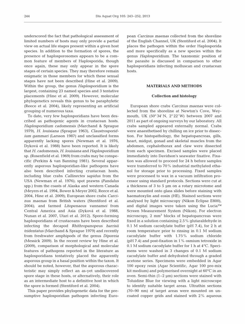
Haplosporidian infection, observed via presence ofopaque haemolymph upon dissection, and associateddistinctive histopathology occurred in 26 of 338 Car -cinus maenas (apparent prevalence 7.6%) sampled
over the study period. Haemocytes, connective tissuecells, reserve inclusion cells, gill epithelial cells andoccasionally muscle fibres contained large, eosino -philic parasitic plasmodia. In late stage infections,liberated uninucleate stages were observed withinthe haemal sinuses of all major organs and tissues(Fig. 1a,b).
Ultrastructural features of the crab haplosporidianwere largely consistent with the previous observa-tions by our laboratory. In brief, 3 distinct life stages:a free uninucleate form, a multinucleate plasmod-ium, and a membrane-bound multicellular plasmo -dial stage (containing uninucleate parasites withsimilar morphology to the free uninucleate stage)were frequently encountered in haemal spaces andassociated with the aforementioned host cell types.Uninucleate forms contained an electron lucent cyto-plasm, dense vesicles (DVs), haplosporosome-likebodies (HLBs) and ovoid lucent vesicles (OLVs).Multinucleate plasmodial stages ranged from binu-cleate forms to large cells containing over 20 nucleiin section. These nuclei often appeared closelyopposed but not connected. As seen in uninucleateforms, DVs, HLBs and OLVs were usually observedwithin the plasmodial cytoplasm. Formation of mem-branes around discrete nuclei (and enclosing HLBs,DVs and OLVs) was once again presumed to pre-empt the re-formation of uninucleate stages (therebycreating the multicellular plasmodial stage. Whilst adetailed description of the ultrastructure of thepathogen is available in previous work from our lab-oratory (Stentiford et al. 2004), a representativeimage of uninucleate and plasmodial life stages isgiven in Fig. 1c.
Molecular taxonomy
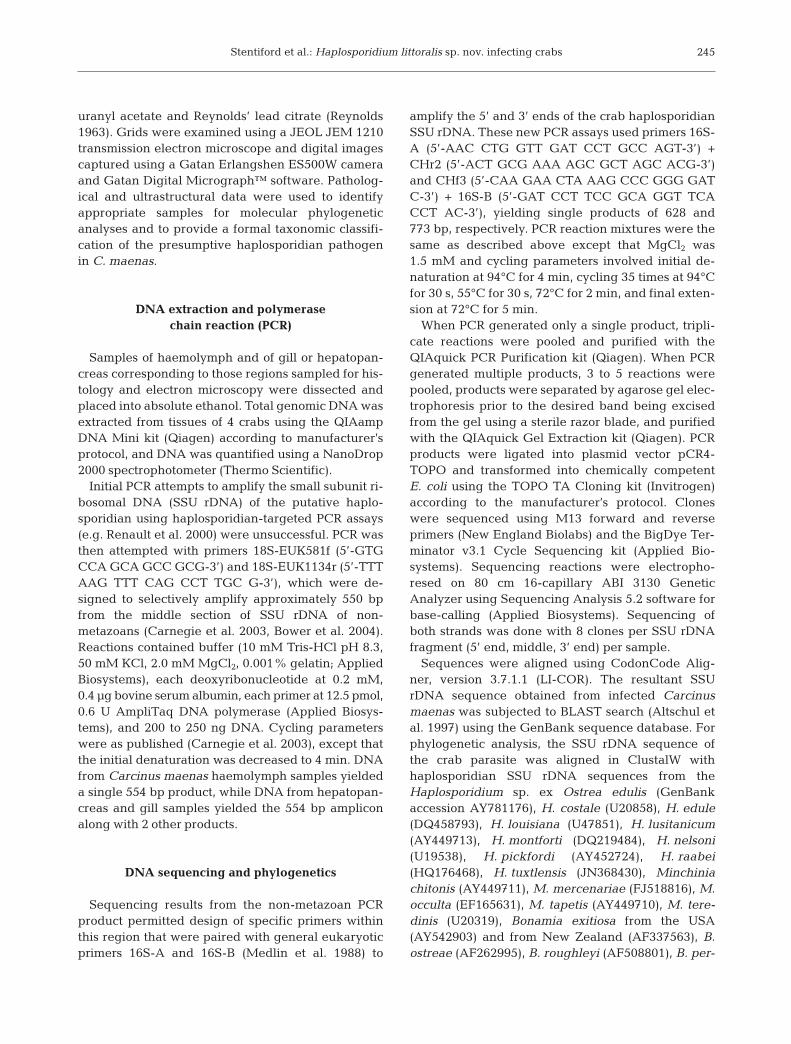
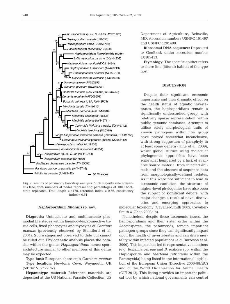
PCR amplification and sequencing of SSU rDNAfrom infected Carcinus maenas yielded one 1736 bpconsensus sequence (GenBank accession JX 18 54 13).BLAST analysis suggested that this sequence washaplosporidian, revealing a 79% maximum identityto both Haplosporidium tuxtlensis (JN368430) andHaplosporidium sp. ex Ostrea edulis (AY 78 11 76).Parsimony bootstrap analysis of a 2167-characterSSU rDNA sequence data set, 838 characters of whichwere parsimony-informative, placed this novel se -quence on a clade comprising the gastropod para-sites H. montforti, H. lusitanicum, H. pickfordi andH. tuxtlensis, and the haplosporidian from the poly-chaete Syllis nipponica (Fig. 2). While the analysissuggested a sister relationship of our sequence to the
246
Stentiford et al.: Haplosporidium littoralis sp. nov. infecting crabs 247
S. nipponica haplo sporidian, this relationship wasnot strongly supported (bootstrap value: 55). Thisclade was sister to another comprising H. costale,H. edule, H. raabei and Haplosporidium sp. exO. edulis. SSU rDNA phylogenetics thus indicatedplacement of the haplo sporidian sequence from C.maenas in the genus Haplosporidium.
In situ hybridisation
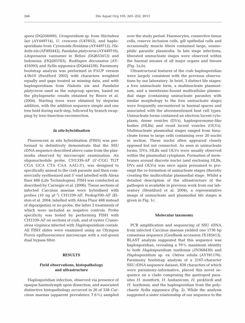
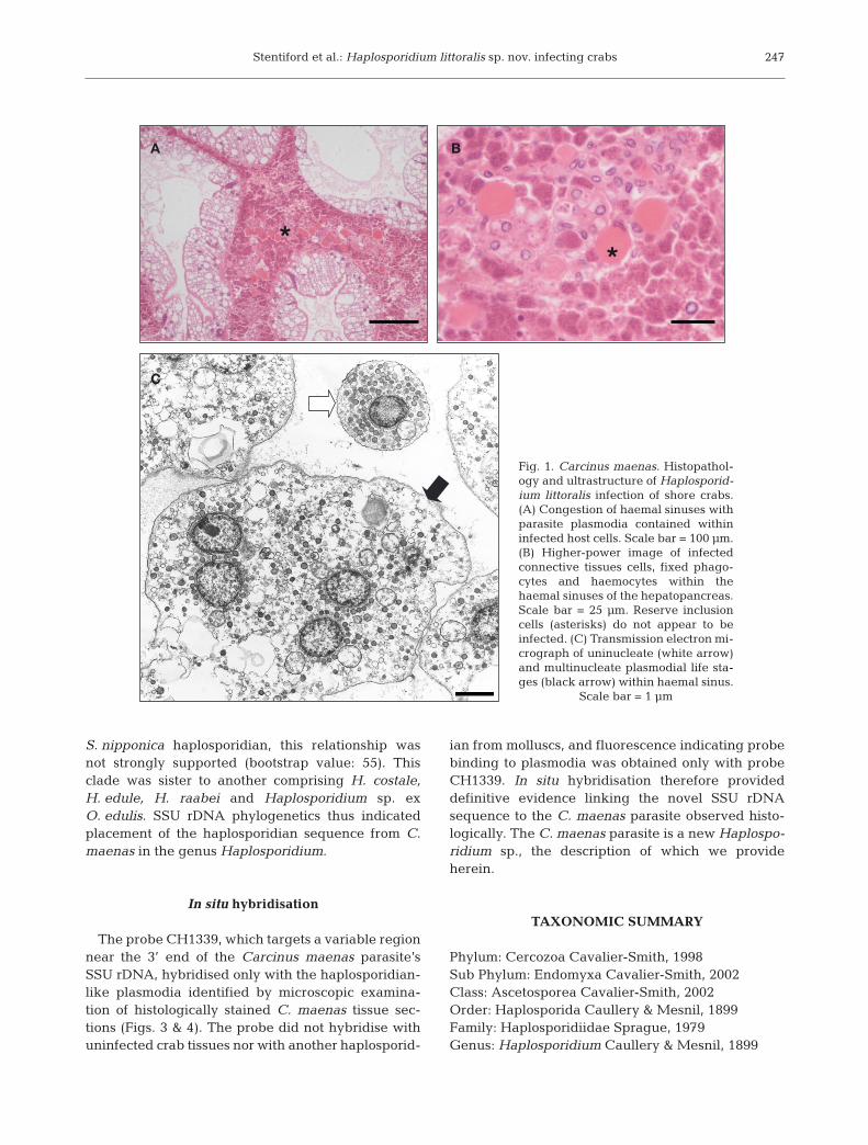
The probe CH1339, which targets a variable regionnear the 3’ end of the Carcinus maenas parasite’sSSU rDNA, hybridised only with the haplosporidian-like plasmodia identified by microscopic examina-tion of histologically stained C. maenas tissue sec-tions (Figs. 3 & 4). The probe did not hybridise withuninfected crab tissues nor with another haplosporid-
ian from molluscs, and fluorescence indicating probebinding to plasmodia was obtained only with probeCH1339. In situ hybridisation therefore provided de finitive evidence linking the novel SSU rDNAsequence to the C. maenas parasite observed histo-logically. The C. maenas parasite is a new Haplo spo -ri dium sp., the description of which we provideherein.
TAXONOMIC SUMMARY
Phylum: Cercozoa Cavalier-Smith, 1998Sub Phylum: Endomyxa Cavalier-Smith, 2002Class: Ascetosporea Cavalier-Smith, 2002Order: Haplosporida Caullery & Mesnil, 1899Family: Haplosporidiidae Sprague, 1979Genus: Haplosporidium Caullery & Mesnil, 1899
C
B
*
A
*
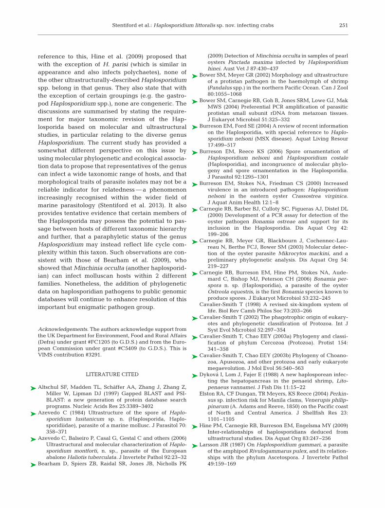
Fig. 1. Carcinus maenas. Histopathol-ogy and ultrastructure of Haplosporid-ium littoralis infection of shore crabs.(A) Congestion of haemal sinuses withparasite plasmodia contained withininfected host cells. Scale bar = 100 µm.(B) Higher-power image of infectedconnective tissues cells, fixed phago-cytes and haemocytes within thehaemal sinuses of the hepatopancreas.Scale bar = 25 µm. Reserve inclusioncells (asterisks) do not appear to beinfected. (C) Transmission electron mi -crograph of uninucleate (white arrow)and multinucleate plasmodial life sta -ges (black arrow) within haemal sinus.
Scale bar = 1 µm
Dis Aquat Org 105: 243–252, 2013
Haplosporidium littoralis sp. nov.
Diagnosis: Uninucleate and multinucleate plas-modial life stages within haemocytes, connective tis-sue cells, fixed phagocytes and myocytes of Carcinusmaenas (previously observed by Stentiford et al.2004). Spore stages not observed to date but cannotbe ruled out. Phylogenetic analysis places the para-site within the genus Haplosporidium; hence sporearchitecture similar to other members of this genusmay be expected.
Type host: European shore crab Carcinus maenasType location: Newton’s Cove, Weymouth, UK
(50° 34’ N, 2° 22’ W)Hepantotype material: Reference materials are
deposited at the US National Parasite Collection, US
Department of Agriculture, Beltsville,MD. Accession numbers USNPC 101497and USNPC 1201498.
Ribosomal DNA sequence: Depositedto GenBank under accession numberJX185413.
Etymology: The specific epithet refersto shore line (littoral) habitat of the typehost.
DISCUSSION
Despite their significant economicimportance and their dramatic effect onthe health status of aquatic inverte-brates, the haplosporidians remain asignificantly understudied group, withrelatively sparse representation withinpublic genomic databases. Attempts toutilise solely morphological traits ofknown pathogens within the grouphave proved somewhat inconclusive,with strong suggestion of paraphyly inat least some genera (Hine et al. 2009),whilst global studies using molecularphylogenetic approaches have beensomewhat hampered by a lack of avail-able source material from infected ani-mals and the absence of sequence datafrom morphologically-defined isolates.As if this were not sufficient to lead totaxonomic confusion, the structure ofhigher-level phylogenies have also beenthe subject of significant debate, withmajor changes a result of novel discov-eries and emerging approaches to
molecular taxonomy (Cavalier-Smith 2002, Cavalier-Smith & Chao 2003a,b).
Nonetheless, despite these taxonomic issues, thehaplosporidians and their sister order within theAscetosporea, the paramyxids, remain importantpathogen groups since they can significantly impactupon the health of invertebrates and can drive mor-tality within infected populations (e.g. Burreson et al.2000). This impact has led to representative members(e.g. Bonamia ostreae and B. exitiosa spp. within theHaplosporida and Marteilia refringens within theParamyxida) being listed in the international legisla-tion of the European Union (Directive 2006/88/EC)and of the World Organisation for Animal Health(OIE 2012). This listing provides an important politi-cal tool by which national governments can control
248
Fig. 2. Results of parsimony bootstrap analysis: 50% majority rule consen-sus tree, with numbers at nodes representing percentages of 1000 boot-strap replicates. Tree length = 4170, retention index = 0.56, consistency
index = 0.51
FE
DC
B
*
A
*
Stentiford et al.: Haplosporidium littoralis sp. nov. infecting crabs
the import of aquatic animals and products derivedfrom them, based upon the important pathogens thatthey may be carrying. In this context, improved taxo -nomy of the group is a key driver in protectingnational biosecurity whilst not unduly restricting freetrade.
The current study has provided such taxonomicclarity for an unclassified ‘haplosporidian-like’pathogen previously described by our laboratory(Stentiford et al. 2004). The parasite, hereby namedHaplosporidium littoralis, causes severe alterationsto the connective tissues and haemolymph of infec -
ted crabs. The prevalence of infection observed here(7.6%) was remarkably similar to our previous obser-vation (7%; Stentiford et al. 2004), and since we con-sider that infection leading to disease would be ter-minal, we continue to assume that the parasite islethal to infected individuals. The parasite thereforeis considered to be a mortality driver in populationsof Carcinus maenas.
Based upon parsimony analysis against the SSUrDNA sequences of other haplosporidian parasites,we report that the crab parasite is sister to the gastro-pod mollusc parasites Haplosporidium montforti,
249
FE
DC
B
*
A
*
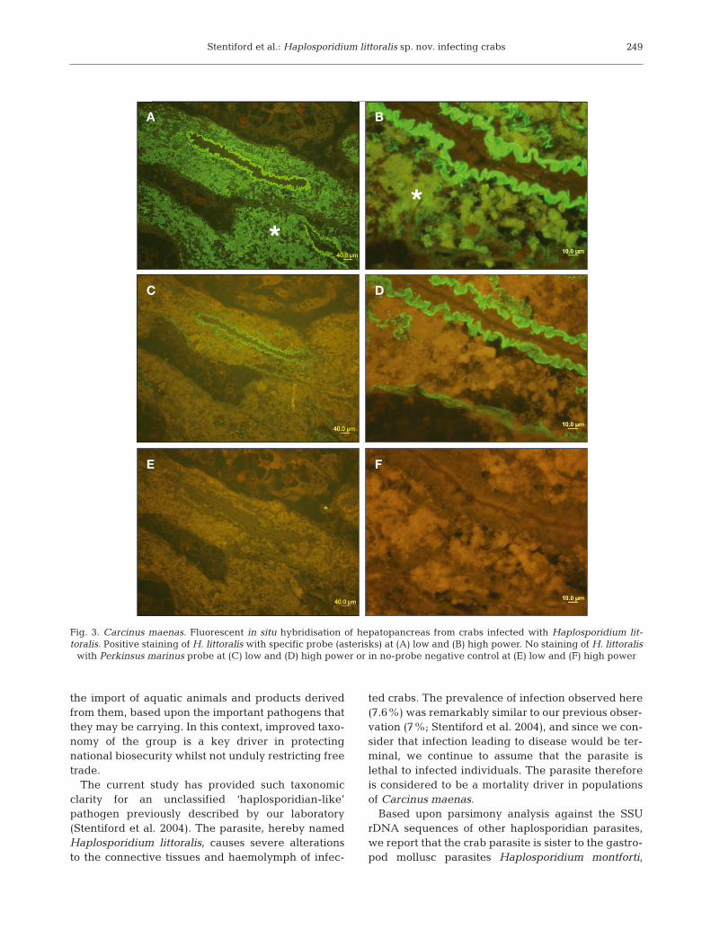
Fig. 3. Carcinus maenas. Fluorescent in situ hybridisation of hepatopancreas from crabs infected with Haplosporidium lit-toralis. Positive staining of H. littoralis with specific probe (asterisks) at (A) low and (B) high power. No staining of H. littoralis
with Perkinsus marinus probe at (C) low and (D) high power or in no-probe negative control at (E) low and (F) high power
D
*
CBA
Dis Aquat Org 105: 243–252, 2013
H. lusitanicum and H. pickfordi, and to a parasitefrom the polychaete Syllis nipponica. The relation-ship of the crab parasite to existing members of thegenus Haplosporidium is somewhat surprising giventhat other known haplosporidian parasites from mar-ine crustaceans, including those from the Pacificwhite shrimp Litopenaeus vannamei (Nunan et al.2007, Utari et al. 2012) and particularly another fromthe spot prawn Pandalus platyceros (Reece et al.2000), were shown to be phylogenetically dissimilarto H. littoralis from European crabs. Despite therather limited literature on this subject, the relativelywide variation in SSU rDNA gene sequence amongthese 3 parasites is suggestive of an underlyingdiversity in the as-yet undiscovered haplosporidianfauna in fecting crustacean hosts. Furthermore, itappears to suggest a closer relationship between cer-tain haplo sporidians infecting ecologically-associ-ated crus tacean and molluscan hosts than to geo-
graphically and ecologically dis-tinct hosts of the same taxa.
Haplosporidium montforti (Aze -vedo et al. 2006), H. lusitanicum(Azevedo 1984) and H. pickfordiBarrow, 1961 are parasites of theEuropean abalone Haliotis tuber-culata, the blue-rayed limpetHelcion pellucidus and the fresh-water Gatineau tadpole snailPhy sella parkeri, re spectively.The hosts H. tuberculata and H.pellucidis are marine shorelinegastro pods found along the coast -lines of Europe. The former has amarginal niche overlap with pop-ulations of Carcinus maenas (e.g.in the Channel Islands of thesouthern English Channel) buthas not been re corded along thecoastline of the northern EnglishChannel (site of the currentstudy). In contrast, H. pellucidis isa common inhabitant of kelp (e.g.Laminaria hyperborea) in thelower eulittoral zone of Europeancoastlines (Marine Life Informa-tion Network: http://www. marlin.ac.uk). The presence of a phylo-genetically similar Haplosporid-ium species in closely associatedorganisms from this shorelinehabitat is intriguing and suggeststhe possibility that haplosporidi-
ans within this genus can passage between mollus-can and crustacean hosts. Furthermore, the potentialfor multi-trophic transfer of haplosporidians couldalso account for the apparent absence of particularlife stages (such as the spore) and or ga nelles (hap-losporosomes) in some of the ‘asporous’ haplosporid-ians of crustacean hosts as defined by Hine et al.(2009).
Numerous authors have reported ultrastructuraland phylogenetic data that show the genus Haplo -sporidium to be paraphyletic (Burreson & Ford 2004,Burreson & Reece 2006, Hine et al. 2009). Evidencefor this is focussed on existing members of the genusoften bearing little morphological resemblance to thetype species of the genus, H. scolopli Caullery andMesnil, 1899 in polychaete worms, and to the basalphylogenetic position of H. nelsoni and H. louisianarelative to not only Haplosporidium spp. but toBonamia spp. and Minchinia spp. as well (Fig. 2). In
250
D
*
CBA
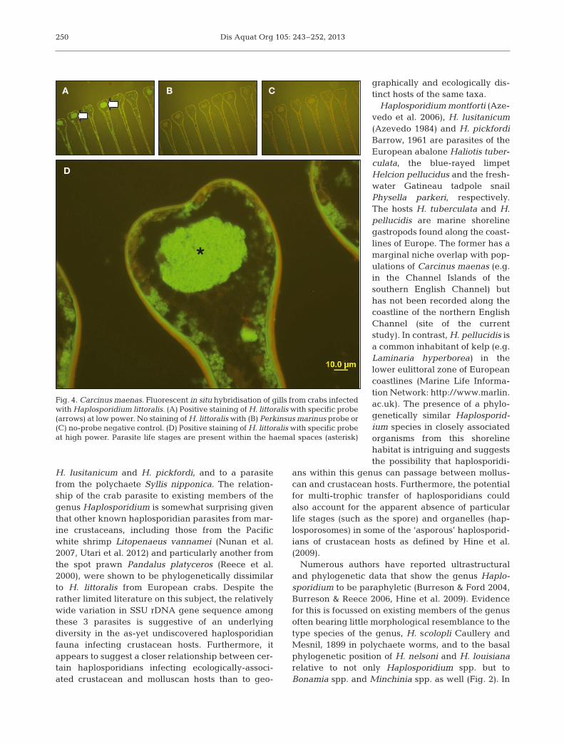
Fig. 4. Carcinus maenas. Fluorescent in situ hybridisation of gills from crabs infectedwith Haplosporidium littoralis. (A) Positive staining of H. littoralis with specific probe(arrows) at low power. No staining of H. littoralis with (B) Perkinsus marinus probe or(C) no-probe negative control. (D) Positive staining of H. littoralis with specific probeat high power. Parasite life stages are present within the haemal spaces (asterisk)
Stentiford et al.: Haplosporidium littoralis sp. nov. infecting crabs 251
reference to this, Hine et al. (2009) proposed thatwith the exception of H. parisi (which is similar inappearance and also infects polychaetes), none ofthe other ultrastructurally-described Haplosporidiumspp. belong in that genus. They also state that withthe exception of certain groupings (e.g. the gastro-pod Haplosporidium spp.), none are congeneric. Thediscussions are summarised by stating the require-ment for major taxonomic revision of the Hap-losporida based on molecular and ultrastructuralstudies, in particular relating to the diverse genusHaplosporidium. The current study has provided asomewhat different perspective on this issue byusing molecular phylogenetic and ecological associa-tion data to propose that representatives of the genuscan infect a wide taxonomic range of hosts, and thatmorphological traits of parasite isolates may not be areliable indicator for relatedness — a phenomenonincreasingly recognised within the wider field ofmarine parasitology (Stentiford et al. 2013). It alsoprovides tentative evidence that certain members ofthe Haplosporida may possess the potential to pas-sage between hosts of different taxonomic hierarchyand further, that a paraphyletic status of the genusHaplosporidium may instead reflect life cycle com-plexity within this taxon. Such observations are con-sistent with those of Bearham et al. (2009), whoshowed that Minchinia occulta (another haplosporid-ian) can infect molluscan hosts within 2 differentfamilies. Nonetheless, the addition of phylogeneticdata on haplosporidian pathogens to public genomicdatabases will continue to enhance resolution of thisimportant but enigmatic pathogen group.
Acknowledgements. The authors acknowledge support fromthe UK Department for Environment, Food and Rural Affairs(Defra) under grant #FC1205 (to G.D.S.) and from the Euro-pean Commission under grant #C5409 (to G.D.S.). This isVIMS contribution #3291.
LITERATURE CITED
Altschul SF, Madden TL, Schäffer AA, Zhang J, Zhang Z,Miller W, Lipman DJ (1997) Gapped BLAST and PSI-BLAST: a new generation of protein database searchprograms. Nucleic Acids Res 25: 3389−3402
Azevedo C (1984) Ultrastructure of the spore of Haplo -sporidium lusitanicum sp. n. (Haplosporida, Haplo -sporidiidae), parasite of a marine mollusc. J Parasitol 70: 358−371
Azevedo C, Balseiro P, Casal G, Gestal C and others (2006)Ultrastructural and molecular characterization of Haplo -sporidium montforti, n. sp., parasite of the Europeanabalone Haliotis tuberculata. J Invertebr Pathol 92: 23−32
Bearham D, Spiers ZB, Raidal SR, Jones JB, Nicholls PK
(2009) Detection of Minchinia occulta in samples of pearloysters Pinctada maxima infected by Haplosporidiumhinei. Aust Vet J 87: 430−437
Bower SM, Meyer GR (2002) Morphology and ultrastructureof a protistan pathogen in the haemolymph of shrimp(Pandalus spp.) in the northern Pacific Ocean. Can J Zool80: 1055−1068
Bower SM, Carnegie RB, Goh B, Jones SRM, Lowe GJ, MakMWS (2004) Preferential PCR amplification of parasiticprotistan small subunit rDNA from metazoan tissues.J Eukaryot Microbiol 51: 325−332
Burreson EM, Ford SE (2004) A review of recent informationon the Haplosporidia, with special reference to Haplo -sporidium nelsoni (MSX disease). Aquat Living Resour17: 499−517
Burreson EM, Reece KS (2006) Spore ornamentation ofHaplo sporidium nelsoni and Haplosporidium costale(Haplosporidia), and incongruence of molecular phylo -geny and spore ornamentation in the Haplosporidia.J Parasitol 92: 1295−1301
Burreson EM, Stokes NA, Friedman CS (2000) Increased virulence in an introduced pathogen: Haplosporidiumnelsoni in the eastern oyster Crassostrea virginica.J Aquat Anim Health 12: 1−8
Carnegie RB, Barber BJ, Culloty SC, Figueras AJ, Distel DL(2000) Development of a PCR assay for detection of theoyster pathogen Bonamia ostreae and support for itsinclusion in the Haplosporidia. Dis Aquat Org 42: 199−206
Carnegie RB, Meyer GR, Blackbourn J, Cochennec-Lau-reau N, Berthe FCJ, Bower SM (2003) Molecular detec-tion of the oyster parasite Mikrocytos mackini, and apreliminary phylogenetic analysis. Dis Aquat Org 54: 219−227
Carnegie RB, Burreson EM, Hine PM, Stokes NA, Aude-mard C, Bishop MJ, Peterson CH (2006) Bonamia per-spora n. sp. (Haplosporidia), a parasite of the oysterOstreola equestris, is the first Bonamia species known toproduce spores. J Eukaryot Microbiol 53: 232−245
Cavalier-Smith T (1998) A revised six-kingdom system oflife. Biol Rev Camb Philos Soc 73: 203−266
Cavalier-Smith T (2002) The phagotrophic origin of eukary-otes and phylogenetic classification of Protozoa. Int JSyst Evol Microbiol 52: 297−354
Cavalier-Smith T, Chao EEY (2003a) Phylogeny and classi -fication of phylum Cercozoa (Protozoa). Protist 154: 341−358
Cavalier-Smith T, Chao EEY (2003b) Phylogeny of Choano -zoa, Apusozoa, and other protozoa and early eukaryotemegaevolution. J Mol Evol 56: 540−563
Dyková I, Lom J, Fajer E (1988) A new haplosporean infec -ting the hepatopancreas in the penaeid shrimp, Lito -penaeus vannamei. J Fish Dis 11: 15−22
Elston RA, CF Dungan, TR Meyers, KS Reece (2004) Perkin-sus sp. infection risk for Manila clams, Venerupis philip-pinarum (A. Adams and Reeve, 1850) on the Pacific coastof North and Central America. J Shellfish Res 23:1101–1105
Hine PM, Carnegie RB, Burreson EM, Engelsma MY (2009)Inter-relationships of haplosporidians deduced fromultrastructural studies. Dis Aquat Org 83: 247−256
Larsson JIR (1987) On Haplosporidium gammari, a parasiteof the amphipod Rivulogammarus pulex, and its relation-ships with the phylum Ascetospora. J Invertebr Pathol49: 159−169
Dis Aquat Org 105: 243–252, 2013
Marchand J, Sprague V (1979) Ultrastructure de Minchiniacadomensis sp. n. (Haplosporida) parasite du décapodeRhithropanopeus harrisii tridentatus Maitland dans lecanal de Caen à la mer (Calvados, France). J Protozool26: 179−185
Medlin L, Elwood HJ, Stickel S, Sogin ML (1988) The char-acterization of enzymatically amplified eukaryotic 16S-like rRNA-coding proteins. Gene 71: 491−499
Messick GA (2009) Haplosporidian parasite in Diporeia spp.amphipods from the Great Lakes region, USA. Dis AquatOrg 83: 153−157
Meyers TR, Lightner DV, Redman RM (1994) A dinoflagel-late-like parasite in Alaskan spot shrimp Pandalusplatyceros and pink shrimp P. borealis. Dis Aquat Org 18: 71−76
Newman MW, Johnson CA, Pauley GB (1976) A Minchinialike haplosporidan parasitizing blue crabs, Callinectessapidus. J Invertebr Pathol 27: 311−315
Nunan LM, Lightner DV, Pantoja CR, Stokes NA, Reece KS(2007) Characterization of a rediscovered haplosporidianparasite from cultured Penaeus vannamai. Dis AquatOrg 74: 67−75
OIE (World Organisation for Animal Health) (2012) Manualof diagnostic tests for aquatic animals, 7th edn, 2012.OIE, Paris
Perkins FO (2000) Phylum Haplosporidia Caullery & Mesnil,1899. In: Lee JJ, Leedale GF, Bradbury P (eds) The illus-trated guide to the Protozoa. Society of Protozoologists,Lawrence, KS, p 1329−1341
Perkins FO, van Banning P (1981) Surface ultrastructure ofspores in three genera of Balanosporida, particularlyMinchinia armoricana van Banning, 1977. The taxo-nomic significance of spore wall ornamentation in theBalanosporida. J Parasitol 67: 866−874
Reece KS, Burreson EM, Bower SM, Dungan CF (2000)Molecular analyses of a parasite in prawns (Pandalus
platyceros) from British Columbia, Canada. J ShellfishRes 19: 647 (Abstract)
Reece KS, Siddall ME, Stokes NA, Burreson EM (2004)Molecular phylogeny of the Haplosporidia based ontwo independent gene sequences. J Parasitol 90: 1111−1122
Renault T, Stokes NA, Chollet B, Cochennec N, Berthe F,Gérard A, Burreson EM (2000) Haplosporidiosis in thePacific oyster Crassostrea gigas from the French Atlanticcoast. Dis Aquat Org 42: 207−214
Reynolds ES (1963) The use of lead citrate at high pH as anelectron-opaque stain in electron microscopy. J Cell Biol17: 208−212
Rosenfield A, Buchanan L, Chapman GB (1969) Comparisonof the fine structure of spores of three species of Min-chinia (Haplosporida, Haplosporidiidae). J Parasitol 55: 921−941
Sprague V (1963) Revision of the genus Haplosporidium andrestoration of the genus Minchinia (Haplosoridia, Haplo -sporidiidae). J Protozool 10, 263–266
Stentiford GD, Feist SW, Bateman KS, Hine PM (2004)Haemolymph parasite of the shore crab Carcinus mae-nas: pathology, ultrastructure and observations on crus-tacean haplosporidians. Dis Aquat Org 59: 57−68
Stentiford GD, Bateman KB, Feist SW, Chambers E, StoneDM (2013) Plastic parasites: extreme dimorphism createsa taxonomic conundrum in the phylum Microsporidia. IntJ Parasitol 43: 339−352
Swofford DL (2002) PAUP*: phylogenetic analysis using par-simony (*and other methods), Version 10. Sinauer Asso-ciates, Sunderland, MA
Utari HB, Senapin S, Jaengsanong C, Flegel TW, Kruatra-chue M (2012) A haplosporidian parasite associated withhigh mortality and slow growth in Penaeus (Litopenaeus)vannamei cultured in Indonesia. J Invertebr Pathol 92: 23−32
252
Editorial responsibility: Sven Klimpel, Frankfurt, Germany
Submitted: March 1, 2013; Accepted: May 16, 2013Proofs received from author(s): July 29, 2013