Embed Size (px)
Citation preview
OR I G I N A L A R T I C L E
Hard and soft tissue analysis of alveolar ridge preservation inesthetic zone using deproteinized bovine bone mineral and asaddle connective tissue graft: A long-term prospective case series
Gaëlle Botilde DDS, MS1,2 | Paul-Emile Colin MD3 |
Oscar González-Martín DMD, PhD, MSc., Clinical Professor4,5,6 |
Geoffrey Lecloux DDS, MS1,2 | Eric Rompen DDS, MS, PhD, Professor1,2 |
France Lambert DDS, MS, PhD, Professor1,2
1Department of Periodontology and Oral
Surgery, Faculty of Medicine, University of
Liege, Liège, Belgium
2Dental Biomaterials Research Unit, University
of Liège, Liège, Belgium
3Department of Radiodiagnostic, Faculty of
Medicine, University of Liege, Liège, Belgium
4Department of Periodontology, University
Complutense of Madrid, Madrid, Spain
5Department of Periodontology, University of
Iowa, Iowa, Iowa
6Private Practice, Gonzalez + Solano Atelier
Dental, Madrid, Spain
Correspondence
Dr. France Lambert, Head of clinic,
Department of Periodontology and Oral
Surgery, Faculty of Medicine, University of
Liege, Service de Medecine Dentaire, Domaine
du Sart Tilman Bat B-35, B-4000 Liege,
Belgium.
Email: [email protected]
Abstract
Aim: Although alveolar ridge preservation (ARP) procedures appear to limit bone
resorption after dental extraction, long-term outcomes remain limited. The objective
of this prospective case series was to evaluate the long-term hard and soft tissue
changes after ARP procedure in the aesthetic area, using deproteinized bovine bone
mineral (DBBM) and saddle connective tissue graft.
Materials and Methods: Fifteen patients were subjected to ARP and impressions and
CT scans were taken at baseline and 3 months. After 5 to 7 years, a secondary long-
term clinical and radiological analysis was carried out. Horizontal alveolar bone
changes, soft tissue profiles and implant outcomes were assessed.
Results: Although a limited hard and soft tissue remodeling occurred during the first
3 months after ARP, from 3 months to the long-term evaluation, the alveolar bone
dimensions remained stable and the soft tissue profiles significantly increased, in the
more cervical levels. The implant survival rate after 5 to 7 years yielded 100% and
peri-implant bone levels and soft tissue health were good.
Conclusion: Within the limits of the study, the present data confirms the long-term
effectiveness of ARP using DBBM and a saddle connective tissue graft offering sta-
ble hard and soft tissue conditions up to 5 to 7 years.
K E YWORD S
aesthetic, alveolar bone, alveolar bone preservation, dimensional change, extraction socket
management, hard tissue volume, long term, soft tissue profile
1 | INTRODUCTION
Despite the technological advances in the field of dentistry, many rea-
sons can lead to tooth loss, such as caries, traumatism, endodontic
failures, or periodontal causes. Tooth removal procedure results in
dimensional hard and soft tissue shrinkage, mainly in the first 3 months
of the healing process.1 According to systematic reviews, evaluating
the dimensional changes in hard and soft tissues following tooth
extraction, alveolar bone remodeling in the maxilla leads to mean hori-
zontal bone loss of 3.8 mm after 6 months.2,3 The severity of this
physiological remodeling depends on several factors such as the tooth
angulation, the facial bone wall thickness, and other differences in the
tooth sites anatomy.4-6 The majority of the horizontal resorption was
proven to occur mainly on the buccal aspect of the ridge as thin
Received: 12 November 2019 Revised: 14 February 2020 Accepted: 10 March 2020
DOI: 10.1111/cid.12899
Clin Implant Dent Relat Res. 2020;1–10. wileyonlinelibrary.com/journal/cid © 2020 Wiley Periodicals LLC 1

buccal plates are tooth-dependent structures.7-10 These dimensional
changes can result, especially in the aesthetic area, in functional and
aesthetic discrepancies and compromise implant placement.
In order to prevent this postextraction bone remodeling, a number
of alveolar ridge preservation (ARP) techniques have been described in
the literature using various biomaterials including autologous bone,
bone substitutes (allografts, xenografts, and alloplasts), bioactive agents,
and autologous blood-derived products.11 Several authors emphasized
that ARP procedures can significantly limit the bone and soft tissue
shrinkage after extraction and although they cannot limit completely
the shrinkage, they may be a valid treatment option in order to avoid
further bone reconstruction.12-19 Furthermore, in order to compensate
for this expected bone remodeling and improve the aesthetic outcomes,
some authors proposed the use of a saddle connective tissue graft in
combination with the biomaterials.20,21
Nevertheless, the studies evaluating the efficacy of ARP were
mostly conducted over a follow-up periods running from 12 weeks to
9 months13,15 and long-term data remains limited.
The aim of the present study was to evaluate the long-term
radiological and clinical outcomes of an ARP technique using
deproteinized bovine bone mineral (DBBM) combined with a saddle
connective tissue graft. The primary objective was to describe the
hard and soft tissue changes from baseline up to a follow-up period
of 5 to 7 years. The secondary objectives were the assessment of
implant survival, peri-implant bone stability, and soft tissue health.
Moreover, long-term aesthetic outcomes based on the Pink Esthetic
Score (PES)22 and patient reported outcomes measures (PROMS)
were investigated.
2 | MATERIALS AND METHODS
2.1 | Study population
Patients needing tooth replacement in aesthetic area (tooth 15-25)
were recruited from the Department of Periodontology and Oral
Surgery of the University of Liège, Belgium. All the patients included
in the study met the following inclusion criteria: good general health
(ASA 1,2), absence of/or controlled periodontitis, at least 18 years
old or with a signed approval document by the parents, and cigarette
F IGURE 1 Surgical procedure and follow-up. A, Atraumatically tooth extraction. B, After granulation removal, connective tissue graft washarvested from the palate. C, D, The socket was filled with Bio-Oss and the graft was inserted into split-thickness buccal and palatal pouches tocover the extraction site. E, 3-month follow-up frontal and F, palatal, view. G, Long-term evaluation frontal, and H, palatal, view. Prosthodontics :Prof. Amélie Mainjot
2 BOTILDE ET AL.
smoking less than 10 per day. The exclusion criteria were: pregnant
or breastfeeding females, patients included in another study at the
same time, patients with bone disease or under bone metabolism-
interfering drugs, patients with a history of head and neck radiother-
apy, and patients presenting dehiscence or fenestration on the bone
wall of the socket.
2.2 | Study design
The present study was designed as a prospective case series.
Between September 2009 and September 2011, all patients included
in the study were subjected to the same ARP technique performed
by two previously calibrated senior periodontists. Radiographic ana-
lyses (CT scans) as well as impressions were performed at baseline
and 3 months thereafter. In June 2016, all patients were recalled for
a secondary long-term analyses during which impression, intraoral
radiography, and cone beam CT (CBCT) were carried out. Alveolar
bone remodeling and soft tissue changes were then evaluated
respectively based on computed axial tomography (CT scans) and
models performed at baseline, 3 months and 5 to 7 years after the
procedure. Moreover, when applicable, implant survival and success
rates, peri-implant bone levels and clinical parameters including PES,
plaque index (PI), bleeding on probing (BOP), and pocket depth
(PD) were recorded in June 2016. Finally, PROMs were evaluated
using VAS questionnaire. This study was performed in full accor-
dance with the declared ethical principles of the World Medical Asso-
ciation Declaration of Helsinki of 1975 (revised in 2008) and the
protocol was approved by the ethical committee of the University of
Liège, Belgium (B707201628853). The study was registered in the
clinical trials registry: www.clinicaltrials.gov (NCT03410251).
2.3 | Surgical procedure
The full surgical procedure was reported in previous articles.20,21 In
brief, single extractions in the anterior maxilla (14-24) were performed
atraumatically and without flap release. After checking the integrity of
the buccal and palatal bone plate, a biomaterial (Bio-Oss; Geistlich
Pharma AG, Wolhusen, Switzerland) was placed into the socket. A
connective tissue graft harvested from the palate was inserted and
sutured in buccal and palatal split-thickness pouches in order to cover
the socket (Figure 1A-D). Tooth brushing was not recommended at
the extraction site for 10 following days and the sutures were
removed 10 days after surgery. Prescribed medication consisted in
Chlorexidine spray (0.12%) BID, Ibuprofen 600 mg TID according to
the needs, and Amoxicillin (500 mg TID) antibiotherapy administrated
for 5 days.
2.4 | Follow-up
Patients were followed at 3 months (Figure 1E,F) and then yearly for
regular check-ups. When indicated, implants were placed 4 to
6 months after the ARP. Implants restorations or conventional fixed
partial denture (FPD) procedures were performed according to the
dentist preference and patient choice. In June 2016, all patients were
recalled for a secondary long-term evaluation (Figure 1G,H).
2.5 | Hard tissue analyses
In order to evaluate the alveolar bone remodeling overtime, the
patients were subjected to CT scan (Somaton Emotion; Siemens,
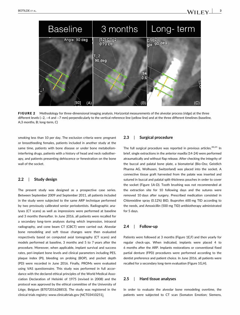
F IGURE 2 Methodology for three-dimensional imaging analysis. Horizontal measurements of the alveolar process (ridge) at the threedifferent levels (−2, −4 and −7 mm) perpendicularly to the vertical reference line (yellow line) and at the three different timelines (baseline,A;3 months, B; long-term, C)
BOTILDE ET AL. 3
Munich, Germany) at baseline and at 3 months. For these examina-
tions, a slide thickness of 0.6 mm was always used. At the long-term
evaluation, for ethical reasons and because of the advances in imaging
technologies, a CBCT (Newtom 5G; Sitech, France) was chosen to
reduce radiation dose. CBCT was performed using a reduced field of
view to cover the desired area at 0.2 mm voxels according to Garib
et al23 and a reduced exposure protocol.
2.5.1 | Methodology for 3D imaging analysis
The measurements were performed by matching and superimposing
baseline and 3-month CT scans as well as long-term CBCT scans,
using three-dimensional (3D) reconstruction software (SyngoVia; Sie-
mens). After an adequate calibration, two independent operators
made all the measurements (GB and PEC). The scans were
automatically superimposed by the software two by two
(baseline/3 months and 3 months/long term). For each case, the mea-
surements were made on a parasagittal section going through the
middle edentulous segment determined on the 3-month CT scan. On
this buccal-palatal section, a vertical reference line was drawn in the
center of the alveolar crest in order to measure the ridge (Figure 2,
yellow line). The horizontal dimensions of the alveolar bone crest
were measured perpendicularly to the vertical reference line at the
three following levels: −2, −4, −7 mm below the most coronal point
of the 3-month section (Figure 2A-C). Thereafter, the same measure-
ments were taken on the baseline CT and long-term CBCT images by
superimposing the 3D images as well as the reference line which was
reported exactly at the same place in the different scans thanks to the
superimposition. Altogether, three measurements were taken at each
time point giving a total of nine measurements per patient. Long-term
bone dimensional changes (expressed both in percentage and in mm)
were then calculated both from baseline to 3 months and from
3 months to the long-term follow-up.
2.6 | Soft tissue outlines volumetric analyses
The impression and pouring protocols were standardized. Baseline,
3month and long-term alginate impressions were taken with the
same impression material (CavexColorChange�, Cavex Holland BV,
Haarlem, The Netherlands) and using an alginate mixer. The impres-
sions were poured in plaster within a period of 24 hours. Two differ-
ent operators took the baseline and 3- month impressions while all
long-term impressions were taken by a third single operator. The
casts were scanned with a 3D laser scanner (D250, 3Shape, Copen-
hagen, Denmark). The .stl files obtained from each model were sub-
sequently transferred to a digital shape sampling and processing
software for re-elaboration of 3D models from the 3D scan data
(Studio, Geomagic, Research Triangle Park, North Carolina). For each
patient, baseline and long-term models were superimposed follow-
ing a previously reported protocol.24 Prior to taking the measure-
ments, the baseline model was set as the reference, while the
3-month and long-term model were set as the test. For each sup-
erimposed model, 2D labio-palatal sections were obtained in the
middle of the extraction area, perpendicular to the alveolar crest.
Subsequently, the linear distance between the preoperative and
postoperative soft tissue profiles was measured. These measure-
ments were taken at the top of the crest and were repeated at 2 and
4 mm in the apical direction (Figure 3).
2.7 | Long-term aesthetic outcomes
At the long-term evaluation, the PES as described by Fürhauser et al
was collected for each patient.22 The contralateral tooth was used as
control whenever it was possible; otherwise, PES was calculated
based on ideal tooth proportions. The threshold for clinical acceptabil-
ity was set at an arbitrary score of 8 out of 14.25
TABLE 1 Patient, sites, and implants characteristics Problem withthe title of part B, must be placed between tabacoo use and teeth.
A. Patient-related descriptive analyses n = 15
Age (years) Mean (±SD)
min-max
43.4 (±13.3)
16.7-62.1
Gender Male 6 (40%)
Female 9 (60%)
Tobacco use (at the cross-sectional
long-term evaluation)
No 9 (60%)
<10c/d 4 (26.7%)
>10c/d 2 (13.3%)
B. Site characteristics n = 15
Teeth Incisors 9 (60%)
Canine 0 (0.0%)
Premolar 6 (40%)
Rehabilitation Implant 12 (80%)
FPD 3 (20%)
C. Implant characteristics and outcomes n = 12
Implant type Straumann BL 10 (83.3%)
Nobel Active 1 (8.3%)
Intralock, MDL (Mini
Drived- Lock) 2.5 mm
1 (8.3%)
PIa Sites with plaque (%) 6/48 (12.5%)
BOPa Sites with BOP 17/48 (35.4%)
PD (mm)a Mean (±SD) 2.96 (±0.84)
min-max 2-4.8
DIB (mm) Mean (±SD) −0.05 (±0.73)
min-max −1.2-1.3
Implant survival
rates
12 (100%)
BOP, bleeding on probing; DIB, distance between implant shoulder and
first bone to implant contact; FPD, fixed partial denture; PD, pocket depth;
PI, plaque index.aAt four aspects around implants (mesial, buccal, palatal, distal).
4 BOTILDE ET AL.

Additionally, aesthetics from a patient perspective were evaluated
using a VAS questionnaire. The following questions were asked:
1. How satisfied are you with your aesthetic rehabilitation?; 2. Do you
have the feeling that the aesthetic appearance of your rehabilitation
has changed overtime?
2.8 | Long-term implants outcomes
At the long-term evaluation, in patients who benefited from an
implant rehabilitation, implant survival, and success rates were
recorded. Success was defined according to the criteria of Buser,26
which are (a) absence of suppuration (recurring peri-implant infection),
(b) absence of persistent complaints like pain, foreign body sensation,
and/or dysaesthesia, (c) absence of continuous radiolucency area
around the implant, (d) absence of implant mobility. Additionally, the
peri-implant bone levels were assessed on periapical radiography
using the parallel technique: the linear distance between the implant
shoulder of the bone level implants and the first bone to implant con-
tact (DIB in mm) was measured at the mesial and distal aspects27
using the specific software Image J64 (National Institutes of Health,
Bethesda, Maryland). The health of peri-implant soft tissues was also
F IGURE 3 Two dimensionalcomparison of the superimposedmodels perpendicular to the alveolarcrest (Baseline- 3 months, top;3 months- long-term, middle;Baseline- long-term, down)
BOTILDE ET AL. 5

assessed at four sites per implant, including: PI, BOP and PD. For PI
and BOP, a dichotomous score was given (PI: 0 = no visible plaque,
1 = plaque at the soft tissue margin; BOP: 0 = no bleeding, 1 = bleed-
ing) and PD was measured by means of a periodontal probe
(CP 15 UNC, Hu-Friedy, Chicago, Illinois) and rounded off to the
nearest millimeter.28
2.9 | Statistical analysis
The results were expressed as means and SD for the continuous vari-
ables and as frequency tables for the categorical variables. The
intraclass correlation coefficient (ICC) was used to test the concor-
dance between the two examiners for each measurement, at each
timeline and for each measurement level. Statistics were performed
on the mean of both examiners for each parameter. The evolution of
the measures between baseline and 3 months, 3 months and long-
term and baseline and long-term data was evaluated by a paired Stu-
dent t test. The analysis of variance was used to compare the levels.
The comparison of the differences between patients with implant and
patients without implant was done by a Student t test. Results were
considered significant at the 5% critical level (P < .05). The calculations
were performed using the SAS version 9.4 for windows (SAS Institute,
Cary, North Carolina).
3 | RESULTS
3.1 | Patient demographics
In total, 29 subjects met the inclusion criteria and were enrolled in this
study. Of these, nine patients dropped out and did not show up for
the secondary long-term analysis, two disagreed to undergo the
CBCT, two had missing data, and one was excluded because a guided
bone regeneration was performed in the neighboring site over the
follow-up period. Therefore, 15 subjects (six men and nine women,
mean age: 43.4 [SD ± 13.3], min: 16.7-max: 62.1) were considered in
this study. The patients were recalled after a mean follow-up of
almost 6 years (mean: 70 months, min: 57.3 months-max:
81.6 months). A dropout analysis emphasized that no significant dif-
ference was found between the dropout and followed patients. No
patient smoked more than 10 cigarettes per day at the time of the
inclusion, but 2 patients started smoking more than 10 cigarettes a
day over the follow-up period (Table 1A). Overall, socket management
procedures were applied in nine incisors and in six premolars sites. To
restore the edentulous spaces, 12 patients received an implant and
3 were managed by a conventional FPD (Table 1B).
3.2 | Hard tissue analyses
The 3D imaging measurements showed high reliability, as inter- and
intraexaminer observations were concordant for all measurements
(ICC mean: 0.98; min: 0.95). The results of measurements are pres-
ented in Table 2.
The measurements of the horizontal bone remodeling revealed
that significant bone loss occurred at the three corono-apical levels
only during the early phase after extraction (baseline—3 months).
Bone losses of −1.41 mm (P < .0001), −0.76 mm (P < .0001), and
−0.45 mm (P = .0003) were respectively found for −2, −4, and −7
levels. Further losses from 3 months to the long-term follow-up
remained below 0.5 mm and were not significant except in the more
apical region (P = .0008) (Table 2).
3.3 | Soft tissue volumetric analyses
Based on the superimposition of the digitalized impressions per-
formed at baseline, 3 months and 5 to 7 years after the ARP, the data
revealed a mild buccal shrinkage of the soft tissue outlines from base-
line to 3 months. From 3 months to the long-term follow-up, a signifi-
cant gain in the soft tissue contours was observed buccally at the
cervical and medial levels. The resulting variation in soft tissue volume
from baseline to 5 years was not significant for each level. The
detailed results are displayed in Table 2.
3.4 | Long-term aesthetic outcomes
The results of PES are displayed in Table 3. The analysis revealed a
mean PES value of 10.9 (min: 8-max: 14) out of 14 points as a maxi-
mum22 and 100% of the implants were considered aesthetically
TABLE 2 Hard and soft tissue remodeling (significant p- value in bold)
n = 15 Baseline-3 months P-value
3 months-
long term P-value
Baseline-
long term P-value
Bucco-
palatal hard tissueremodeling mm (±SD)
Cervical level −2 mm −1.41 (±0.64) <.0001 0.01 (±0.78) .97 −1.40 (±0.86) <.0001
Medial level −4 mm −0.76 (±0.36) <.0001 −0.24 (±0.58) .13 −1.00 (±0.69) <.0001
Apical level −7 mm −0.45 (±0.36) .0003 −0.43 (±0.37) .0008 −0.88 (±0.48) <.0001
Buccal soft tissueremodeling mm (±SD)
Cervical level 0 mm −0.6 (±0.97) .032 +0.89 (±1.32) .024 +0.26 (±0.76) .202
Medial level −2 mm −0.52 (± 0.58) .004 +0.58 (± 0.97) .036 +0.06 (±0.63) .708
Apical level −4 mm −0.29 (± 0.4) .015 +0.31 (± 0.81) .164 +0.02 (± 0.66) .893
6 BOTILDE ET AL.
acceptable according to the criteria. The PROMs related to aesthetic
satisfaction (see question 1) reached a mean VAS score of 9.13 (min:
7-max: 10) out of 10 and the one assessing aesthetic stability (see
question 2) over time displayed a mean VAS score of 9.20 (min:
7-max: 10) out of 10. The detailed aesthetic outcomes based on
patient opinions are displayed in Table 4.
3.5 | Long-term implant outcomes
Detailed implant characteristics and outcomes are listed in Table 1C.
All of the 12 implants were osseointegrated at the long-term evalua-
tion time point, leading to an implant survival rate of 100%. The mean
DIB value calculated on the long-term peri-apical radiographs was
−0.05 mm (SD ± 0.73). Implants demonstrated fairly healthy peri-
implant soft tissues. Local plaque deposit was registered in 6 out of
48 (12.5%) sites around the implants and BOP was found in 17 out of
48 (35.4%) sites. Finally, the mean PD value was 2.96 mm (SD ± 0.84).
4 | DISCUSSION
This study demonstrated that extraction socket management combin-
ing the use of DBBM and a saddle connective tissue graft limited the
bucco-palatal bone shrinkage in the 3 first months after the ARP
(1.4 mm in the cervical area) when compared to the mean remodeling
of 3.8 mm described in the literature after extraction alone.3 Thereaf-
ter, the hard tissue dimensions remained stable up to 7 years, except
in the most apical region where a fairly low additional loss of 0.4 mm
was observed. Moreover, the connective tissue graft used to compen-
sate for the expected bone remodeling, and insure proper soft tissue
outlines, was found to be effective in the long term.
According to recent systematic reviews, even though the benefits
of ARP procedures are nowadays recognized, follow-up studies
evaluating the possible influence of these treatments on the long-
term aesthetic and implant outcomes are lacking.12,13,16,17 To the best
of our knowledge, the present report is one of the few studies29
assessing long-term outcomes of alveolar ridge preservation proce-
dures and the first study assessing long-term hard tissue changes on
3D images.
4.1 | Hard tissue remodeling
In the present study, the main bone dimensional changes were
observed during the first 3 months following the ARP procedure and
afterwards they remained stable up to 5 to 7 years. A recent literature
review assessing the hard tissue shrinkage after ARP procedures using
a xenograft emphasized a mean bucco-palatal bone loss of 1.3 mm
after 3 months.30 Although, the heterogeneity of the extracted tooth
types and of measurements protocols limits the comparison, these
results are in agreement with the present results. Moreover, several
studies describe higher horizontal bone remodeling in the cervical
region and the values decrease progressively towards the apical
level.6,20,31 The present study described a similar outcome from base-
line to 3 months but interestingly, the pattern is inverted from
3 months to the long-term follow-up. Indeed, from 3 months to the
long-term follow-up, the bone dimensions were found to be very sta-
ble in the cervical and medial levels while a further horizontal loss
(0.4 mm) was observed at the apical level. Although significant, this
additional loss is extremely limited from a clinical point of view. The
dimensional stability in the more cervical levels might be related to
the nonresorbable characteristics of the biomaterial used in the proce-
dures.32 The initial remodeling would be the consequence of the buc-
cal bone plate resorption and then33 the nonresorbable or slowly
resorbable biomaterial placed in the socket would provide the long-
term dimensional stability. The slight resorption observed in the apical
region might be related to a physiological remodeling related to facial
growth in absence of biomaterials in this apical region.34 Therefore,
the nonremodeling properties of the chosen biomaterial might be of
capital importance for the long-term stability of ARP. Further compar-
ative studies would be, however, necessary to confirm this
hypothesis.
4.2 | Soft tissue remodeling
Looking at the soft tissue outlines, it is interesting to observe a loss of
volume from baseline to 3 months and a gain from 3 months to the
TABLE 3 Pink Esthetic Score values
PES
score
Mesial
papilla Distal papilla
Level soft
tissue margin
Soft tissue
contour
Alveolar
process
Soft tissue
color
Soft tissue
texture Total PES
0 0/15 (0%) 1/15 (6.6%) 2/15 (13.3%) 1/15 (6.6%) 0/15 (0%) 0/15 (0%) 1/15 (6.6%) 10.9 (min: 8-max: 14)
1 6/15 (40%) 6/15 (40%) 7/15 (46.6%) 8/15 (53.3%) 1/15 (6.6%) 3/15 (20%) 6/15 (40%)
2 9/15 (60%) 8/15 (53.3%) 6/15 (40%) 6/15 (40%) 14/15 (93.3%) 12/15 (80%) 8/15 (53.3%)
TABLE 4 Patient reported outcomes measures (PROMs) (n = 16)
How satisfied are youwith the aesthetic ofyour rehabilitation?
Did you have the feelingthat the aesthetic
appearance of yourrehabilitation decreasesovertime?
1 = not satisfied
100 = very satisfied
1 = yes, a lot
100 = Not at all
Mean (±SD) 91.3 (±9.15) 92.0 (±9.41)
Min-max 70-100 70-100
BOTILDE ET AL. 7

long-term follow-up. This observation might be attributed to two dis-
tinct phenomena. On the one hand, the loss of transgingival support
at the time of extraction and restoration of a transgingival part at
implant loading may have an influence on the buccal gingival out-
lines.35 On the other hand, the tissue creeping after connective tissue
graft described by some authors may also be responsible for this gain
of volume in the long term.36 However, an impression after the abut-
ment connection and the restoration of the implant would have been
necessary to accurately assess the effect of these hypotheses.
4.3 | Implant survival and success
This study demonstrated that single implant placement 4 months after
ridge preservation procedure provides successful long-term outcomes.
Indeed, the implant survival rate was 100% and a mean DIB value of
−0.05 mm (SD ± 0.73) was found. One of the limitations of this study
is the absence of a standardized x-ray after the implant placement;
however, the DIB was already proposed by some authors to assess
the peri-implant bone environment.27 These results corroborate the
findings of a recent study demonstrating equal success when implants
were placed in preserved vs nonpreserved alveolar ridges.37 Addition-
ally, the peri-implant soft tissue health is considered to be an impor-
tant criterion of implant success.38 Comparable PI, BOP, and PD
values where found in a prospective study on immediate implants
placed in the aesthetic area at 5-year follow-up,28 and these clinical
findings were considered as successful implant outcomes. However,
better BOP (18.4%) results were found in a 10-year follow-up pro-
spective study29 on implants placed after ridge preservation using a
xenograft as well. The better BOP described in that study may be
associated with the strict maintenance care program that patients
received, which indeed may be paramount for long-term implant
success.
4.4 | Aesthetic outcomes
At the long-term follow-up, the PES value reached 10.9 out of
14, which can be considered as a very good score according to some
authors.22,28 Moreover, similar or even a better cervical level of the
mucosal margin was found compared to the contralateral tooth, while
mid-facial recession is considered as frequent complication of immedi-
ate implant placement in the aesthetic zone.25,28,39-42 These observa-
tions provide some evidence that, in the aesthetic area, socket
management procedure using CTG and delayed implantation could be
more predictable for long-term stability of facial hard and soft tissues.
However, these results have to be interpreted carefully in the absence
of PES baseline values. Moreover, it is important to mention that out
of the 15 patients, 3 were restored with a conventional bridge while
the PES as described by Fürhauser et al was developed to assess the
aesthetics of implant rehabilitations.
Although the white esthetic scores (WES) were not considered in
the present study as the restorative protocol was not standardized,
the aesthetic outcomes from a patient's perspective displayed excel-
lent results (>9 out of 10). It means that patients were highly satisfied
with the aesthetic appearance of their implant rehabilitation in the
long term, which is also an important aspect of success.
The present protocol should also be compared to alternative
implant treatment options in the aesthetic zone such as extraction
and early or delayed implant placement combined with lateral Guided
Bone Regeneration (GBR).43,44 This approach was widely described
and seems to display short- and long-term effectiveness as well as
hard and soft tissue dimension stability over time.45 However, com-
pared to GBR technique, the advantage of the present protocol is the
absence of flap release and need for a membrane, and therefore
potentially reducing costs and patient morbidity.46 As suggested by
some authors, to further reduce the morbidity, the connective tissue
graft might be performed only if necessary at the time of implant
placement.47 In this case, at the time of extraction, it might be rele-
vant to use a connective or a collagen plug to cover the socket and
protect the biomaterials underneath.48
4.5 | Study limitations
The present study suffers from several limitations that should be
highlighted. First of all, the study was designed as a case series and
does not allow a comparison with another treatment concept. More-
over, the limited samples size and the significant number of dropouts
are further weakness of the present study. Although the dropout anal-
ysis did not emphasize any significant difference between the dropout
and followed patients, this limitation should be considered when
interpreting the results.
Finally, from a methodological perspective, the use of two different
radiographic methods (CT and CBCTs) may also be critical; however,
some authors demonstrated no statistically significant difference in terms
of linear measurements accuracy between CT and CBCT scans.15,49 Addi-
tionally, the use of 3D imaging to evaluate ARP has already been
described in the literature and consists in a well-established method for
the detection of bone dimensional changes.20,30,50
Despites these limitations, the study still provides relevant long-
term information on extraction socket management with DDBM and
saddle connective tissue graft.
5 | CONCLUSION
Despite the limitations of the present study, the management of
intact extraction socket with DBBM and saddle connective tissue
graft seems to be effective for the preservation of alveolar hard and
soft tissues up to 5-7 years post-treatment. The present long-term
study revealed bone dimensional changes only during the 3 first
months after the ARP procedure and stable soft tissue outlines from
baseline up to 5 to 7 years. Additionally, this surgical technique
allowed implant placement after a follow-up period of 4 to 6 months
without any further bone regeneration therapy, as well as long-term
8 BOTILDE ET AL.

implant success rates and aesthetic outcomes. This long-term study
therefore suggests that this treatment modality is a reliable option for
the replacement of missing teeth in the aesthetic area. However, fur-
ther investigations and controlled studies are warranted.
CONFLICT OF INTEREST
The authors declare no potential conflict of interest.
ORCID
France Lambert https://orcid.org/0000-0002-1018-2544
REFERENCES
1. Schropp L, Wenzel A, Kostopoulos L, Karring T. Bone healing and soft
tissue contour changes following single-tooth extraction: a clinical
and radiographic 12-month prospective study. Int J Periodontics
Restorative Dent. 2003;23:313-323.
2. Tan WL, Wong TLT, Wong MCM, Lang NP. A systematic review of
post-extractional alveolar hard and soft tissue dimensional changes in
humans. Clin Oral Implants Res. 2012;23:1-21.
3. Van der Weijden F, Dell'Acqua F, Slot DE. Alveolar bone dimensional
changes of post-extraction sockets in humans: a systematic review.
J Clin Periodontol. 2009;36:1048-1058.
4. Cardaropoli D, Tamagnone L, Roffredo A, Gaveglio L. Relationship
between the buccal bone plate thickness and the healing of
postextraction sockets with/without ridge preservation. Int J Peri-
odontics Restorative Dent. 2014;34:211-217. http://www.quintpub.
com/journals/prd/abstract.php?article_id=13987.
5. Chappuis V, Engel O, Reyes M, Shahim K, Nolte LP, Buser D. Ridge
alterations post-extraction in the esthetic zone: a 3D analysis with
CBCT. J Dent Res. 2013;92:195-201.
6. Misawa M, Lindhe J, Araújo MG. The alveolar process following
single-tooth extraction: a study of maxillary incisor and premolar sites
in man. Clin Oral Implants Res. 2015;27:884-889.
7. Araújo MG, Lindhe J. Dimensional ridge alterations following tooth
extraction. An experimental study in the dog. J Clin Periodontol. 2005;
32:212-218.
8. Botticelli D, Berglundh T, Lindhe J. Hard-tissue alterations following
immediate implant placement in extraction sites. J Clin Periodontol.
2004;31:820-828.
9. Lekovic V, Camargo PM, Klokkevold PR, et al. Preservation of alveolar
bone in extraction sockets using bioabsorbable membranes. J Periodontol.
1998;69:1044-1049. https://doi.org/10.1902/jop.1998.69.9.1044.
10. Lekovic V, Kenney EB, Weinlaender M, et al. A bone regenerative
approach to alveolar ridge maintenance following tooth extraction.
Report of 10 cases. J Periodontol. 1997;68:563-570.
11. Darby I, Chen ST, Buser D. Ridge preservation techniques for implant
therapy. Int J Oral Maxillofac Implants. 2009;24:260-271.
12. Avila-Ortiz G, Elangovan S, Kramer KWO, Blanchette D, Dawson DV.
Effect of alveolar ridge preservation after tooth extraction: a system-
atic review and meta-analysis. J Dent Res. 2014;93(10):950-958.
13. Brandam L, Malmstrom H, Javed F, Calvo-Guirado J-L, Romanos GE.
Ridge preservation techniques in the anterior esthetic zone. Implant
Dent. 2015;24(6):699-712.
14. Horváth A, Mardas N, Mezzomo LA, Needleman IG, Donos N. Alveo-
lar ridge preservation. A systematic review. Clin Oral Investig. 2013;
17:341-363.
15. Macbeth N, Trullenque-Eriksson A, Donos N, Mardas N. Hard and
soft tissue changes following alveolar ridge preservation: a systematic
review. Clin Oral Implants Res. 2016;1-23.
16. Vignoletti F, Matesanz P, Rodrigo D, Figuero E, Martin C, Sanz M.
Surgical protocols for ridge preservation after tooth extraction. A sys-
tematic review. Clin Oral Implants Res. 2012;23:22-38.
17. Avila-Ortiz G, Chambrone L, Vignoletti F. Effect of alveolar ridge
preservation interventions following tooth extraction: a systematic
review and meta-analysis. J Clin Periodontol. 2019;46(Suppl 21):
195-223.
18. Iocca O, Farcomeni A, De Virgilio A, et al. Prognostic significance of
lymph node yield and lymph node ratio in patients affected by squa-
mous cell carcinoma of the oral cavity and oropharynx: study protocol
for a prospective, multicenter, observational study. Contemp Clin Tri-
als Commun. 2019;14:100324.
19. Vittorini Orgeas G, Clementini M, De Risi V, de Sanctis M. Surgical
techniques for alveolar socket preservation: a systematic review. Int J
Oral Maxillofac Implants. 2013;28:1049-1061.
20. Lambert F, Vincent K, Vanhoutte V, Seidel L, Lecloux G, Rompen E. A
methodological approach to assessing alveolar ridge preservation pro-
cedures in humans: hard tissue profile. J Clin Periodontol. 2012;39:
887-894.
21. Vanhoutte V, Rompen E, Lecloux G, Rues S, Schmitter M, Lambert F.
A methodological approach to assessing alveolar ridge preservation
procedures in humans: soft tissue profile. Clin Oral Implants Res.
2014;25:304-309.
22. Fürhauser R, Florescu D, Benesch T, Haas R, Mailath G, Watzek G.
Evaluation of soft tissue around single-tooth implant crowns: the pink
esthetic score. Clin Oral Implants Res. 2005;16:639-644.
23. Garib DG, Calil LR, Leal CR, Janson G. Is there a consensus for CBCT
use in orthodontics? Dental Press J Orthod. 2014;19:136-149.
24. Gonzalez-Martin O, Veltri M, Moraguez O, Belser UC. Quantitative
three-dimensional methodology to assess volumetric and
profilometric outcome of subepithelial connective tissue grafting at
pontic sites: a prospective pilot study. Int J Periodontics Restorative
Dent. 2014;34:673-679.
25. Cosyn J, Eghbali A, De Bruyn H, Collys K, Cleymaet R, De Rouck T.
Immediate single-tooth implants in the anterior maxilla: 3-year results
of a case series on hard and soft tissue response and aesthetics. J Clin
Periodontol. 2011;38:746-753.
26. Buser D, Weber H-P, Lang NP. Tissue integration of non-submerged
implants. 1-year results of a prospective study with 100 iti hollowcylinder
and hollow-screw implants. Clin Oral Implants Res. 1990;1:33-40.
27. Buser D, Halbritter S, Hart C, et al. Early implant placement with
simultaneous guided bone regeneration following single-tooth
extraction in the esthetic zone: 12-month results of a prospective
study with 20 consecutive patients. J Periodontol. 2009;80:
152-162.
28. Cosyn J, Eghbali A, Hermans A, Vervaeke S, De Bruyn H, Cleymaet R.
A 5-year prospective study on single immediate implants in the aes-
thetic zone. J Clin Periodontol. 2016;43:702-709.
29. Roccuzzo M, Gaudioso L, Bunino M, Dalmasso P. Long-term stability
of soft tissues following alveolar ridge preservation: 10-year results
of a prospective study around nonsubmerged implants. Int J Periodon-
tics Restorative Dent. 2014;34:795-804. http://www.quintpub.com/
journals/prd/abstract.php?article_id=14902 - .VGypnhavueU.
30. Jambhekar S, Kernen F, Bidra AS. Clinical and histologic outcomes of
socket grafting after flapless tooth extraction: a systematic review of
randomized controlled clinical trials. J Prosthet Dent. 2015;113:371-
382. https://doi.org/10.1016/j.prosdent.2014.1012.1009.
31. Jung RE, Philipp A, Annen BM, et al. Radiographic evaluation of dif-
ferent techniques for ridge preservation after tooth extraction: a
randomized controlled clinical trial. J Clin Periodontol. 2013;40:
90-98.
32. Traini T, Valentini P, Iezzi G, Piattelli A. A histologic and
histomorphometric evaluation of anorganic bovine bone retrieved
9 years after a sinus augmentation procedure. J Periodontol. 2007;78:
955-961.
33. Araujo MG, Lindhe J. Ridge preservation with the use of Bio-Oss col-
lagen: a 6-month study in the dog. Clin Oral Implants Res. 2009;20:
433-440.
BOTILDE ET AL. 9
34. Daftary F, Mahallati R, Bahat O, Sullivan RM. Lifelong craniofacial
growth and the implications for osseointegrated implants. Int J Oral
Maxillofac Implants. 2013;28:163-169. http://www.quintpub.com/
journals/find_article.php?doi=110.11607/jomi.12827.
35. Su H, Gonzalez-Martin O, Weisgold A, Lee E. Considerations of
implant abutment and crown contour: critical contour and subcritical
contour. Int J Periodontics Restorative Dent. 2010;30:335-343.
36. Harris RJ. Creeping attachment associated with the connective tissue
with partial-thickness double pedicle graft. J Periodontol. 1997;68:
890-899.
37. Cardaropoli D, Tamagnone L, Roffredo A, Gaveglio L. Evaluation of
dental implants placed in preserved and nonpreserved postextraction
ridges: a 12-month postloading study. Int J Periodontics Restorative
Dent. 2017;35:677-685. http://quintpub.com/journals/prd/abstract.
php?iss672_id=1326&article_id=15605.
38. Jepsen S, Berglundh T, Genco R, et al. Primary prevention of peri-
implantitis: managing peri-implant mucositis. J Clin Periodontol. 2015;
42(Suppl 16):S152-S157.
39. Chen ST, Darby IB, Reynolds EC. A prospective clinical study of non-
submerged immediate implants: clinical outcomes and esthetic
results. Clin Oral Implants Res. 2007;18:552-562.
40. De Rouck T, Collys K, Cosyn J. Immediate single-tooth implants in the
anterior maxilla: a 1-year case cohort study on hard and soft tissue
response. J Clin Periodontol. 2008;35:649-657.
41. Kan JY, Rungcharassaeng K, Lozada JL, Zimmerman G. Facial gingival
tissue stability following immediate placement and provisionalization
of maxillary anterior single implants: a 2- to 8-year follow-up. Int J
Oral Maxillofac Implants. 2011;26:179-187.
42. Raes S, Eghbali A, Chappuis V, Raes F, De Bruyn H, Cosyn J. A long-
term prospective cohort study on immediately restored single tooth
implants inserted in extraction sockets and healed ridges: CBCT ana-
lyses, soft tissue alterations, aesthetic ratings, and patient-reported
outcomes. Clin Implant Dent Relat Res. 2018;20:522-530.
43. Buser D, Chappuis V, Belser UC, Chen S. Implant placement post
extraction in esthetic single tooth sites: when immediate, when early,
when late? Periodontol 2000. 2017;73:84-102.
44. Chappuis V, Rahman L, Buser R, Janner SFM, Belser UC, Buser D.
Effectiveness of contour augmentation with guided bone regenera-
tion: 10-year results. J Dent Res. 2018;97:266-274.
45. Graziani F, Chappuis V, Molina A, et al. Effectiveness and clinical per-
formance of early implant placement for the replacement of single
teeth in anterior areas: a systematic review. J Clin Periodontol. 2019;
46(Suppl 21):242-256.
46. Frost NA, Banjar AA, Galloway PB, Huynh-Ba G, Mealey BL. The
decision-making process for ridge preservation procedures after
tooth extraction. Clin Adv Periodontics. 2014;4:56-63. https://doi.org/
10.1902/cap.2013.130013.
47. Seyssens L, Eghbali A, Christiaens V, De Bruyckere T, Doornewaard R,
Cosyn J. A one-year prospective study on alveolar ridge preservation
using collagen-enriched deproteinized bovine bone mineral and saddle
connective tissue graft: a cone beam computed tomography analysis.
Clin Implant Dent Relat Res. 2019;21(5):853-861.
48. Fickl S, Kauffmann F, Stappert CF, Kauffmann A, Schlagenhauf U.
Scar tissue formation following alveolar ridge preservation: a
case control study. Int J Periodontics Restorative Dent. 2018;38:
e1-e7.
49. Fatemitabar SA, Nikgoo A. Multichannel computed tomography ver-
sus cone-beam computed tomography: linear accuracy of in vitro
measurements of the maxilla for implant placement. Int J Oral Maxil-
lofac Implants. 2010;25:499-505.
50. Morimoto T, Tsukiyama Y, Morimoto K, Koyano K. Facial bone alter-
ations on maxillary anterior single implants for immediate placement
and provisionalization following tooth extraction: a superimposed
cone beam computed tomography study. Clin Oral Implants Res.
2015;26:1383-1389.
How to cite this article: Botilde G, Colin P-E, González-
Martín O, Lecloux G, Rompen E, Lambert F. Hard and soft
tissue analysis of alveolar ridge preservation in esthetic zone
using deproteinized bovine bone mineral and a saddle
connective tissue graft: A long-term prospective case series.
Clin Implant Dent Relat Res. 2020;1–10. https://doi.org/10.
1111/cid.12899
10 BOTILDE ET AL.


![Histological assessment of the palatal mucosa and greater ... · exposed root, alveolar ridge, or soft tissue around implants [2, 3]. Successful surgery depends on several factors,](https://img.pdfslide.net/doc/110x75/5e070bb6a07c0d7ce307230d/histological-assessment-of-the-palatal-mucosa-and-greater-exposed-root-alveolar.jpg)