Embed Size (px)
Citation preview

Review ArticleA Review on Oxidative Stress, Diabetic Complications, and theRoles of Honey Polyphenols
Visweswara Rao Pasupuleti ,1,2,3 Chandra Sekhar Arigela ,1 Siew Hua Gan ,4
Sirajudeen Kuttulebbai Nainamohamed Salam ,5 Kumara Thevan Krishnan ,1
Nurhanan Abdul Rahman ,1 and Mohammad Saffree Jeffree 6
1Faculty of Agro-Based Industry, Universiti Malaysia Kelantan, Campus Jeli, 17600 Jeli, Malaysia2Department of Biomedical Sciences and Therapeutics, Faculty of Medicine & Health Sciences, Universiti Malaysia Sabah, Malaysia3Department of Biochemistry, Faculty of Medicine and Health Sciences, Abdurrab University, Jl Riau Ujung No. 73,Pekanbaru 28292, Riau, Indonesia4School of Pharmacy, Monash University Malaysia, Jalan Lagoon Selatan, 47500 Bandar Sunway, Selangor, Malaysia5Department of Basic Medical Sciences, Kulliyyah of Medicine, International Islamic University Malaysia (IIUM),Bandar Indera Mahkota, 25200 Kuantan, Pahang, Malaysia6Department of Community and Family Medicine, Faculty of Medicine and Health Sciences, Universiti Malaysia Sabah,Kota Kinabalu, 88400 Sabah, Malaysia
Correspondence should be addressed to Visweswara Rao Pasupuleti; [email protected] Nurhanan Abdul Rahman; [email protected]
Received 6 August 2020; Revised 18 October 2020; Accepted 23 October 2020; Published 23 November 2020
Academic Editor: Yueliang Zhao
Copyright © 2020 Visweswara Rao Pasupuleti et al. This is an open access article distributed under the Creative Commons AttributionLicense, which permits unrestricted use, distribution, and reproduction in any medium, provided the original work is properly cited.
Despite the availability of various antidiabetic drugs, diabetes mellitus (DM) remains one of the world’s most prevalent chronicdiseases and is a global burden. Hyperglycaemia, a characteristic of type 2 diabetes mellitus (T2DM), substantially leads to thegeneration of reactive oxygen species (ROS), triggering oxidative stress as well as numerous cellular and molecular modificationssuch as mitochondrial dysfunction affecting normal physiological functions in the body. In mitochondrial-mediated processes,oxidative pathways play an important role, although the responsible molecular mechanisms remain unclear. The impairedmitochondrial function is evidenced by insulin insensitivity in various cell types. In addition, the roles of master antioxidantpathway nuclear factor erythroid 2-related factor 2 (Nrf2)/Kelch-like ECH-associated protein 1 (Keap1)/antioxidant responseelements (ARE) are being deciphered to explain various molecular pathways involved in diabetes. Dietary factors are known toinfluence diabetes, and many natural dietary factors have been studied to improve diabetes. Honey is primarily rich incarbohydrates and is also abundant in flavonoids and phenolic acids; thus, it is a promising therapeutic antioxidant for variousdisorders. Various research has indicated that honey has strong wound-healing properties and has antibacterial, anti-inflammatory,antifungal, and antiviral effects; thus, it is a promising antidiabetic agent. The potential antidiabetic mechanisms of honey wereproposed based on its major constituents. This review focuses on the various prospects of using honey as an antidiabetic agent andthe potential insights.
1. Introduction
Diabetes mellitus is characterized mainly by hyperglycaemiaoccurring due to defects in insulin secretion, insulin action,or both. According to the International Diabetes Federation(IDF), 463 million people globally have been diagnosed with
diabetes in 2019 and the figure is expected to rise to 700million by 2040 [1]. In fact, type 2 diabetes mellitus (T2DM)is one of the leading causes of illness and premature death inthe world, with 4.2 million fatalities reported in 2019 [1].The disease mainly affects both developed regions (“Occiden-tal World”), as well as developing countries, contributed by
HindawiOxidative Medicine and Cellular LongevityVolume 2020, Article ID 8878172, 16 pageshttps://doi.org/10.1155/2020/8878172

unhealthy lifestyles, such as physical inactivity and high-fatand sugar consumption [2] .
In Asia, the numbers of diabetics in China (>113.9million diabetics), India (>62 million), and Malaysia (3.5million) render the region a critical “hot spot” for diabetes[3]. Some of the factors contributing to the accelerated trajec-tory of T2DM include (i) poor nutrition in utero and in earlylife and (ii) overnutrition in later lives (especially in popula-tions undergoing rapid nutrition transitions, involvingchanged food habits and reduced physical activity). Interest-ingly, the prevalence of T2DM is slightly higher in men thanin women [4].
Besides the environment, T2DM is a multifactorial dis-ease involving genetic influence that has a “nature vs. nurtureoutcome.” The pathophysiological changes of diabetes arecharacterized by (1) β-cell dysfunction in the periphery(e.g., the liver, skeletal muscle, and adipose tissue) and (2)inflammation-dependent ROS generated locally by tissue ordue to immune cells interacting with the insulin receptor(IR) as well as their downstream signalling pathways, result-ing in a failure to respond adequately to insulin levels. All ofthese factors form a basis for insulin resistance and chronicinflammation which progressively hamper blood glucosecontrol, leading to the development of micro- and macrovas-cular complications.
With respect to hyperglycaemia, it has been reported that atleast eight distinct pathophysiological abnormalities contributeto impaired glucose homeostasis; this has been well establishedearly in the natural history of T2DM [5]. In addition to the“ominous octet,” two additional pathophysiological abnormal-ities, namely, (1) activation of inflammatory pathways and (2)impaired insulin-mediated vasodilation, contribute to muscleinsulin resistance. More recently, our understanding of thepathophysiology of T2DM has been further aided by thediscovery of novel disease biomarkers where high blood con-centrations of proinflammatory cytokines, such as C-reactiveprotein (CRP), interleukin-6 (IL-6), and tumour necrosis factor(TNF) are associated with an increased risk of T2DM [6] .
Oxidative stress is a condition that emerges from theimbalance between the formation of reactive oxygen species(ROS) and the ability of the antioxidant system to neutralizethese compounds. Increased oxidative stress is the result ofenhanced ROS production and/or decreased ROS scavengingability resulting in tissue damage [7]. In fact, the pathology ofa number of diseases including diabetic, vascular, and neuraldisorders are greatly influenced by oxidative stress.
Antioxidants are compounds that can help reduce oxida-tive stress. They can be (1) endogenous (reduced glutathioneand antioxidant enzymes such as catalase, superoxidedismutase, glutathione peroxidase, and reductase) or (2) exog-enous (antioxidant vitamins like vitamins A, C, and E). Bothtypes of antioxidants can help prevent the formation of freeradicals by scavenging or promoting their decomposition[8]. Additionally, they can transform the reactive metabolitesinto less reactive molecules [9]. As further insights into themolecular underpinnings of this disease and its destructivesequelae are gained, more opportunities in the developmentof novel, targeted approaches for diabetes treatment, progno-sis and, ultimately, prevention exist.
Natural products have become a powerful tool to combatagainst oxidative stress since phytochemicals provide themain source for antioxidants. These antioxidants help inimproving insulin secretion and hepatic glycogen storageand reduce oxidative stress [10]. To date, researchers haveobserved that honey is useful in preventing oxidative damageto various organs, including the brain and the heart [11, 12].In particular, the health benefits of stingless bee honey(SLBH) have gained considerable attention in recent years[13], with the Nrf2/ARE antioxidant pathway emerging asone of the promising mechanisms. In this review, the rolesof insulin resistance, oxidative stress, honey as an antidia-betic, and the Nrf2/ARE pathway involved in T2DM, as wellas the current state of efforts aimed at exploiting naturalproducts to increase the number of armamentariums againstdiabetes are reviewed.
2. The Role of Insulin Resistance inDiabetic Complications
2.1. Insulin. Insulin is a pancreatic β-cell anabolic peptidehormone, where its mature forms occur in two stages [14].Pre–pro-insulin is processed first, resulting in the formationof proinsulin [15] from the signal fragment. Following this,the centre fragment (C-chain-35aa) is cut off, forming dipep-tides via two chains of disulphides (A-21aa, B-30aa) [15].Insulin is an important protein, playing several roles inregulating carbohydrate and lipid metabolisms, where thepostprandial rise in blood glucose levels is the most significantstimulus for its production. By enhancing insulin output aswell as its effects on the effector cells (myocytes, adipocytes,and hepatocytes), glucose transport into the cells is enhanced,thus decreasing the blood glucose. The event is accomplishedby enhanced translocation of insulin-dependent glucose car-riers (GLUT), with GLUT4 found in skeletal muscle, hepato-cytes, and adipocytes [16].
When the glucose level in the small intestine exceeds30mM, glucose transport to the inside of the pancreatic βcells will be initiated in an insulin-independent manner viaGLUT2. GLUT2 promotes transportation of glucose basedon a concentration gradient. In the cell, glucose is convertedinto glucose-6-phosphate, which minimises equalization ofglucose concentrations and prolonged transport of glucoseinto the cells. Further, glycolysis of glucose-6 phosphate leadsto the formation of ATP, the level of which increases slowlydue to a constant glucose supply. ATP inhibits the potassiumchannel because it blocks the outflow of potassium (K+ ions)from cells. Therefore, the level of K+ ions increases in the cell,which becomes more and more electropositive until themembrane charges are aligned, and the membrane is depo-larized. The voltage-dependent calcium channel activatesdue to depolarisation, thus encouraging cell ion influx ofcalcium ions (Ca2+). Ca2+ activates the ryanodine channelin the membrane of insulin-accumulating vesicles, causingmigration and the release of insulin molecules into the cellmembrane [17].
2.2. Insulin Signalling Pathway. The released insulin isinvolved in many metabolic processes, including (1)
2 Oxidative Medicine and Cellular Longevity

deposition of glycogen in the liver and skeletal muscle, (2)inhibition of lipolysis, (3) enhancement of lipogenesis, (4) sup-pression of gluconeogenesis in the liver, and (5) enhanced glu-cose absorption via the insulin receptor signalling pathway[18]. Signal transmission from the blood into the cell is com-plicated and is a highly integrated process where hormonebinding to the insulin regulator (IR) occurs, leading to the for-mation of a major large protein signal complex below the sur-face of the cell membrane [19].
IRs are heterotetrameric glycoproteins which are com-posed of two extra- (ш) and intracellular (β) subunits. Theyexist primarily on the cell layer of metabolically active tissuesincluding bone, liver, and fat. Mostly, they occur on thesurfaces of metabolic tissues such as liver, muscle, and fat.Insulin binding by extracellular subunits contributes to IRdimerization, thus allowing binding of ATP to β-subunits[20] which triggers the activation of the tyrosine kinase cata-lytic regions in the cytoplasm [21]. In the first phase, receptorautophosphorylation occurs, followed by phosphorylation ofseveral substrate proteins where the most significant proteinsappear to be the insulin receptor substrate (IRS). Subse-quently, phosphorylation happens on tyrosine residues.Then, IRS-phosphorylated proteins can trigger two signifi-cant signalling mechanisms: (1) The first significant signal-ling mechanism leads from Ras to the mitogen-activatedkinase (MAPK), thus contributing to the expression and/orregulation of genes important for cell development anddifferentiation. (2) The second significant signalling mecha-nism involves phosphatidylinositol 3 kinase (PI3K), whichinduces AKT/PKB receptor phosphorylation and is requiredfor insulin’s metabolic activity.
Upon activation of the PI3K/AKT pathway, IRS proteinsbind to PI3 kinase regulatory subunits via the SH2 domain.The PI3 kinase phosphorylates 4,5-biphosphate phos-phatidylinositol (PIP2) and activates phosphatidyl inositol(3,4,5)-triphosphate (Pip3). This in turn contributes to theactivation of the kinases of the PIP3-dependent kinases(PDK-1 and PDK-2) and ultimately subsequent activation ofthe AKT/PKB kinase and atypical protein kinase C (PKCs)[22]. Eventually, AKT then catalyses the phosphorylation ofAS160 substrate proteins, which stimulates the translocationof GLUT glucose transporters from the cytoplasmic vesiclesto the surface of the cell membrane, thus improving the trans-port of insulin-dependent glucose into the cell. GLUT4 occursprimarily inside the nonstimulated cells owing to two differentactions: (1) slow exocytosis and (2) quick endocytosis. AS160increases exocytosis of GLUT4 and inhibits its endocytosisinto the adipocytes through its downstream target, Rab10,which results in the accumulation of GLUT4 in the plasmamembrane [23]. In addition to activating insulin-dependentglucose uptake via GLUT4, AKT has many other intracellulartargets and mediates various metabolic effects. For example,AKT triggers the phosphorylation of glycogen synthase kinase3 (GSK3), allowing the liver and skeletal muscles to stimulatefurther glycogen synthesis as depicted in Figure 1 [24].
2.3. Insulin Resistance. Glucose is the primary energy sourceof our body, and is in fact, the only energy source for thebrain. Nevertheless, homeostasis of glucose must be finely
regulated with blood glucose levels maintained at specificlevels (70-90mg/dL). The key pathological features ofT2DM are (1) insulin resistance, (2) a decrease in insulinresponse, and (3) a compensated increase in pancreatic β-cellinsulin secretion to maintain euglycemia. Insulin resistance,if left unchecked, leads to the exhaustion of the β-cell and areduced insulin secretion, leading to hyperglycaemia andacute diabetes.
Signalling for insulin deficiency is triggered by severalfactors, including IRS or downstream-located effector mole-cules as well as posttranslational changes in mutations andin insulin receptors. The most prevalent changes in insulinresistance include (1) a reduction in the number of insulinreceptors and their catalytic activities; (2) enhanced Ser/Thrphosphorylation in the insulin receptors and IRS; (3)enhanced Tyr phosphatase activity, in particular PTP-1Binvolving receptors and IRS dephosphorylation; (4) loweredPI3K and AKT kinase activity; and (5) defects in functionsand expression of GLUT4 [25]. These modifications cause adecrease in glucose uptake into the adipose and musculartissues to stimulate metabolic changes and promote changesat a metabolic level.
Insulin resistance is distinguished by a series of intracel-lular changes such as (1) a decrease in receptor concentrationand its kinase activity; (2) phosphorylation of IRS-1 and IRS-2; (3) PI3K activation; (4) activation of several other molecu-lar factors that may be interrelated with the etiology of insu-lin resistance in liver, muscle, and fat; (5) modifications inmitochondrial respiration, inflammation, and endoplasmicreticulum (ER) stress; and (6) concomitant activation ofSer/Thr protein kinases, for example, the c-Jun N-terminalkinase (JNK), the IκB kinase, (IKK), and the protein kinaseC (PKC). Nevertheless, the key integrative mediators forthe development of insulin resistance are thought to be thereactive oxygen species (ROS).
2.4. In Vitro Mechanism of Insulin Resistance Initiated byROS. There are various mechanisms by which mitochondrialROS may contribute to insulin resistance, with differingprocesses based on tissue type. ROS activates MAPKs suchas JNK and p38. For example, ROS activates JNK by oxi-dizing and inactivating MAPK phosphatases (MKPs)(Figure 1), which dephosphorylate and inactivate JNK [26].Additionally, ROS may oxidize thioredoxin (TRX), resultingin TRX dissociation and apoptotic signal-regulating kinase 1(ASK1; kinase MAPK) complexes and a consequent JNKsignalling activation [27]. Finally, the JNK inhibitor glutathi-one S-transferase Pi is oxidized by ROS and promotes JNKdissociation [28].
Although ROS activates IKKβ via some mechanisms thatremain unknown, in some cases, some mechanisms depen-dent on tyrosine phosphorylation may play a role. Specifi-cally, Src protein tyrosine kinase ROS-mediated activationleads to activation of tyrosine phosphorylation and proteinkinase D, which in turn phosphorylates and activates IKKβ[29]. Regardless of the exact mechanisms by which JNKand IKKβ are activated by ROS, several studies have revealedthat the protein kinases contribute significantly to thedevelopment of insulin resistance in diabetes [30]. In the case
3Oxidative Medicine and Cellular Longevity

of JNK deficiency, specific muscles prevent high-fat diet-(HFD-) induced muscle insulin resistance [31], while cell-permeable JNK inhibitory peptide administration improvesglucose tolerance and causes increased insulin sensitivity asseen in obese db/db C57BLKS/J diabetes mice [32]. In thesame way, the overexpression of hepatocyte-specific IKKβcreates resistance to hepatic insulin [33], while IKKβ hepaticimpairment protects the mice against resistance to facilitatehepatic insulin as induced by HFD. [34]. Subsequently, JNKand IKK enter the insulin pathway during the IRS-1 phase[30]. IKK and JNK phosphorylate IRS-1 on serines 302, 307,and 612 to prevent the attachment of IRS-1 to the insulinreceptor, thus promoting IRS-1 degradation and reducingthe binding of phosphatidylinositol 3 kinase (PI3K) as wellas the activation of important metabolic pathways, includingactivation of AKT. A study has shown that non-IRS-1-depen-dent tyrosine kinases receptors are also defective in AKTsignalling when the cells become insulin resistant [35] indicat-ing that while oxidative stress and increase of insulin resis-tance via IRS-1 Ser phosphorylation can happen in obeseand diabetic states, it is possible that ROS and downstreamIRS-1 serine phosphorylation will promote insulin resistance.Among the various proinflammatory cytokines, TNF-α isone of the most important proinflammatory cytokines, whichpromotes the development of insulin resistance and T2DM. In
adipocytes and peripheral tissue, the high level of TNF-α fromthe macrophages of tissues [36] induces insulin resistance byaltering insulin signalling via serine phosphorylation, again,leading to the development of T2DM [37].
Diacylglycerol- (DAG-) PKC signalling alterations in themuscle, liver, and adipose tissues are correlated with insulinresistance contributed by aging, high fructose, and fat feedingas well as obesity. Additionally, glucose-induced formation ofDAG, which (1) improves phosphorylation, (2) decreases IRregulation, and (3) enhances glucose flux during hypergly-caemia occurring via the hexosamine biosynthesis pathway,can block glucose transport by activating PKC [38]. PKChas also been directly involved in lipolysis and metabolismof adipose tissue [39] where transfected hepatocytes thatoverexpress PKC tend to become resistant to insulin [40].
Various trials have shown a close relationship betweenER stress, inflammatory response, and insulin resistance.ER stress is believed to contribute to the development ofinsulin resistance. Three stress-sensor kinases are activatedin reaction to protein misfolding in the ER, namely, (1)protein kinase R-like endoplasmic reticulum kinase (PERK),(2) endoribonuclease inositol-requiring enzyme 1 (-1), and(3) activating transcription factor 6 (ATF-6), all of whichdetect protein misfolding and initiate a process recognizedas unfolded protein response (UPR). These kinases also
Glycogen synthesis
Lipolysis inhibition
Endoplasmicreticulum
Mitochondria
ETC
SOD1
SOD2
Protein synthesis
DAG
mTORC1
PI3kinases
PDK1
PIP3 PIP2
Fatty acid Insulin
PKC
NOX
Cytokines Glucose,fatty acidTNF
H2O2
H2O2
H2O2
H2O2 O2–
O2–
O2–
O2
JNK, p38
MKPs
AS160 GSK3
PP
FOXO PDE3B
GLUT4
PKB
pY pYpY
pY
pYIRS1/2
Figure 1: In the insulin signalling pathway, insulin attaches to insulin receptors triggering its dimerization and the intracellularautophosphorylation of their tyrosine residues, which constitute an attachment for IRS proteins. These molecules also undergophosphorylation and form a complex with PI3K utilizing SH2 domains. PI3K phosphorylates PIP2, which results in the formation of PIP3and activation of PDK1/2. AKT is then phosphorylated and activated by PDK1/2, subsequently eliciting phosphorylation of AS160. Thelatter is responsible for GLUT4 translocation to the cellular membrane as well as glucose inflow. Nutrient overload, inflammation, andendoplasmic reticulum (ER) stress contribute to the pathological generation of ROS in obesity and T2DM. The mitochondria are aprimary source of oxygen (O2-), which is dismutated into hydrogen peroxide (H2O2) by superoxide dismutase 1 (SOD1) in themitochondrial intermembrane space and SOD2 in the mitochondrial matrix. H2O2 diffuses across the membranes to promote theactivation of Ser/Thr protein kinases such as PKC, MAPKs, JNK, and p38 (activation of which may be partly mediated by oxidation andinactivation of MAPK phosphatases (MKPs)), which phosphorylate IRS-1/2 to inhibit the association of IRS-1/2 with the IR, thuspromoting IRS-1/2 degradation and suppressing the recruitment and activation of PI3K. Proinflammatory cytokines such as TNF promotemitochondrial ROS generation and activate nicotinamide adenine dinucleotide phosphate oxidase (NOX) enzymes on the plasmamembrane to contribute to the development of insulin resistance.
4 Oxidative Medicine and Cellular Longevity

encourage processes that lead to the synthesis of inflamma-tory cytokines or activation of kinases that regulate IRSactivity. PERK supports the release of NF-κB from its inhib-itor (IκB), thus triggering its translocation to the nucleus,stimulating the production of defensive cytokines such asTNF-α, IL-1β, and IL-6 [41]. As a result, various Ser kinasesincluding JNK [42, 43] and PKC-θ [44] are triggered, leadingto IRS-1 phosphorylation on Ser, which results in insulinresistance. IRE-1 kinase phosphorylates and activates JNK,which in turn phosphorylates IRS-1 to Ser, an impact thatdecreases its ability to bind with other signalling proteins.With PI3K/AKT and MAP kinase pathways being impaired,insulin resistance occurs [45].
3. The Role of Oxidative Stress inDiabetic Complications
Although large amounts of cellular radical generation may beharmful, should there be a substantial rise in free radicalgeneration or a reduction in free radical elimination fromthe cells, oxidative cellular stress occurs [46]. Therefore, it isessential to balance free radical generation with elimination.To date, there are sufficient experimental and clinicalevidences that point to the increased formation of reactiveoxygen species (ROS) in diabetes and that the developmentof diabetes is strongly linked to oxidative stress [47].
Oxidative stress causes elevated ROS and/or reactivespecies of oxygen (RNS) [47] which include (1) charged spe-cies like hydroxyl radical and superoxide and (2) unchargedspecies including peroxide of hydrogen and singlet oxygen[48]. The autooxidation of glucose; changes in redox balance;decreases in low-molecular-weight antioxidant substanceslike glutathione (GSH) and vitamin E; and impaired antioxi-dant defence operations, such as SOD and CAT, may beplausible causative factors of oxidative stress in diabetes[49]. Additionally, high-glucose-generated ROS is causallylinked to elevated glucose and other metabolic abnormalitiesthat are crucial to the development of diabetic complications.However, the precise mechanisms by which oxidative stresscan lead to the progression of diabetic problems remainunknown [50].
In the last few decades, rising evidence has associatedoxidative stress with a multitude of pathological conditionsincluding cancer, cardiovascular disease, chronic inflamma-tory disease, postischemic organ injury, diabetes mellitus,and xenobiotic and rheumatoid arthritis [51]. The role of freeradicals and oxidative stress in the pathogenesis and develop-ment of diabetic retinopathy, nephropathy, neuropathy, andrapid coronary artery disease has also been confirmed withconvincing evidence [51]. In fact, several experiments haveshown that increased extra- and intracellular glucose levelsresult in oxidative stress, both in experimental diabetes ani-mals and in diabetic patients [51]. A cascade of ROS leakingfrom the mitochondria can be a source of oxidative stresswhich contributes to the (1) development of type 1 diabetes(T1DM) via apoptosis of pancreatic beta cells and (2) onsetof T2DM through insulin resistance [52]. The fundamentalprocesses in the development of diabetes are complicated,since hyperglycaemia may also be due to the cause-effect rela-
tionship of enhanced oxidative stress. Oxidative stress bio-markers are normally measured by lipid peroxidationindicators and protein oxidation, both in T1DM andT2DM [52].
Normally, oxidative stress in diabetes is contributed byvarious mechanisms, including excessive oxygen radicalformation from the autooxidation of glucose [53], glycatedprotein formation, and antioxidant enzyme glycation whichrestrict the ability of antioxidants to detoxify the free radicals[54]. In contrast to these mechanisms, two other mechanismshave been proposed to be responsible for producing oxygenradicals in diabetes. First, Jain indicated that elevated glucoseconcentrations can boost cytochrome P450-like activity dueto the production of excessive nicotinamide adenine dinucle-otide phosphate oxidase (NADPH) generated by glucosemetabolism [55]. Secondly, ketosis, a classic and specificexample of T1DM, may also enhance the generation of freeradicals in diabetic patients [56].
Many reports have attributed the common pathogenic ele-ment in endothelial and beta-cell dysfunction to oxidativestress [30]. For example, beta-cell dysfunction caused byprolonged exposure to high glucose raised free fatty acid(FFA) levels [57]. Beta cells are especially susceptible to ROSbecause they are low in enzymes such as CAT, glutathioneperoxidase (GPx), and SOD; they are also low in free radicalquenching (antioxidants). It is therefore not surprising thatoxidative stress can damage mitochondria and significantlyblunt insulin secretion [58]. It has been shown that oxidativestress caused by a limited exposure from beta-cell preparationsto hydrogen peroxide (H2O2) can increase the output of pro-tein cycline-dependent kinase inhibitor 1 (p21) and decreaseinsulin messenger ribonucleic acid (mRNA), cytosolic adeno-sine triphosphate (ATP), and calcium flux in the cytosol andmitochondria [59]. In fact, many experimental data have dem-onstrated that different kinds of vascular cells can generateROS under hyperglycaemic situations [54].
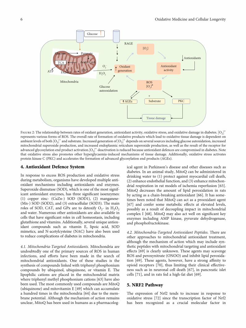
ROS levels are tightly controlled by the antioxidantand nonenzyme antioxidant protective measures in normaland healthy cells. However, excessive ROS concentrationsare contributed by hyperglycaemia in diabetes, leading toa significant complication of DM. In the case of diabetesor insulin resistance, a higher oxidative glucose metabo-lism itself increases the mitochondrial production of O2
•,which is then converted to OH• and H2O2 [60]. In addi-tion to glucose, ROS generation by mitochondrial FFA isalso augmented (Figure 2) [61]. Overall, the overexpres-sion and activation of mitochondrial inner membraneuncoupling enzymes (UCPs) have been suggested to con-tribute to an increase in superoxide production under dia-betic conditions [62].
NADPH oxidase is a significant source of ROS produc-tion in diabetes. NADPH oxidase is present in the plasmamembrane of several renal cell types, including mesangialand proximal tubular cells, endothelial cells, fibroblastcells, and vascular smooth muscle cells. The production ofNADPH oxidase-dependent ROS oxidation plays a majorpart in encouraging oxidative stress caused by hyperglycae-mia. In fact, NADPH oxidase improves oxidative stress andultimately leads to diabetic nephropathy in rats.
5Oxidative Medicine and Cellular Longevity
4. Antioxidant Defence System
In response to excess ROS production and oxidative stressduring metabolism, organisms have developed multiple anti-oxidant mechanisms including antioxidants and enzymes.Superoxide dismutase (SOD), which is one of the most signif-icant antioxidant enzymes, has three significant isoenzymes:(1) copper zinc- (CuZn-) SOD (SOD1), (2) manganese-(Mn-) SOD (SOD2), and (3) extracellular (SOD3). The mainroles of SOD, CAT, and GPx are to detoxify O2- in H2O2and water. Numerous other antioxidants are also available incells that have significant roles in cell homeostasis, includingglutathione and vitamins. Additionally, several unique antiox-idant compounds such as vitamin E, lipoic acid, SODmimetics, and N-acetylcysteine (NAC) have also been usedto reduce complications of diabetes in mitochondria.
4.1. Mitochondria-Targeted Antioxidants. Mitochondria areundoubtedly one of the primary sources of ROS in humaninfections, and efforts have been made in the search ofmitochondrial antioxidants. One of these studies is thesynthesis of compounds linked with triphenyl phosphoniumcompounds by ubiquinol, ubiquinone, or vitamin E. Thelipophilic cations are placed in the mitochondrial matrixwhere triphenyl methyl phosphonium cations [63] have alsobeen used. The most commonly used compounds are MitoQ(ubiquinone) and mitovitamin E [49] which can accumulatea hundred times in the mitochondria [64] due to the mem-brane potential. Although the mechanism of action remainsunclear, MitoQ has been used in humans as a pharmacolog-
ical agent in Parkinson’s disease and other diseases such asdiabetes. In an animal study, MitoQ can be administered indrinking water to (1) protect against myocardial cell death,(2) enhance endothelial function, and (3) enhance mitochon-drial respiration in rat models of ischemia reperfusion [65].MitoQ decreases the amount of lipid peroxidation in ratsby acting as a chain-breaking antioxidant [66]. It has some-times been noted that MitoQ can act as a prooxidant agent[67] and confer some metabolic effects at elevated levels,possibly as a result of decoupling impact in mitochondrialcomplex I [68]. MitoQ may also act well on significant keyenzymes including AMP kinase, pyruvate dehydrogenase,and phosphofructokinase.
4.2. Mitochondria-Targeted Antioxidant Peptides. There areother approaches to mitochondrial antioxidant treatment,although the mechanism of action which may include syn-thetic peptides with mitochondrial targeting and antioxidanteffects [69] is clearly unknown. These agents may scavengeROS and peroxynitrate (ONOO) and inhibit lipid peroxida-tion [69]. These agents, however, have a strong affinity toopioid receptors [70], thus limiting their clinical effective-ness such as in neuronal cell death [67], in pancreatic isletcells [71], and in rats fed a high-fat diet [69].
5. NRF2 Pathway
The expression of Nrf2 tends to increase in response tooxidative stress [72] since the transcription factor of Nrf2has been recognized as a crucial molecular factor in
ATP FADH2 ETC
Acetyl CoAOxidant
generation
Glucoseautooxidation
H2O2
O2– Antioxidant
activity
Antioxidants
PKCSubstrate+
Tissue damage
AGEs
ROS[O2]
RAGE
Glucose
[O2]PyruvateGlucose
Mitochondria
TCA
∗
Figure 2: The relationship between rates of oxidant generation, antioxidant activity, oxidative stress, and oxidative damage in diabetes. ½O2�∗represents various forms of ROS. The overall rate of formation of oxidative products which lead to oxidative tissue damage is dependent onambient levels of both ½O2�∗ and substrate. Increased generation of ½O2�∗ depends on several sources including glucose autoxidation, increasedmitochondrial superoxide production, and increased endoplasmic reticulum superoxide production, as well as the result of the receptor foradvanced glycosylation end product activation.½O2�∗ deactivation is reduced because antioxidant defences are compromised in diabetes. Notethat oxidative stress also promotes other hyperglycaemia-induced mechanisms of tissue damage. Additionally, oxidative stress activatesprotein kinase C (PKC) and accelerates the formation of advanced glycosylation end products (AGEs).
6 Oxidative Medicine and Cellular Longevity

orchestrating the adaptive cellular reactions under a broadrange of cellular environments (either extracellular or intra-cellular) [73]. To date, both clinical and research communi-ties are highly interested in understanding the upregulationof Nrf2 and/or its upstream antioxidant genes that are acti-vated due to hyperglycaemia. The Nrf2 is a key regulatorfor cellular detoxification and redox status as well as for pro-tection against oxidative stress [71]. Given that the molecularmechanisms for diabetic complications are not well under-stood, research can play a major role in explaining the molec-ular pathways and in developing strategies for macro- andmicrovascular diseases, including diabetes complications,prevention, therapy, and management.
5.1. Implications of Nrf2 Transcription Factor in DiabeticComplications. Nrf2 gene, which is also known as NFE2L2,encodes for a transcription factor that controls for ARE genesin the promoter regions. These genes encode for proteinsproduced in response to environmental stress, metabolicenzymes, injury, and inflammation as well as detoxifyingenzymes, including free radical production. Nrf2 in the cyto-plasm is linked to Kelch-like ECH-associated protein 1(Keap1) in unstressed circumstances, avoiding its transloca-tion to the nucleus as illustrated in Figure 3 [72].
During stressed conditions (either electrophilic or oxi-dative stress), the Keap1/Nrf2 complex receives a signal
involving phosphorylation and/or redox modificationresulting in translocation of Nrf2 into the nucleus. Keap1,however, mediates the rapid ubiquitination and subsequentNrf2 degradation by the proteasome 26S under basal cir-cumstances. Cullin 3-based ubiquitin E3 (Cul-E3) ligasecomplex ubiquitinates Nrf2 while Keap1 acts as a substrateadapter which promotes the ubiquitination of Nrf2 by cul-lin 3. Consequently, Nrf2 has a short half-life (only 20minutes) under normal circumstances. Oxidative stress isresponsible for destroying critical cysteine residues inKeap1, disrupting the Keap1-Cul3 ubiquitination mecha-nism. However, if Nrf2 is not ubiquitinated, it builds upin the cytoplasm and is translocated to the nucleus (illus-trated in Figure 3) [74, 75].
Nrf2 associates with the small protein called Maf presentin its nucleus to create a heterodimer and initiate transcrip-tion in the upstream promoter region of several cytoprotec-tive genes. The cytoprotective genes include the encodingantioxidant and phase-II detoxifying enzymes like superox-ide dismutases (SODs), catalase (CAT), NAD(P)H dehydro-genase, γ-glutamyl cysteine synthase (γ-GCS), NQO-1,hemeoxygenase-1 (HO-1), glutathione peroxidase-1 (GPx-1), and glutathione S-transferase (GST). The response shownby the antioxidants to the signalling pathways NFE2L2 andNFE1-NFE2L2/ARE is considered a promising target againstdiabetic complications affecting the pulmonary, hepatic,
Normal conditions
Proteasomedegradation
Nrf2
Nrf2
Nrf2
ARE
PAntioxidant genes
Antioxidant enzymes (Ho-1,MnSOD, NQO-1 GSTA2)(i) Inhibition of inflammation(ii) Free radical metabolism
Pancreatic beta-cell damage(i) Insulin resistance
DNA damage
Ub Cul3
Cul3
Keap1
Keap1ERK
Reactive oxygenspecies (stress)
HyperglycaemiaHyperlipidaemia
Endogenous activatorMitochondrial dysfunction
AKT
P
Cytoplasm
Nucleus Nrf2Maf
Figure 3: The Nrf2/Keap1/ARE pathway in T2DM. (a) Under diabetic conditions, the Nrf2 transcription factor is covalently bonded tocysteine residues on its native repressor Keap1 in the cytoplasm, which results in the (1) constitutive ubiquitination and degradation ofNrf2 in the proteasome and (2) inhibition of the antioxidant response. (b) During a stressful condition of electrophilic or oxidative stress,cysteine residues on Keap1 are modified, resulting in the stabilization and translocation of Nrf2 into the nucleus where it can bind to thepromoter region of ARE and initiate the transcription of various cytoprotective enzymes which can promote cellular survival through avariety of mechanisms including the upregulation of antioxidant function, inflammatory inhibition, and the transport of toxic metabolites.These cellular adaptations have been shown to improve a wide array of tissue damage underlying the pathogenesis and progression ofdiabetes. There are three major mechanisms of Nrf2 induction by small molecule activators: (1) upstream kinases such as AKT and ERK,which phosphorylate Nrf2 at specific sites, favoring its release by Keap1 and nuclear translocation; (2) modification of Keap1 cysteineresidues, which disrupts the Nrf2-Keap1 complex, favoring dissociation of Nrf2 and subsequent nuclear translocation; (3) inhibition ofNrf2 ubiquitination by Keap1 and degradation by the proteasome augments Nrf2 availability, thus favoring nuclear translocation of Nrf2.Ub: ubiquitination; P: phosphorylation.
7Oxidative Medicine and Cellular Longevity

digestive, neural, and cardiovascular systems as well asdiabetic nephropathy [76, 77].
Animal studies have demonstrated that the Nrf2/Kea-p1/ARE system is a crucial defensive pathway to protectpancreatic β-cells both physiologically and pathologically.Nrf2 depletion can reduce cytoprotective antioxidant expres-sion in β-cell transgenic mice and exacerbate oxidative β celldamage, where Nrf2 induction suppressed accumulation ofROS, ROS-induced DNA formations, and pancreative β-cellapoptosis in the islets [78]. In Nrf2/Keap1/ARE, pancreaticβ-cell protection does not only include free radical scaveng-ing activity but also an inflammatory reduction via the NF-κB pathway [79]. A new study in murine models illustratesthat increased signalling of Nrf2 can also improve insulinresistance via suppression of hypothalamic oxidative stress,a phenomenon that can affect systemic metabolic regulation[80]. Murine studies have also confirmed that Nrf2 inductioncan suppress weight gain and increase the consumption ofskeletal muscle oxygen, mitochondrial redox homeostasis,and ATP production as well as increase the intake of cellularglucose in type II diabetes [81].
6. Honey Polyphenols as Antidiabetic Agents
Scientists look into alternative medicines for diabetic treat-ment because of the adverse effects conferred by conven-tional allopathic treatments as well as the economic burdenof the conventional diabetic medications. Natural productshave always been used in the treatment of diabetes, manyof them being polyphenols which are bioactive [82–86].Dietary flavonoids have been considered a related risk-reduction factor for T2D [87]. For many years, people havebelieved that diabetic patients cannot consume honey dueto its high sugar content. However, a series of questions haveemerged: “Can honey replace sugar in diabetic diet?” “Is thesugar in honey key in prevention and treatment of diabetesmellitus?” Interestingly, several researchers worldwide havefocused on the characterization of honey from differentsources and determining its biological properties. This studyprovides stronger evidence to support the function of fruc-tose in mediating the hypoglycemic effect of honey. Fructoseand glucose are the major monosaccharides with the samemolecular formula but with a different structural formulapresent in honey [88]. Several studies reported that fructosereduces glucose levels or hyperglycaemia in diabetes patientsand diabetic rat models [42, 89–91]. Evidence showed thatgastric emptying is prolonged by the intake of fructose [92],which may slow down the rate of intestinal absorption [93].In addition to delaying absorption, fructose intake reducesthe intake of food [11], which is often due to delayed gastricemptying [94]. Fructose does not increase plasma glucoseand does not require insulin secretion to its metabolism.Glucokinase, a vital enzyme for the intracellular metabolismof glucose, is known to be activated by dietary fructose [95].Glucokinase further catalyses glucose converted to glucose-6 phosphate, which decreases blood glucose [17], and fruc-tose stimulates secretion of insulin from an isolated pancreas[96]. Curry et al. observed no insulin reaction to fructose inrat pancreas preparation when very low glucose is present
in the medium; however, when higher glucose concentrationis present, insulin reaction to fructose was observed. Variouspreclinical and clinical studies have highlighted the efficacyand use of honey in diabetic patients and in animal models[97–100]. Nevertheless, although the hypoglycemic effect ofhoney remains unclear, numerous experiments have indi-cated that it is so [100, 101].
Although honey holds several types of polyphenols, onlya few, such as kaempferol, catechin, quercetin, luteolin, rutin,and apigenin, have been shown to decrease blood glucoselevels through various mechanisms [102]. Some of the mech-anisms include (1) inhibition of enzymes such as α-glucosideand α-amylase [81]; (2) improved pancreatic β-cell protec-tion by (i) reducing oxidative stress, (ii) increased secretion,and (iii) increased insulin activity [85]; (3) higher absorptionof glucose and production of insulin receptor and GLUT4;(4) inhibition of gluconeogenic enzymes; and (5) inhibitionof the enzyme aldose reductase. Plasma glucose levels havesignificantly been reduced due to honey administration andas a result of enhanced insulin secretion. In an experimenton normal rats which received honey supplementation(10%), the glycated haemoglobin was considerably reduced[103], where α-glucosidase and α-amylase were inhibited,the crucial enzymes involved in ameliorating blood glucoselevel elevation as a result of carbohydrate breakdown. Theeffect is purported to be the effect of honey polyphenolsand other polyphenols as proposed by several investigations[83, 104]. Thus, the inhibition of both enzymes is consideredan efficient method to decrease blood sugar concentrations indiabetic patients [104].
A few experiments using animal models have reportedsome reduction in blood glucose due to the presence of fruc-tose, which may contribute to the decreased food intake,reduced rate of intestinal absorption, and a prolonged gastricemptying time. Fructose triggers glucokinase’s activity in thehepatocytes, involved in both the absorption and storage ofglucose by the liver as glycogen. Glucose, unlike fructose,enhances the absorption of fructose and improves hepaticfunction by enhancing fructose delivery to the liver.
The pancreas is one of the most important organsinvolved in diabetic management, due to the fact that insulinand glucagon are produced by the pancreatic cells. The hypo-glycaemic effect of honey may be attributed to its antioxidantmolecules that help defend against oxidative stress and dam-age. Insulin response and glucose homeostasis in normal ratsare enhanced by fructose intake or by a combination ofsucrose molecules in comparison with glucose intake in rats.Notably, it has been demonstrated that honey administeredin sufficient doses, can confer hypoglycaemic effects in ani-mal models of types 1 and 2 diabetes as, respectively, inducedby alloxan and streptozotocin [100, 105, 106].
In another study, honey and fructose were fed to type 1diabetic (alloxan-induced) and healthy rats. The formershowed a significant reduction in glucose levels, when com-pared to the latter in control rats [107, 108]. It was alsoshown that honey effectively brought about glucose homeo-stasis when given in the diet for diabetics (or hypertriglyc-eridemia) and in healthy patients in contrast to a diet thatis rich in sucrose and dextrose. In diabetic patients, plasma
8 Oxidative Medicine and Cellular Longevity

glucose levels were substantially decreased with honeyadministration when compared to dextrose consumption.Interestingly, it was observed that in normal subjects, C-reactive protein, blood lipids, and homocysteine werereduced after regular consumption of honey when comparedto sucrose consumption [108, 109].
Honey can noticeably reduce fructosamine concentrationwhich is a glucose-modified protein; this is an added meta-bolic advantage for diabetes mellitus and is not generallyyielded with other hypoglycaemic agents. Fructosamine canundergo oxidative cleavage resulting in the formation ofROS and has been shown to be substantially increased in ratswith diabetes mellitus and/or their complications [110],implicating that fructosamine is an important molecularplayer in diabetes. Antioxidants in honey scavenge the freeradicals crucial in various inflammatory cascades andsuppress the generation of reactive oxygen species (ROS)catalysed by metal ions such as iron and copper. These metalions can be reached with flavonoids and other polyphenoliccompounds found in honey [111]. Thus, honey possessesan antioxidant activity mediated by flavonoids and polyphe-nols. Additionally, honey can mediate insulin secretion dueto the presence of minerals including zinc, manganese,copper, selenium, chromium, calcium, and potassium, whichimplies the antidiabetic activity of honey [112]. A clinicalresearch [113] has demonstrated that early modifications ofspecial trace elements can lead to impaired insulin and glu-cose metabolisms, while on the other hand, diabetes mellitusis said to be linked to trace element disturbance [113].
Deficiencies in some elements such as zinc (Zn),chromium, and manganese can enhance an individual’s riskof glycemia and diabetic complications. For example, Zn isdirectly engaged with insulin synthesis, storage, secretion, andconformational integrity. Zn assembles into crystalline insulinas a dimeric structure for storage and secretion. In people withtype-2 diabetes mellitus, a lower zinc concentration may influ-ence the capacity of pancreatic cells to synthesize and secreteinsulin [114].
T2DM can be induced by streptozotocin (STZ) by dam-aging β-cells of islets of Langerhans in the pancreas. Thesecells produce insulin to enhance glucose cellular uptake.There was a significant decrease in blood glucose concentra-tions three days following administration of Mad honey pro-duced from the flower of rhododendrons in STZ-induceddiabetic rats and nondiabetic rats [115].
Honey’s antidiabetic effect is also explained by its antiox-idant capacity and modulation of adiponectin concentra-tions. Adipose tissue secretes adiponectin hormone, whichregulates both glucose and lipid metabolisms and is shownto be significantly reduced in diabetic patients [116]. Impor-tantly, oxidative stress-mediated lipid peroxidation wasdirectly implicated in the development of diabetic complica-tions [117]. High concentrations of adiponectin decrease sys-temic inflammation and increase insulin sensitivity [118].
In another type of honey, Gelam honey extracts were usedto protect the pancreatic hamster cells from hyperglycaemicconditions. Gelam honey significantly reduced production ofROS, glucose-induced lipid peroxidation, increased insulincontent, and cell viability under hyperglycaemic conditions
[119]. Additionally, Jujube honey has been shown tomodulatethe key enzymes involved in glucose metabolism, namely, glu-cokinase and glucose 6-phosphatase in rats. It was observedthat Jujube honey reduced malondialdehyde (MDA) levelsbut significantly increased total antioxidant capacity in dia-betic rats (p < 0:05). Interestingly, heat shock protein(HSP70) and glucose expression of 6-phosphatase werereduced as well, while glucokinase expression was increasedin the treatment group [120].
In 2015, Hemmati et al. have demonstrated that honeyorally administered (1.0 and 2.0 g/kg/day) to STZ-induceddiabetic rats for 21 days has resulted in a substantial rise inadiponectin concentrations (by 4:5 ± 0:2 and 4:2 ± 0:3mg/L, respectively) with a marked reduction in malondialdehyde(MDA) concentrations as compared to control rats. Theincrease is linked to a substantial enhancement in the levelof fasting blood sugar (FBS) and lipid profiles in honey-supplemented diabetic rats [121].
Multiple evidences have suggested that the antidiabeticand hypoglycaemic abilities of honey is dependent on itsantioxidant capacity; indeed, the pathogenesis of diabetesmellitus, especially that of type 2, appears to be closely linkedto oxidation stress and ROS in different organs and tissues[122]. A rise in glucose absorption by the adipose tissuesand muscles increases ROS production, leading to oxidativestress, a process that affects glycogen synthesis and glucoseuptake. Oxidative stress may also lead to insulin resistancedue to insulin signalling impairment, which can be restoredthrough honey treatment [123].
In pancreatic β-cells, oxidative stress affects cell function-ality, resulting in inadequate insulin secretion and a high rateof β-cell apoptosis. The scavenger activity of honey isnormally confirmed to enhance pancreatic oxidative stress[124]. Lipid metabolism is impaired in diabetes mellitus,which shows a high concentration of low-density lipopro-teins (LDLs), which are oxidized and glycated in oxidizedLDLs, thus leading to endothelial damage. In this case too,honey antioxidants help to inhibit lipid oxidative degrada-tion in patients with type 2 diabetes mellitus [125].
The antioxidant impact of Tualang honey (TH) in thepancreas of diabetic rats induced by STZ has been explored.Following supplementation of TH (1.0 g/kg/day) for 28 days,there was substantial downregulation of SOD and MDA,together with increased pancreatic CAT activity (p < 0:05)in diabetic rats as compared to control rats. It is plausible thatthe antioxidant effects of TH protected the pancreatic cellsfrom oxidative stress which led to an enhancement of FBSin diabetic rats when compared with control rats (median(IQR): 8.8 (5.8) and 17.9 (2.6) mmol/L, respectively) [126].Similar hypoglycaemic impact is also exerted by other typesof honey, such as honey from Nigeria. In fact, alloxan-induced diabetic rats supplemented with honey for 21 days attwo doses (1.0 and 2.0 g/kg/day) had considerably (p < 0:05)decreased FBS as compared to control [127].
In another study, honey collected from Ilam forest, Ilamprovince, Iran, has been shown to enhance glycemic control.Consumption of natural honey (1.0 g/kg) along with metfor-min (100mg/kg) once daily for four weeks tends to be moreeffective than administering metformin alone (100mg/kg).
9Oxidative Medicine and Cellular Longevity

Ilam honey was shown to be rich in antioxidants and ismore effective than Tualang honey in treating lipid abnor-malities [128].
It has been shown that polyphenols present in honeyhave protective activity on pancreatic β-cells, thus alleviatingdiabetes. In streptozotocin-induced diabetes in rats, theregeneration of pancreatic β-cells improved by administeringquercetin at a dose of 10-15mg/kg for ten days [129–131]and increased glucose uptake in insulin-resistant tissues[132]. Apigenin and rutin were shown to have guarded pan-creatic β-cells by elevating the levels of antioxidants, antiox-idant enzymes, and insulin in rat models of streptozotocin-induced diabetes [133]. In another research, quercetinprotected and retained the structure and integrity of thepancreatic β-cells while improving insulin secretion instreptozotocin-induced diabetes in rats [130]. Kaempferol, aflavanol found in honey, was reported to have prevented pan-creatic β cell dysfunction in middle-aged obese mice withdiabetes, and enhanced peripheral insulin sensitivity [131].Clinical studies indicate that both honey and other sourcesof polyphenols decrease oxidative stress in diabetic animals(Figure 4). Quercetin, found in honey, decreased oxidativestress in streptozotocin-induced diabetes in rats by loweringblood glucose concentrations and lipid peroxidation, andraising levels of ascorbic acid, vitamin E, and tissue antioxi-dant activity [134]. Luteolin is a flavone, reported to bepresent in honey, which has been shown to reduce oxidativestress in type 1 diabetic cardiomyopathy to ameliorate heartfailure [135].
In DM, glucose usage is reduced in both insulin and non-insulin-susceptible tissues. Dietary polyphenols found inhoney are reported to boost peripheral absorption of glucoseinto these tissues. Two other plant phenolic compounds alsodiscovered in honey are chlorogenic and ferulic acids that canincrease glucose uptake and be more efficient as two hypogly-caemic medicines called metformin and 2,4-thiozolodine-dione (THZ) [136]. Chlorogenic acid stimulates glucoseuptake through the expression of the PI3K-independentpathways of the GLUT4 and peroxisome proliferator-
activated gamma receptors (PPAR-γ). Ferulic acid, however,promotes glucose uptake by expressing GLUT4 and PI3Ktranscripts via PI3K process-dependent pathways [136].
Improving the activity of insulin secretion is the mostsignificant goal in the treatment of DM. It was demonstratedthat daily administration of rutin 25-100mg/kg for 45 dayscould enhance insulin secretion and reduce fasting bloodglucose concentrations in streptozotocin-induced diabetesin rats [137]. Quercetin protected pancreas β-cells againsthydrogen peroxide (H2O2) and the activation of ERK-1/2(extracellular signal-regulated kinases) pathway triggered byglucose and glibenclamide in insulin-secreting cell lines(INS-1) [138]. Luteolin, a flavonoid, was shown to reducehyperglycaemia by raising insulin secretion in a diabetic ani-mal model [139]. The process of activation of peroxisomeproliferator-activated receptor gamma (PPAR-γ) [140] alsoprevented insulin resistance in T2DM patients by the forma-tion of adipokines, which implies that honey may be helpfulin modulating adiponectin mediated by PPAR-γ [116], thusalleviating insulin resistance.
A nonrandomized open clinical trial single-arm phase Icohort prospective study on the effects of honey reported thathoney consumption resulted in hyperglycaemia in type 2diabetic patients. Interestingly, regular and long-term honeyconsumption was shown to decrease the body weight andblood pressure in the same cohort [141] of subjects. Anotherrandomized controlled trial was conducted in type 2diabetics to investigate the effect of daily ingestion of kanukahoney blended with “cinnamon,” “chromium,” and “magne-sium” on glucose metabolism and lipid variables [142].However, the blended honey supplementation did not pro-duce any statistically significant change in HbA1c or fastinginsulin in the subjects, although the supplementation hasalleviated abnormal lipid variables, improved high-densitylipoprotein, and led to a reduction in body weight. The twostudies have shown contrasting findings on the antidiabeticactivity of honey, although they also showed lipid profileimprovement and body weight reduction in the subjects.However, further clinical studies have to be conducted to
Nrf2 activation, NF-𝜅B inhibition
Cellular antioxidant response
CATSOD GPx
ROS
↑ Insulin sensitivity, ↑ Insulin secretion,↓𝛽-Cell dysfunction
Antioxidant effect of honeypolyphenols
Antioxidant enzymes
Oxidantenzymes
Figure 4: Potential mechanisms contributing to the beneficial effects of polyphenols by protecting cells against oxidative stress. Abbreviations:CAT—catalase; GPx—glutathione peroxidase; NF-κB—nuclear factor kappa light-chain enhancer of activated B-cells; Nrf-2—nuclear factorerythroid 2; ROS—reactive oxygen species; SOD—superoxide dismutase.
10 Oxidative Medicine and Cellular Longevity

gain insights into the antidiabetic activity of honey with morestringent criteria to select honey and subjects for inclusion inthe studies.
Based on the data from the experimental studies andreports, it is clear that honey is useful in the treatment ofdiabetes mellitus patients. Although some studies havesuggested contrasting views on the usage of honey, mostscientists agree that honey is useful in improving hypergly-caemia and diabetic problems on various organs and mayreduce complications.
7. Conclusion and Future Directives
The burden in the community is increasing with chronic ill-nesses including diabetes mellitus, hypertension, atherosclero-sis, and cancer. The mortality resulting from these diseases hasalso increased. The evidence that oxidative stress plays asubstantial role in pathogenesis or disorders suggests that anti-oxidants can be beneficial. However, a variety of antidiabeticdrugs may also contribute to cardiovascular complications.Natural substances have been used for a long time to curedifferent types of diseases, including T2DM. Honey is anatural product, and it has several biochemical and biologicalroles in both humans and animals. Honey is a major source ofpolyphenols, and the level of each polyphenol varies consider-ably. The polyphenols inhibit the development of differentdiseases through several specific mechanisms, for example,regulating the expression of a certain gene or encouraging/-blocking certain steps on a metabolic pathway. Various typesof honey have been investigated for their antimicrobial, antidi-abetic, anti-hyper-cholesterol, anti-inflammatory, antioxidant,and wound-healing effects. Moreover, their credibility islimited owing to a relatively low number of clinical trials.Further investigation is required to determine specific mecha-nisms. It is highly imperative that several clinical and experi-mental trials are carefully designed and planned since theyare often required to verify the efficacy of honey either aloneor as an adjuvant treatment.
Conflicts of Interest
The authors declare no conflict of interest.
Authors’ Contributions
Pasupuleti Visweswara Rao and Chandra Sekhar Arigelacontributed equally to this work.
Acknowledgments
The corresponding author would like to thank the researchfunding by Fundamental Research Grant (FRGS) (grantnumber R/FRGS/A1300/01095A/003/2018/00551).
References
[1] P. Saeedi, I. Petersohn, P. Salpea et al., “Global and regionaldiabetes prevalence estimates for 2019 and projections for2030 and 2045: Results from the International Diabetes Fed-
eration Diabetes Atlas, 9th edition,” Diabetes Research andClinical Practice, vol. 157, article 107843, 2019.
[2] Y. Zheng, S. H. Ley, and F. B. Hu, “Global aetiology and epi-demiology of type 2 diabetes mellitus and its complications,”Nature Reviews Endocrinology, vol. 14, no. 2, pp. 88–98, 2018.
[3] P. Hossain, B. Kawar, andM. El Nahas, “Obesity and diabetesin the developing world—a growing challenge,” New EnglandJournal of Medicine, vol. 356, no. 3, pp. 213–215, 2007.
[4] F. Aguiree, A. Brown, N. H. Cho et al., IDF Diabetes Atlas,International Diabetes Federation, 2013.
[5] R. DeFronzo, “Insulin resistance, lipotoxicity, type 2 diabetesand atherosclerosis: the missing links. The Claude BernardLecture 2009,” Diabetologia, vol. 53, no. 7, pp. 1270–1287,2010.
[6] X. Wang, W. Bao, J. Liu et al., “Inflammatory markers andrisk of type 2 diabetes: a systematic review and meta-analy-sis,” Diabetes Care, vol. 36, no. 1, pp. 166–175, 2013.
[7] P. Sytze van Dam, “Oxidative stress and diabetic neuropathy:pathophysiological mechanisms and treatment perspectives,”Diabetes/Metabolism Research and Reviews, vol. 18, no. 3,pp. 176–184, 2002.
[8] A. K. Tiwari, “Imbalance in antioxidant defence and humandiseases: multiple approach of natural antioxidants therapy,”Current Science, vol. 81, no. 9, pp. 1179–1187, 2001.
[9] C. K. Sen, “Oxygen toxicity and antioxidants: state of the art,”Indian Journal of Physiology And Pharmacology, vol. 39,no. 3, pp. 177–196, 1995.
[10] H. M. A. Abdelrazek, O. E. Kilany, M. A. A. Muhammad,H. M. Tag, and A. M. Abdelazim, “Black seed thymoquinoneimproved insulin secretion, hepatic glycogen storage, andoxidative stress in streptozotocin-induced diabetic male Wis-tar rats,” Oxidative Medicine and Cellular Longevity,vol. 2018, Article ID 8104165, 10 pages, 2018.
[11] O. O. Erejuwa, S. A. Sulaiman, and M. S. AbWahab, “Honey:a novel antioxidant,”Molecules, vol. 17, no. 4, pp. 4400–4423,2012.
[12] V. R. Pasupuleti, L. Sammugam, N. Ramesh, and S. H. Gan,“Honey, propolis, and royal jelly: a comprehensive review oftheir biological actions and health benefits,” Oxidative Medi-cine and Cellular Longevity, vol. 2017, Article ID 1259510, 21pages, 2017.
[13] I. A. A. da Silva, T. M. S. da Silva, C. A. Camara et al., “Phe-nolic profile, antioxidant activity and palynological analysisof stingless bee honey from Amazonas, Northern Brazil,”Food Chemistry, vol. 141, no. 4, pp. 3552–3558, 2013.
[14] F. G. Banting and C. H. Best, “The internal secretion of thepancreas,” Indian Journal of Medical Research, vol. 125,no. 3, p. L251, 2007.
[15] G. Dodson and D. Steiner, “The role of assembly in insulin’sbiosynthesis,” Current Opinion in Structural Biology, vol. 8,no. 2, pp. 189–194, 1998.
[16] A. L. Olson and J. E. Pessin, “Structure, function, and regula-tion of the mammalian facilitative glucose transporter genefamily,” Annual Review of Nutrition, vol. 16, no. 1, pp. 235–256, 1996.
[17] G. L. Kellett, E. Brot-Laroche, O. J. Mace, and A. Leturque,“Sugar absorption in the intestine: the role of GLUT2,”Annual Review of Nutrition, vol. 28, no. 1, pp. 35–54, 2008.
[18] E. Newsholme and G. Dimitriadis, “Integration of biochemi-cal and physiologic effects of insulin on glucose metabolism,”
11Oxidative Medicine and Cellular Longevity

Experimental and Clinical Endocrinology & Diabetes,vol. 109, Supplement 2, pp. S122–S134, 2001.
[19] A. Ullrich and J. Schlessinger, “Signal transduction by recep-tors with tyrosine kinase activity,” Cell, vol. 61, no. 2, pp. 203–212, 1990.
[20] R. Perlman, D. Bottaro, M. White, and C. Kahn, “Conforma-tional changes in the alpha- and beta-subunits of the insulinreceptor identified by anti-peptide antibodies,” Journal ofBiological Chemistry, vol. 264, no. 15, pp. 8946–8950, 1989.
[21] V. Baron, P. Kaliman, N. Gautier, and E. Van Obberghen,“The insulin receptor activation process involves localizedconformational changes,” Journal of Biological Chemistry,vol. 267, no. 32, pp. 23290–23294, 1992.
[22] P. Manna and S. K. Jain, “PIP3 but not PIP2 increases GLUT4surface expression and glucose metabolism mediated byAKT/PKCζ/λ phosphorylation in 3T3L1 adipocytes,”Molecularand Cellular Biochemistry, vol. 381, no. 1-2, pp. 291–299, 2013.
[23] H. Sano, L. Eguez, M. N. Teruel et al., “Rab 10, a target of theAS160 Rab GAP, is required for insulin-stimulated transloca-tion of GLUT4 to the adipocyte plasma membrane,” CellMetabolism, vol. 5, no. 4, pp. 293–303, 2007.
[24] M. Friedrichsen, J. B. Birk, E. A. Richter et al., “Akt 2 influ-ences glycogen synthase activity in human skeletal musclethrough regulation of NH2-terminal (sites 2+2a) phosphory-lation,” American Journal of Physiology-Endocrinology andMetabolism, vol. 304, no. 6, pp. E631–E639, 2013.
[25] J. Olivares-Reyes, “Bases moleculares del síndrome metabó-lico y resistencia a la insulina,” in Obesidad en la edad pediá-trica: prevención y tratamiento, G. N. Garibay Nieto and S.García Velasco, Eds., pp. 185–214, Corinter, Ciudad de Méx-ico, Mexico, 2012.
[26] H. Kamata, S.-i. Honda, S. Maeda, L. Chang, H. Hirata, andM. Karin, “Reactive oxygen species promote TNFα-induceddeath and sustained JNK activation by inhibiting MAP kinasephosphatases,” Cell, vol. 120, no. 5, pp. 649–661, 2005.
[27] H. Liu, H. Nishitoh, H. Ichijo, and J. M. Kyriakis, “Activationof apoptosis signal-regulating kinase 1 (ASK1) by tumornecrosis factor receptor-associated factor 2 requires prior dis-sociation of the ASK1 inhibitor thioredoxin,” Molecular andCellular Biology, vol. 20, no. 6, pp. 2198–2208, 2000.
[28] V. Adler, Z. Yin, S. Y. Fuchs et al., “Regulation of JNK signal-ing by GSTp,” The EMBO Journal, vol. 18, no. 5, pp. 1321–1334, 1999.
[29] P. Storz and A. Toker, “Protein kinase D mediates a stress-induced NF-kappaB activation and survival pathway,” TheEMBO Journal, vol. 22, no. 1, pp. 109–120, 2003.
[30] P. Gual, Y. Le Marchand-Brustel, and J.-F. Tanti, “Positiveand negative regulation of insulin signaling through IRS-1phosphorylation,” Biochimie, vol. 87, no. 1, pp. 99–109,2005.
[31] G. Sabio, N. J. Kennedy, J. Cavanagh-Kyros et al., “Role ofmuscle c-Jun NH2-terminal kinase 1 in obesity-induced insu-lin resistance,” Molecular and cellular biology, vol. 30, no. 1,pp. 106–115, 2010.
[32] H. Kaneto, Y. Nakatani, T. Miyatsuka et al., “Possible noveltherapy for diabetes with cell-permeable JNK-inhibitory pep-tide,” Nature Medicine, vol. 10, no. 10, pp. 1128–1132, 2004.
[33] D. Cai, M. Yuan, D. F. Frantz et al., “Local and systemic insu-lin resistance resulting from hepatic activation of IKK-β andNF-κB,” Nature Medicine, vol. 11, no. 2, pp. 183–190, 2005.
[34] M. C. Arkan, A. L. Hevener, F. R. Greten et al., “IKK-β linksinflammation to obesity-induced insulin resistance,” NatureMedicine, vol. 11, no. 2, pp. 191–198, 2005.
[35] K. L. Hoehn, C. Hohnen-Behrens, A. Cederberg et al., “IRS1-independent defects define major nodes of insulin resis-tance,” Cell Metabolism, vol. 7, no. 5, pp. 421–433, 2008.
[36] B. M. De Taeye, T. Novitskaya, O. P. McGuinness et al.,“Macrophage TNF-α contributes to insulin resistance andhepatic steatosis in diet-induced obesity,” American Journalof Physiology-Endocrinology and Metabolism, vol. 293, no. 3,pp. E713–E725, 2007.
[37] G. S. Hotamisligil, P. Arner, J. F. Caro, R. L. Atkinson, andB. M. Spiegelman, “Increased adipose tissue expression oftumor necrosis factor-alpha in human obesity and insulinresistance,” The Journal of Clinical Investigation, vol. 95,no. 5, pp. 2409–2415, 1995.
[38] A. Filippis, S. Clark, and J. Proietto, “Increased flux throughthe hexosamine biosynthesis pathway inhibits glucose trans-port acutely by activation of protein kinase C,” BiochemicalJournal, vol. 324, no. 3, pp. 981–985, 1997.
[39] H.-L. Vikman, J. M. Kaartinen, R. K. Tuominen, and J. J. Ohi-salo, “A possible role for protein kinase C in the regulatorydifferences between intra-abdominal and subcutaneoushuman adipose tissue,” Clinical Science, vol. 85, no. 3,pp. 265–268, 1993.
[40] J. Chin, M. Dickens, J. Tavare, and R. Roth, “Overexpressionof protein kinase C isoenzymes alpha, beta I, gamma, andepsilon in cells overexpressing the insulin receptor. Effectson receptor phosphorylation and signaling,” Journal of Bio-logical Chemistry, vol. 268, no. 9, pp. 6338–6347, 1993.
[41] F. Urano, X. Wang, A. Bertolotti et al., “Coupling of stress inthe ER to activation of JNK protein kinases by transmem-brane protein kinase IRE1,” Science, vol. 287, no. 5453,pp. 664–666, 2000.
[42] S.-R. Won, D.-C. Lee, S. H. Ko, J.-W. Kim, and H.-I. Rhee,“Honey major protein characterization and its applicationto adulteration detection,” Food Research International,vol. 41, no. 10, pp. 952–956, 2008.
[43] J. Hirosumi, G. Tuncman, L. Chang et al., “A central role forJNK in obesity and insulin resistance,” Nature, vol. 420,no. 6913, pp. 333–336, 2002.
[44] G. Perseghin, K. Petersen, and G. I. Shulman, “A central rolefor JNK in obesity and insulin resistance,” Nature, vol. 420,no. 6913, pp. 333–336, 2003.
[45] U. Özcan, Q. Cao, E. Yilmaz et al., “Endoplasmic reticulumstress links obesity, insulin action, and type 2 diabetes,” Sci-ence, vol. 306, no. 5695, pp. 457–461, 2004.
[46] M. Valko, D. Leibfritz, J. Moncol, M. T. Cronin, M. Mazur,and J. Telser, “Free radicals and antioxidants in normal phys-iological functions and human disease,” The InternationalJournal of Biochemistry & Cell Biology, vol. 39, no. 1,pp. 44–84, 2007.
[47] J. S. Johansen, A. K. Harris, D. J. Rychly, and A. Ergul, “Oxi-dative stress and the use of antioxidants in diabetes: linkingbasic science to clinical practice,” Cardiovascular Diabetol-ogy, vol. 4, no. 1, p. 5, 2005.
[48] P. Rösen, P. Nawroth, G. King, W. Möller, H. J. Tritschler,and L. Packer, “The role of oxidative stress in the onset andprogression of diabetes and its complications: asummary ofa Congress Series sponsored byUNESCO-MCBN, the Amer-ican Diabetes Association and the German Diabetes Society,”
12 Oxidative Medicine and Cellular Longevity

Diabetes/Metabolism Research and Reviews, vol. 17, no. 3,pp. 189–212, 2001.
[49] V. M. Victor, M. Rocha, R. Herance, and A. Hernandez-Mijares, “Oxidative stress and mitochondrial dysfunction intype 2 diabetes,” Current Pharmaceutical Design, vol. 17,no. 36, pp. 3947–3958, 2011.
[50] R. A. Kowluru and P.-S. Chan, “Oxidative stress and diabeticretinopathy,” Experimental Diabetes Research, vol. 2007,Article ID 043603, 2007.
[51] D. M. Niedowicz and D. L. Daleke, “The role of oxidativestress in diabetic complications,” Cell Biochemistry and Bio-physics, vol. 43, no. 2, pp. 289–330, 2005.
[52] J. Cederberg, S. Basu, and U. J. Eriksson, “Increased rate oflipid peroxidation and protein carbonylation in experimentaldiabetic pregnancy,” Diabetologia, vol. 44, no. 6, pp. 766–774,2001.
[53] S. Wolff, “Diabetes mellitus and free radicals: free radicals,transition metals and oxidative stress in the aetiology of dia-betes mellitus and complications,” British Medical Bulletin,vol. 49, no. 3, pp. 642–652, 1993.
[54] D. Giugliano, A. Ceriello, and G. Paolisso, “Diabetes mellitus,hypertension, and cardiovascular disease: which role for oxi-dative stress?,”Metabolism, vol. 44, no. 3, pp. 363–368, 1995.
[55] S. K. Jain, “Hyperglycemia can cause membrane lipid perox-idation and osmotic fragility in human red blood cells,” Jour-nal of Biological Chemistry, vol. 264, no. 35, pp. 21340–21345,1989.
[56] S. K. Jain, K. Kannan, and G. Lim, “Ketosis (acetoacetate) cangenerate oxygen radicals and cause increased lipid peroxida-tion and growth inhibition in human endothelial cells,” FreeRadical Biology & Medicine, vol. 25, no. 9, pp. 1083–1088,1998.
[57] J. L. Evans, I. D. Goldfine, B. A. Maddux, and G. M. Grodsky,“Are oxidative stress-activated signaling pathways mediatorsof insulin resistance and β-Cell Dysfunction?,” Diabetes,vol. 52, no. 1, pp. 1–8, 2003.
[58] T.-H. Lu, C.-C. Su, Y.-W. Chen et al., “Arsenic induces pan-creatic β-cell apoptosis via the oxidative stress-regulatedmitochondria-dependent and endoplasmic reticulum stress-triggered signaling pathways,” Toxicology Letters, vol. 201,no. 1, pp. 15–26, 2011.
[59] P. Maechler, L. Jornot, and C. B. Wollheim, “Hydrogen per-oxide alters mitochondrial activation and insulin secretionin pancreatic beta cells,” Journal of Biological Chemistry,vol. 274, no. 39, pp. 27905–27913, 1999.
[60] T. Nishikawa, D. Edelstein, X. L. du et al., “Normalizing mito-chondrial superoxide production blocks three pathways ofhyperglycaemic damage,” Nature, vol. 404, no. 6779,pp. 787–790, 2000.
[61] J. L. Evans, I. D. Goldfine, B. A. Maddux, and G. M. Grodsky,“Oxidative stress and stress-activated signaling pathways: aunifying hypothesis of type 2 diabetes,” Endocrine Reviews,vol. 23, no. 5, pp. 599–622, 2002.
[62] A. P. Rolo and C. M. Palmeira, “Diabetes and mitochondrialfunction: role of hyperglycemia and oxidative stress,” Toxi-cology and Applied Pharmacology, vol. 212, no. 2, pp. 167–178, 2006.
[63] S. González-Rubio, A. B. Hidalgo, G. Ferrín et al., “Mitochon-drial-driven ubiquinone enhances extracellular calcium-dependent nitric oxide production and reduces glycocheno-deoxycholic acid-induced cell death in hepatocytes,” Chemi-
cal Research in Toxicology, vol. 22, no. 12, pp. 1984–1991,2009.
[64] A. M. James, R. A. Smith, and M. P. Murphy, “Antioxidantand prooxidant properties of mitochondrial Coenzyme Q,”Archives of Biochemistry and Biophysics, vol. 423, no. 1,pp. 47–56, 2004.
[65] A. K. Doughan and S. I. Dikalov, “Mitochondrial redoxcycling of mitoquinone leads to superoxide production andcellular apoptosis,” Antioxidants & Redox Signaling, vol. 9,no. 11, pp. 1825–1836, 2007.
[66] V. J. Adlam, J. C. Harrison, C. M. Porteous et al., “Targetingan antioxidant to mitochondria decreases cardiac ischemia-reperfusion injury,” The FASEB Journal, vol. 19, no. 9,pp. 1088–1095, 2005.
[67] D. A. Thomas, C. Stauffer, K. Zhao et al., “Mitochondrial tar-geting with antioxidant peptide SS-31 prevents mitochon-drial depolarization, reduces islet cell apoptosis, increasesislet cell yield, and improves posttransplantation function,”Journal of the American Society of Nephrology, vol. 18, no. 1,pp. 213–222, 2006.
[68] S. Dröse and U. Brandt, “The mechanism of mitochondrialsuperoxide production by the cytochrome bc1 complex,”Journal of Biological Chemistry, vol. 283, no. 31, pp. 21649–21654, 2008.
[69] K. Zhao, G.-M. Zhao, D. Wu et al., “Cell-permeable peptideantioxidants targeted to inner mitochondrial membraneinhibit mitochondrial swelling, oxidative cell death, andreperfusion injury,” Journal of Biological Chemistry,vol. 279, no. 33, pp. 34682–34690, 2004.
[70] C. Thiemermann, “Membrane-permeable radical scavengers(tempol) for shock, ischemia-reperfusion injury, and inflam-mation,” Critical Care Medicine, vol. 31, Supplement,pp. S76–S84, 2003.
[71] E. J. Anderson, M. E. Lustig, K. E. Boyle et al., “MitochondrialH2O2 emission and cellular redox state link excess fat intaketo insulin resistance in both rodents and humans,” The Jour-nal of Clinical Investigation, vol. 119, no. 3, pp. 573–581,2009.
[72] B. Li, S. Liu, L. Miao, and L. Cai, “Prevention of diabetic com-plications by activation of Nrf2: diabetic cardiomyopathy andnephropathy,” Experimental Diabetes Research, vol. 2012,Article ID 216512, 7 pages, 2012.
[73] D. Ayers, B. Baron, and T. Hunter, “miRNA influences inNRF2 pathway interactions within cancer models,” Journalof Nucleic Acids, vol. 2015, Article ID 143636, 6 pages, 2015.
[74] M. Kobayashi, L. Li, N. Iwamoto et al., “The antioxidantdefense system Keap1-Nrf2 comprises a multiple sensingmechanism for responding to a wide range of chemical com-pounds,” Molecular and Cellular Biology, vol. 29, no. 2,pp. 493–502, 2009.
[75] B.-h. Choi, K.-S. Kang, and M.-K. Kwak, “Effect of redoxmodulating NRF2 activators on chronic kidney disease,”Molecules, vol. 19, no. 8, pp. 12727–12759, 2014.
[76] T. Yamamoto, T. Suzuki, A. Kobayashi et al., “Physiologicalsignificance of reactive cysteine residues of Keap1 in deter-mining Nrf2 activity,” Molecular and Cellular Biology,vol. 28, no. 8, pp. 2758–2770, 2008.
[77] J.-T. Cheng, C.-C. Huang, I.-M. Liu, T.-F. Tzeng, and C. J.Chang, “Novel mechanism for plasma glucose–loweringaction of metformin in streptozotocin-induced diabetic rats,”Diabetes, vol. 55, no. 3, pp. 819–825, 2006.
13Oxidative Medicine and Cellular Longevity

[78] Y. Yagishita, T. Fukutomi, A. Sugawara et al., “Nrf2 protectsPancreatic β-Cells From Oxidative and nitrosative stress indiabetic model mice,” Diabetes, vol. 63, no. 2, pp. 605–618,2014.
[79] M.-Y. Song, E.-K. Kim,W.-S. Moon et al., “Sulforaphane pro-tects against cytokine-and streptozotocin-induced β-celldamage by suppressing the NF-κB pathway,” Toxicologyand Applied Pharmacology, vol. 235, no. 1, pp. 57–67, 2009.
[80] Y. Yagishita, A. Uruno, T. Fukutomi et al., “Nrf2 improvesleptin and insulin resistance provoked by hypothalamic oxi-dative stress,” Cell Reports, vol. 18, no. 8, pp. 2030–2044,2017.
[81] S. E. Kahn, R. L. Hull, and K. M. Utzschneider, “Mechanismslinking obesity to insulin resistance and type 2 diabetes,”Nature, vol. 444, no. 7121, pp. 840–846, 2006.
[82] D. Patel, R. Kumar, D. Laloo, and S. Hemalatha, “Diabetesmellitus: an overview on its pharmacological aspects andreported medicinal plants having antidiabetic activity,” AsianPacific Journal of Tropical Biomedicine, vol. 2, no. 5, pp. 411–420, 2012.
[83] R. J. Marles and N. R. Farnsworth, “Antidiabetic plants andtheir active constituents,” Phytomedicine, vol. 2, no. 2,pp. 137–189, 1995.
[84] J.-M. Meng, S.-Y. Cao, X.-L. Wei et al., “Effects and mecha-nisms of tea for the prevention and management of diabetesmellitus and diabetic complications: an updated review,”Antioxidants, vol. 8, no. 6, p. 170, 2019.
[85] G. R. Gandhi, A. B. S. Vasconcelos, D.-T. Wu et al., “Citrusflavonoids as promising phytochemicals targeting diabetesand related complications: a systematic review of in vitroand in vivo studies,” Nutrients, vol. 12, no. 10, p. 2907, 2020.
[86] B.-Y. Li, X.-Y. Xu, R.-Y. Gan et al., “Targeting gut microbiotafor the prevention and management of diabetes mellitus bydietary natural products,” Foods, vol. 8, no. 10, p. 440, 2019.
[87] Y. Liu, D. Li, Y. Zhang, R. Sun, and M. Xia, “Anthocyaninincreases adiponectin secretion and protects againstdiabetes-related endothelial dysfunction,” American Journalof Physiology-Endocrinology and Metabolism, vol. 306, no. 8,pp. E975–E988, 2014.
[88] M. Aurongzeb and M. K. Azim, “Antimicrobial properties ofnatural honey: a review of literature,” Pakistan Journal Bio-chemistry and Molecular Biology, vol. 44, no. 3, pp. 118–124, 2011.
[89] S. E. A. Mohammed and M. K. Azim, “Characterisation ofnatural honey proteins: implications for the floral and geo-graphical origin of honey,” International Journal of Food Sci-ence & Technology, vol. 47, no. 2, pp. 362–368, 2012.
[90] M. T. Iglesias, C. de Lorenzo, M. d. C. Polo, P. J. Martín-Álva-rez, and E. Pueyo, “Usefulness of amino acid composition todiscriminate between honeydew and floral honeys. Applica-tion to honeys from a small geographic area,” Journal of Agri-cultural and Food Chemistry, vol. 52, no. 1, pp. 84–89, 2004.
[91] J. Graham, The Hive and the Honey Bee, Dadant & Sons, Inc,Hamilton, IL, USA, 1992.
[92] M. Khalil, N. Alam, M. Moniruzzaman, S. Sulaiman, andS. Gan, “Phenolic acid composition and antioxidant proper-ties of Malaysian honeys,” Journal of Food Science, vol. 76,no. 6, pp. C921–C928, 2011.
[93] J. Topliss, A. Clark, E. Ernst et al., “Natural and synthetic sub-stances related to human health (IUPAC Technical Report),”
Pure and Applied Chemistry, vol. 74, no. 10, pp. 1957–1985,2002.
[94] N. Vaisman, E. Niv, and Y. Izkhakov, “Catalytic amounts offructose may improve glucose tolerance in subjects withuncontrolled non-insulin-dependent diabetes,” ClinicalNutrition, vol. 25, no. 4, pp. 617–621, 2006.
[95] T. H. Moran and P. R. McHugh, “Distinctions among threesugars in their effects on gastric emptying and satiety,” Amer-ican Journal of Physiology-Regulatory, Integrative and Com-parative Physiology, vol. 241, no. 1, pp. R25–R30, 1981.
[96] L. Thibault, “Dietary carbohydrates: effects on self-selection,plasma glucose and insulin, and brain indoleaminergic sys-tems in rat,” Appetite, vol. 23, no. 3, pp. 275–286, 1994.
[97] D. L. Curry, K. P. Curry, and M. Gomez, “Fructose potentia-tion of insulin secretion,” Endocrinology, vol. 91, no. 6,pp. 1493–1498, 1972.
[98] D. L. Curry, “Fructose potentiation of mannose-inducedinsulin secretion,” American Journal of Physiology-LegacyContent, vol. 226, no. 5, pp. 1073–1076, 1974.
[99] O. O. Erejuwa, S. A. Sulaiman, and M. S. A. Wahab, “Hon-ey—a novel antidiabetic agent,” International Journal of Bio-logical Sciences, vol. 8, no. 6, pp. 913–934, 2012.
[100] M. A. Abd Jalil, A. R. Kasmuri, and H. Hadi, “Stingless beehoney, the natural wound healer: a review,” Skin Pharmacol-ogy and Physiology, vol. 30, no. 2, pp. 66–75, 2017.
[101] O. O. Erejuwa, S. A. Sulaiman, and M. S. A. Wahab, “Oligo-saccharides might contribute to the antidiabetic effect ofhoney: a review of the literature,” Molecules, vol. 17, no. 1,pp. 248–266, 2011.
[102] P. V. Rao, K. T. Krishnan, N. Salleh, and S. H. Gan, “Biolog-ical and therapeutic effects of honey produced by honey beesand stingless bees: a comparative review,” Revista Brasileirade Farmacognosia, vol. 26, no. 5, pp. 657–664, 2016.
[103] L. Chepulis and N. Starkey, “The long-term effects of feedinghoney compared with sucrose and a sugar-free diet on weightgain, lipid profiles, and DEXAmeasurements in rats,” Journalof Food Science, vol. 73, no. 1, pp. H1–H7, 2007.
[104] R. Tundis, M. Loizzo, and F. Menichini, “Natural products asα-amylase and α-glucosidase inhibitors and their hypoglycae-mic potential in the treatment of diabetes: an update,” Mini-Reviews in Medicinal Chemistry, vol. 10, no. 4, pp. 315–331,2010.
[105] O. Bobiş, D. S. Dezmirean, and A. R. Moise, “Honey and dia-betes: the importance of natural simple sugars in diet for pre-venting and treating different type of diabetes,” OxidativeMedicine and Cellular Longevity, vol. 2018, Article ID4757893, 12 pages, 2018.
[106] M. Abdulrhman, M. el Hefnawy, R. Ali, I. Abdel Hamid,A. Abou el-Goud, and D. Refai, “Effects of honey, sucroseand glucose on blood glucose and C-peptide in patients withtype 1 diabetes mellitus,” Complementary Therapies in Clini-cal Practice, vol. 19, no. 1, pp. 15–19, 2013.
[107] F. Adesoji and A. Oluwakemi, “Differential effect of honey onselected variables in alloxan-induced and fructose-induceddiabetic rats,” African Journal of Biomedical Research,vol. 11, no. 2, 2010.
[108] S. Samarghandian, T. Farkhondeh, and F. Samini, “Honeyand health: a review of recent clinical research,” Pharmacog-nosy Research, vol. 9, no. 2, pp. 121–127, 2017.
[109] N. S. Al-Waili, “Natural honey lowers plasma glucose, C-reactive protein, homocysteine, and blood lipids in healthy,
14 Oxidative Medicine and Cellular Longevity

diabetic, and hyperlipidemic subjects: comparison with dex-trose and sucrose,” Journal of Medicinal Food, vol. 7, no. 1,pp. 100–107, 2004.
[110] L. Kennedy and J. Baynes, “Non-enzymatic glycosylation andthe chronic complications of diabetes: an overview,” Diabeto-logia, vol. 26, no. 2, pp. 93–98, 1984.
[111] S. Z. A. Makawi, E. A. Gadkariem, and S. M. H. Ayoub,“Determination of antioxidant flavonoids in Sudanese honeysamples by solid phase extraction and high performance liq-uid chromatography,” Journal of Chemistry, vol. 6, Article ID382504, 9 pages, 2009.
[112] S. Bogdanov, T. Jurendic, R. Sieber, and P. Gallmann, “Honeyfor nutrition and health: a review,” Journal of the AmericanCollege of Nutrition, vol. 27, no. 6, pp. 677–689, 2008.
[113] A. H. Zargar, M. I. Bashir, S. R. Masoodi et al., “Copper, zincand magnesium levels in type-1 diabetes mellitus,” SaudiMedical Journal, vol. 23, no. 5, pp. 539–542, 2002.
[114] R. A. DiSilvestro, “Zinc in relation to diabetes and oxidativedisease,” The Journal of Nutrition, vol. 130, no. 5,pp. 1509S–1511S, 2000.
[115] N. Öztaşan, K. Altinkaynak, F. Akçay, F. Göçer, and Ş. Dane,“Effects of mad honey on blood glucose and lipid levels in ratswith streptozocin-induced diabetes,” Turkish Journal of Vet-erinary and Animal Sciences, vol. 29, no. 5, pp. 1093–1096,2005.
[116] S. Li, H. J. Shin, E. L. Ding, and R. M. van Dam, “Adiponectinlevels and risk of type 2 diabetes: a systematic review andmeta-analysis,” JAMA, vol. 302, no. 2, pp. 179–188, 2009.
[117] R. Srivatsan, S. Das, R. Gadde et al., “Antioxidants and lipidperoxidation status in diabetic patients with and withoutcomplications,” Archives of Iranian Medicine, vol. 12, no. 2,pp. 121–127, 2009.
[118] Y. Matsuzawa, T. Funahashi, and T. Nakamura, “The conceptof metabolic syndrome: contribution of visceral fat accumu-lation and its molecular mechanism,” Journal of Atherosclero-sis and Thrombosis, vol. 18, no. 8, pp. 629–639, 2011.
[119] K. Batumalaie, R. Qvist, K. M. Yusof, I. S. Ismail, and S. D.Sekaran, “The antioxidant effect of the Malaysian Gelamhoney on pancreatic hamster cells cultured under hypergly-cemic conditions,” Clinical and Experimental Medicine,vol. 14, no. 2, pp. 185–195, 2014.
[120] M. Gholami, M. Hemmati, A. Taheri-Ghahfarokhi,R. Hoshyar, and M. Moossavi, “Expression of glucokinase,glucose 6-phosphatase, and stress protein in streptozotocin-induced diabetic rats treated with natural honey,” Interna-tional Journal of Diabetes in Developing Countries, vol. 36,no. 1, pp. 125–131, 2016.
[121] Mina Hemmati, Mehdi Karamian, and Mohammad Maleka-neh, “Anti-atherogenic potential of natural honey: anti-diabetic and antioxidant approaches,” Journal of Pharmacyand Pharmacology, vol. 3, no. 6, pp. 278–284, 2015.
[122] F. Folli, D. Corradi, P. Fanti et al., “The role of oxidative stressin the pathogenesis of type 2 diabetes mellitus micro- andmacrovascular complications: avenues for a mechanistic-based therapeutic approach,” Current Diabetes Reviews,vol. 7, no. 5, pp. 313–324, 2011.
[123] J. S. Kim, V. Saengsirisuwan, J. A. Sloniger, M. K. Teachey,and E. J. Henriksen, “Oxidant stress and skeletal muscle glu-cose transport: roles of insulin signaling and p38 MAPK,”Free Radical Biology & Medicine, vol. 41, no. 5, pp. 818–824, 2006.
[124] O. O. Erejuwa, S. A. Sulaiman, M. S. A. Wahab, K. N. S. Sir-ajudeen, M. S. M. Salleh, and S. Gurtu, “Antioxidant protec-tive effect of glibenclamide and metformin in combinationwith honey in pancreas of streptozotocin-induced diabeticrats,” International Journal of Molecular Sciences, vol. 11,no. 5, pp. 2056–2066, 2010.
[125] R. Rahimi, S. Nikfar, B. Larijani, and M. Abdollahi, “A reviewon the role of antioxidants in the management of diabetesand its complications,” Biomedicine & Pharmacotherapy,vol. 59, no. 7, pp. 365–373, 2005.
[126] O. O. Erejuwa, S. A. Sulaiman, M. S. Wahab, K. N. S. Sirajud-een, M. S. M. D. Salleh, and S. Gurtu, “Antioxidant protectionof Malaysian tualang honey in pancreas of normal andstreptozotocin-induced diabetic rats,” Annales d'endocrinolo-gie, vol. 71, no. 4, pp. 291–296, 2010.
[127] O. O. Erejuwa, N. Nwobodo, J. Akpan et al., “Nigerian honeyameliorates hyperglycemia and dyslipidemia in alloxan-induced diabetic rats,” Nutrients, vol. 8, no. 3, p. 95, 2016.
[128] O. Nasrolahi, R. Heidari, F. Rahmani, and F. Farokhi, “Effectof natural honey from Ilam and metformin for improvingglycemic control in streptozotocin-induced diabetic rats,”Avicenna Journal of Phytomedicine, vol. 2, no. 4, p. 212, 2012.
[129] M. Vessal, M. Hemmati, and M. Vasei, “Antidiabetic effectsof quercetin in streptozocin-induced diabetic rats,” Compar-ative Biochemistry and Physiology Part C: Toxicology & Phar-macology, vol. 135, no. 3, pp. 357–364, 2003.
[130] S. O. Adewole, E. A. Caxton-Martins, and J. A. O. Ojewole,“Protective effect of quercetin on the morphology of pancre-atic β-cells of streptozotocin-treated diabetic rats,” AfricanJournal of Traditional, Complementary and Alternative Med-icines, vol. 4, no. 1, pp. 64–74, 2007.
[131] H. Alkhalidy, W. Moore, Y. Zhang et al., “Small moleculekaempferol promotes insulin sensitivity and preserved pan-creatic β-cell mass in middle-aged obese diabetic mice,” Jour-nal of Diabetes Research, vol. 2015, Article ID 532984, 14pages, 2015.
[132] C.-C. Lee, W.-H. Hsu, S.-R. Shen, Y.-H. Cheng, and S.-C.Wu,“Fagopyrum tataricum (buckwheat) improved high-glucose-induced insulin resistance in mouse hepatocytes and diabetesin fructose-rich diet-induced mice,” Experimental DiabetesResearch, vol. 2012, Article ID 375673, 10 pages, 2012.
[133] M. A. Esmaeili and H. Sadeghi, “Pancreatic Β-cell protectiveeffect of rutin and apigenin isolated from Teucrium polium,”Pharmacology Online, vol. 2, pp. 341–353, 2009.
[134] O. Coskun, M. Kanter, A. Korkmaz, and S. Oter, “Quercetin,a flavonoid antioxidant, prevents and protectsstreptozotocin-induced oxidative stress and β-cell damagein rat pancreas,” Pharmacological Research, vol. 51, no. 2,pp. 117–123, 2005.
[135] G.Wang, W. Li, X. Lu, P. Bao, and X. Zhao, “Luteolin amelio-rates cardiac failure in type I diabetic cardiomyopathy,” Jour-nal of Diabetes and its Complications, vol. 26, no. 4, pp. 259–265, 2012.
[136] P. K. Prabhakar and M. Doble, “Synergistic effect of phyto-chemicals in combination with hypoglycemic drugs on glu-cose uptake in myotubes,” Phytomedicine, vol. 16, no. 12,pp. 1119–1126, 2009.
[137] N. Kamalakkannan and P. S. M. Prince, “Antihyperglycaemicand antioxidant effect of rutin, a polyphenolic flavonoid, instreptozotocin-induced diabeticWistar rats,” Basic & ClinicalPharmacology & Toxicology, vol. 98, no. 1, pp. 97–103, 2006.
15Oxidative Medicine and Cellular Longevity

[138] E. Youl, G. Bardy, R. Magous et al., “Quercetin potentiatesinsulin secretion and protects INS-1 pancreatic β-cellsagainst oxidative damage via the ERK1/2 pathway,” BritishJournal of Pharmacology, vol. 161, no. 4, pp. 799–814, 2010.
[139] A. Zarzuelo, I. Jiménez, M. J. Gámez et al., “Effects of luteolin5-O-β-rutinoside in streptozotocin-induced diabetic rats,”Life Sciences, vol. 58, no. 25, pp. 2311–2316, 1996.
[140] L. Ding, D. Jin, and X. Chen, “Luteolin enhances insulin sen-sitivity via activation of PPARγ transcriptional activity in adi-pocytes,” The Journal of Nutritional Biochemistry, vol. 21,no. 10, pp. 941–947, 2010.
[141] M. A. Abdulrhman, “Honey as a sole treatment of type 2 dia-betes mellitus,” Endocrinology & Metabolic Syndrome, vol. 5,no. 2, 2016.
[142] P. Whitfield, A. Parry-Strong, E. Walsh, M. Weatherall, andJ. D. Krebs, “The effect of a cinnamon-, chromium- andmagnesium-formulated honey on glycaemic control, weightloss and lipid parameters in type 2 diabetes: an open-labelcross-over randomised controlled trial,” European Journalof Nutrition, vol. 55, no. 3, pp. 1123–1131, 2016.
16 Oxidative Medicine and Cellular Longevity