Embed Size (px)
Citation preview
Headache With Right Upper Extremity Headache With Right Upper Extremity Weakness and Dysphasia in an Weakness and Dysphasia in an
AdolescentAdolescent
Headache With Right Upper Extremity Headache With Right Upper Extremity Weakness and Dysphasia in an Weakness and Dysphasia in an
AdolescentAdolescent
Andy Jagoda, MD, FACEPProfessor of Emergency MedicineMount Sinai School of Medicine
New York, New York
Andy Jagoda, MD, FACEP
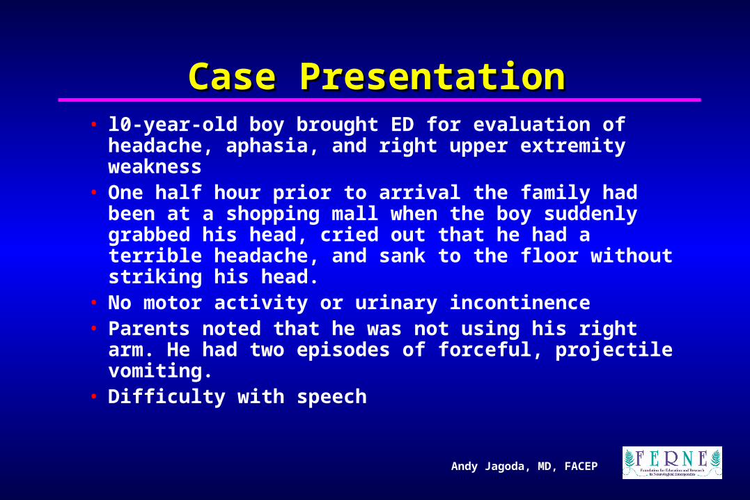
Case PresentationCase Presentation• l0-year-old boy brought ED for evaluation of
headache, aphasia, and right upper extremity weakness
• One half hour prior to arrival the family had been at a shopping mall when the boy suddenly grabbed his head, cried out that he had a terrible headache, and sank to the floor without striking his head.
• No motor activity or urinary incontinence• Parents noted that he was not using his right arm.
He had two episodes of forceful, projectile vomiting. • Difficulty with speech

Andy Jagoda, MD, FACEP
Case PresentationCase Presentation
• No PMH except for a heart murmur –Echocardiographically proven to
be due to a bicuspid aortic valve. • No history of trauma, headaches, or
drug use• There was no significant family
history.
Andy Jagoda, MD, FACEP
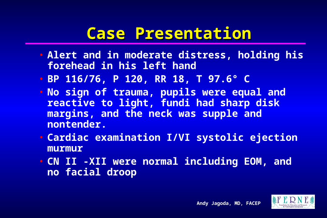
Case PresentationCase Presentation• Alert and in moderate distress, holding his
forehead in his left hand• BP 116/76, P 120, RR 18, T 97.6° C• No sign of trauma, pupils were equal and
reactive to light, fundi had sharp disk margins, and the neck was supple and nontender.
• Cardiac examination I/VI systolic ejection murmur
• CN II -XII were normal including EOM, and no facial droop

Andy Jagoda, MD, FACEP
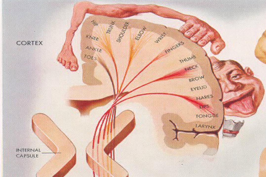
Case PresentationCase Presentation• Motor 5/5 strength in all muscles on the left side and
the right leg, but there was 0/5 strength in all the muscles groups in the right upper extremity
• DTRs were +2 on the left: o/2 on the RUE; toes were downgoing
• Sensation intact; right upper extremity localized pinprick
• No meningeal signs• The patient could understand and carry out three-step
commands• Speech: Difficulty with naming and with repetition
Andy Jagoda, MD, FACEP
Case PresentationCase Presentation
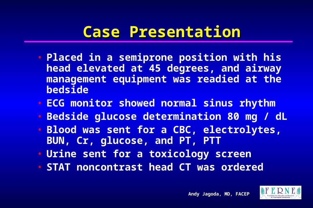
• Placed in a semiprone position with his head elevated at 45 degrees, and airway management equipment was readied at the bedside
• ECG monitor showed normal sinus rhythm• Bedside glucose determination 80 mg / dL• Blood was sent for a CBC, electrolytes, BUN, Cr,
glucose, and PT, PTT• Urine sent for a toxicology screen• STAT noncontrast head CT was ordered
Andy Jagoda, MD, FACEP
Case PresentationCase Presentation
• The patient remained stable over the first hour in the ED except for one more episode of vomiting
• His aphasia persisted• The arm weakness began to resolve • All of the laboratory tests returned
within normal limits
Andy Jagoda, MD, FACEP
The initial ED differential diagnosis of this patient’s The initial ED differential diagnosis of this patient’s presentation includes all of the following except:presentation includes all of the following except:
A. Hemorrhagic stroke
B. Embolic stroke
C. Migraine headache
D. Complex partial seizure
Andy Jagoda, MD, FACEP
One of the more common focal neurologic One of the more common focal neurologic findings reported in SAH is:findings reported in SAH is:
A. Dilated unilateral pupil
B. Unilateral facial droop
C. Deviated tongue
D. Dysphagia

Andy Jagoda, MD, FACEP
Speech is usually controlled by which Speech is usually controlled by which side of the brain?:side of the brain?:
A. Right
B. Left
Andy Jagoda, MD, FACEP
Patients presenting within on hour of symptom onset with a Patients presenting within on hour of symptom onset with a suspected SAH who have a negative head CT and normal CSF suspected SAH who have a negative head CT and normal CSF
analysis are best managed:analysis are best managed:
A. Head CT with contrast
B. Emergency cerebral angiogram
C. Discharged with close follow-up
D. Repeat LP at 12 hours post symptom onset
Andy Jagoda, MD, FACEP
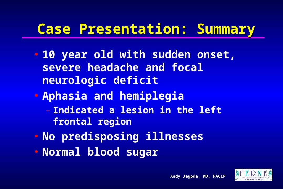
Case Presentation: SummaryCase Presentation: Summary
• 10 year old with sudden onset, severe headache and focal neurologic deficit
• Aphasia and hemiplegia – Indicated a lesion in the left frontal region
• No predisposing illnesses
• Normal blood sugar
Andy Jagoda, MD, FACEP
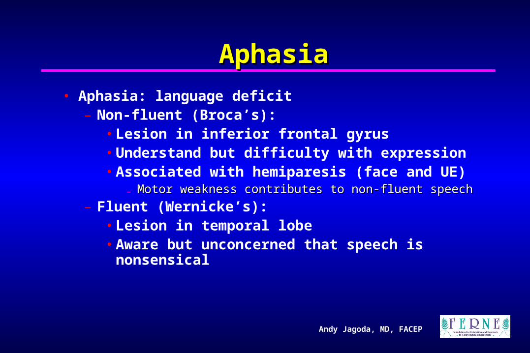
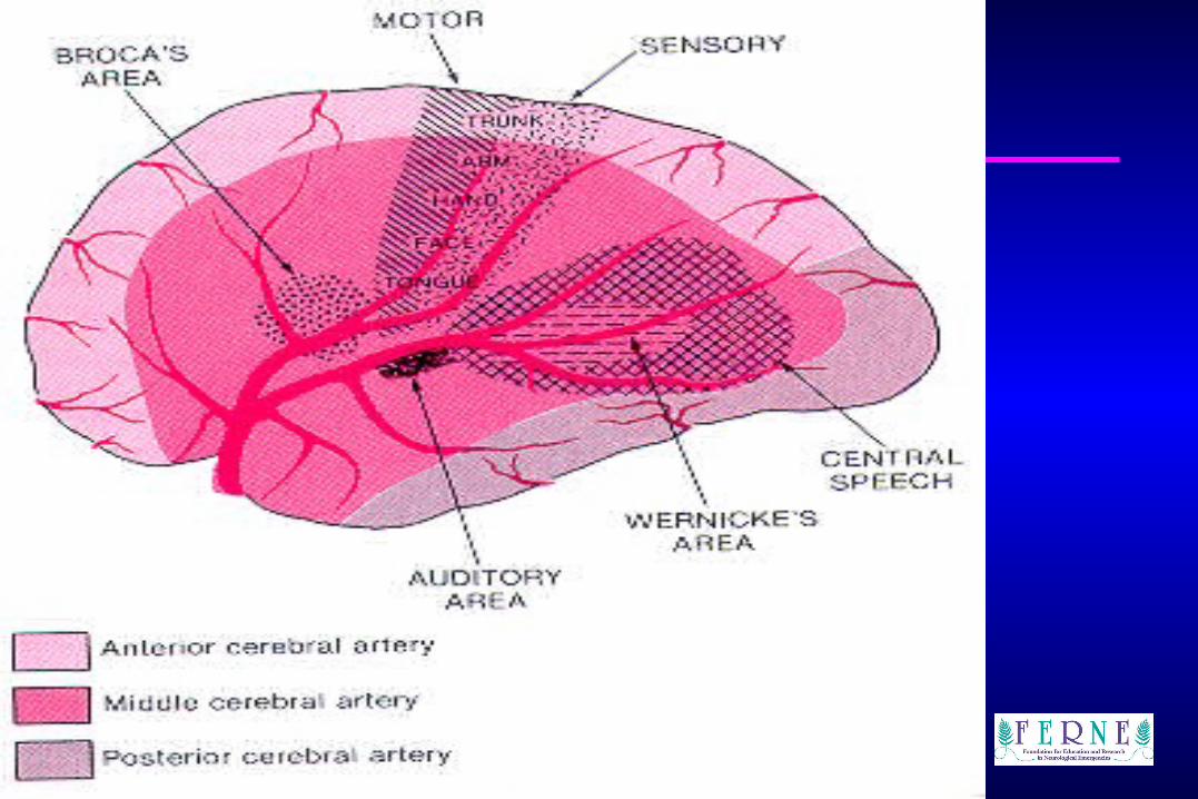
AphasiaAphasia
• Aphasia: language deficit– Non-fluent (Broca’s):
• Lesion in inferior frontal gyrus• Understand but difficulty with expression• Associated with hemiparesis (face and UE)
_ Motor weakness contributes to non-fluent speechMotor weakness contributes to non-fluent speech
– Fluent (Wernicke’s): • Lesion in temporal lobe• Aware but unconcerned that speech is
nonsensical
Andy Jagoda, MD, FACEP
Andy Jagoda, MD, FACEP
Andy Jagoda, MD, FACEP
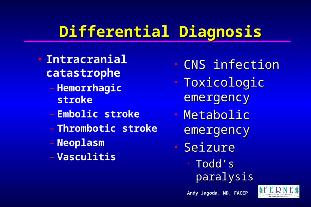
Differential DiagnosisDifferential Diagnosis
• Intracranial catastrophe– Hemorrhagic
stroke– Embolic stroke– Thrombotic stroke– Neoplasm– Vasculitis
• CNS infectionCNS infection• Toxicologic Toxicologic
emergencyemergency• Metabolic Metabolic
emergencyemergency• SeizureSeizure
• Todd’s paralysisTodd’s paralysis
Andy Jagoda, MD, FACEP
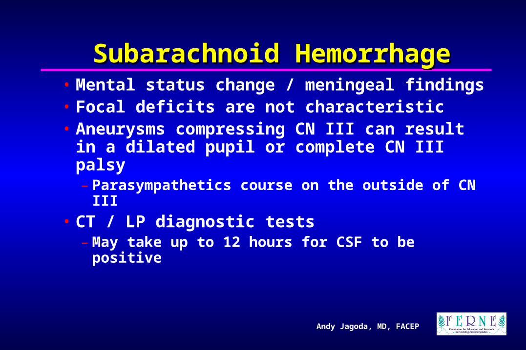
Subarachnoid HemorrhageSubarachnoid Hemorrhage• Mental status change / meningeal findings• Focal deficits are not characteristic• Aneurysms compressing CN III can result in a
dilated pupil or complete CN III palsy– Parasympathetics course on the outside of CN III
• CT / LP diagnostic tests– May take up to 12 hours for CSF to be positive

Andy Jagoda, MD, FACEP
Embolic Stroke in ChildrenEmbolic Stroke in Children
• Cardiac valve disease
• Septal defects
• Arrhythmias
• Carotid / Vertebral artery dissections

Andy Jagoda, MD, FACEP
Thrombotic StrokeThrombotic Stroke
• Sickle cell disease
• Myelodysplasias / Neoplastic disease
• Vasospasm / vasculitis
Andy Jagoda, MD, FACEP
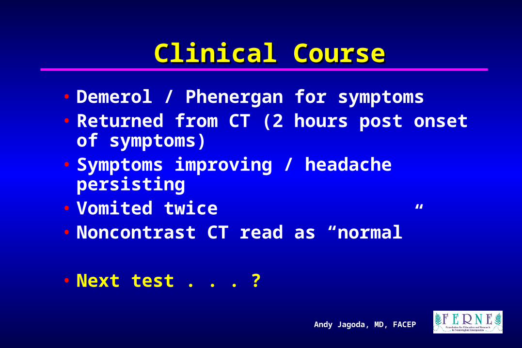
Clinical CourseClinical Course
• Demerol / Phenergan for symptoms • Returned from CT (2 hours post onset of
symptoms)• Symptoms improving / headache persisting• Vomited twice• Noncontrast CT read as “normal”
• Next test . . . ?
Andy Jagoda, MD, FACEP
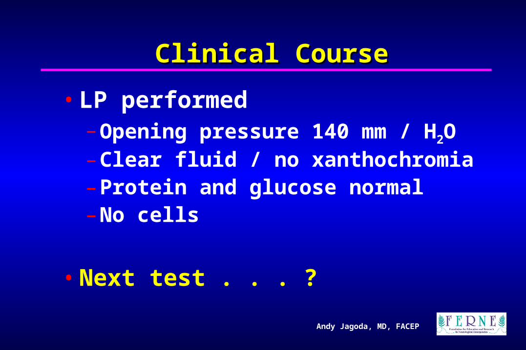
Clinical CourseClinical Course
• LP performed– Opening pressure 140 mm / H2O– Clear fluid / no xanthochromia– Protein and glucose normal– No cells
• Next test . . . ?

Andy Jagoda, MD, FACEP
Decision MakingDecision Making
• Clinical presentation was not characteristic of a SAH or of a MCA stroke
• If SAH was a consideration, options included: – Observation and repeat LP – Angiogram
Andy Jagoda, MD, FACEP
Clinical CourseClinical Course
• Admitted for observation• Symptoms resolved overnight with
normal examination in the am• EEG showed no abnormalities
• Final diagnosis: Hemiplegic migraine• 3 months later, patient had a similar
episode

Andy Jagoda, MD, FACEP
MigraineMigraine
• Migraine with aura• Migraine without aura• Complicated migraines
– Auras lasting more than one hour• Opthalmoplegic• Hemiplegic
• Migraine equivalents
Andy Jagoda, MD, FACEP
Migraine without auraMigraine without aura• At least 5 attacks fullfilling the following• Durations of 4 – 72 hours • Presence of at least 2 of the following:
– Unilateral location– Pulsating quality– Moderate or severe intensity– Aggrevation by routine physical activity
• Presence of at least one of the following:– Nausea and / or vomiting– Photophobia– Phonophobia

Andy Jagoda, MD, FACEP
Migraine with auraMigraine with aura• At least 2 attacks fullfilling the following• All aura symptoms are fully reversible• Aura symptoms indicate focal cerebral cortical
and / or brainstem dysfunction• At least 1 aura symptom develops gradually over
>4 minutes or, > 2 symptoms occur in succession
• No one aura symptom lasts > 60 minutes• Headache follows aura with a free interval of < 60
minutes
Andy Jagoda, MD, FACEP
Migraine: PathophysiologyMigraine: Pathophysiology• Vascular theory (not confirmed by blood flow studies)
– Aura due to vasoconstriction– Headache due to vasodilatation
• Neural hypthosis– Symptoms due to abnormal function of the cerebral
cortex and not due to vasospasm / dilatation– Mediated by serotonin
• Neurovascular hypothesis– Vasodilatation and extravasation of neuropeptides– Neurogenic inflammation

Andy Jagoda, MD, FACEP
Hemiplegic MigraineHemiplegic Migraine
• Two types:– Familial– Non-familial (FHM) or sporadic
• Headache plus visual, sensory, aphasic, and or motor symptoms – Usually two aura types are present– Headache usually begins at the same time of
the aura

Andy Jagoda, MD, FACEP
Hemiplegic MigraineHemiplegic Migraine
• FHM is autosomal dominant, inherited subtype– Gene mutation within the neuronal calcium
channel– Aura is generally prolonged
• Usually begin before age 25 • Female:Male 3:1 • MRA demonstrated constriction / vasodilatation• Prolonged symptoms (days) and infarction
have been reported

Andy Jagoda, MD, FACEP
Hemiplegic Migraine: TreatmentHemiplegic Migraine: Treatment
• Serotonin receptor modulators– No studies using sumitriptan in children– Study in adolescents (12-18 years) has shown
DHE and metoclopramide to be effective in 90%– Promethazine has anecdotely been advocated as
the anti-emetic of choice in children
• Case reports suggest calcium channel blockers (verapamil) to be effective in FHM

Andy Jagoda, MD, FACEP
ConclusionsConclusions• Sudden severe headache suggests a vascular etiology• Strokes involving the MCA will usually involve face and
arm• Aphasia associated with a MCA stroke is described as
“non-fluent” involving naming and difficulty with repetition (exacerbated by motor compromise)
• Hemiplegic migraine is a type of complicated migraine with a prolonged aura due to genetic mutation of the neuronal calcium channel
• Treatment of hemiplegic migraine involve serotonin modulating drugs; Ca channel blockers are a consideration