Embed Size (px)
Citation preview
Headaches in the Emergency Room
Dr Jane AndersonAddenbrooke’s Hospital, Cambridge
Princess Alexandra Hospital, Harlow
UK
25 April 2019

Disclosures
I have received funding from the following companies for educational grants, providing
educational sessions, and received travel grants to attend conferences:
• Allergan
• Merz Pharmaceuticals
• Teva

Overview
Thunderclap headache Helpful clinical signs
Predicting probability of SAH
beyond SAH
Treatment in the ER
Future therapies coming our way

Setting the scene…
68yr ♀, fit & well, smoker
5days prior in Spain:
EtOH ++ in last 24hrs
sunbathing at time
Headache abrupt & severe, within 10mins
10/10
L occipital frontal with facial ache
throbbing pain
assoc vomiting, marked photophobia, pre-
syncopal
Seen A&E: ?migraine and DC with
codeine and paracetamol
Presented NNUH on return:
Unwell with headache
on-going & severe at 8/10 –worse in
morning
Neck pain
Vomiting daily; poor oral intake
Photophobia
Functionally disabled
Collapsed with brief LOC
O/E
Nil focal neuro, fundi N, nuchal rigidity
Temp 37.8oC; weight reduction of 4kg
BP 156/105; HR114; ECG N

68yr ♀, fit & well, smoker
5days prior in Spain:
EtOH ++ in last 24hrs
sunbathing at time
Headache abrupt & severe, within 10mins
10/10
L occipital frontal with facial ache
throbbing pain
assoc vomiting, marked photophobia, pre-
syncopal
Seen A&E: ?migraine and DC with
codeine and paracetamol
Presented NNUH on return:
Unwell with headache
on-going & severe at 8/10 –worse in
morning
Neck pain
Vomiting daily; poor oral intake
Photophobia
Functionally disabled
Collapsed with brief LOC
O/E
Nil focal neuro, fundi N, nuchal rigidity
Temp 37.8oC, weight reduction 4kg
BP 156/105; HR114; ECG N
French guidelines for the emergency management of headache_Moisset et al 2016
Setting the scene…

Papilloedema
6th Cranial nerve plasy
C
Neema Kasravi et al. CMAJ 2010;
182:E373-E377
Horners
+
B
helpful clinical signs….the eyes have it
Ptosis & conjunctival inj
4. Cluster headache
3. Venous sinus thrombosis
6. IIH
1. Sub-arachnoid haemorrhage
5. SOL
2. Cervical arterial dissection
ASubhyaloid haemorrhage
D

Ptosis & conjunctival inj
4. Cluster headache
A
helpful clinical signs….the eyes have it

Neema Kasravi et al. CMAJ 2010;
182:E373-E377
Horners
+
2. Cervical arterial
dissection
helpful clinical signs….the eyes have it
B

Caplan.
Nature
2008
headache in 60-95% & thunderclap in 1/5
ipsilateral
usually headache/ cranio-cervical pain plus [Silbert et al 1995])
Horner's
pulsatile tinnitus,
amaurosis fugax
cerebral ischaemic symptoms
ipsilateral cranial palsies in 12% [Mokri et al 1996]
5% lower cranial nerve involvement- usually XII
4% palsy of cranial nerve V
3% ocular motor palsies
Neema Kasravi et al. CMAJ 2010;
182:E373-E377
Horners
+
2. Cervical arterial
dissection
B
helpful clinical signs….the eyes have it

Papilloedema
6th Cranial nerve plasy
6. IIH
3. Venous sinus thrombosis
5. SOL
helpful clinical signs….the eyes have it
C

Papilloedema
6th Cranial nerve plasy
Venous sinus thrombosis
• headache in 90%
• postural
• valsalva triggered
• visual obscuration
• PULSATILE tinnitus
• diplopia (CN6 palsy as false localiser)
• typically progressive but can be thunderclap
• seizures in 40%
• bilateral brain involvement
• risk factors:
smoker
COCP & pregnancy/postpartum
cancer
dehydration & hyperviscosityGregory Piazza Circulation. 2012;125:1704-1709
Copyright © American Heart Association, Inc. All rights
reserved.
helpful clinical signs….the eyes have it
C

Subhyaloid haemorrhage
1. Sub-arachnoid
haemorrhage
helpful clinical signs….the eyes have it
D

Leakage of blood into CSF space with meningeal irritation:
85% Saccular Aneurysm, 10% perimensephalic, 5% other
Headache in isolation in up to 50-70%
Thunderclap (peaks within mins & lasts at least 1hr)
‘Worst Ever’ with:
• Vomiting in 70% ( vs 42% in benign)
• Meningism; photophobia
• LOC: typically transient at onset (26-50%)
• Transient focal neurology (33%)
• Subhyaloid haemorrhage (17%)
• Delirium (16%)
• Epileptic seizures (6-9%)
• Sudden death (10%)
helpful clinical signs….the eyes have it
Subhyaloid haemorrhage
1. Sub-arachnoid
haemorrhage
D

Ptosis & conjunctival inj
Cluster headache
Neema Kasravi et al. CMAJ 2010;
182:E373-E377
Horners
+
Cervical arterial
dissection
Papilloedema
6th Cranial nerve plasy
IIH
Venous sinus thrombosis
SOL
Subhyaloid haemorrhage
Sub-arachnoid
haemorrhage
helpful clinical signs….the eyes have it

helpful clinical sign?…. meningeal irritation
meningitis MRI
– T1 + Contrast
normal MRI
– T1 + Contrast
SAH on CT
Thomas et al 2002 - evaluating Kernig’s & Brudzinski sign in suspected meningitis:
sensitivity 5%
specificity 95%
not helpful when negative
↑ temp even when no CNS drive can cause headache
↑ temp will occur in both bacterial & chemical meningitis
high frequency of pyrexia in SAH but typically delayed
and prominent 72 hrs on

Index of suspicion
vulnerable group
age>50
tempo
landscape
triggerschange
systemic symp
neuro signs
Threshold of suspicion…..

Threshold of suspicion….. Ottawa (adult) SAH rule Perry et al 2013, JAMA
Alert with non-traumatic severe ha peaking within 1 hr w/o deficit
Negative Predictive Value 100%;
high sensitivity ~100%
but very poor specificity (15% reducing to 7% on validation testing_Bellolio et al 2015, Am J Em Med)
Headachein the ER
trauma
cranial & cervical vascular
non-vascular
intracranial
infectiondrugs
facial / other
cranial structure
primary

Tempo: abrupt, 1st & worst with a progressive course

CT v CSF debate in the diagnosis of Subarachnoid haemorrhage
<6 hrs <12 hrs <24hrs <1wk <2wk 3wk 4wk
CT >98% 95% 90-93% 50% poor poor
CSF xanthochromia 100% 100% 100% ~100% ~100% 70% 40%
Timeline of Xanthochromia in SAH: Vermeulen, Van Gin, JNNP 1990; 53; 365-372 & JNNP 1989; 52; 826-828
Timeline of CT in SAH: Van Ginjn, Van Dongen, Neuroradiol 1982 23: 153-6

CT for SAH in patients with acute headache stratified by timing of scan _Perry et al BMJ 2011
retrospective, n729 (88 in house)_Alons et al, 2018
CTA for severe acute non-traumatic headache + normal exam +normal CT
7.4% vascular abnormality but only 1.6% thought to be causative
Time fromonset to scan
N % sensitivity(95% CI)
% specificity(95% CI)
PPV(95% CI)
NPV(95% CI)
All patients 3132 92.9(89 to 95.5)
100(99.9 to 100)
100(98.3 to 100)
99.4(99.1 to 99.6)
≤ 6 hrs 953 100(97.0 to 100)
100(99.5 to 100)
100(96.9 to 100)
100(99.5 to 100)
> 6 hrs 2179 85.7(78.3 to 90.9)
100(99.8 to 100)
100(96.3 to 100)
99.2(98.7 to 99.5)
but need real world DGH reporting to insure can extrapolate
Reliability of CT alone if done immediately?
CT angio to avoid LP?

SAH: need for improved management_NCEPOD 2013
care unsatisfactory in 42% of cases [177/427];30% requiring improvement in clinical approach
43% of cases [32/75] in 1o care diagnosis overlooked [sig impact on outcome in 23]
18% no formal neuro exam performed or documented in initial presentation in secondary care
In 2o care
no SAH management protocol = 27%
no acute severe headache protocol = 32%

Sudden onset headache
Normal
CSF studies
CT head
Abnormal
Normal Abnormal
SAH
Stroke
CVST
SOL etc
seek APPROPRIATE
OPINION
LP: haemorrhage
LP: elevated WBC
LP: high opening pressure
SAH- refer NEUROSURGEON +start Rx
?meningitis- refer ID +start Rx
Thunderclap beyond SAH
Anderson_manuscript in prep

Sudden onset headache
Normal
CSF studies
CT head
Ipsilateral:
cranio-cervical pain
+horner’s or other CN Abnormal
Postural
Valsalva trigger
Visual obscuration
pulsatile tinnitus
Papilloedema
Recent recurrent
thunderclap
MR angiogram ±
dissection seq
Normal Abnormal
MR venogram ±
with contrast
SAH
Stroke
CVST
SOL etc
seek APPROPRIATE
OPINION
LP: haemorrhage
LP: elevated WBC
LP: high opening pressure
SAH- refer NEUROSURGEON +start Rx
?meningitis- refer infectious disease +start
Rx
RCVS- refer NEURO + stop driving factor
Dissection- refer NEURO + start anticoag
Normal ?Primary
IIH/ SIH
CVST
SOL
Anderson_manuscript in prep

Limited evidence of benefit of shunting or stenting for headache alone
post shunt ongoing headache in 68% at 6 months and 72% at 2 yrs
Visual led pathway
IIH Consensus guidelines _Mollan et al jnnp 2017
Acute headache exacerbation in the IIH shunted patient

Drug Dose NNT 2hr pain freedom
Paracetamol 100mg po 12
Naproxen 500mg po 11
Aspirin 900mg po 8.1
Ibuprofen 400mg po 7.2
Prochlorperazine 10mg iv 17
metoclopramide 20mg iv 17
Prochlorperazine + aspirin 10mg po + 900mg po ≡sumatriptan 100mg po
Metoclopramide + paracetamol 20mg po +1000mg po ≡sumatriptan 100mg po
Sumatriptan 50mg po 6.1
Sumatriptan +naproxen 50mg +500mg po 4.9
Sumatriptan 100mg po 4.7
Sumatriptan 6mg s/c 6mg subcut 2.3
Treatment in the ER
SIGN 155 guidelines _migraine

Drug Dose NNT 2hr pain freedom
Prochlorperazine + aspirin 10mg po + 900mg po ≡ sumatriptan 100mg po
Metoclopramide + paracetamol 20mg po +1000mg po ≡ sumatriptan 100mg po
Sumatriptan +naproxen 50mg +500mg po 4.9
Sumatriptan 100mg po 4.7
Sumatriptan 6mg s/c 6mg subcut 2.3
Treatment in the ER_ aim for at least 1 cured out of every 5 treated
SIGN 155 guidelines _migraine

Treatment in the ER_head to head supports avoiding opiates
Friedman et al Neurology 2017
• n127 patients enrolled & trial halted by data monitoring committee
• 1mg IV hydromorphone v 10mg IV prochlorperazine
• 1o outcome: patients with mild or no headache at 2 hrs
• significant superiority established for prochlorperazine: achieved in 37 of 62 (60%) v 20 of 64 (31%)
• Conclusions: IV hydromorphone is substantially less effective than IV prochlorperazine for the
treatment of acute migraine in the ED and should not be used as first-line therapy.
Class 1 evidence
Friedman et al Neurology 2017

Proposed treatment for acute headache in the ER
FRONT DOOR
• IV/IM metoclopramide or prochlorperazine
• IV paracetamol 1000mg
• IV fluid support
• environment
No active vascular concerns
• add SC/PO Sumatriptan
Once haemorrhage
excluded
• Regular: PO ibuprofen or aspirin +anti-emetic
• ± PRN: PO/ IV paracetamol

The future is bright…..
Lasmitidan
• oral
• selective serotonin 5HT1F agonist
• CNS penetrance
• 2hr pain freedom comparable to triptans
• response not dependent on prior triptan response
• no vasoconstrictor effects
0
5
10
15
20
25
30
35
40
45
SPARTAN SAMURAI
Placebo
Lasmitidan 200mg
% h
eadache f
ree a
t 2 h
rs
Placebo n=1262
Lasmitidan n=1258
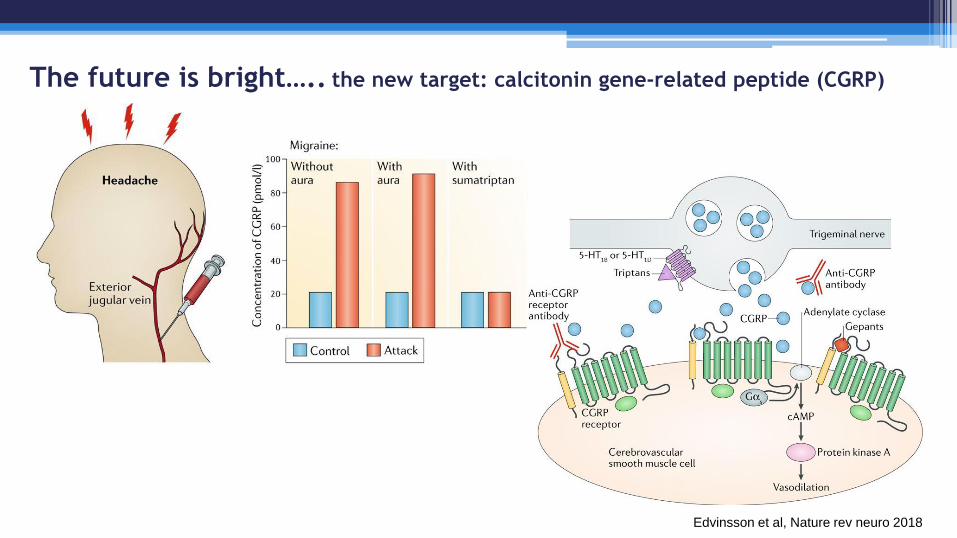
The future is bright….. the new target: calcitonin gene-related peptide (CGRP)
Edvinsson et al, Nature rev neuro 2018

The future is bright…..Phase 3 CGRP mAb’s & small molecule antagonist (EM)
50%
responder
rate
Ereneumab
Aviomig
Fremanezumab
Ajovy
Galcanezumab
Emgality
Eptinezumab
target CGRP Receptor CGRP CGRP CGRP
Route of
admin
SC (mthly) SC (mthly) SC (2 wkly or
mthly)
IV
T ½ (days) 21 45 28 31
EM phase 3 STRIVE
LIBERTY (failed 4 prev)
ARISE
HALO EM EVOLVE 1&2 PROMISE 1
arms P, 70mg, 140mg P, 225, 675mg P, 120mg, 240mg P, 100mg, 300mg
Ubrogepant
(small molecule)
CGRP Receptor
PO
?
ACHIEVE 1&2
ACHIEVE 1: P, 50mg, 100mg
ACHIEVE 2: P, 25,mg 50mg

The future is bright….. Phase 3 small molecule antagonist (EM)
Ubrogepant
(small molecule)
CGRP Receptor
PO
?
ACHIEVE 1&2
ACHIEVE 1: P, 50mg, 100mg
ACHIEVE 2: P, 25,mg 50mg0
5
10
15
20
25
ACHIEVE 1
Placebo
50mg
100mg
Placebo n=1436
Ubrogepant n=1327
% h
eadache f
ree a
t 2 h
rs

The future is bright…..Phase 3 CGRP monoclonal Ab (EM)
50%
responder
rate
0
10
20
30
40
50
60
70 Placebo
Dose 1
Dose 2
n873 n825 n900n955

FRONT DOOR
• IV/IM metoclopramide or prochlorperazine
• IV paracetamol 1000mg
• IV fluid support
• environment
vascular concerns or failed triptan
• add PO Lasmitidan
Once haemorrhage
excluded
• Regular: PO ibuprofen or aspirin +anti-emetic
• ± PRN: PO/ IV paracetamol
If all excluded
and migraine
• Consider CGRP blockers

Thank you for listening


Reversible Cerebral vasoconstriction syndrome (RCVS)
• majority spontaneous but can be triggered
eg. vasoactive drugs (aka Call Fleming) or post-partum
• monophasic course
• recurrent thunderclap over wks; spontaneously resolves (4-6 wks)
• transient disturbance in the control of cerebral vascular tone
• early complication: cortical SAH, ICH, seizures and PRES
• on cerebral angio ‘beading’ which fully reverses on f/u
Ducros & Bousser_Practical Neurology 2009

Thunderclap headache
Peaks within minutes -80% within 1st minute1
1o v 2o : clinically cannot differentiate1
Primary TCH diagnosis of exclusion
SAH CT/LP earlier or CTA later
Arterial Dissection focal Neuro signs, MRA
Venous Sinus Thrombosis raised CSF OP, CTV
Pituitary Apoplexy
Spontaneous Intracranial Hypotension
3rd ventricle colloid cyst
Reversible cerebral vasoconstriction syndrome (RCVS)
EMERGENCY Referral required

Cervical arterial dissection
Headache occurs in 60-95%
typically progressive but thunderclap in up to 1/5
ipsilateral
usually headache plus (develops 4 days post [Silbert et al 1995])
Horner's
pulsatile tinnitus,
amaurosis fugax
cerebral ischaemic symptoms
ipsilateral cranial palsies in 12% [Mokri et al 1996]
5% lower cranial nerve involvement- usually XII
4% palsy of cranial nerve V
3% ocular motor palsies
Treatment: anticoagulation/ antiplatelet
Caplan.
Nature
2008
CT Angiogram –
Dissection ‘flap’
CT Angiogram CT Angio 3D MR –T1 fat suppression

Q4. differential for severe headache, visual loss, diplopia?
Pituitary apoplexy
IIH
GCA
SOL; PCOM aneurysmal mass
cervical arterial dissection

Cerebral venous sinus thrombosis
Gregory Piazza Circulation. 2012;125:1704-1709 Copyright © American Heart Association, Inc. All rights reserved.
Saposnik et al 2011

Cerebral venous sinus thrombosis
Non contrast CT demonstrating hyperdensities along
the left tentorium (arrows, A) and involving the left
sigmoid sinus (arrowheads, B). MRV demonstrating
thrombosis of the left transverse (arrowheads), sigmoid
sinus and proximal jugular vein (arrows) in the axial (C)
and coronal (D) planes*Piazza, 2012, circulation*
have a low index of suspicion especially in progressive states
/seizures
direct relevant investigations
look for papilloedema
Present in up to 25% of CVT[Crassard et al 2005]
n131 papilloedema/?IIH- 10% CVT [Ferro et al 2004]
>80% will have elevated OP on LP but not always when hyper-acute
direct appropriate imaging to include venography if
suspected

Idiopathic intracranial hypertension
Young
high BMI
COCP
Vit A, tetracyclines,
IIH Consensus guidelines _Mollan et al jnnp 2017

Idiopathic intracranial hypertension
Young
high BMI
COCP
Vit A, tetracyclines,
Radiological findings
• empty sella
• dilated optic nerve sheaths
• optic nerve tortuosity
• posterior globe flattening
• protrusion optic nerve head
• narrowing of Meckel’s cave
• reduction in the diameter of the cavernous sinus
• venous flow voids/ stenoses

Idiopathic intracranial hypertension_management
IIH Consensus guidelines _Mollan et al jnnp 2017
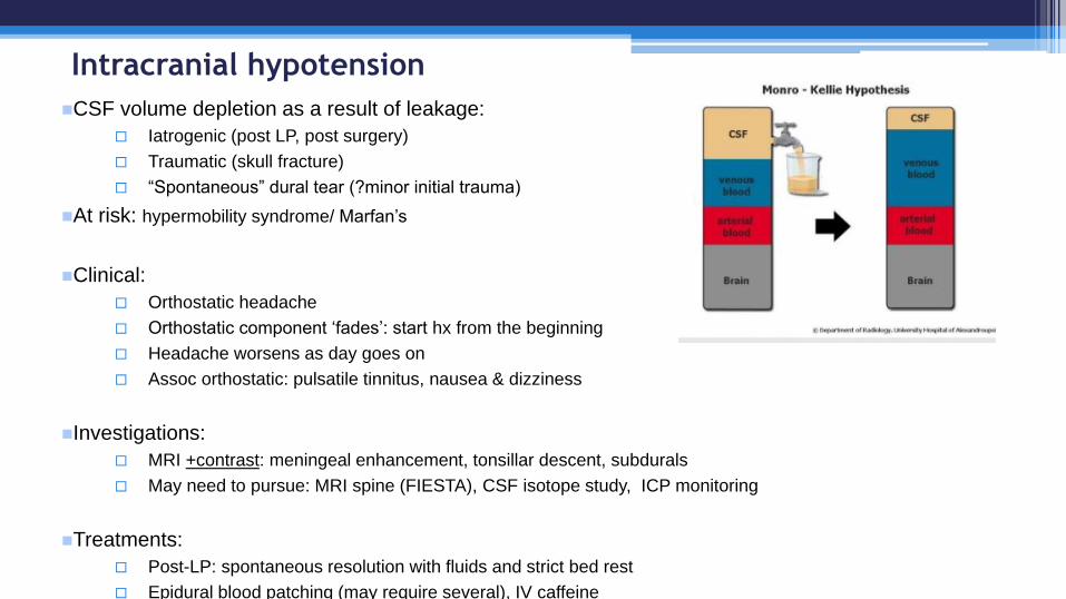
Intracranial hypotension
CSF volume depletion as a result of leakage:
Iatrogenic (post LP, post surgery)
Traumatic (skull fracture)
“Spontaneous” dural tear (?minor initial trauma)
At risk: hypermobility syndrome/ Marfan’s
Clinical:
Orthostatic headache
Orthostatic component ‘fades’: start hx from the beginning
Headache worsens as day goes on
Assoc orthostatic: pulsatile tinnitus, nausea & dizziness
Investigations:
MRI +contrast: meningeal enhancement, tonsillar descent, subdurals
May need to pursue: MRI spine (FIESTA), CSF isotope study, ICP monitoring
Treatments:
Post-LP: spontaneous resolution with fluids and strict bed rest
Epidural blood patching (may require several), IV caffeine

Dr Anvekar, Neuroradiology Unit, S P Institute of Neurosciences,Solapur,Maharashtra, INDIA
Intracranial hypotension

Temporal arteritis
Critical to identify as 13% permanent visual loss
Ha characteristics unhelpful other than allodynia:
Headache, scalp tenderness, jaw claudication, weight loss, low grade fever, non-specifically unwell
Clinical suspicion high Consider in >50yrs; mean age 71yrs
Most helpful clinical symptoms/signs are:
Diplopia
Jaw claudication
Temporal artery beading/ Palpable enlarged temporal artery
Temporal artery tenderness
Absent temporal artery pulse
Excess of vertebrobasilar territory ischaemic events:7% vestibular symptoms [Caselli 1998]
ESR/Plt and CRP Normal < 1%
Temporal Artery Biopsy as soon as possible– but can be patchy; (?role of US)
Treatment: High dose steroids 1mg/kg- initial improvement of PMR by 48hrs and ESR by 72 hrs
BUT steroids produce non-specific improvement in many types headaches & this does NOT confirm the diagnosis

Temporal arteritis
US of greater sensitivity but not greater specificity cf biopsy
-role for proceeding to biopsy in US negative group TABUL study Luqmani et at 2016
Normal EVG
Fragmentation /distortion of internal elastic lamina
TA EVG
Normal H&E
TA H&E
Intimal thickening & transmural inflam.
Obliteration of the lumen.
Dense inflammatory infiltrate
with giant multinucleated cells