Embed Size (px)
Citation preview

2
Department of Surgery, Cardiothoracic Division,
The Laboratory of Transplantation Pathology, University of Helsinki
The Institute of Biotechnology, Helsinki, and
the Institute of Biomaterials, Tampere University of Technology, Tampere,
Finland
Healing of airway anastomoses and
stenting of airway stenosis
Antti Korpela
Academic dissertation
To be presented with the permission of the Medical Faculty of the University of
Helsinki, for public examination in Auditorium Xlll, Unioninkatu 34, University of
Helsinki, on June 16th 2000, at 12:00 noon
Helsinki 2000
3
Supervised by:
Professor Ari L. J. Harjula, M.D.
Department of Surgery, Cardiothoracic Division,
Helsinki University Hospital
and
Docent Pertti Aarnio, M.D.
Department of Surgery,
Satakunta Central Hospital, Pori
Reviewed by:
Docent Paula Maasilta, M.D.
Department of Medicine,Pulmonary Division,
Helsinki University Hospital
and
Docent Timo Savunen, M.D.
Department of Surgery, Cardiothoracic Division,
Turku University Hospital
Discussed with:
Docent Matti Tarkka, M.D.
Department of Surgery, Cardiothoracic Division
Tampere University Hospital
ISBN 952-91-2233-0 (nid.)
ISBN 952-91-2234-9 (verkkojulkaisu, pdf)
ISBN 952-91-2235-7 (verkkojulkaisu, html)
http://ethesis.helsinki.fi
Helsinki 2000
Yliopistopaino
4
CONTENTS
1. List of original publications....................................................................7
2. Introduction............................................................................................8
3. Review of the literature........................................................................11
3.1. The bronchial circulation...............................................................................11
3.2. Measurement of blood perfusion by laser Doppler flowmetry (LDF)............12
3.3. Laryngotracheal stenosis..............................................................................12
3.4. Experimental airway stenosis........................................................................12
3.5. Lung transplantation......................................................................................13
3.5.1. Airway complications..............................................................................13
3.6. Bronchial graft...............................................................................................16
3.7. Management of tracheobronchial complications...........................................17
3.8. Stenting of the airways..................................................................................18
3.8.1. Stent materials.......................................................................................19
3.8.1.1. Silicone...........................................................................................19
3.8.1.2. Metallic............................................................................................21
3.8.1.3. Biodegradable.................................................................................23
4. Aims of the study.................................................................................26
5. Material and methods..........................................................................27
5.1. Test animals..................................................................................................27
5.2. Anesthesia.....................................................................................................27
5.3. Surgical procedures......................................................................................28
5.3.1. Measurement of bronchial mucosal blood flow by laser Doppler
flowmetry...............................................................................................28
5.3.1.1. Operation principle of laser Doppler flowmetry...............................28
5.3.2. Bronchial autograft and allograft in an animal model.............................29
5
5.3.3. Surgically implanted self-reinforced poly-l-lactide bronchial stent in
pigs........................................................................................................30
5.3.4. Self-reinforced poly-l-lactide and silicone stents in rabbit trachea.........30
5.3.5. Stenting of experimental tracheal stenosis............................................31
5.4. Histology.......................................................................................................32
5.5. Scanning electron microscopy......................................................................33
5.6. Statistical analysis.........................................................................................33
6. Results.................................................................................................34
6.1. Bronchial mucosal blood perfusion measured by laser Doppler flowmetry
(Study l)........................................................................................................34
6.2. Healing of a totally avascular bronchial graft (Study ll).................................34
6.3. Tissue reactions to self-reinforced poly-l-lactide bronchial stent
(Study lll).......................................................................................................35
6.4. Tissue reactions to intratracheal stents in rabbits (Study lV)........................36
6.5. Results of management of experimental tracheal stenosis by dilatation
and intratracheal stents (Study V)................................................................37
7.
Discussion................................................................................................41
7.1. The blood supply of the lung.........................................................................41
7.2. Airway stenosis.............................................................................................41
7.3. Laser doppler flowmetry................................................................................42
7.3.1. Limitations of LDF..................................................................................42
7.4. Problems in healing of bronchial anastomoses............................................43
7.5. Methods of handling airway stenosis............................................................45
7.6. Stenting of the airways..................................................................................45
7.6.1. Silicone stent..........................................................................................46
7.6.2. Metallic stent..........................................................................................47
7.6.3. Biodegradable stent...............................................................................48
6
8. Conclusions.........................................................................................49
9. Summary.............................................................................................50
10. Acknowledgements...........................................................................53
11. References........................................................................................55
Original publications
7
1. LIST OF ORIGINAL PUBLICATIONS
This thesis is based on the following publications, referred to hereafter by their
Roman numerals.
I Korpela A, Aarnio P, Harjula A: Evaluation of bronchial mucosal blood flow by
laser doppler flowmeter. International Journal of Angiology 1995;4:110-112
II Korpela A, Aarnio P, Taskinen E, Ikonen T, Harjula A: The healing of bronchial
allografts in pigs. Scand Cardiovasc J (in press)
III Korpela A, Aarnio P, Sariola H, Törmälä P, Harjula A: Bioabsorbable bronchial
stent in an animal model. Journal of Bronchology 1998;5:9-13
IV Korpela A, Aarnio P, Sariola H, Törmälä P, Harjula A: Comparison of tissue
reactions in the tracheal mucosa surrounding a bioabsorbable and silicon airway
stents. Ann Thorac Surg 1998;66:1772-1776
V Korpela A, Aarnio P, Sariola H, Törmälä P, Harjula A: Bioabsorbable self-
reinforced poly-l-lactide, metallic and silicon stents in the management of
experimental tracheal stenosis. Chest 1999;115(2):490-495
8
2. INTRODUCTION
Airway stenosis may be caused by the postsurgical state, by malacia or by
obstructing tumours or extrinsic compression, any of which may induce progressive
dyspnea and hypoxemia. The most common indication for tracheal resection and
reconstruction is still post-intubation tracheal stenosis (Grillo et al. 1995).
Lung transplantations in human dates from 1963 when J. D. Hardy undertook the
first human single lung transplantation (Hardy et al. 1963). In the early days; most
patients undergoing pulmonary transplantation suffered from bronchial anastomotic
complications; since then, results have improved. Shennib and associates (1994)
reviewed airway complications in lung transplantation, estimating that the lethal
tracheal dehiscense rate in heart-lung transplantation is currently less than 3% and
the nonlethal complication rate resulting in tracheal stenosis is from 7.5% to 14%.
The incidence of lethal complications is similar in isolated lung transplantation, but
the figure for nonlethal complications appears to be higher, from 12% to 17%.
In addition to impaired bronchial circulation, rejection and infection can affect the
healing of a bronchial anastomosis. Although in the transplantation procedure the
bronchial circulation is cut off, the bronchial arteries can be revascularised, and
some authors have recommended transplantation with revascularisation of the
bronchial artery (Couraud et al. 1992, Laks et al. 1991, Schreinemakers et al. 1990).
However experimental studies have shown that ischemia of the donor bronchus is an
important factor underlying bronchial complications (Legal et al. 1985, Turrentine et
al. 1990, Mills et al. 1970). Wrapping with omentum (Lima et al. 1982, Morgan et al.
1982, Turrentine et al. 1990), intercostal muscle flaps (Legal et al. 1985, Turrentine
et al. 1990) or internal mammary artery pedicle (Turrentine et al. 1990) has been
used to protect and revascularise bronchial anastomoses and this has been shown
to promote local revascularisation, although it may take several days (Siegelman et
al. 1977, Rabinovich 1972); during this time the donor bronchus is dependent on the
blood supply from collaterals of the pulmonary circulation. Rejection of the bronchus
9
itself will however, impair the local revascularisation process, and avoidance of
rejection may thus also be important in preventing ischemic complications of the
bronchi in the early postoperative period after transplantation (Davreux et al. 1993).
A totally ischemic canine bronchial autograft model has been used to study the
healing and revascularisation process in the bronchus and bronchial anastomosis
(Milhiet et al. 1968, Fell et al. 1985, Morgan et al. 1982).
Surgical resection and end-to-end anastomosis is the method of choice in handling
airway stenosis, but this is not always possible due to the nature and cause of the
stenosis and the general state of the patient; tracheobronchial malacia is also
difficult to correct surgically. There have been attempts to substitute the trachea
with tracheal prostheses or homografts. Endobronchial methods developed to deal
with airway stenosis include laser surgery, cryotherapy, endobronchial resection,
dilatation, brachytherapy when malignant obstruction is concerned, and stenting of
the stenotic area in the airways (Hetzell et al. 1991).
Different types of stents have been developed for the treatment of airway stenosis.
The Montgomery T-tube, made of silicone, introduced by W.W. Montgomery in 1965
(Montgomery 1965) can be used in high laryngotracheal and tracheal stenoses but
requires tracheostomy; it is still widely used with satisfactory results (Gaissert et al.
1994). The most common types of stents in use are made of silicone (Dumon 1990)
or metallic wire (Rousseau et al. 1993). The ideal stent for tracheobronchial stenosis
has yet to be designed.
Bioabsorbable airway stents may offer benefits. Compared to the devices currently
available for clinical use, these need not be extracted, and after resorption do not
interfere with the normal function of the airways. The absorption time can be
modified and is dependent on the choice of basic molecule, the shape of the stent,
the degree of its polymerisation, the internal arrangement of the material
components, and the site of implantation.
10
The aim of this present study was first to examine the real bronchial mucosal
perfusion by laser Doppler flowmetry (LDF), when the bronchial circulation is cut off;
secondly, to study the effects of rejection on bronchial healing. The biocombatibility
of stents made of SR-PLLA applied in the airway was also tested. Finally, the
possibilities of treating surgically induced tracheal stenoses were studied with a
bioabsorbable SR-PLLA stent compared with stents made of silicon and metal.
11
3. REVIEW OF THE LITERATURE
3.1. The bronchial circulation
The bronchial arteries have been studied in human cadavers (Cauldwell et al. 1948).
Their origin from the aorta varies, but the types comprising two left and one right,
one left and one right or two arteries to each side, constitute 82.67 % of all patterns.
Right bronchial arteries arise in 88.7 % in a common stem with an aortic intercostal
artery, from which the first aortic intercostal participates in the common
intercostobronchial stem in 78 %. In only 4 % does a left bronchial artery arise in
common with an intercostal artery. Widespread anastomoses occur between
bronchial and other arteries such as the esophageal, subclavian, internal mammary,
superior intercostal, pericardiophrenic and aortic mediastinals. Bronchial arteries
form both the peribronchial and submucosal vascular plexus along the bronchial tree
(Deffebach et al. 1987).
Anastomoses between the bronchial and pulmonary arteries have been shown in
studies with a corrosion model of the human lungs ( Pump 1963). The diameter of
the anastomotic vessels ranged from 0.05 mm to 0.5 mm, and their length from 0.1
mm to 10 mm. They were found on bronchi 1.60 mm to 3.50 mm in diameter.
Anastomoses were found throughout the lung parenchyma except for a small area
surrounding the hilum. Subsequently the same author (Pump 1972) showed
anastomoses between smaller precapillary-sized bronchopulmonary and pulmonary
arterial vessels, diameters ranging from 24 µm to 48 µm.
Reconstitution of the bronchial artery in a canine left lung allotransplantation model
has been shown to reduce bronchial complications to 20 %, while 82 % of animals
without reconstitution developed bronchial complications (Mills et al. 1970). The
authors took these data to indicate that an attempt at reconstitution of the bronchial
arterial supply should be made in humans receiving lung transplants.
12
3.2. Measurement of blood perfusion by LDF
Laser Doppler flowmetry (LDF) was first applied in measurement of the peripheral
circulation of the skin in 1975 (Stern 1975). Since then the technique has been
used to measure the tissue perfusion of heart (Ahn et al. 1988), kidney (Stern et al.
1979), liver, digestive mucosa (Ahn et al. 1985) and bronchial mucosa (Aoki et al.
1991, Inui et al. 1990, Inui et al.1993, Takao et al. 1991, Yokomise et al. 1991,
Yokomise et al. 1989, Tanabe et al. 1991, Takao et al. 1990, Hyytinen et al. 1999).
3.3.Laryngotracheal stenosis
At the beginning of this century, the most frequent source of laryngotracheal
stenosis was the destructive process of some primary disease, for example,
diphtheria, lues, tuberculosis and typhoid fever. Subsequently came the problem of
cicatricial stenosis caused by high tracheotomy (Jackson 1921). Postintubation
tracheal stenosis is to this day the most common indication for tracheal resection
and reconstruction, despite identification of the causes of these lesions and the
development of means of avoiding them. The strategy for treatment of airway
stenoses is now well established. Segmental tracheal resection and end-to-end
anastomosis remains the preferred definitive treatment for postintubation stenosis
(Grillo et al. 1995, Har-El et al. 1993, Bisson et al. 1992).
3.4.Experimental airway stenosis
Extramucosal resection of the cartilaginous arches of the trachea in pigs has been
used to create an experimental malacia (Johnston et al. 1980). When Marquette
resected three tracheal rings and used caustic agent intrabronchially during rigid
bronchoscopy to create tracheal stenosis, clinically significant stenosis developed in
adult minipigs within eight weeks (Marquette et al. 1995).
13
3.5. Lung transplantation
The first attempt at clinical lung transplantation was made by Hardy and coworkers
in 1963. The subject died of renal insufficiency on the 18th postoperative day.
Postmortem examination revealed a 0.5 cm defect which had caused a postoperative
airleak in the membranous portion of the bronchus of the transplanted lung. The
authors concluded that the bronchial anastomosis proved to represent perhaps the
weakest element among the several anastomoses. Thus every means should be
exploited to reinforce this anastomosis by covering it with viable pedicles of
surrounding autogenous tissue (Hardy et al. 1963).
The first single lung transplantation for pulmonary fibrosis in humans with successful
long-term survival was performed in 1983 by the Toronto Lung Transplant Group
(1983). Since then increasing numbers of lung transplantations have been reported,
with improving results. An important factor in this was the introduction of a new
immunosuppressive drug, the fungal metabolite cyclosporine, introduced in 1977
(Borel et al. 1977). Lung transplantation has become a viable option for patients with
some end stage pulmonary diseases (Kaiser et al. 1991, Marinelli et al. 1992, De
Leval et al. 1991, Ramirez et al. 1992, Calhoon et al. 1991, Shennib et al. 1992,
Patterson 1997). There remain a number of problems with lung transplantation;
infection, rejection, immunosuppressive drug therapy and the general capacity of the
patient can adversely affect the healing process postoperatively.
3.5.1. Airway complications
Airway complications have constituted a problematic sequela after transplantation.
At the beginning of the era of lung transplantation the major causes of morbidity and
mortality were complications at the bronchial anastomotic site, due probably to
ischemia. Most of a cohort of 38 patients who underwent lung transplantation before
January 1981 and survived ten days or longer suffered from airway complications
which contributed directly to death (Veith et al. 1983).
14
The prevalence of airway complications is lowest in heart-lung transplantations:
tracheal stenosis develops in 7.5% to 14% of patients; the risk of any kind of airway
complication in single-lung or bilateral sequential-lung transplantation ranges from
12 % to 17%. The overall incidence of lethal airway complications is estimated to be
2% to 3%, whereas that of late stricture is 7% to 14% (Shennib et al. 1994).
Although tracheal complications and stenosis are very rare in heart-lung
transplantation, bronchial complications and stenosis are not uncommon following
single lung and double lung transplantations, especially if the bronchial circulation is
not re-established. (McCarty et al. 1990, Schafers et al. 1991, Patterson et al. 1990,
Shennib et al. 1994, Haydock et al. 1992, Hoyos et al, 1992, Pettersson et al. 1994,
Kshettry et al. 1997, Date et al. 1995).
In the transplantation operation the systemic blood supply from the bronchial
circulation is interrupted, and if it is not re-established during surgery, the donor
bronchus is at risk of ischemia before local revascularisation at anastomosis. This
healing and revascularisation process can take 12-15 days (Siegelman et al. 1977,
Rabinovich 1972), during which time the viability of the donor bronchus depends on
retrograde collateral perfusion from the pulmonary circulation.
Experimental studies have revealed that ischemia of the donor bronchus to be an
important factor making for bronchial complications (Legal et al. 1985, Mills et al.
1970, Turrentine et al. 1990). Reducing the length of the transplanted bronchus has
been shown to reduce the prevalence of bronchial healing disturbance (Pinsker et
al. 1979). Omental or internal mammary artery wrapping (Khaghani et al. 1994),
telescope anastomosis (Calhoon et al. 1991) and reversed telescope anastomosis
(Rabinov et al. 1996) have been used to counter anastomotic complications in lung
transplantation.
Wrapping with omentum (Lima et al. 1982, Turrentine et al. 1990, Morgan et al.
1982), internal mammary artery pedicle (Turrentine et al. 1990) or intercostal muscle
flaps (Legal et al. 1985, Turrentine et al. 1990) has been used to protect and
revascularise bronchial anastomoses, and the approach has allowed local

15
revascularisation, although it took several days ((Legal et al. 1985, Laks et al. 1991).
During this time the donor bronchus receives its blood supply from collaterals of the
pulmonary circulation. Wilson and collegues in 1996 reviewed retrospectively their
123 assessable bronchial anastomoses accomplished in lung transplantation. At the
outset they used wrapping of the anastomosis with omentum, pericardium or
intercostal muscle; in the last 89 anastomoses they used only apposition of
peribronchial tissue over the anastomosis, but their airway complication rate, as low
as 1.6%, was not affected by the presence of a wrap (Wilson et al. 1996).
More recently, bronchial arterial revascularisation has been shown to be associated
with good bronchial healing. Methods for direct revascularisation of bronchial
arteries have been developed and they have been shown to facilitate bronchial
anastomotic healing. Some authors have recommended transplantation with
revascularisation of the bronchial artery (Couraud et al. 1992, Laks et al. 1991,
Schreinemakers et al. 1990, Couraud et al. 1992). Nørgaard and associates
attempted bronchial artery revascularisation with the left internal thoracic artery in 63
patients, 53 of whom were evaluated by angiography. Fifty patients showed
successful revascularisation, poor bronchial healing being noted in three patients in
the series, two of whom evinced no communication to the bronchial arteries although
the internal thoracic artery was open (Nørgaard et al. 1996).
At the beginning of the era of lung transplantations it was sought to avoid the use of
corticosteroids during the first few days after the operation, as canine lung
transplantation models had shown their use to hamper healing of the bronchus (Lima
et al. 1981, Goldberg et al. 1983). Subsequent results suggested that preoperative
and postoperative use of steroids does not preclude normal tracheal healing (Novich
et al. 1991) and in a canine allograft model steroid immunosuppression was found to
be compatible with excellent bronchial healing (Auteri et al. 1992). Nowadays, many
transplantation groups such as the San Antonio group and the Toronto lung
transplant group use corticosteroids routinely during the posttransplantation period,
with excellent results (Calhoon et al. 1991, Novich et al. 1991, Hoyos et al. 1992).
16
Rejection of the bronchus itself impairs the local revascularisation process;
avoidance of rejection may thus also be of significance in preventing ischemic
complications of the bronchi in the early postoperative period after transplantation
(Davreux et al. 1993).
3.6. Bronchial graft
A totally ischemic canine bronchial autograft model has been used to study the
healing and revascularisation process in the bronchus and bronchial anastomosis
(Morgan et al. 1982, Milhiet et al. 1968, Fell et al. 1985). The initial study with the
bronchial autograft model, showed good autograft healing without mortality in any of
the 11 dogs (Milhiet et al. 1968). Later the same experimental method was used, but
in contrast to the results of the preceding series all five dogs in the autograft group
died within five days of operation. Three died of major bronchopleural fistula and two
of mediastinitis. All five autografts in the omental pedicle flap group healed without
complications. It was concluded that in a severe ischemic bronchial autograft model
in dogs, an omental pedicle flap established revascularisation as early as on the
fourth postoperative day and maintained viability in this otherwise lethal condition
(Morgan et al. 1982).
In another study all five dogs with postpneumonectomy bronchial autografts died of
bronchopleural fistulas due to autograft necrosis, while nine animals with bronchial
autografts wrapped with the intercostal pedicle flap healed well. Histologic
examination revealed healed suture lines of the bronchial autografts, with normal
mucosa and viable bronchial cartilage. The intercostal pedicle flap was thus a
reliable means of providing neovascularity and mechanical reinforcement to an
ischemic bronchial anastomosis. Its effect on bronchial anastomotic healing was not
diminished by administration of corticosteroids. In the same study a different free
bronchial autograft model was also used without pulmectomy. A 1.5 cm ring of the
left main bronchus was reimplanted in six dogs. All the autografts healed well.
Bronchoscopic and gross examinations when the animals were put sacrified
demonstrated intact bronchial mucosa with no evidence of stricture. Wrapping and
17
isolation of the segmental bronchial autograft from adjacent pulmonary and
mediastinal tissues with polytetrafluoroethylene caused necrosis of the graft in all
four animals 4 to 12 days after operation (Fell et al. 1985).
A different method had previously been used in a study by where a 1 cm ring of the
left main bronchus was reimplanted in seven dogs. All seven autografts failed owing
to ischemic necrosis within seven days after the operation (Kiriluk et al. 1953). An
experimental postpneumonectomy bronchial autograft method has been used,
telescoping the proximal end of the bronchus into the distal end. Ten dogs in the
bronchial autograft group survived the autotransplantation as well as animals in
groups where the bronchial autograft was wrapped with a free omental pedicle, a
detached omental pedicle or a Gelfoam sponge soaked in porcine omental extract.
Wrapping of the anastomosis tended to increase the narrowing of the ischemic
bronchial segment in all groups in question. This was possibly related to the bulk of
the wrap. It was concluded that wrapping of a telescoped anastomosis is not
necessary to prevent early complications; however, no method completely eliminates
stenosis (LoCicero lll et al. 1992).
3.7. Management of tracheobronchial complications
Therapeutic options available in the management of bronchial strictures include
surgical methods such as resection and reanastomosis or completion
pneumonectomy if possible after sleeve resection. There are a variety of
endoscopic methods (Mehta et al. 1995): endobronchial resection (Mathisen et al.
1989), bronchoscopic dilatation (Cohen et al. 1984, Colt et al. 1992, Carre et al.
1994, Sheski et al. 1998), cryotherapy (Mathur et al. 1996, Marasso et al. 1993,
Maiwand et al. 1997), brachytherapy (Cavaliere et al. 1996), laser therapy (Hetzel et
al. 1985, Cavaliere et al. 1988) and stenting of the stenotic area (Carre et al. 1994,
Carrasco et al. 1994, Monnier et al. 1996). An anastomotic stenosis is a potentially
serious complication after lung transplantation and bronchial resections, but can
usually be successfully treated by conservative means.
18
3.8. Stenting of the airways
Endoscopic intubation of malignant tumors of the tracheobronchial tree with a
Souttar tube without tracheostomy was introduced by Clarke in 1980 (Clarke 1980).
Editorials in Chest conclude that the very existence of a plethora of surgical
techniques and prosthetic materials for treating stenosis of the tracheobronchial
tree, would indicate that all methods have limited success rates. Complicated
tracheobronchial problems not amenable to resection and reanastomosis, with or
without prostheses, could however be overcome stenting of the narrowed airways
(Unger 1990).
Bronchial stenosis after lung transplantation is a specific problem, often due to scar
stenosis at anastomotic level with or without previous anastomotic dehiscence.
Apart from endobronchial laser or cryotherapy, useful for some short stenoses and
granulomas, these are best handled by insertion of a silicone or metallic stent
(Brichon et al. 1992). The advent of lung transplantation has increased the potential
demand for tracheobronchial stents, since airway anastomosis still constitutes a
surgical weak point (Zannini et al. 1994). A rate of 7% to 14% of bronchial
complications is reported in most series, mainly from dehiscence, stenosis or
malacia of the anastomosis (Schafers et al. 1991, Shennib et al. 1994, Kshettry et al.
1997, Date et al. 1995). In a series of 57 patients treated with a silicone stent
(Sonett et al. 1995), the conclusion was that endoscopic stenting provides effective
palliation of tracheobronchial stenoses of both benign and malignant causes. They
considered such stenting to be the primary management option for airway
obstruction after lung transplantation.
Both silicone (Sonett et al. 1995) and self-expandable metallic stents (Rousseau et
al. 1993, Brichon et al. 1992) have thus performed satisfactorily in the treatment of
postransplantation airway stenosis.
19
3.8.1. Stent materials
Different types of silicone (Cooper et al. 1989, Gaer et al.1992, Tsang et al. 1989,
Tsang et al. 1992, Montgomery 1974, Westaby et al. 1982, Dumon 1990) and
metallic (Spatenka et al. 1991, Varela et al. 1990, Simonds et al. 1989, Clarce 1980,
George et al. 1990, Wallace et al. 1986, Pagliero et al.1974, Tsugava et al. 1997,
Vinograd et al. 1994) internal stents are available in the treatment of airway
stenosis. Teflon (Amemiya et al. 1985), polymeric woven (Mair et al. 1990) and the
Tracheoflex tracheostomy tube (Orlowski 1987, Orlowski 1997) have also been used
to stent obstructed airways, and combinations of different materials in the same stent
have recently been introduced (Freitag et al. 1997, Wassermann et al. 1997, Bolliger
et al. 1996).
3.8.1.1. Silicone
The silicone T-tube for stenting the airway after repair of stenosis in the low
subglottic region was developed and described by Dr. William Montgomery in 1965
(Montgomery 1965). This Montgomery T-tube calls for permanent tracheostomy.
The type is still widely used in tracheal obstruction not amenable to surgical
reconstruction (Gaissert et al. 1994). Silicone stents have since been used in
tackling anastomotic problems in the tracheobronchial tree. In spite of their wide
application, however, they have certain disadvantages which render them
unsatisfactory in long-term use. Because they are relatively thick, their internal
diameter is smaller than the normal airway lumen; they interfere with normal airway
mucociliary function, and thus tend to become occluded by mucous secretions,
making regular bronchoscopies necessary to see whether cleaning or replacement is
needed (Tsang et al. 1992). Silicone stents are difficult to place in the right main
stem bronchus without obstructing the orifice of the upper lobe and after insertion
they also have tendency to migrate (Higgins et al. 1994). On the other hand,
silicone stents are usually well tolerated and can be readjusted after insertion if
necessary.
20
In addition to the Montgomery T-tube, one of the most widely used types of silicone
stents was designed in 1990 (Dumon 1990). Dumon stents were used in the
management of benign tracheobronchial stenosis in 63 patients (Martinez-Ballarin et
al. 1996). Temporary stents were removed after 18 months in 21 cases, and
although stenosis recurred in 4 patients, the therapy proved successful in the
remaining 17. The complication rate associated with stents was altogether 31.7%,
problems including migration of the stent in 11 patients, granuloma formation in 4,
and airway obstruction due to heavy secretion in 4; one of the patients died of
hypersecretion and obstruction of the airways. The complication rate was no greater
than that with the T-tube, and the Dumon prosthesis itself is not a cause of increased
damage to the airways. Silicone stents can thus be used in benign tracheal
stenoses without the need of tracheostomy.
Patients with malignant airway obstruction may suffer from infections, dyspnea and
symptoms of suffocation. In view of the poor prognosis in such cases, palliative
methods are needed to maintain airway patency. Bolliger and coworkers (1993) used
38 Dumon stents in 31 patients for endoscopic palliation of malignant airway
stenosis. Their patients first had laser resection and insertion of the stent, followed
by percutaneous or endobronchial radiotherapy. Stent-related complications were
encountered with 10 stents, the most common problem being migration. Both
Dumon silicone and metallic stents with or without a silicone covering were used to
treat patients with severe dyspnea arising from airway stenosis caused by malignant
tumours. It was concluded that stents make a significant contribution to the
alleviation of respiratory symptoms and the quality of life of patients with malignant
tracheobronchial stenosis not amenable to operative cure. Different types of stents
have been used as palliative treatment of malignant obstructions in the carinal
region (Shiraishi et al. 1998). Bare metallic stents are effective in extrinsic
compression, but Dumon stents or covered metallic stents are preferable for
intraluminal stenosis (Tojo et al. 1996).
21
The Dynamic stent designed by Freitag is a bifurcated silicone device whose
tracheal section is horseshoe-shaped and supported with incorporated steel struts,
except for the posterior wall made of flexible silicone. The collapsible posterior wall
mimics the mechanism of the posterior wall of the trachea (Freitag et al. 1994,
Freitag et al. 1997).
Experimental use of a novel thin-walled self-expandable prototype silicone airway
stent has been reported (Bolliger et al. 1999). This Polyflex stent is made of a
texture of tightly woven polyester filaments and covered with a silicone layer. Clinical
use of this stent in 19 patients has also been reported, 17 of them with
tracheobronchial complications of infiltrating cancer and two with benign
postintubation stricture and malacia (Wassermann et al. 1997).
3.8.1.2. Metallic
Harkins (1952) reported the use of a metal alloy tube to stent benign tracheal
stenoses, however the patient developed obstruction of mucus and edema at either
end of the stent. Belsey (1951) introduced a new method of reconstructing airways
following surgical resection, with a free graft of fascia sutured around a wire
”skeleton” made of 32-cauge stainless steel. Self-expanding metallic stents were
first developed for intraluminal use to prevent restenosis after percutaneous
transluminal angioplasty (Sigwart et al. 1987, Wright et al. 1985, Palmaz et al.
1985). This type have also been used in biliary (Gillams et al. 1990) and urethral
(Milroy et al. 1988, Milroy et al. 1989) strictures. The disadvantage of metallic stents
is that once they are implanted their removal may call for open surgery.
Metallic expandable wire stents have a low internal-to-external diameter ratio. They
cannot usually migrate after insertion, and the mucociliary clearance function is
better maintained (de Souza et al. 1994). The usefulness of the expandable metallic
stent was attested in 1986 by Wallace and associates whose stent was constructed
of 0.018-inch thick stainless steel wire formed in a cylindrical zigzag configuration of
five to ten bends (Wallace et al. 1986). These stents were first implanted into the
22
trachea and bronchi of 11 dogs. This so-called Gianturco stent was then used to
treat two patients with bronchial stenosis and bronchomalacia after tracheobronchial
reconstruction. In 1990, Varela’s group successfully treated 5 patients suffering
from tracheobronchial pathology with the Gianturco stent. None had complications
consequent upon stent placement, and their clinical problems were satisfactorily
resolved, although the longest follow-up time was only 12 months (Varela et al.
1990). This type has since been used in airway stenosis (Wilson et al. 1996, Bolot
et al. 1998). However complications such as migration and/or rupture of the metallic
mesh associated with Gianturco stents have been reported (Rousseau et al. 1993,
Hind et al. 1992, Schäfers et al. 1992, Hramiec et al. 1997).
The Wallstent, developed for endovascular use, has also been used in treating
airway stenosis (Rousseau et al. 1993, Brichon et al. 1992, Dasgupta et al. 1998)
and is gaining increasing acceptance. In 1993 Rousseau and collegues used 39
Wallstents and 35 Gianturco stents in 55 patients with 33 tracheal and 29 bronchial
noninflammatory (except one) lesions, the cause of which was postsurgical in 13,
chronic obstructive pulmonary disease in 14, posttransplantation in 11,
postintubation in 7, tumoral compression in 5, postinfectious in 4, and Wegeners
syndrome in one. The Wallstent group yielded a total of six complications, with one
tumoral proliferation successfully treated by laser endoscopically. Two stenoses due
to inadequate covering of the original lesion were treated with a second stent, and
for three restenoses the stent was handled with balloon dilatation in two cases and
insertion of a second stent in one. The Gianturco device was used exclusively for
tracheobronchomalacia; in this group a high (31%) rate of complications was noted,
including braces in the filament branches with or without migration of the stent,
potentially leading to obstruction or wall perforation. Wallstent insertion was
deemed a safe procedure and a good alternative to silicone stent insertion for the
treatment of posttransplantation bronchial stenoses without inflammatory lesions
(Rousseau et al. 1993).
23
The Airway Wallstent, with a polyurethane covering on the outside, has been used
as a palliative treatment in patients with inoperable malignant lesions in central
airways (Bolliger et al. 1996).
The Palmaz stent is a balloon-expandable metallic mesh cylinder device used mainly
in pediatric patients and only recently in adults in airway position (Filler et al. 1995,
Filler et al. 1998, Slonim et al. 1998, Susanto et al. 1998). This stent has no intrinsic
radial force and sufficiently strong compressive forces may lead to possible
complications due to the collapse and loss of function of the stent.
The Strecker is likewise a balloon-expandable stent made of knitted tantalum wire
mounted on an expansion balloon. The Accuflex stent is a self-expanding wire mesh
made of nitinol. Both of these have been employed in intrabronchial position in
malignant airway stenosis (Hauch et al. 1997).
3.8.1.3. Biodegradable
Polyglycolic acid (PGA) and polylactid acid (PLA) are the most important poly-alpha-
hydroxy acids used in surgery. They have plastic properties and were synthesized in
the 1950s. Polylactic acid was introduced as a suture material in 1966 (Kulkarni et
al. 1966). Advances in the manufacture of PGA sutures led to the first commercial
synthetic bioabsorbable suture (Dexon®) in 1970 (Schmitt et al. 1967).
PLA can be synthesised to high molecular weight polymers through direct
condensation polymerisation of lactic acid or in the anionic ring opening
polymerisation of the cyclic diester, lactide (Bischoff et al. 1893, Schneider 1955).
Polylactic acid has two enantiomeric forms, the stereo-isomers D- and L-lactide (Fig.
1). When manufactured by non-reinforcing techniques these partially crystalline,
linear chain absorbable polymers evince only modest mechanical strength
(Vainionpää et al. 1989), In self-reinforced biodegradable composites the polymeric
reinforcing fibers and the binding polymeric matrix have the same chemical element
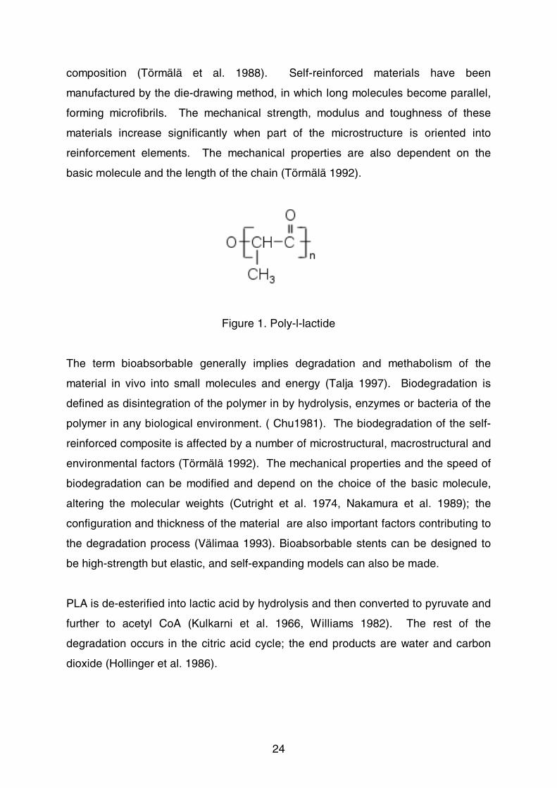
24
composition (Törmälä et al. 1988). Self-reinforced materials have been
manufactured by the die-drawing method, in which long molecules become parallel,
forming microfibrils. The mechanical strength, modulus and toughness of these
materials increase significantly when part of the microstructure is oriented into
reinforcement elements. The mechanical properties are also dependent on the
basic molecule and the length of the chain (Törmälä 1992).
Figure 1. Poly-l-lactide
The term bioabsorbable generally implies degradation and methabolism of the
material in vivo into small molecules and energy (Talja 1997). Biodegradation is
defined as disintegration of the polymer in by hydrolysis, enzymes or bacteria of the
polymer in any biological environment. ( Chu1981). The biodegradation of the self-
reinforced composite is affected by a number of microstructural, macrostructural and
environmental factors (Törmälä 1992). The mechanical properties and the speed of
biodegradation can be modified and depend on the choice of the basic molecule,
altering the molecular weights (Cutright et al. 1974, Nakamura et al. 1989); the
configuration and thickness of the material are also important factors contributing to
the degradation process (Välimaa 1993). Bioabsorbable stents can be designed to
be high-strength but elastic, and self-expanding models can also be made.
PLA is de-esterified into lactic acid by hydrolysis and then converted to pyruvate and
further to acetyl CoA (Kulkarni et al. 1966, Williams 1982). The rest of the
degradation occurs in the citric acid cycle; the end products are water and carbon
dioxide (Hollinger et al. 1986).
25
Biodegradable stents offer certain theoretical benefits when compared to the types
now available for clinical use; extraction of the stent is not necessary and after
resorption there is no interference with normal function of the airways.
For suture purposes biodegradable materials have been in clinical use for over 20
years and have evinced good biocompatibility properties.Their biocompatibility has
been relatively good in organs other than respiratory. Biodegradable plates and
screws have been used for years in clinical settings in orthopedic and plastic
surgery, with favorable results (Rokkanen et al. 1985, Majola et al. 1991, Törmälä
1992). Self-reinforced poly-l-lactide spiral stents have proved to possess good
biocompatibility in the rabbit urethra compared to stents made of stainless steel
(Kemppainen et al. 1993). Spiral stents made of self-reinforced polyglycolic acid
have been used in the human urethra to prevent postoperative urinary retention after
visual laser ablation, again with good results (Talja et al. 1995). Bioabsorbable self-
expandable SR-PLLA urethral stents have been used for recurrent urethral
strictures, preliminary results being favorable (Isotalo et al. 1998).
Several other types of stents have also been developed for intratracheal and
intrabronchial use. All types have their limitations, and the ideal stent for use in the
tracheobronchial tree has yet to be developed. Since stenting of a tracheobronchial
stenosis following lung transplantation can only be a temporary measure (Sonett et
al. 1995), bioabsorbable stents may be optimal.
26
4. AIMS OF THE STUDY
The objectives in the present series were:
1. To evaluate the bronchial mucosal blood flow by laser Doppler flowmetry, and
the relative importance of the bronchial and the pulmonary artery on the
bronchial mucosal blood supply in an animal model.
2. To investigate the healing potential of totally ischemic bronchial grafts and the
effects of rejection and immunosuppressive treatment on the healing process in
an experimental model in pigs.
3. To examine possible adverse tissue reactions and the biocombatibility of a
bronchial stent made of bioabsorbable self-reinforced poly-l-lactide in the
bronchial mucosa in an animal model.
4. To examine the suitability of self-reinforced poly-l-lactide as material for an
airway stent compared with silicone stents in the rabbit trachea.
5. To examine in rabbits the biocompatibility and the suitability of a bioabsorbable
spiral stent made of self-reinforced poly-l-lactide in the management of
experimental tracheal stenosis compared to stents made of metallic and
silicone.
27
5. MATERIAL AND METHODS
5.1. Test animals
In studies l, ll and lll, altogether 27 randomly bred domestic piglets two to three
months of age, weighing 11-26 kg were used as test animals. In studies lV and V
altogether 50 rabbits weighing 2.1-4.0 kg were used.
All animals received humane care in compliance with the European convention for
protection of vertebrate animals used for experimental and other scientific purposes,
ratified in Strasbourg, 1986, by the Council of Europe. The study protocol was
approved by the institutional committee for test animal research and by the
Provincial Government of Uusimaa.
5.2. Anesthesia
In Studies I, II and III the pigs were premedicated with ketamine sulfate (30 mg/kg)
and atropine sulfate (0.05 mg/kg) administered intramuscularly. They were
anesthetised with sodium pentobarbital (8 mg/kg) and pancuronium chloride (0.1
mg/kg) given intravenously, intubated and connected to a Servo 900 C respirator
(Siemens-Elema, Sweden) with a tidal volume of 15 ml/kg and a respiratory
frequency of 18 per minute. The animals were ventilated with 50 % oxygen and
anesthesia was maintained with 50 % N20 and 1 % halothane. Postoperative pain
was treated with diclophenac acid (12.5-25 mg i.m.). For infection prophylaxis, all
animals were given 500 mg ceftriaxone intramuscularly immediately prior to the
operation.
In Studies IV and V the rabbits were anesthetised with a combination of atropine
0.75 mg, ketamine 20 mg, medetomide 300 µg, and diazepam 1.0 mg per kilogram
body weight. Procaine penicillin 150 000 IU served as antibiotic prophylaxis. All
drugs were administered subcutaneously. The animals maintained spontaneous
28
breathing during the operations. They were killed when necessary with an overdose
of sodium pentobarbital.
5.3. Surgical procedures
5.3.1. Measurement of bronchial mucosal blood flow by laser
Doppler flowmeter
Twenty pigs weighing 12 to 21 kg (mean 16.8 kg) underwent left thoracotomy.
Periflux PF3 laser doppler flowmeter (Perimed Co., Sweden) was used in this study.
The left stem bronchus and the pulmonary vessels were dissected free avoiding
damage to the bronchial artery. The measurement probe for LDF was inserted to
contact the bronchial mucosa of the membranous portion of the bronchus, being
passed through a small hole made in the left stem bronchus. A standard endoscopic
probe was used. Baseline bronchial mucosal blood flow was measured first. The
probe was held in place long enough to allow the measurement level to stabilise.
The microvascular perfusion of the bronchial mucosa was then measured during
occlusion of the pulmonary artery by a vascular clamp or during the occlusion of the
bronchial artery by clamping the bronchus tightly with the bronchial artery. Finally,
both were occluded and the bronchial mucosal blood flow measured.
5.3.1.1. Operation principle of LDF
The Periflux PF3 laser Doppler flowmeter (Perimed Co., Sweden) was used in this
study. The equipment comprises 2 mW helium-neon laser, with a wavelength of
632.8 nm. In this technique, a narrow beam of monochromatic light is carried by an
optical fiber to the tissue studied. Light hitting moving blood cells will undergo a
slight Doppler shift (change in wavelength). The magnitude and frequency
distribution of the laser shift is directly related to the number and velocity of blood
cells, but virtually unrelated to the direction in which they move. The back-scattered,
29
Doppler shifted light is measured. LDF measures microvascular perfusion; the
measured values are relative in nature but comparable at any given timepoint and in
the same object.
5.3.2. Bronchial autograft and allograft in an animal model
Twenty-one pigs weighing 11-21 kg underwent left standard thoracotomy. The left
pulmonary artery and veins were ligated and divided and the left stem bronchus was
dissected free up to the lobar bronchi. The bronchus was closed at this level using
an RL 30 stapler (Ethicon, USA), and cut distally to the stapler line; the lung was
then removed. The left main bronchus was clamped distally to the tracheal
bifurcation and completely transected. In group A (6 pigs) this totally ischemic 2.5 cm
long bronchial graft was reanastomosed with a continuous monofilament
polypropylene suture. In group B (8 pigs) the procedure was otherwise the same
apart from the fact that the bronchial stump was anastomosed into another pig as an
allograft.
In group C seven animals with bronchial allografts were medicated with
immunosuppressive drugs, cyclosporin A 15 mg/kg, azathioprine 2 mg/kg and
methylprednisolone 30 mg/kg given intravenously during induction and operation.
Postoperatively the animals daily received cyclosporin A 15 mg/kg, azathioprine 2
mg/kg and methylprednisolone 2 mg/kg orally. For infection prophylaxis, all animals
were given 500 mg ceftriaxone intramuscularly immediately before the operation.
The healing of the bronchial stump was controlled by repeated bronchoscopy under
general anesthesia. Animals were sacrificed if they showed signs of infection or were
not otherwise progressing well during the follow-up. After sacrifice, the bronchial
stump with proximal anastomosis was excised and fixed in a 4 % formalin solution.
30
5.3.3. Surgically implanted self-reinforced poly-l-lactide bronchial
stent in pigs
This study involved six pigs with a mean weight of 22.3 kg (15-26 kg). Via left
standard thoracotomy, the left stem bronchus was dissected free and opened
transversally for about two thirds of its circumference. Unnecessary dissection was
avoided so as not to interfere with the blood circulation of the bronchus. An
absorbable stent was implanted inside the bronchus and fixed with an absorbable
PDS suture (Ethicon, USA), which was also used to close the bronchotomy. As the
stent was not self-expanding model open surgery was used to ensure that it was
properly in place.
Poly-L-lactide was chosen for the material of the stent, as having the longest
biodegradation time among the basic molecules available. The molecular weight of
the PLLA stent was 660 000, and its material was reinforced to increase strength
and retention in vivo. It´s mechanical construction was a simple helical spiral of 1.1
mm wire. The length of the device was 15 mm, outer diameter 9.5 mm. The stents
were gamma-X-ray sterilised. Healing and macroscopic tissue reactions and
possible narrowing or obstructing material around the stent were checked by
bronchoscopy at two and six weeks postoperatively. After six months´ clinical
observation the animals were sacrificed and the left stem bronchus excised and fixed
in 4 % formalin solution for histologic studies.
5.3.4. Self-reinforced poly-l-lactide and silicone stents in rabbit
trachea
In this study rabbits were used as test animals. The cervical trachea was prepared in
the midline into sight for implantation of the stent, unnecessary dissection being
avoided in order not to interfere with the blood circulation in the trachea. The
trachea was opened transversally between cartilage rings for two-thirds of its
31
circumference and the stent implanted intratracheally and fixed with the same
continuous 5-0 polypropylene suture used to close the tracheotomy.
Self-reinforced poly-l-lactide was chosen as material in the spiral stents, made of
0.7-mm wire with an outer diameter of 5.5 mm and a length of 12 mm. Silicone, the
longest used and most common material for tracheobronchial stents, was chosen as
control material. The silicone stents were tubes 1 mm thick with a 5- or 6-mm outer
diameter and a length of 12 mm.
Test animals were allocated to two groups. Group A, with silicone stents, comprised
nine rabbits with a mean weight of 3.3 kg (range 2.5-4.0 kg). Group B, with SR-PLLA
stents, comprised nine rabbits, mean weight 3.3 kg (range 2.1-4.0 kg).
Bronchoscopic examinations were not undertaken in view of the difficulties
encountered with the rabbit oropharyngeal anatomy. After a mean follow-up of 14
(15,16,12), 24 (24,24,24) and 36 (33, 38, 38) weeks in group A with silicone stents
and 12.7 (12,12,14), 24 (24,24,24) and 40 (40,40,40) weeks in group B with SR-
PLLA stents, the animals were killed with an overdose of sodium pentobarbital and
the cervical trachea excised and prepared for histologic and SEM studies.
5.3.5. Stenting of experimental tracheal stenosis
In this study rabbits were used as test animals. The cervical trachea of the animals
was prepared midline, and three to five cartilage arches were excised submucously
for half of the circumference of the trachea, leaving an area of about 5 X 8 mm
without the support of the cartilage. The wound was closed in layers. After follow-up
of four to eight weeks, the rabbits were reoperated. The previously operated area
was dissected free and the trachea opened transversally for about two-thirds of the
circumference between the cartilages distal to the stenosed area where the cartilage
had been partly excised. A 6-mm thick angioplasty balloon catheter was inserted
through the tracheotomy in the stenosed area, which was then dilatated with the
filled balloon. The pressure of the balloon was regulated manually under visual
control in order to avoid overdistension and extra damage to the bronchial wall. The

32
stent was then placed in the dilatated area, the tracheotomy was closed with an
uninterrupted 5-0 polypropylene suture and the wound closed in layers.
Altogether 32 rabbits underwent the first operation in which the arch of the tracheal
cartilage was partly excised in order to create a tracheal stenosis. One animal died
before insertion of the stent and another during that procedure. Group A, receiving
silicone stents, comprised ten rabbits, mean weight 2.5 kg (range 2.1-3.0 kg); group
B, with SR-PLLA stents, 11 animals, mean weight 2.7 kg (range 2.2-3.5 kg); and
group C, metallic stents, nine rabbits, mean weight 2.7 kg (range 2.1-3.0 kg).
Of the three types of stents, the silicone stents were tubes 1.0 mm thick with a 5- or
6-mm outer diameter and a length of 12 mm. The reinforced poly-l-lactide (SR-
PLLA) stents were made of 0.7-mm wire and were 5.5 mm in outer diameter and 12
mm in length. The length of the metallic Schneider Wallstent (Pfizer, Bern,
Switzerland) was 12 mm and the nominal outer diameter 6 mm. The rabbits were
killed if they had stridor or difficulties with breathing; otherwise they were followed 3,
6 or 9 months postoperatively. After follow-up, they were sacrificed and the cervical
trachea prepared for further examinations.
5.4. Histology
Specimens were fixed in a 4 % formalin solution. From paraffin embedded tissue
material, 4 µm thick sections were cut and stained with hematoxylin and eosin. The
magnitude of inflammation, necrosis and fibrosis in the epithelium, lamina propria,
adventitia, glandular structures and cartilage, and proliferation of the cartilage, were
assessed semiquantitatively using a four-stage grading system as follows:
O=normal, 1 =mild changes, 2=moderate changes, 3=severe changes.
33
5.5. Scanning electron microscopy
The tissue samples for SEM studies were dehydrated in ethanol and critical-point-
dried ( Balzers CPD 020, Balzers Union Ltd., Liechtenstain ), mounted on aluminum
studs and coated with gold by means of a Jeol JFC-1100 sputtering device ( Jeol
Ltd., Tokyo, Japan ). A Jeol JSEM-820 scanning electron microscope at 5 kV
acceleration voltage in study IV and a Zeiss DSM 962 digital scanning electron
microscope at 10 kV acceleration voltage in study V, were used in SEM studies at
the Electron Microscopy Unit of the Institute of Biotechnology, University of Helsinki.
Changes in the epithelial cell layer and ciliated cells were evaluated. Due to the
nature of findings in histological and SEM studies, quantitative analysis was not
undertaken.
5.6. Statistical analysis
In study l the collected data were expressed as mean ± standard deviation of the
measured bronchial mucosal blood flow. Statistical analysis between groups was
carried out using paired t-test with Systat Statistical software; p-values less than
0.05 were considered significant. In study ll statistical analysis was made by Mann-
Whitney U-test. Significance levels of p< 0.05 were considered significant.
34
6. RESULTS
6.1. Bronchial mucosal blood perfusion measured by laser
Doppler flowmetry (study l)
In the baseline perfusion group, when both pulmonary artery and bronchial artery
were open (group 1), the measured LDF mean value was 3.6 +- 1.1 perfusion units.
When the pulmonary artery was clamped (group 2), the LDF mean value was 2.4 +-
1. 1 perfusion units, and when the bronchial artery was clamped (group 3), the value
was 1.2 +- 0.7 perfusion units. When both circulation systems were clamped (group
4), the measured LDF mean value was 0.8 +- 0.4 perfusion units. A statistically
highly significant (p<0.05) difference was noted when group 1 was compared to
groups 2, 3 and 4, when group 2 was compared to groups 3 and 4, and when group
3 was compared to group 4. In other words bronchial mucosal blood perfusion
constituted about one-third of the normal values when the bronchial circulation was
closed.
6.2. Healing of a totally avascular bronchial graft (study ll)
On histologic evaluation in five pigs in group A with bronchial autograft, the bronchial
walls were found to be normal apart from some modest inflammatory alterations. The
first animal, sacrified after seven days of observation evinced greater histological
changes, including epithelial necrosis, than the other four with longer observation
periods (mean 28 days) in the same group. Among these the bronchial epithelium
was comparable to the normal airway epithelium; slight signs of inflammatory
reactions were found in the lamina propria. Proliferation of the chondrocytes in the
bronchial cartilage was seen in four animals. In two piglets slight inflammatory
infiltration was seen in the glandular structures and adventitia.
In group B (mean follow-up 13 days), animals with bronchial allograft without
immunosuppressive medication, all allotransplanted bronchi were rejected. The
35
eight animals suffered from severe bronchopleural fistula and empyema and had to
be sacrificed. Macroscopically the stump was totally necrotic and there was a direct
connection from the bronchus to the pleural cavity. Microscopic examination of six
pigs revealed severe necrosis through all layers of the bronchial wall.
Three of the seven animals in group C (mean follow-up 18 days) with bronchial
allografts and triple drug immunosuppression healed well without infection or other
complications. At autopsy some degree of stricture of the bronchial stump was
found, however without fistulae. In three pigs out of six the microscopic findings
were comparable to those in group A. The other four animals showed a clinical
course and histological findings similar to those in group B.
Necrosis of the respiratory epithelium and the deeper layers of the bronchial wall, as
well as inflammatory infiltrations in the lamina propria, were significantly more
pronounced in allograft group B compared to those in the autograft group A.
Statistically no significant histological differences emerged between autograft group
A and immunosuppressed allograft group C. On the other hand, necrosis of the
bronchial epithelium and the lamina propria were significantly more prominent in
non-treated animals (group B) compared to those in the treated (group C).
6.3. Tissue reactions to self-reinforced poly-l-lactide
bronchial stent (study lll)
All stents were seen at the site of implantation in the left main bronchus in
bronchoscopic checks at two and six weeks postoperatively. No evidence of
granulation tissue could be seen intrabronchially at the site of the stent. At the time
of sacrifice at six months postoperatively only one stent was attached to the place of
implantation. The healing of the bronchial wound was macroscopically satisfactory.
No bronchial stenosis or macroscopical tissue reactions were observed apart from
the bronchotomy scar. Histological evaluation showed fibrosis through all layers of
the bronchial wall due to scar tissue formation at the bronchotomy site. No signs
were seen of the PDS suture used to the closure of the bronchotomy and fixation of

36
the stent. Proximal to the bronchotomy scar, where the stent had been lying,
histologic evaluation showed only minor inflammatory reaction and fibrosis. No
additional tissue reactions or foreign-body reaction could be observed caused by the
stent and different from the normal bronchial epithelium. Apart from the epithelium,
also the lamina propria, adventitia, glandular structures and cartilage showed only
minor changes and were comparable to the normal bronchial wall in pigs.
6.4. Tissue reactions to intratracheal stents in rabbits
(study lV)
In group A with silicone stents, two animals (6- and 9-months´ follow-up) had stridor
from excitement at the time of killing. In macroscopic dissection the bronchial wound
was seen to have healed well in all animals, with no stenosis developing at the
bronchotomy site. After a follow-up of 6 and 9 months, light brown obstructive
material was visible inside the stent. Mucosal hyperplasia had also developed at
both ends of the stents.
In group B with self-reinforced poly-l-lactide stents likewise two animals (3- and 6-
month follow-up) had excitement-induced stridor when killed. In all cases the
bronchial lumen was fully open. After 10 months’ follow-up, all stents had
disappeared except for a short part of the spiral which was fixed to the bronchial wall
with nonabsorbable suture. In three animals (6- and 6- and 3-months´ follow-up) the
stents had been compressed to the same level as the surrounding bronchial
mucosa. The remaining devices were detached when the specimens and the spirals
were cut at the same time for fixation. Grooves made by the stents in the bronchial
wall were visible. All stents in rabbits killed at 6 months postimplantation had lost
some of their mechanical strength.
Histological examination of group A, with silicone stents showed ulceration of the
epithelium under the stent. At the ends of the device, reserve cell hyperplasia had
developed forming a hyperplastic polyp. In the same area in the submucosa there
37
was moderate chronic lymphocytic inflammation and moderate eosinophilia. In
group B, with SR-PLLA stents, the spirals of the stent had compressed the adjacent
tissue, causing ulcerations of the epithelium and between these reserve-cell
hyperplasia was observed. This same area showed mild to moderate eosinophilia
and chronic lymphocytic inflammation. The foreign-body reaction was minimal in
both groups. The silicone group evinced slightly more histological changes such as
eosinophilia and lymphocytic chronic inflammation, but no notable differences
emerged between the groups. To sum up, both types of stents were well tolerated,
except that the silicone type had a tendency to become occluded by encrustation
material.
In the scanning electron microscopic studies, group B, with SR-PLLA stents, showed
an intact epithelial cell layer with ciliated and nonciliated cells three months
postoperatively, and with a uniform carpet of ciliated cells between the spirals of the
stent at the end of follow-up, comparable to the normal tracheal epithelium in rabbits.
In group A, silicone stents, the epithelium had disappeared in some areas, showing
only uncovered basal membrane and cell debris; part of the surface was covered
with flat cells with or without microvilli and with solitary ciliated cells. The
assessment was silmilar for group B regarding areas lying under the spirals of the
stent.
6.5. Results of management of experimental tracheal
stenosis by dilatation and intratracheal stents (study V)
All rabbits developed a clinically significant tracheal stenosis within 8 weeks,
apparent in the second operation before balloon dilatation, with more than 50%
stenosis. As clinical symptoms the animals had stridor in both inspirium and
expirium in consequence of excitement.
All animals in group A except one were put to death because of stridor and
difficulties in breathing (mean follow-up 22 weeks). Macroscopically at autopsy light
38
brown obstructive material was apparent inside the lumen of the stent, and mucosal
hyperplasia had developed at the ends of the stents, stenosing the tracheal lumen.
In group B (mean follow-up 24 weeks), one rabbit died of uncertain cause 24 weeks
post-implantation. The stent had disappeared, but the lumen was well open and
otherwise finding were unexceptional. Another animal died 31 weeks after surgery
and in the lumen of the trachea parts of the spirals of the stent were discovered
which could have caused the death of the animal. One rabbit was killed 26 weeks
postoperatively because of stridor induced by parts of the spirals occluding the
lumen of the trachea. The finding at necropsy in the other seven animals was
acceptable: two stents had disappeared, but each lumen was still open and in five
cases the stents were in place, the lumen was fully open and no restenosis had
developed.
In group C, all the stents were satisfactorily in place at autopsy, partly covered by
the growing epithelium, and the lumen was open with no restenosis having
developed (mean follow-up 25 weeks).
In histological studies, epithelial ulceration was noted under the silicone stents as
well as under the spirals of the SR-PLLA and under the filaments of the metallic
stents. Masses of polypoid granulation tissue and moderate eosinophilia were
observed at the ends of the silicone stents and moderate chronic lymphocytic
inflammation under the stents. In the self-reinforced poly-l-lactide group B, reserve-
cell hyperplasia was detected between the spirals; some degree of eosinophilia and
chronic lymphocytic inflammation was apparent in the same areas. Between the
metallic stent filaments reserve cell hyperplasia and eosinophilia also appeared
together with some degree of chronic lymphocytic inflammation. No foreign-body
reaction occurred in any group; all stents were histologically well tolerated, although
the self-reinforced poly-l-lactide and metallic devices seemed to cause somewhat
less eosinophilia and chronic lymphocytic inflammation than did stents made of
silicone.
39
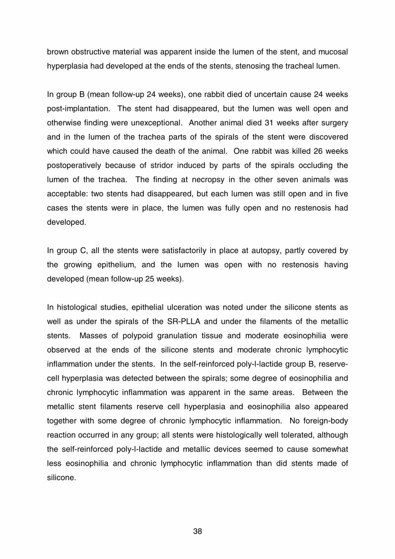
Observations in SEM studies showed a well-preserved epithelial cell layer and an
uninterrupted carpet of ciliated cells between the spirals of the SR-PLLA stents; in
grooves where the spirals had rested the ciliated cells had disappeared. The
findings in the ciliated cell area were comparable to the normal tracheal epithelium
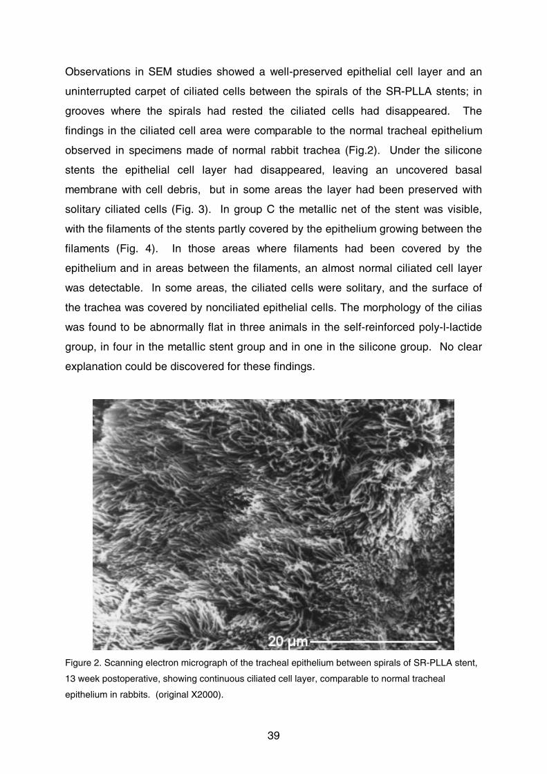
observed in specimens made of normal rabbit trachea (Fig.2). Under the silicone
stents the epithelial cell layer had disappeared, leaving an uncovered basal
membrane with cell debris, but in some areas the layer had been preserved with
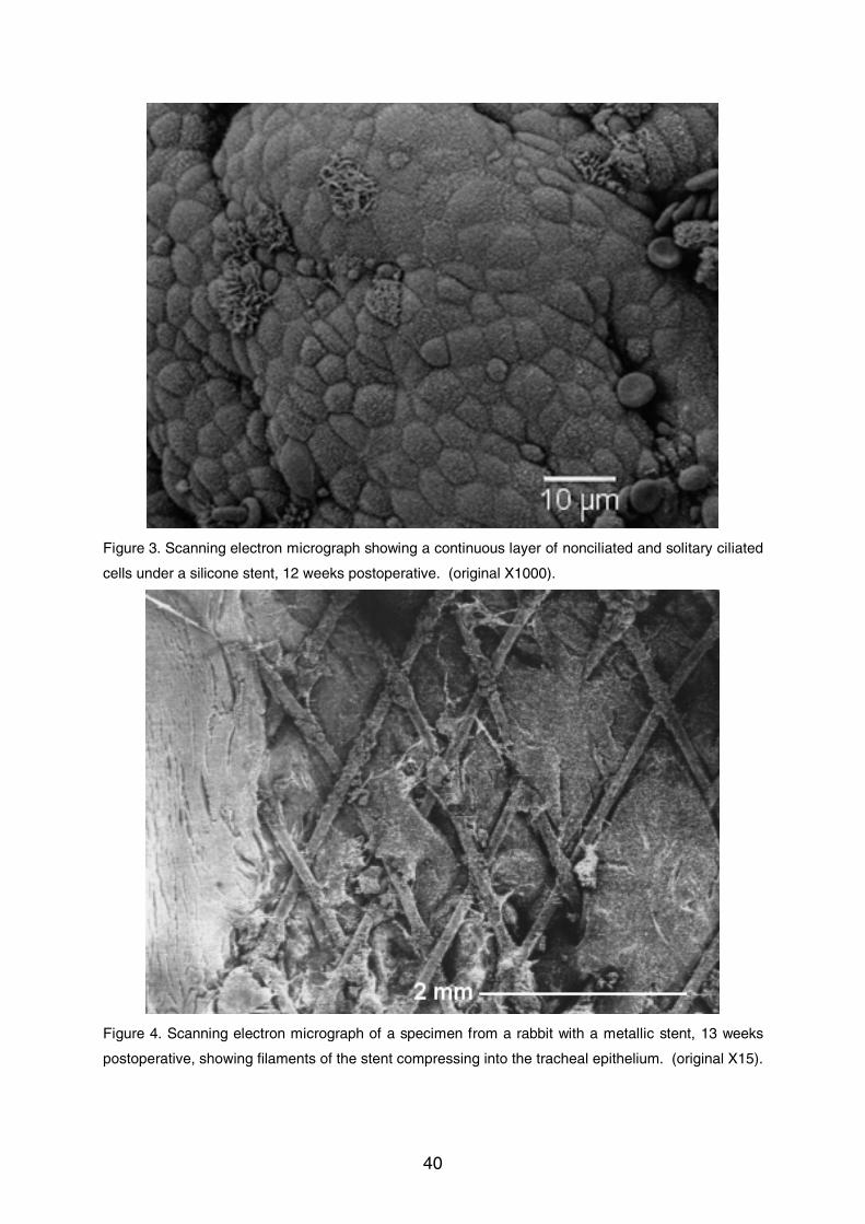
solitary ciliated cells (Fig. 3). In group C the metallic net of the stent was visible,
with the filaments of the stents partly covered by the epithelium growing between the
filaments (Fig. 4). In those areas where filaments had been covered by the
epithelium and in areas between the filaments, an almost normal ciliated cell layer
was detectable. In some areas, the ciliated cells were solitary, and the surface of
the trachea was covered by nonciliated epithelial cells. The morphology of the cilias
was found to be abnormally flat in three animals in the self-reinforced poly-l-lactide
group, in four in the metallic stent group and in one in the silicone group. No clear
explanation could be discovered for these findings.
Figure 2. Scanning electron micrograph of the tracheal epithelium between spirals of SR-PLLA stent,
13 week postoperative, showing continuous ciliated cell layer, comparable to normal tracheal
epithelium in rabbits. (original X2000).
40
Figure 3. Scanning electron micrograph showing a continuous layer of nonciliated and solitary ciliated
cells under a silicone stent, 12 weeks postoperative. (original X1000).
Figure 4. Scanning electron micrograph of a specimen from a rabbit with a metallic stent, 13 weeks
postoperative, showing filaments of the stent compressing into the tracheal epithelium. (original X15).
41
7. DISCUSSION
7.1. The blood supply of the lung
The blood supply of the lung is ensured by pulmonary and bronchial arteries, and
there are differences between these two vascular systems. The bronchial arteries
carry oxygenated arterial blood at systemic blood pressure, while the pulmonary
arteries carry deoxygenated venous blood at low pressure with a much larger blood
volume. Normally the bronchial circulation is less than 1% of cardiac output. The
system takes care of the nutrition and energy supply of the ciliated columnar airway
epithelium and its mucous and serosal glands (Deffebach et al. 1987). The anatomy
of the human bronchial arteries and their anastomoses with pulmonary ateries are
well documented (Cauldwell et al. 1948, Pump 1963, Pump 1972, Schreinemakers et
al. 1990).
7.2. Airway stenosis
Airway stenosis may be a complication of prolonged intubation or tracheostomy or
may develop after bronchial or tracheal resections. Tracheobronchomalacia,
intrabronchial tumors or extrinsic compression can cause obstruction of the airways.
Airway stenosis may result in progressive dyspnea and hypoxemia. Airway
complications following lung transplantation constitute a specific problem due to the
impaired circulation of the donor bronchus resulting from interruption of the vascular
supply to the bronchial tree if the bronchial arterial circulation is not re-connected
during the operation. As early as 1950 the experimental method of restoring the
bronchial arterial circulation with a cuff of the aorta was presented (Metras et al.
1950). On the basis of experimental findings in dogs, it was concluded that all
humans receiving lung allografts should undergo reconstitution of the bronchial
arterial system (Mills et al. 1970).
42
7.3. Laser Doppler flowmetry
LDF has been applied in the measurement of tissue perfusion of different tissues
since it was first used by Stern to measure the peripheral circulation of the skin
(Stern 1975). The value of LDF as a means of measuring bronchial mucosal blood
flow has been documented (Yokomise et al. 1991). The collateral flow in the central
airways with isolated perfusion of the lungs in an animal model has been measured
by LDF, perfusion being about 50% of the normal values at the level of the main
carina (Yokomise et al. 1989). The effect of reimplantation of the bronchial artery on
the bronchial mucosal blood flow has been studied after modified unilateral lung
transplantation in pigs, measurement being made by LDF and radioisotope studies
(Aoki et al. 1991). Reconstruction of the bronchial circulation was found to result in
significant improvement of the graft bronchial blood flow, although normal levels of
bronchial perfusion could not be achieved. This was taken to be due to surgical
trauma and defects in microcirculation within the bronchial arterial bed. In an
experimental study in a left lung allotransplantation model in dogs, Takao and
associates could show that the bronchial mucosal blood flow of donor bronchus
diminishes in accordance with the degree of lung rejection and that it increases to a
value within normal range with the reversal of rejection. The blood flow was
measured by LDF, using L/C ratio, in which L was measured at the second carina of
the transplanted lung and C at the carina. It was concluded that the L/C ratio
appears to be a sensitive and specific index of acute rejection in the transplanted
lung (Takao et al. 1991) . The advantages of LDF are noninvasiveness and
reproductibility. LDF measurements can be safely made in conjunction with other
bronchoscopic studies and the approach gives immediate results.
7.3.1. Limitations of LDF
The values measured by LDF are relative in nature; however, they are comparable
at any given time-point in the same object. The problem of relativity can be reduced
by correlating the measured values to values measured at the same time from
another point in the tissue investigated (Takao et al. 1991). The method is fairly
43
sensitive and can be affected by the contact pressure of the probe or movement of
the airway wall in response to heartbeat or respiration of the subject. Measured
values can also be influenced by other factors, such as hemodynamic changes and
hematocrit which are dependent on the individual animal under investigation.
7.4. Problems in healing of bronchial anastomoses
Ischemia, rejection and infection of the donor bronchus are believed to be important
causes of impaired healing of bronchial anastomoses in clinical lung
transplantation. Complications resulting from defective healing of anastomosis were
the major problems in lung transplantation before cyclosporine was available. The
majority of a cohort of 38 patients who underwent lung transplantation before
January 1981 and survived ten days or more died of complications directly
attributable to problems in the healing of the bronchial anastomosis, including
anastomotic disruption with air leakage, bleeding, mucosal necrosis and stenosis
(Veith et al. 1983).
Bronchial stenosis following lung transplantation is a specific problem, often arising
from scar stenosis at the anastomotic site with or without previous anastomotic
dehiscence. The process of healing of an airway anastomosis can be adversely
affected by rejection, infection, drug therapy and the general condition of the patient
(Morgan et al. 1982). Many operative methods have been adopted to avoid airway
anastomotic complications. Shortening of the transplanted bronchus (Pinsker et al.
1979), use of a telescopic anastomotic technique (Calhoon et al. 1991, Rabinov et
al. 1996), or wrapping of the anastomosis with a vascularised tissue pedicle such as
omentum, intercostal or internal mammary artery pedicle. Randomised clinical trials
have shown, however, that vascular pedicles do not improve the bronchial
anastomotic healing after single lung transplantation (Khaghani et al. 1994).
Increasing experience, meticulous surgical technique, and careful postoperative care
have significantly reduced the incidence of airway complications after pulmonary
transplantation (Date et al. 1995). Anastomotic complications arise in general less
frequently, but the advent of lung transplantation has increased the number of
44
potential candidates for anastomotic stenosis, since airway anastomosis still
constitutes a surgical weak-point (Zannini et al. 1994).
Study l showed the bronchial mucosal blood flow to be reduced by two-thirds when
the bronchial circulation was closed. When both bronchial and pulmonary
circulations were closed, a small amount of perfusion could still be measured, this
possibly to be explained by the sensitivity of the measuring method. The blood
perfusion from the collateral pulmonary circulation is only one-third compared to the
normal perfusion when the bronchial circulation is intact. Besides this, the collateral
blood may not be adequately saturated and blood pressure may be lowered. The
viability of a bronchial anastomosis depends on this collateral blood flow from the
pulmonary circulation during the first days after operation and untill the bronchial
circulation is restored.
Experimental methods have been deviced to study the healing and revascularisation
process in bronchial anastomoses in dogs. The first totally ischemic bronchial
autograft model was reported by Millhiet and collegues (1968). They reported good
autograft healing without any mortality in all 11 of their dogs. On the other hand
Morgan and associates, using the same experimental method reported that all five
dogs in their bronchial autograft group died within five days of their operation
(Morgan et al. 1982).
To establish whether the bronchial autograft model works in pigs study ll was
undertaken. Surprisingly it emerged that a totally ischemic bronchial autograft
healed well both clinically and histologically in all six animals used. This would
indicate that without rejection the healing capacity and the revascularisation process
in the bronchial autograft in this model sufficed for survival of the graft. Rejection, in
addition to total ischemia, led to necrosis of the graft and to clinical complications in
the group of bronchial allografts without immunosuppression. Immosuppressive
medication helped in healing in three out of seven cases in preventig the rejection
process. As to the reason for the still poor healing in four cases, one possible
explanation could be inadequacy of immunosuppression.
45
7.5. Methods of handling airway stenosis
The most common indication for tracheal segmental resection and reconstruction is
still post-intubation tracheal stenosis. The principles of tracheal reconstruction
include avoidance of excessive anastomotic tension, maintenance of tracheal blood
supply and meticulous dissection and anastomosis with absorbable sutures (Grillo et
al. 1995). Whereas postintubation stenoses and some malignant lesions of the
trachea are best treated by resection, most primary tumors are inoperable at the time
of presentation, by reason of invasion of local structures and mediastinal lymph node
metastases (Houston et al. 1969).
Many nonsurgical palliative methods have been developed for cases where surgical
corrections of airway stenosis are not feasible. These include resection of the
stenosis with diathermy, laser, cryosurgery or forceps, dilatation of the stenosis,
brachytherapy and stenting of the stenosed zone of the tracheobronchial tree or
combinations of different methods.
Bronchial stenoses after lung transplantation are best handled by insertion of a
silicone or metallic stent, with the exception of some short stenoses and granulomas
which can be treated with endobronchial laser or cryotherapy (Brichon et al. 1992).
7.6. Stenting of the airways
The era of aiway stenting began in 1965 when Montgomery introduced his T-tube for
treating subclottic stenosis (Montgomery 1965). Since then stents made of silicone
have been used in most series. Subsequently a variety of materials and
constructions have been tried in airway stents, although the perfect stent has not yet
been designed. Some authors have considered stenting of tracheobronchial
stenoses to be the primary management option for airway obstruction after lung
transplantation (Sonett et al. 1995). Colt and Dumon (1995) have listed the
demands of an ideal stent. The device needs to fit snugly into the airway lumen
46
without causing irreversible mucosal injury; it should remain in place without external
fixation. Satisfactory expansion of the stricture must be achieved regardless of its
origin and the forces causing extrinsic compression. Granulation tissue formation
caused by airway inflammation must be minimal. The stent should not damage
adjacent cartilage or vascular structures. It must be easily tailored to fit the entire
stricture, solid enough to resist extrinsic compression, and easy to replace in cases
of obstruction by tumor overgrowth or secretions. Bare metallic stents are effective
in extrinsic compression, but silicone stents or covered metallic stents are preferable
for intraluminal stenoses (Tojo et al. 1996). Insertion of airway stents has been
shown in respiratory functional tests to improve pulmonary function (Vergnon et al.
1995, Eisner et al. 1999).
7.6.1. Silicone stent
Although silicone stents are widely used, they have certain disadvantages which
render them unsuitable in long-term use. Insertion of silicone stents usually requires
rigid bronchoscopy under general anesthesia. Because these stents are relatively
thick, their internal diameter is smaller than the normal airway lumen. They interfere
with normal airway mucociliary function, which can result in accumulation of
secretions inside the stent and obstruction of its lumen. Silicone stents may have a
tendency to stimulate the formation of granulation tissue distal to the stent (Spatenka
et al. 1991), and regular bronchoscopic examinations are therefore recommended to
see whether the stent needs cleaning or replacement (Tsang et al. 1992) Silicone
stents are difficult to place in the right main stem bronchus without obstructing the
orifice of the upper lobe. After insertion they may also have a tendency to migrate
(Higgins at al. 1994, Bolliger et al. 1993). Stents positioned in the trachea usually
need removal before patients can be safely intubated.
Our studies also confirmed that silicon stents have tendency to occlude due to
encrustation material inside the stents and hyperplastic polyps at the ends of the
stent. Also the ciliated cell layer disappeares under the stents. On the other hand,
47
silicon stents are usually clinically well tolerated and can be cleaned and readjusted
after insertion. They are usually also easy to remove and replace if necessary.
7.6.2. Metallic stent
Metallic expandable wire stents have a low internal-to-external diameter ratio. They
offer greater flexibility than silicon tube prostheses, a small surface area in contact
with the tracheobronchial mucosa and minimal risk of occluding bronchial orifices if
misplaced (Simonds et al. 1989). They cannot usually migrate after insertion, and
the mucociliary clearance function is better maintained. Metallic stents can usually
be inserted during flexible bronchoscopy under fluoroscopic control and they are
clearly visible in radiographic examinations. Patients can if necessary be intubated
through the stent placed in the trachea. The disadvantage of the metallic stents is
difficult extraction of the device. Once implanted they can be removed by rigid
bronchoscopy, but extraction may need open surgery.
The usefulness of the expandable metallic device as an airway stent was reported in
1986 by Wallace and collegues (1986). Complications have however been reported
associated with this so-called Gianturco stent (Rousseau et al. 1993, Hind et al.
1992, Nashef et al. 1992). Palmaz stent has been used in experimental studies
(Fraga et al. 1993) and in clinical settings as an airway stent mainly in children
(Filler et al. 1995, Filler et al. 1998, Santoro et al. 1995) with favorable results. More
recently it has also been used in adults, but stent collapse has been found to be a
potential complication of this type of stent (Hautman et al. 1996, Susanto et al.
1998). Metallic stents have been employed to facilitate extubation in patients with
substantial airway obstruction (Shaffer et al. 1998). The Wallstent, developed for
endovascular use, has also been used in the treatment of airway stenosis and is
gaining increasing acceptance (Rousseau et al. 1993, Brichon et al. 1992, Monnier
et al. 1996). A metallic self-expanding stent is possibly the best choice if permanent
stenting of the airways is needed. In study V the metallic stent was well tolerated
and no complications were recorded due to the stent. The filaments of the stent
48
were partly covered with the ciliated epithelium, indicating that mucociliary function
was preserved.
7.6.3. Biodegradable stent
Bioabsorbable materials for suture have been used since 1960s. In orthopedic
surgery devices made of bioabsorbable materials have been in clinical use for over a
decade, with favorable results. A bioabsorbable self-reinforced poly-l-lactide
material has recently been tested in animal studies and showed no acute toxicity,
thus qualifying as biocompatible (Isotalo et al. 1999). The same author presented
promising preliminary results when recurrent urethral strictures were treated with
self-expandable self-reinforced poly-l-lactide spiral stents (Isotalo et al. 1998).
Because the need for stenting of the airways may be only temporary, particularly in
benign and post-transplantation stenosis, an airway stent made of biodegradable
material could offer benefits compared to other materials, in that there is no need for
subsequent extraction.
Our present experimental studies III, IV, and V showed that airway stents made of
SR-PLLA possess good biocompatibility and clinical prospects compared to silicone
and metallic stents. In SEM studies it emerged that ciliated epithelium was
preserved between the stent spirals. This may suggest that the mucociliary function
is already viable before the epithelium has fully covered the stent. At least after
resorption of the whole device, the ciliated epithelium preserves its normal function.
Konrad and associates in a prospective study in intubated patients found, that
impaired mucociliary function was associated with the loss rather than with
ultrastructural abnormalities of the cilia (Konrad et al. 1995). The absorption time
and mechanical properties can be modified and different constructions such as self-
expanding models of biodegradable stents can be made by different processing
methods. Biodegradable stents can even be used as a local drug delivery device.
The present model of the SR-PLLA stent cannot be recommended in malignant
airway stenoses by reason of the possible growth of the tumor between the spirals to
the lumen; such lesions are best handled with silicone or covered metallic stents.
49
8. CONCLUSIONS
1. Bronchial mucosal blood flow as measured by LDF diminishes by two thirds
compared to normal values when the bronchial circulation is closed in an animal
model. After lung transplantation, the donor bronchus has to manage with this
remaining blood perfusion until the bronchial circulation is re-established unless the
bronchial circulation is restored during the operation.
2. The healing capacity of a totally avascular bronchial graft in pigs is excellent
when the graft is autotransplanted, but very poor when it is allotransplanted without
immunosuppressive treatment. Immunosuppressive medication promotes the
healing process of allotransplanted bronchi.
3. A bioabsorbable airway stent made of self-reinforced poly-l-lactide causes no
adverse tissue reactions in the bronchial mucosa when implanted intrabronchially in
pigs. Stents made of SR-PLLA might be suitable for treatment of airway stenosis.
4. An airway stent made of SR-PLLA showed minimal adverse reactions in the
tracheal mucosa in histologic and SEM studies. The biocompatibility of SR-PLLA is
good sufficiently to make it a promising material for an airway stent compared to
silicone stents.
5. In the management of experimental tracheal stenosis in an animal model silicone
stents showed a tendency to become occluded, and those made of metal and of SR-
PLLA were well tolerated. SR-PLLA is a promising material and worth further
investigation as a material for airway stenting.
50
9. SUMMARY
Tracheobronchial stenosis and its management remains problematic in that stenoses
are not always amenable to surgical resection and reconstruction. This entails, in
particular, anastomotic problems and stenoses after lung transplantation. A stenosis
can be handled by stenting, although the ideal stent has yet to be developed; all
available stents have their disadvantages. Since the need for stenting of the airways
may be only temporary, devices made of bioabsorbable materials offer theoretical
benefits.
The aim of this present study was to evaluate bronchial mucosal blood flow when the
bronchial circulation is closed, to investigate the healing of bronchial grafts in an
experimental model in pigs, to examine the biocombatibility of and possible adverse
tissue reactions in the airways to a bioabsorbable bronchial stent made of self-
reinforced poly-l-lactide, and possibilities of managing tracheal stenosis with
different types of stents.
Laser Doppler flowmetry (LDF) was applied to measure bronchial mucosal blood
perfusion when the bronchial circulation is closed in an animal model. LDF studies
showed that the blood perfusion in the bronchial mucosa diminished by one third
when the pulmonary artery was clamped and by two thirds when the bronchial artery
was clamped. The viability of a bronchial anastomosis depends on this collateral
blood flow from the pulmonary circulation during the first days after operation unless
the bronchial circulation is restored at surgery.
Auto-or allotransplantation of a totally avascular bronchial graft was performed on
domestic pigs. Six pigs with autotransplanted bronchi healed eminently satisfactorily
without infection. Eight cases of allotransplanted bronchi without
immunosuppression ended in rejection and all of these animals suffered from major
bronchopleural fistula. Seven pigs with allotransplanted bronchi received triple drug
immunosuppressive medication. Three of them healed well without infection, but the
51
anastomoses were slightly stenosed by the time of sacrifice (mean 30 days). Four
allotransplanted immunosuppressed animals suffered from bronchopleural fistula.
This study with totally avascular bronchial grafts showed that the healing potential of
the totally avascular bronchial graft in pigs is excellent when autotransplanted, but
poor when allotransplanted without immunosuppressive treatment.
Both silicone and metallic internal stents are available for the treatment of bronchial
stenosis. Absorbable stents may offer some benefits compared to others available.
Tissue reactivity has been minimal when absorbable materials have been used in
other organs and when extraction of the stent is not needed, and the airway
preserves its normal function after stent resorption.
Bioabsorbable 15-mm long spiral stents were implanted intrabronchially via standard
left thoracotomy in six pigs (15-26 kg). Macroscopically healing and tissue reactions
around the stents were observed by repeated bronchoscopy. Histological studies
were made at the time of sacrifice after six months’ follow-up. Bioabsorbable and
silicone stents were compared in a study where spiral airway stents made of 0.7-mm
wire of self-reinforced poly-l-lactide were implanted operatively intratracheally in nine
rabbits the silicone stents serving as controls. Tracheal stenosis was created in the
rabbits by extramucosal resection of cartilaginous arches of the cervical trachea.
After a few weeks the animals were reoperated, and those stenoses which had
developed were dilated with balloon dilatation. Stents were then implanted at the
stenosed site to keep the dilated trachea open. All the animals in the group with
silicone stents had to be sacrificed because of respiratory difficulties, their stents
having a tendency to become occluded with encrustation material inside, a
hyperplastic polyp developing at the ends of the stents.
The present studies showed that airway stents made of self-reinforced poly-l-lactide
are biocombatible and cause no adverse tissue reaction. Silicone stents had a
tendency to become stenosed with encrustation material and to develop a
hyperplastic polyp at both ends. Self-reinforced poly-l-lactide stents were well

52
tolerated and caused no foreign-body reaction, and they evinced a tendency to
penetrate to the tracheal wall. Silicone stents were likewise seen to have a
tendency to occlude, while metallic stents and stents made of self-reinforced poly-l-
lctide proved to be well tolerated and could be used in the management of airway
stenosis.
53
10. ACKNOWLEDGEMENTS
This study was carried out at the Department of Thoracic and Cardiovascular
Surgery, the Laboratory of Transplantation Pathology, Helsinki University Central
Hospital, The Institute of Biotechnology, Helsinki, and the Institute of Biomaterials,
Tampere University of Technology, Tampere between 1991 and 1999.
I wish to express my gratitude to Professor Severi Mattila for the possibility of
carrying out this project in his department and for providing me with the facilities of
the Test Animal Laboratory during these years.
Sincere thanks go to my supervisor Professor Ari Harjula, who gave me the topic of
this thesis and whose guidance and encouragement has enabled me to accomplish
the undertaking.
I am greatly indebted to my other supervisor Docent Pertti Aarnio, whose enthusiasm
and guidance have helped me throughout.
I am grateful to Professor Pertti Törmälä for support and ideas during this study.
My sincere thanks are due to Professor Hannu Sariola and Docent Eero Taskinen
for their guidance and analysis of histological samples.
I wish to express special thanks to Docent Risto Mokka for his encouragement and
for providing me with the opportunity to complete this study.
I also wish to thank my co-worker Tuija Ikonen M.D. for her help in bringing the work
to its conclusion.
My special thanks to Antero Sahlman M.D. for the statistical analysis in study l.
54
For co-operation and help with the stents, my special thanks to Tero Välimaa M.Sci.
(Eng.).
Thanks are due to Docent Paula Maasilta and Dosent Timo Savunen for revision
and constructive criticism of this thesis.
My special expression of gratitude is due to Ms Pirkko Uhlbäck, whose assistance in
the Test Animal Laboratory has been of the greatest value.
My sincere thanks to Robert MacGilleon M.A. for his skilful linquistic revision of the
manuscript.
I take this opportunity of acknowledging the support and education of all my previous
colleagues in The Department of Thoracic and Cardiovascular Surgery at the
Helsinki University Central Hospital during these years.
My deepest thanks are due to my parents Kaisa and Mauno for providing a loving
and supportive home.
Finally, I wish to express warmest gratitude to my beloved wife Tarja and our
daughters Emilia and Erika for their love, understanding and support throughout
these years.
This study was financially supported by The Finnish Society of Angiology and the
Recearch Fund of Helsinki University Hospital.
Lahti, May 2000
Antti Korpela
55
11. REFERENCES
Ahn H, Lindhagen J, Nilsson GE, Salerud EG, Jodal M, Lundgren O. Evaluation of laser Doppler
flowmetry in the assessment of intestinal blood flow in cat. Gastroenterology 88:951-7, 1985
Ahn HC, Ekroth R, Nilsson GE, Svedjeholm R, Thelin S. Laser Doppler flowmetry estimating
myocardial perfusion after internal mammary artery grafting. Scand J Thor Cardiovasc Surg 22:281-
4, 1988
Amemiya R, Matsushima Y, Kunii T, Taira O, Oho K, Hayata Y. Palliative tracheal tube stent without
tracheotomy in tracheal stenosis. J Thorac Cardiovasc Surg 90:631-2, 1985
Anderson MB, Kriett JM, Harrell J, Smith C, Kapelanski DP, Tarazi RY, Perricone A, Jamieson SW.
Techniques for bronchial anastomosis. J Heart Lung Transplant 14:1090-4, 1995
Aoki M, Schäfers H-J, Inui K, Klipsch N, Demertzis S, Kotzerke J, Haverich A, Wada H. Bronchial
circulation after experimental lung transplantation: the effect of direct revascularization of a bronchial
artery. Eur J Cardio-thorac Surg 5:561-5, 1991
Auteri JS, Jeevanandam V, Sanches JA, Marboe CC, Kirby TJ, Smith CR. Normal bronchial healing
without bronchial wrapping in canine lung transplantation. Ann Thorac Surg 53:80-4, 1992
Bando K, Paradis IL, Similo S, Konishi H, Komatsu K, Zullo TG, Yousem SA, Close JM, Zeevi A,
Duquesnoy RJ, Manzetti J, Keenan RJ, Armitage JM, Hardesty RL, Griffith BP. Obliterative
bronchiolitis after lung and heart-lung transplantation; an analysis of risk factors and management. J
Thorac Cardiovasc Surg 110:4-14, 1995
Belsey R. Resection and reconstruction of the intrathoracic trachea. Br J Surg 51:200-5, 1951
Bischoff CA, Walden P, Cited by Higgins in: USPat 2,676,945, 1954
Bisson A, Bonnette B, Ben El Cadi N, Leroy M, Colchen A, Personne C, Toty L, Herzog P. Tracheal
sleeve resection for iatrogenic stenoses (subclottic laryngeal and tracheal). J Thorac Cardiovasc Surg
104:882-7, 1992
Bolliger CT, Heitz M, Hauser R, Probst R, Perruchoud AP. An airway Wallstent for the treatment of
tracheobronchial malignancies. Thorax 51:1127-9, 1996

56
Bolliger CT, Probst R, Tschopp K, Soler M, Perruchoud AP. Silicone stents in the management of
inoperable tracheobronchial stenoses. Chest 104:1653-9, 1993
Bolliger CT, Wyser C, Wu X, Hauser R, Studer W, Dalquen P, Perruchoud AP. Evaluation of a new
self-expandable silicone stent in an experimental tracheal stenosis. Chest 115:496-501, 1999
Bolot G, Poupart M, Pignat J-C, Bertocchi M, Wiesendanger T, Thevenet F, Gamondes J-P, Mornex
J-F. Self-expanding metal stents for the management of bronchial stenosis and bronchomalacia after
lung transplantation. Laryngoscope 108:1230-3, 1998
Borel JF, Feurer C, Magnee C, Stahelin H. Effects of the new antilymphocytic peptide cyclosporin A
in animals. Immunology 32:1017-25, 1977
Brichon PY, Blanc-Jouvan F, Rousseau H, Pison C, Pin I, Barnoud D, Dumon JF, Thony F, Dahan M,
Didier A, Noirclerc M, Joffre F. Endovascular stents for bronchial stenosis after lung transplantation.
Transplant proc 24:2656-9, 1992
Calhoon JH, Grover FL, Gibbons WJ, Bryan CL, Levine SM, Bailey SR, Nichols L, Lum C, Trinkle JK.
Single lung transplantation; alternative indications and technique. J Thorac Cardiovasc Surg 101:816-
25, 1991
Carrasco CH, Nesbitt JC, Charnsangavej C, Ryan MB, Walsh GL, Yasumori K, Lawrence DD,
Wallace S. Management of tracheal and bronchial stenoses with the Gianturco stent. Ann Thorac
Surg 58:1012-7, 1994
Carre P, Rousseau H, Lombart L, Didier A, Dahan M, Fournial G, Leophonte P, the Toulouse Lung
Transplantation Group. Balloon dilatation and self-expanding metal Wallstent insertion. Chest
105:343-8, 1994
Cauldwell EW, Siekert RG, Lininger RE, Anson BJ. The Bronchial Arteries; an anatomic study of 150
human cadavers. Surg Gynec Obst 86:395-412, 1948
Cavaliere S, Foccoli P, Farina PL. Nd:YAG laser bronchoscopy: A five-year experience with 1,396
applications in 1,000 patients. Chest 94:15-21, 1988
Cavaliere S, Venuta F, Foccoli P, Toninelli C, La Face B. Endoscopic treatment of malignant airway
obstruction in 2008 patients. Chest 110:1536-42, 1996
Chu CC. Hydrolytic degradation of polyglycolic acid: tensile strength and crystallinity study. J Appl
Polym Sci 26:1727-34, 1981
57
Clarke DB. Palliative intubation of the trachea and main bronchi. J Thorac Cardiovasc Surg 80:736-
41, 1980
Cohen MD, Weber TR, Rao CC. Balloon dilatation of tracheal and bronchial stenosis. AJR 142:477-8,
1984
Colt HG, Dumon J-F. Tracheobronchial stents: indications and applications. Lung cancer 9:301-6,
1993
Colt HG, Dumon J-F. Airway stents. Clin Chest Med 16:465-78, 1995
Colt HG, Janssen JP, Dumon J-F, Noirclerc MJ. Endoscopic management of bronchial stenosis after
douple lung transplantation. Chest 102:10-6, 1992
Cooper JD, Pearson FG, Patterson GA, Todd TRJ, Ginsberg RJ, Goldberg M, Waters P. Use of
silicone stents in the management of airway problems. Ann Thorac Surg 47:371-8, 1989
Couraud L, Baudet E, Martigne C, Roques X, Velly J-F, Laborde N, Dubrez J, Clerc F, Dromer C,
Vallieres E, The Bordeaux Lung And Heart-Lung Transplant Group. Bronchial revascularization in
douple-lung transplantation: A series of 8 patients. Ann Thorac Surg 53:88-94, 1992
Couraud L, Baudet E, Nashef SAM, Martigne C, Roques X, Velly J-F, Laborde N, Dubrez J, Clerc F.
Lung transplantation with bronchial revascularisation. Eur J Cardio-thorac Surg 6:490-5, 1992
Cutright DE, Perez B, Beasley JD, Larson WJ, Posey WR. Degradation rates of polymers and
copolymers of polylactic and polyglycolic acids. Oral Surg 37:142-52, 1974
Dasgupta A, Dolmatch BL, Abi-Saleh WJ, Mathur PN, Mehta AC. Self-expandable metallic airway
stent insertion employing flexible bronchoscopy; preliminary results. Chest 114:106-9, 1998
Date H, Trulock EP, Arcidi JM, Sundaresan S, Cooper JD, Patterson GA. Improved airway healing
after lung transplantation; an analysis of 348 bronchial anastomoses. J Thorac Cardiovasc Surg
110:1424-33, 1995
Davreux CJ, Chu NH, Waddell TK, Mayer E, Patterson A. Improved tracheal allograft viability in
immunosuppressed rats. Ann Thorac Surg 55:131-4, 1993
Deffebach MK, Charan NB, Lakshminaryan S, Butler J. The bronchial circulation: small but a vital
attribute of the lung. Am Rev Respir Dis 135:463-81, 1987
58
Dumon J-F. A dedicated tracheobronchial stent. Chest 97:328-32, 1990
Eisner MD, Gordon RL, Webb WR, Gold WM, Hilal SE, Edinburgh K, Golden JA. Pulmonary function
improves after expandable metal stent placement for benign airway obstruction. Chest 115:1006-11,
1999
Fell SC, Mollenkopf FP, Montefusco CM, Mitsudo S, Kamholz SL, Goldsmith J, Veith FJ.
Revascularization of ischemic bronchial anastomoses by an intercostal pedicle flap. J Thorac
Cardiovasc Surg 90:172-8, 1985
Filler RM, Forte V, Chait P. Tracheobronchial stenting for the treatment of airway obstruction. J
Pediatr Surg 33:304-11, 1998
Filler RM, Forte V, Fraga JC, Matute J. The use of expandable metallic airway stents for
tracheobronchial obstruction in children. J Pediatr Surg 30:1050-6, 1995
Fraga JC, Filler RM, Forte V, Bahoric A, Smith C. Experimental trial of balloon-expandable, metallic
Palmaz stent in the trachea. Arch Otolaryngol Head Neck Surg 119:522-8, 1993
Freitag L, Eicher R, Linz B, Greschuchna D. Theoretical and experimental basis for the development
of a dynamic airway stent. Eur Respir J 7:2038-45, 1994
Freitag L, Tekolf E, Stamatis G, Greschuchna D. Clinical evaluation of a new bifurcated dynamic
airway stent: a 5-year experience with 135 patients. Thorac Cardiovasc Surgeon 45:6-12, 1997
Gaer JAR, Tsang V, Khaghani A, Gillbe CE, Townsend ER, Fountain SW, Yacoub MH. Use of
endotracheal silicone stents for relief of tracheobronchial obstruction. Ann Thorac Surg 54:512-6,
1992
Gaissert HA, Grillo HC, Mathisen DJ, Wain JC. Temporary and permanent restoration of airway
continuity with the tracheal T-tube. J Thorac Cardiovasc Surg 107:600-6, 1994
George PJM, Irving JD, Mantell BS, Rudd RM. Covered expandable metal stent for recurrent tracheal
obstruction. Lancet 335:582-4, 1990
Gillams A, Dick R, Dooley JS, Wallsten H, El-Din A. Self-expandable stainless steel braided
endoprosthesis for biliary strictures. Radiology 174:137-40, 1990
59
Goldberg M, Lima O, Morgan E, Ayabe HA, Luk S, Ferdman A, Peters WJ, Cooper JD. A comparison
between cyclosporin A and methylprednisolone plus azathioprine on bronchial healing following
canine lung autotransplantation. J Thorac Cardiovasc Surg 85:821-6, 1983
Griffith BP, Hardesty RL, Armitage JM. A decade of lung transplantation. Ann Surg 218:310-20, 1993
Grillo HC, Donahue DM, Mathisen DJ, Wain JC, Wright CD. Postintubation tracheal stenosis;
treatment and results. J Thorac Cardiovasc Surg 109:486-93, 1995
Hardy JD, Webb WR, Dalton Jr ML, Walker Jr GR. Lung homotransplantation in man. JAMA
186:1065-74, 1963
Har-El G, Shaha A, Chaudry R, Lucente FE. Resection of Tracheal Stenosis with end-to-end
anastomosis. Ann Otol Rhinol Laryngol 102:670-4, 1993
Harkins WB. An endotracheal metallic prosthesis in the treatment of stenosis of the upper trachea.
Ann Otol Rhinol Laryngol 61:663-75, 1952
Hauck RW, Lembeck RM, Emslander HP, Schömig A. Implantation of Accuflex and Strecker stents in
malignant bronchial stenosis by flexible bronchoscopy. Chest 112:134-44, 1997
Hautmann H, Huber RM. Stent flexibility: an essential feature in the treatment of dynamic airway
collapse. Eur Respir J 9:609-11, 1996
Haydock DA, Trulock EP, Kaiser LR, Ettinger NA, Triantifillou AN, Ochoa LL, Pasque MK, Knight SR,
Cooper JD, The Washington University Lung Transplant Group. Lung transplantation; Analysis of
thirty-six consecutive procedures performed over a twelve-month period. J Thorac Cardiovasc Surg
103:329-40, 1992
Hetzel MR, Nixon C, Edmondstone WM, Mitchell DM, Millard FJC, Nanson EM, Woodcock AA,
Bridges CE, Humberstone AM. Laser therapy in 100 tracheobronchial tumours. Thorax 40:341-5,
1985
Hetzel MR, Smith SGT. Endoscopic palliation of tracheobronchial malignancies. Thorax 46:325-33,
1991
Higgins R, McNeil K, Dennis C, Parry A, Large S, Nashef SAM, Wells FC, Flower C, Wallwork J.
Airway stenoses after lung transplantation: management with expanding metal stents. J Heart Lung
Transplant 13:774-8, 1994

60
Hind CRK, Donnelly RJ. Expandable metal stents for tracheal obstruction: permanent or temporary?
A cautionary tale. Thorax 47:757-8, 1992
Hollinger JH, Battistone GC. Biodegradable bone repair materials; synthetic polymers and ceramics.
Clin Orthop 207:290-305, 1986
Houston HE, Payne WS, Harrison EGJ, Olsen AM. Primary cancers of the trachea. Arch Surg 99:132-
9, 1969
Hoyos de AL, Patterson GA, Maurer JR, Ramirez JC, Miller JD, Winton TL, The Toronto lung
transplant group. Pulmonary transplantation; early and late results. J Thorac Cardiovasc Surg
103:295-306, 1992
Hramiec JE, Haasler GB. Tracheal wire stent complications in malacia: implications of position and
design. Ann Thorac Surg 63:209-12, 1997
Hyytinen TA, Halme M. Laser doppler flowmetry detects early risk of tracheal anastomotic
complications after lung transplantation. Scand Cardiovasc J :, 1999
Inui K, Schäfers H-J, Aoki M, Becker V, Ongsiek B, Kemnitz J, Haverich A, Borst HG. Bronchial
circulation after experimental lung transplantation; the effect of long-term administration of
prednisolone. J Thorac Cardiovasc Surg 105:474-9, 1993
Inui K, Wada H, Yokomise H, Lee M, Yamazaki F, Aoki M, Hitomi S. Evaluation of a bronchial
anastomosis by laser Doppler velocimetry. J Thorac Cardiovasc Surg 99:614-9, 1990
Isotalo T, Halasz A, Talja M, Tammela TLJ, Paasimaa S, Törmälä P. Tissue biocompatibility of a new
caprolactone coated self-reinforced self-expandable poly-l-lactic acid (SR-PLLA) bioabsorbable
urethral stent. J Endourol in Press, 1999
Isotalo T, Tammela TLJ, Talja M, Välimaa T, Törmälä P. A bioabsorbable self-expandable, self-
reinforced poly-l-lactic acid urethral stent for recurrent urethral strictures: A preliminary report. J Urol
160:2033-6, 1998
Jackson C. High tracheotomy and other errors the chief causes of chronic laryngeal stenosis. Surg
Gynec Obst 32:392-8, 1921
Johnston MR, Loeber N, Hillyer P, Stephenson LW, Edmunds Jr LH. External stent for repair of
secondary tracheomalacia. Ann Thorac Surg 30:291-6, 1980
61
Kaiser LR, Pasque MK, Trulock EP, Low DE, Dresler CM, Cooper JD. Bilateral sequential lung
transplantation: The procedure of choice for douple-lung replacement. Ann Thorac Surg 52:438-46,
1991
Kemppainen E, Talja M, Riihelä M, Pohjonen T, Törmälä P, Alftan O. A bioresorbable urethral stent.
Urol Res 21:235-8, 1993
Khaghani A, Tadjkarimi S, Al-Kattan K, Banner N, Daly R, Theodoropoulos S, Madden B, Yacoub M.
Wrapping the anastomosis with omentum or an internal mammary artery pedicle does not improve
bronchial healing after single lung transplantation: Results of a randomized clinical trial. J Heart Lung
Transplant 13:767-73, 1994
Kiriluk LB, Merendino KA. An experimental evaluation of bronchial anastomosis and healing with
special consideration of the plane of transection. Surg Gynec Obst 96:175-82, 1953
Konrad F, Schiener R, Marx T, Georgieff M. Ultrastructure and mucociliary transport of bronchial
respiratory epithelium in intubated patients. Intensive Care Med 21:482-9, 1995
Kshettry VR, Kroshus TJ, Hertz MI, Hunter DW, Shumway SJ, Bolman RM lll. Early and late airway
complications after lung transplantation: incidence and management. Ann Thorac Surg 63:1576-83,
1997
Kulkarni RK, Pani KC, Neuman C, Leonard F. Polylactic acid for surgical implants. Arch Surg 93:839-
43, 1966
Laks H, Louie HW, Haas GS, Drinkwater DC, Lewis W, Permut LC, Stein DG, Chang P. New
technique of vascularization of the trachea and bronchus for lung transplantation. J Heart Lung
Transplant 10:280-7, 1991
Legal YM, Chittal SM, Wright ES. Early bronchial revascularization with an intercostal pedicle graft
following canine lung autotransplantation. Can J Surg 28:518-22, 1985
Leval de MR, Smyth R, Whitehead B, Scott JP, Elliott MJ, Sharples L, Caine N, Helms P, Martin IR,
Higenbottam T, Wallwork J. Heart and lung transplantation for terminal cystic fibrosis. J Thorac
Cardiovasc Surg 101:633-42, 1991
Lima O, Cooper JD, Peters WJ, Ayabe H, Townsend E, Luk SC, Goldbrg M. Effects of
methylprednisolone and azathioprine on bronchial healing following lung autotransplantation. J
Thorac Cardiovasc Surg 82:211-5, 1981

62
Lima O, Goldberg M, Peters WJ, Ayabe H, Townsend E, Cooper JD. Bronchial omentopexy in canine
lung transplantation. J Thorac Cardiovasc Surg 83:418-21, 1982
LoCicero lll J, Massad M, Oba J, Bresticker M, Greene R. Short-term and long-term results of
experimental wrapping techniques for bronchial anastomosis. J Thorac Cardiovasc Surg 103:763-6,
1992
Mair EA, Parsons DS, Lally KP. Treatment of severe bronchomalacia with expanding endobronchial
stents. Arch Otolaryngol Head Neck Surg 116:1087-90, 1990
Mair EA, Parsons DS, Lally KP, Van Dellen AF. Comparison of expandable endotracheal stents in the
treatment of surgically induced piglet tracheomalacia. Laryngoscope 101:1002-8, 1991
Maiwand MO, Zehr KJ, Dyke CM, Peralta M, Tadjkarimi S, Khagani A, Yacoub MH. The role of
cryotherapy for airway complicatoions after lung and heart-lung transplantation. Eur J Cardio-thorac
Surg 12:549-54, 1997
Majola A, Vainionpää S, Vihtonen K, Mero M, Vasenius J, Törmälä P, Rokkanen P. Absorption,
biocombatibility, and fixation properties of polylactic acid in bone tissue: An experimental study in
rats. Clin Orthop 268:260-9, 1991
Marasso A, Gallo E, Massaglia GM, Onoscuri M, Bernardi V. Cryosurgery in bronchoscopic treatment
of tracheobronchial stenosis; indications, limits, personal experience. Chest 103:472-4, 1993
Marinelli WA, Hertz MI, Shumway SJ, Fox JMK, Henke CA, Harmon KR, Savik K, Bolman RM lll.
Single lung transplantation for severe emphysema. J Heart Lung Transplant 11:577-83, 1992
Marquette CH, Mensier E, Copin M-C, Desmidt A, Freitag L, Witt C, Petyt L, Ramon P. Experimental
models of tracheobronchial stenoses: A useful tool for evaluating airway stents. Ann Thorac Surg
60:651-6, 1995
Martinez-Ballarin JI, Diaz-Jimenez JP, Castro MJ, Moya JA. Silicone stents in the management of
benign tracheobronchial stenoses; tolerance and early results in 63 patients. Chest 109:626-9, 1996
Mathisen DJ, Grillo HC. Endoscopic relief of malignant airway obstruction. Ann Thorac Surg 48:469-
75, 1989
Mathur PN, Wolf KM, Busk MF, Briete WM, Datzman M. Fiberoptic bronchoscopic cryotherapy in the
management of tracheobronchial obstruction. Chest 110:718-23, 1996
63
McCarthy PM, Starnes VA, Theodore J, Stinson EB, Oyer PE, Shumway NE. Improved survival after
heart-lung transplantation. J Thorac Cardiovasc Surg 99:54-60, 1990
Mehta AC, Dasgupta A. Airway stents. Clin Chest Med 20:139-51, 1999
Mehta AC, Harris RJ, De Boer GE. Endoscopic management of benign airway stenosis. Clin Chest
Med 16:401-13, 1995
Metras H. Note preliminaire sur la greffe totale du poumon chez la chien. Proc Acad Sci 231:1176-
1177, 1950, (Translation from French: J Heart Lung Transplant 6:1213-1216, 1992)
Milhiet H, Renault P, Drutel P, Chartier P, Jager P, Timsit G. Contribution a la greffe pulmonaire
experimentale explication de la revascularisation bronchique du greffon par l'etude d'auto-greffons
bronchiques libres. Ann Chir Thor Car 7:1195-201, 1968
Mills NL, Boyd AD, Gheranpong C. The significance of bronchial circulation in lung transplantation. J
Thorac Cardiovasc Surg 60:866-74, 1970
Milroy EJG, Chapple CR, Eldin A, Wallsten H. A new stent for the treatment of urethral strictures;
preliminary report. Br J Urol 63:392-6, 1989
Milroy EJG, Cooper JE, Wallsten H, Chapple CR, Eldin A, Seddon AM, Rowles PM. A new treatment
for urethral strictures. Lancet :1424-7, 1988
Monnier P, Mudry A, Stanzel F, Haeussinger K, Heitz M, Probst R, Bolliger CT. The use of the
covered Wallstent for the palliative treatment of inoperable tracheobronchial cancers; a prospective,
multicenter study. Chest 110:1161-8, 1996
Montgomery WW. T-tube tracheal stent. Arch Otolaryng 82:320-1, 1965
Montgomery WW. Silicone tracheal T-tube. Ann Otol Rhinol Laryngol 83:71-5, 1974
Morgan E, Lima O, Goldberg M, Ferdman A, Luk SK, Cooper JD. Succesful revascularization of
totally ischemic bronchial autografts with omental pedicle flaps in dogs. J Thorac Cardiovasc Surg
84:204-10, 1982
Nakamura T, Hitomi S, Watanabe S, Shimizu Y, Jamshidi K, Hyon SH, Ikada Y. Bioabsorption of
polylactides with different molecular properties. J Biomed Mater Res 23:1115-30, 1989
64
Nashef SA, Dromer C, Velly J-F, Labrousse L, Couraud L. Expanding wire stents in benign
tracheobronchial disease: indications and complications. Ann Thorac Surg 54:937-40, 1992
N∅rgaard MA, Olsen PS, Svendsen UG, Pettersson G. Revascularizationof the bronchial arteries in
lung transplantation: An overview. Ann Thorac Surg 62:1215-21, 1996
Novick RJ, Menkis AH, McKenzie FN, Reid KR, Pflugfelder PW, Kostuk WJ, Ahmad D. The safety of
low-dose prednisone before and immediately after heart-lung transplantation. Ann Thorac Surg
51:642-5, 1991
Orlowski TM. Palliative intubation of the tracheobronchial tree. J Thorac Cardiovasc Surg 94:343-8,
1987
Orlowski TM. The management of tracheal obstruction. Rocz Akad Med 42 suppl 1:318-39, 1997
Pagliero KM, Shepherd MP. Use of stainless steel wire coil prosthesis in treatment of anastomotic
dehiscence after cervical tracheal resection. J Thorac Cardiovasc Surg 67:932-5, 1974
Palmaz JC, Sibbitt RR, Reuter SR, Tio FO, Rice WJ. Expandable intraluminal graft: A preliminary
study. Radiology 156:73-7, 1985
Patterson GA. Indications;Unilateral, bilateral, heart-lung, and lobar transplant procedures. Clin Chest
Med 18:225-30, 1997
Patterson GA, Todd TR, Cooper JD, Pearson FG, Winton TL, Maurer J, The Toronto Lung Transplant
Group. Airway complications after douple lung transplantation. J Thorac Cardiovasc Surg 99:14-21,
1990
Pettersson G, Arendrup H, Mortensen SA, Aldershvile J, Thiis JJ, Aggestrup S, Svendsen UG, Efsen
F. Early experience of douple-lung transplantation with bronchial artery revascularization using
mammary artery. Eur J Cardiothorac Surg 8:520-4, 1994
Pinsker KL, Koerner SK, Kamholz SL, Hagstrom JWC, Veith FJ. Effect of donor bronchial length on
healing; a canine model to evaluate bronchial anastomotic problems in lung transplantation. J Thorac
Cardiovasc Surg 77:669-73, 1979
Pump KK. The bronchial arteries and their anastomoses in the human lung. Dis Chest 43:245-55,
1963
Pump KK. Distribution of Bronchial Arteries in the Human Lung. Chest 62:447-51, 1972
65
Rabinov M, Esmore DS, Snell GI, Salomonsen RF, Griffiths A, Williams T. Reverse telescope
anastomotic technique reduces the incidence of bronchial stricture. J Heart Lung Transplant 15:243-8,
1996
Rabinovich JJ. Re-establishment of bronchial arteries after experimental lung lobe
autotransplantation. J Thorac Cardiovasc Surg 64:119-26, 1972
Ramirez JC, Patterson GA, Winton TL, De Hoyos AL, Miller JD, Maurer JR, The Toronto Lung
Trasnsplant Group. Bilateral lug transplantation for cystic fibrosis. J Thorac Cardiovasc Surg 103:287-
94, 1992
Rokkanen P, Böstman O, Vainionpää S, Vihtonen K, Törmälä P, Laiho J, Kilpikari J, Tamminmäki M.
Biodegradable implants in fracture fixation: Early results of treatment of fractures of the ankle.
Lancet:1422-4, 1985
Rousseau H, Dahan M, Lauque D, Carre P, Didier A, Bilbao I, Herrero J, Blancjouvant F, Joffre F.
Self-expandable prostheses in the tracheobronchial tree. Radiology 188:199-203, 1993
Santoro G, Picardo S, Testa G, Formigari R, Marianeschi S, Catena G, Ballerini L. Balloon-
expandable metallic stents in the management of tracheomalacia in neonates. J Thorac Cardiovasc
Surg 110:1145-8, 1995
Schäfers H-J, Hamm M, Wagner TOF. Gianturco self-expanding metallic stents. Eur J Cardio-thorac
Surg 6:278, 1992
Schafers HJ, Haydock DA, Cooper JD. The prevalence and management of bronchial anastomotic
complications in lung transplantation. J Thorac Cardiovasc Surg 101:1044-52, 1991
Schmitt EE, Polistina RA. Surgical sutures. US Pat 3,297,033, 1967
Schneider AK. Polymers of high melting lactide. US Pat 2,703,316, 1955
Schreinemakers HHJ, Weder W, Miyoshi S, Harper BD, Shimokawa S, Egan TM, McKnight R,
Cooper JD. Direct revascularization of bronchial arteries for lung transplantation: An anatomical
study. Ann Thorac Surg 49:44-54, 1990
Shaffer JP, Allen JN. The use of expandable metal stents to facilitate extubation in patients with large
airway obstruction. Chest 114:1378-82, 1998
66
Shennib H, Massard G. Airway complications in lung transplantation. Ann Thorac Surg 57:506-11,
1994
Shennib H, Noirclerc M, Ernst P, Metras D, Mulder DS, Giudicelli R, Lebel F, Dumon J-F, and the
cystic fibrosis transplant study group. Douple-lung transplantation for cystic fibrosis. Ann Thorac Surg
54:27-32, 1992
Sheski FD, Mathur PN. Long-term results of fiberoptic bronchoscopic balloon dilatation in the
management of benign tracheobronchial stenosis. Chest 114:796-800, 1998
Shiraishi T, Kawahara K, Shirakusa T, Inada K, Okabayashi K, Iwasaki A. Stenting for airway
obstruction in the carinal region. Ann Thorac Surg 66:1925-9, 1998
Siegelman SS, Hagstrom WC, Koerner SK, Veith FJ. Restoration of bronchial artery circulation after
canine lung allotransplantation. J Thorac Cardiovasc Surg 73:792-5, 1977
Sigwart U, Puel J, Mirkovitch V, Joffre F, Kappenberger L. Intravascular stents to prevent occlusion
and restenosis after transluminal angioplasty. N Engl J Med 316:701-6, 1987
Simonds AK, Irving JD, Clarke SW, Dick R. Use of expandable metal stents in the treatment of
bronchial obstruction. Thorax 44:680-1, 1989
Slonim SM, Razavi M, Kee S, Semba CP, Dake MD. Transbronchial palmaz stent placement for
tracheo-bronchial stenosis. JVIR 9:153-60, 1998
Sonett JR, Keenan RJ, Ferson PF, Griffith BP, Landreneau RJ. Endobronchial management of
benign, malignant, and lung transplantation airway stenoses. Ann Thorac Surg 59:1417-22, 1995
Souza de AC, Keal R, Hudson N, Leverment JN, Spyt TJ. Use of expandable wire stents for
malignant airway obstruction. Ann Thorac Surg 57:1573-8, 1994
Spatenka J, Khaghani A, Irving JD, Theodoropoulos S, Slavik Z, Yacoub MH. Gianturco self-
expanding metallic stents in treatment of tracheobronchial stenosis after single lung and heart and
lung transplantation. Eur J Cardio-thorac Surg 5:648-52, 1991
Stern MD. In vivo evaluation of microcirculation by coherent light scattering. Nature 254:56-8, 1975
Stern MD, Bowen PD, Parma R, Osgood RW, Bowman RL, Stein JH. Measurement of renal cortical
and medullary blood flow by laser-Doppler spectroscopy in the rat. Am J Physiol 236:F80-7, 1979
67
Susanto I, Peters JI, Levine SM, Sako EY, Anzueto A, Bryan CL. Use of balloon-expandable metallic
stents in the management of bronchial stenosis and bronchomalacia after lung transplantation. Chest
114:1330-5, 1998
Takao M, Katayama Y, Onoda K, Tanabe H, Hairaiwa T, Mizutani T, Yada I, Namikawa S, Yuasa H,
Kusagawa M. Significance of measurements of bronchial mucosal blood flow for the monitoring of
acute rejection of transplanted lungs. Transplantation 50:345-8, 1990
Takao M, Katayama Y, Onoda K, Tanabe H, Hiraiwa T, Mizutani T, Yada I, Namikawa S, Yuasa H,
Kusagawa M. Significance of bronchial mucosal blood flow for the monitoring of acute rejection in
lung transplantation. J Heart Lung Transplant 10:956-67, 1991
Takao M, Katayama Y, Tanabe H, Hiraiwa T, Mizutani T, Yada I, Namikawa S, Yuasa H, Kusagava
M. Histologic changes in donor bronchi may explain the reduced mucosal blood flow seen during
acute lung allograft rejection. J Heart Lung Transplant 11:994-1000, 1992
Talja M, Tammela T, Petas A, Välimaa T, Taari K, Viherkoski E, Törmälä P. Biodegradable self-
reinforced polyglycolic acid spiral stent in prevention of postoperative urinary retention after visual
laser ablation of the prostate-laser prostatectomy. J Urol 154:2089-92, 1995
Talja M, Välimaa T, Tammela T, Petas A, Törmälä P. Bioabsorbable and biodegradable stents in
urology. J Endourol 11:391-7, 1997
Tanabe H, Takao M, Hiraiwa T, Mizutani T, Yada I, Namikawa S, Yuasa H, Kusagawa M. New
diagnostic method for pulmonary allograft rejection by measurement of bronchial mucosal blood flow.
J Heart Lung Transplant 10:968-74, 1991
Törmälä P. biodegradable self-reinforced composite materials; manufacturing structure and
mechanical properties. Clin Mater 10:29-34, 1992
Törmälä P, Rokkanen P, Laiho J, Tamminmäki M, Vainionpää S. Material for ostheosynthetic
devices. US Pat 4,743,257, 1988
Tojo T, Iioka S, Kitamura S, Maeda M, Otsuji H, Uchida H, Mori T, Furuse K. Management of
malignant tracheobronchial stenosis with metal stents and Dumon stents. Ann Thorac Surg 61:1074-
8, 1996
Toronto Lung Transplant Group. Unilateral lung transplantation for pulmonary fibrosis. N Engl J Med
314:1140-5, 1986

68
Tsang V, Goldstraw P. Endobronchial stenting for anastomotic stenosis after sleeve resection. Ann
Thorac Surg 48:568-71, 1989
Tsang V, Williams AM, Goldstraw P. Sequential silastic and expandable metal stenting for
tracheobronchial strictures. Ann Thorac Surg 53:856-60, 1992
Tsugawa C, Nishijima E, Muraji T, Yoshimura M, Tsubota N, Asano H. A shape memory airway stent
for tracheobronchomalacia in children: an experimental and clinical study. J Pediatr Surg 32:50-3,
1997
Turrentine MW, Kesler KA, Wright CD, McEven KE, Faught PR, Miller ME, Mahomed Y, King H,
Brown JW. Effect of omental, intercostal, and internal mammary artery pedicle wraps on bronchial
healing. Ann Thorac Surg 49:574-9, 1990
Unger M. Tracheobronchial stents and stunts. Chest 97:260-1, 1990
Vainionpää S, Rokkanen P, Törmälä P. Surgical applications of biodegradable polymers in human
tissue. Prog Polym Sci 14:679-716, 1989
Varela A, Maynar M, Irving D, Dick R, Reyes R, Rousseau H, Lopez L, Pulido-Duque JM, Letourneau
JG, Castaneda-Zuniga WR. Use of Gianturco self-expandable stents in the tracheobronchial tree.
Ann Thorac Surg 49:806-9, 1990
Veith FJ, Kamholz SL, Mollenkopf FP, Montefusco CM. Lung transplantation 1983. Transplantation
35:271-8, 1983
Vergnon JM, Costes F, Bayon MC, Emonot A. Efficacy of tracheal and bronchial stent placement on
respiratory functional tests. Chest 107:741-6, 1995
Vinograd I, Klin B, Brosh T, Weinberg M, Flomenblit Y, Nevo Z. A new intratracheal stent made from
nitinol, an alloy with "shape memory effect". J Thorac Cardiovasc Surg 107:1255-61, 1994
Välimaa T. Polylaktidikuidun valmistus ekstruusiopolymeroinnilla. Diplomityö Tampere University of
Technology, Tampere, 1993
Wallace MJ, Charnsangavej C, Ogawa K, Carrasco CH, Wright KC, McKenna R, McMurtrey M,
Gianturco C. Tracheobronchial tree: Expandable metallic stents used in experimental and clinical
applications. Radiology 158:309-12, 1986
69
Wanamaker JR, Eliachar I. An overview of treatment options for lower airway obstruction. Otolaryngol
Clin North Am 28:751-70, 1995
Wassermann K, Koch A, Muller-Ehmsen J, Reuter M, Michel O, Eckel H-E. Clinical and laboratory
evaluation of a new thin-walled self-expanding tracheobronchial silicone stent: Progress and pitfalls.
J Thorac Cardiovasc Surg 114:527-34, 1997
Westaby S, Jackson JW. A bifurcated silicone rubber stent for relief of tracheobronchial obstruction. J
Thorac Cardiovasc Surg 83:414-7, 1982
Williams DF. Biodegradation of surgical polymers. J Mater Sci 17:1233-46, 1982
Wilson GE, Walshaw MJ, Hind CRK. Treatment of large airway obstruction in lung cancer using
expandable metal stents inserted under direct vision via the fibreoptic bronchoscope. Thorax 51:248-
52, 1996
Wilson IC, Hasan A, Healey M, Villaquiran J, Corris PA, Forty J, Hilton CJ, Dark JH. Healing of the
bronchus in pulmonary transplantation. Eur J Cardio-thorac Surg 10:521-7, 1996
Wright KC, Wallace S, Charnsangavej C, Carrasco CH, Gianturco C. Percutaneous endovascular
stents: An experimental evaluation. Radiology 156:69-72, 1985
Yokomise H, Cardoso PFG, Kato H, Keshavjee SH, Wada H, Slutsky AS, Patterson GA. The effect of
pulmonary arterial flow and positive end-expiratory pressure on retrograde bronchial mucosal blood
flow. J Thorac Cardiovasc Surg 101:201-8, 1991
Yokomise H, Wada H, Inui K, Yamazaki F, Lee M, Aoki M, Hitomi S. Application of laser Doppler
velocimetry to lung transplantation. Transplantation 48:550-4, 1989
Zannini P, Melloni G, Chiesa G, Carretta A. Self-expanding stents in the treatment oftracheobronchial obstruction. Chest 106:86-90, 1994