Embed Size (px)
Citation preview
Revision 6.001 March 2016
AustralasianHealth Facility Guidelines
Part B - Health Facility Briefing and Planning0600 - Radiation Oncology Unit
Australasian Health Facility Guidelines
Part B - Health Facility Briefing and Planning Page 20600 - Radiation Oncology Unit, Revision 6.0, 01 March 2016
COPYRIGHT AND DISCLAIMER
Copyright
© 2015 Australasian Health Infrastructure Alliance The Australasian Health Facility Guidelines (AusHFG) and the information in them are the copyright of theAustralasian Health Infrastructure Alliance (AHIA). The information in the AusHFG is made freely available. Australasian Health Facility Guidelines Address: PO Box 1060, North Sydney NSW 2059Website: http://www.healthfacilityguidelines.com.auEmail: [email protected] The AusHFGs are an initiative of the Australasian Health Infrastructure Alliance (AHIA). AHIA membershipis comprised of representatives from government health infrastructure planning and delivery entities in alljurisdictions in Australia and New Zealand.
Disclaimer
AHIA gives no warranty or guarantee that the information in the AusHFG is correct, complete or otherwisesuitable for use. AHIA shall not be liable for any loss howsoever caused whether due to negligence orotherwise arising from the use of or reliance on this information. AHIA recommends that those seeking to rely on the information in the AusHFG obtain their own independentexpert advice.
Australasian Health Facility Guidelines
Part B - Health Facility Briefing and Planning Page 30600 - Radiation Oncology Unit, Revision 6.0, 01 March 2016
Index
01 INTRODUCTION 401.01 Preamble 401.02 Introduction 401.03 Policy Framework 501.04 Description 5
02 PLANNING 802.01 Operational Models 802.02 Operational Policies 802.03 Planning Models 1002.04 Functional Areas 1102.05 Functional Relationships 14
03 DESIGN 1503.01 Accessibility 1503.02 Parking 1503.03 Disaster Planning 1503.04 Infection Control 1603.05 Environmental Considerations 1603.06 Space Standards and Components 1703.07 Safety and Security 1703.08 Finishes 1803.09 Fixtures, Fittings & Equipment 1903.10 Building Service Requirements 19
04 COMPONENTS OF THE UNIT 2204.01 Standard Components 2204.02 Non-Standard Components 22
AX APPENDICES 30AX.01 Schedule of Accommodation 30AX.02 Functional Relationships / Diagrams 35AX.03 Checklists 35AX.04 References 36AX.05 Further Reading 37AX.06 Brachytherapy 39
Australasian Health Facility Guidelines
Part B - Health Facility Briefing and Planning Page 40600 - Radiation Oncology Unit, Revision 6.0, 01 March 2016
01 INTRODUCTION
01.01 Preamble
This Health Planning Unit (HPU) has been developed by the Australasian Health Infrastructure Alliance(AHIA). This revision has been informed by an extensive consultation process during 2014 which includedclinical experts.
This document is intended to support the planning and design process for the design team, projectmanagers and end users.
01.02 Introduction
GENERAL
Treatments for cancer may include surgery, chemotherapy, radiation therapy and supportive interventionssuch as palliative care, allied health and psycho-oncology. These treatments may also be used incombination.
Radiation therapy is the medical use of ionising radiation as part of cancer treatment of tumours and dividesinto two main types:
• external beam radiation therapy (EBRT) - delivered by linear accelerators; and
• brachytherapy using radioactive material (mostly a sealed source) brought into close contact witha treatment area (often by surgical means).
Superficial radiation therapy and orthovoltage radiation therapy refer to low energy, low penetrationtreatments for skin lesions and tumours just under the skin.
Radiation therapy may be used for definitive or curative cancer treatment and is also used as a palliativetreatment with the aim of local disease control or symptom relief.
This document should be read in conjunction with the Australasian Health Facility Guidelines (AusHFG)generic requirements and Standard Components described in:
• Part A: Introduction and Instructions for Use;
• Part B: Section 80: General Requirements and Section 90: Standard Components, Room DataSheets and Room Layout Sheets;
• Part C: Design for Access, Mobility, OHS and Security;
• Part D: Infection Prevention and Control;
• Part E: Building Services and Environmental Design; and
• Part F: Project Implementation.
This HPU outlines specific requirements for a Radiation Oncology Unit. Should this service be part of acancer centre, refer to HPU 155 Ambulatory Care Unit for details relating to day therapy treatment servicessuch as chemotherapy and outpatient clinics.
TERMINOLOGY
Radiation therapy uses terms including :
• course: a planned series of treatment sessions for either new or repeat patients;
• fraction: a patient treatment session, representing a single-treatment delivery of one radiationdose; and
Australasian Health Facility Guidelines
Part B - Health Facility Briefing and Planning Page 50600 - Radiation Oncology Unit, Revision 6.0, 01 March 2016
• field: an individual beam of a radiation delivered to a specific area as part of a treatment fraction.There may be one or more beams of radiation delivered to make a fraction.
The process of patient treatment occurs in several phases.
01.03 Policy Framework
SPECIFIC POLICIES/GUIDELINES
Prior to undertaking a project, planners and project staff should familiarise themselves with individual stateand territory specific policies, and with the following publications:
• Radiation Oncology Practice Standards (Tripartite Committee in Radiation Oncology, 2011); and
• Radiation Oncology Practice Standards Supplementary Guide (Tripartite Committee onRadiotherapy, 2011).
These publications are a Tripartite Initiative of the Royal Australian and New Zealand College of Radiologists(Faculty of Radiation Oncology), the Australian Institute of Radiography and the Australian College ofPhysical Scientists and Engineers in Medicine.
In 2013, an advisory group to the Ministry of Health New Zealand adapted these standards for use in NewZealand.
• Radiation Oncology Practice Standards for New Zealand (Tripartite Committee on Radiotherapy,2013); and
• Radiation Oncology Practice Standards, Supplementary Guide New Zealand (TripartiteCommittee on Radiotherapy, 2013, 2013).
A range of standards and guidelines relating to Radiation Oncology are listed in the Further Reading sectionof the Appendices.
01.04 Description
DEFINITION OF HEALTH PLANNING UNIT (HPU)
This HPU provides the information necessary to plan and design a Radiation Oncology Unit. A RadiationOncology Unit may be provided as:
• a stand-alone unit, operating as a ‘hub’ centre;
• a ‘spoke’ service of a ’hub’ centre; or
• a component of an integrated cancer centre.
Facility design should, where appropriate, meet all necessary criteria to reach accreditation standards withregards to design, equipment and radiation safety.
MODEL OF CARE
The diagnosis, treatment and management of cancer requires coordinated, multidisciplinary care.
Multidisciplinary patient centred clinics and case review meetings, often organised to focus on specific'tumour groups', is considered a standard of care (refer to clause 600.006.007 in this document).
Radiation therapy is a major treatment modality. Approximately 48.3% of patients with a diagnosis of cancershould ideally have radiation therapy at some time during their treatment. Refer to Review of OptimalRadiotherapy Utilisation Rates (Barton, M et al, 2013).
Ideally, cancer services will be either collocated or located nearby related services to promote coordinationand easy access for patients. Services in non-metropolitan locations will generally be networked to a major
Australasian Health Facility Guidelines
Part B - Health Facility Briefing and Planning Page 60600 - Radiation Oncology Unit, Revision 6.0, 01 March 2016
site. Telemedicine is needed to support these hub-and-spoke models to ensure both patients and clinicalstaff has access to expert advice.
All Radiation Oncology Units will provide radiation therapy to adults.
Additional services provided by selected Radiation Oncology Units may include:
• total body irradiation (TBI) to prepare the body to receive a bone marrow transplant;
• total body electrons (TBE) for treating the entire skin surface;
• paediatric radiation oncology (usually one or two centres per state);
• stereotactic radiosurgery, which is a specialised type of external beam radiation therapy thatfocuses beams to target a well-defined tumour with extreme accuracy;
• fractionated stereotactic radiation therapy;
• brachytherapy: low dose rate brachytherapy for prostate seed implants and high dose ratebrachytherapy (refer to Appendix for further details); and
• intraoperative radiation therapy.
The major phases of radiation therapy are described below.
PHASE 1 – INITIAL CONSULTATION
Patients are referred to a radiation oncologist by members of the multidisciplinary team for an initialconsultation to assess their suitability for radiation therapy treatment.
PHASE 2B – TREATMENT PLANNING
Planning utilises information obtained during CT simulation to design and calculate the optimal treatmentconfiguration for the patient. A three-dimensional treatment planning system is vital to ensure that radiationis delivered to precise parts of the body and that adjacent organs are protected. The planning process maytake up to several days to complete and the patient is not in attendance during this process.
Treatment planning results in the establishment of a treatment plan which determines the dosage, numberof treatment sessions, fields required etc. that comprise the 'course'. This includes dosimetry and includestarget delineation, beam design and dose calculation to produce an optimal plan for delivering the dose tothe tumour prescribed by a radiation oncologist.
A three-dimensional planning system requires a computer workstation, which is generally larger than aconventional PC. Sufficient workspace is vital for radiation therapists to perform radiation range of activitiesincluding engaging in multi-disciplinary discussion on planning issues.
Increasingly, major centres will utilise additional planning information using MRI and PET.
PHASE 2C – PRE-TREATMENT VERIFICATION
Quality assurance activities associated with a patient’s treatment plan including a ‘dry run’ of plannedtreatment, independent checking of the plan, monitor unit calculations and measurements.
PHASE 3 - TREATMENT
Treatment commences once the final treatment plan has been approved by a radiation oncologist. Atreatment course may vary from one treatment attendance to a course totalling up to 40 treatments oversix to eight weeks with daily or twice daily attendance. Each daily treatment attendance will usually takebetween 10 and 30 minutes.
PHASE 4 – POST TREATMENT FOLLOW-UP
Following treatment, patients are reviewed by the radiation oncologist at between two and six weeks tomonitor for side effects. Patients may then be seen at three, six and 12 monthly intervals and then otherspecialists and/or GP shared care arrangements are usually commenced. Ongoing follow-up may occur withchildren and patients on clinical trial protocols.
Australasian Health Facility Guidelines
Part B - Health Facility Briefing and Planning Page 70600 - Radiation Oncology Unit, Revision 6.0, 01 March 2016
FUTURE TRENDS
Planning for a radiation oncology service may consider the following trends which may in turn impact on thefacility design:
• combined modality treatment such as surgery and/or chemotherapy and radiation therapyoccurring concurrently (e.g. intraoperative radiation therapy including intra-beam for the treatmentof breast cancer);
• moving to CITRIX and cloud based computing, which should alleviate some local computingrequirements in planning while increasing server requirements;
• expanding medical imaging requirements including increased use of 3D/4D PET CT and/or MRI.MRI-based simulation and planning is an emerging standard and should be considered;
• increased formal networking and exchange of clinical data between units, extended computernetworks to rural and remote communities. This has major implications for bandwidth;
• increased complexity of individual treatment plans (and number of plans per patient). This willimpact radiation therapist and physics resources towards the increased load and patient focusedquality assurance;
• increased requirement for accuracy in treatment;
• technological advances in treatment improving the success rate of radiation therapy andexpanding the number of cancer cases for which radiation therapy can be beneficial. Mostjurisdiction have planning parameters and targets;
• capability for medium to long-term inclusion of newer technologies (e.g. VERO, helicaltomography, cyber knife, MR linacs / cobalt). Particular attention should be made to shieldingdesign for rotational therapy, room size to hold additional devices like Calypso or ultrasound,depth of floor concrete and weight capacity, extra conduit space, ceiling mounts etc.; and
• use of endorectal ultrasound for staging / treatment decision-making for patients with rectalcancers.
PATIENT NEEDS
Recognising the often depleted physical and emotional state of patients, their families and carers, it isimportant to develop a quality built environment that not only eases patient and carer anxiety but alsoprovides staff with a work environment conducive to delivering optimal patient care. As far as is practicable anon-clinical, restful environment should be encouraged by wall paintings, soft colours etc.
Planning must recognise the need for patients and their families to discuss personal matters in a private andconfidential environment and to minimise concerns regarding appearance and loss of self-esteem.
Access is required to services including:
• support and assistance with regard to affordable accommodation and travel that may be requiredfor the duration of treatment particularly for patients from rural and remote areas;
• supportive care including palliative care, allied health and psycho-oncology;
• advice on available complimentary therapies (massage, stress management etc.) and provision ofwigs. These services are usually provided as part of a broader integrated cancer service;
• patient and family counselling;
• education / information resources - brochures, computer access, support organisations, etc. isprovided; and
• an interpreter service for patients from non-English background.
It must be noted that increasing survival due to early diagnosis and constantly improving technology isleading to an increase in the ongoing requirement for supportive care.
Australasian Health Facility Guidelines
Part B - Health Facility Briefing and Planning Page 80600 - Radiation Oncology Unit, Revision 6.0, 01 March 2016
02 PLANNING
02.01 Operational Models
Where Radiation Oncology Units are provided as part of an integrated cancer centre, opportunities to shareselected facilities should be explored to increase opportunities for collaboration and reduce duplication.Examples include:
• outpatient clinics;
• patient holding areas;
• meeting rooms to support training and multidisciplinary team approaches;
• resources and wellness areas for patients and carers; and
• research including clinical trials and data management.
02.02 Operational Policies
GENERAL
Operational policies have a major impact on design requirements as well as capital and recurrent costsfor health care facilities. Operational policies should be established at the earliest stages in planning withconsideration given to local jurisdictional policies.
Unit specific operational policies are detailed below; a list of general operational policies is available fromPart B: Section 80 General Requirements.
HOURS OF OPERATION
Opening hours are usually 8.00am to 5.00pm Monday to Friday. However, extended hours of operationproviding sessions into the evening and on Saturdays may occur. Regular 'down time' is required formaintenance of major medical equipment. After-hours emergency access may be required for radiationtherapy.
MULTIDISCIPLINARY CASE REVIEW
Cancer treatment is increasingly provided by multidisciplinary teams that may include medical oncology,radiation oncology, haematology and surgical oncology, radiologists, pathologists, palliative care, alliedhealth, nursing and radiation technicians. The Radiation Oncology Unit may provide rooms for thesemultidisciplinary case reviews to occur where not located as part of an integrated cancer service. The casereviews may involve face-to-face interaction or may involve videoconferencing and viewing of imagesand other data remotely, particularly for ‘spoke’ services. Additional requirements will usually include amicroscope.
These rooms can also be used for meetings, staff and student education and video conferences with othercentres and clinicians.
ANAESTHESIA AND RECOVERY
Selected centres will provide specialist services including brachytherapy and radiation therapy for children.Anaesthesia, when needed, will routinely be delivered in the bunker (or brachytherapy room) and patientswill be recovered in the patient holding area.
CLINICAL TRIALS
Clinical trials will be conducted in all centres and provision is needed for staff including office space andstorage for pharmaceuticals, patient files and source documents. Trials involving patients will be conductedin the outpatient clinics and other planning and treatment areas of the Unit.
Australasian Health Facility Guidelines
Part B - Health Facility Briefing and Planning Page 90600 - Radiation Oncology Unit, Revision 6.0, 01 March 2016
MEDICAL RECORDS, IMAGE AND DATA STORAGE
Ideally, an electronic record system will be in place. Some hard copy storage of existing paper records thatmay need to be accessed for historical reasons. The retention and disposal of medical records, includingrational dose information, is subject to the legislative and regulatory requirements.
Image management and data storage should ideally be a picture archiving computer system (PACS).Provision should be made for the storage of some historical images retained on disk or CD. Radiationoncology information systems (ROIS) should have online storage and back-up capability.
For further information, refer to:
• Guidelines for Medical and Dosimetry Record Storage (RANZCR, 2011); and
• jurisdictional policies.
EDUCATION AND TRAINING
The extent of undergraduate and post-graduate training for all disciplines to be undertaken in the RadiationOncology Unit will need to be established to ensure that the necessary teaching and office space isprovided. Most tertiary radiation oncology services will be affiliated with a university.
FOOD AND NUTRITION SERVICES
Provision of beverages and vending machines for outpatients and visitors are essential either within the Unitor located nearby.
Light refreshments should be available for patients who may be in the Unit for extended periods whenreceiving multiple treatments or extended stays in the patient holding / recovery area.
Storage may be required for dietary supplements and resource material provided by a dietician.
MAINTENANCE
Each item of treatment and associated equipment should have a program of planned maintenance followingmanufacturer's recommendations. Additional days planned for quality assurance and technique developmentare preferred.
Service contracts should be in place for all major equipment (including software), or provided by radiationoncology-trained biomedical engineers or technicians to undertake adjustments and normal maintenance.
Space will be required to accommodate spare parts and vendor engineer desk space.
MANAGEMENT OF CHILDREN, ADOLESCENTS AND YOUNG PEOPLE
Children will only receive treatment at centres designated for the purpose.
The design of Radiation Oncology Units that treat children should consider the needs of children and theirfamilies such as the provision of private, discreet waiting areas close to the treatment area, and suitabledistractions such as toys.
Units routinely treating adolescents and young adults should provide access to age-appropriate waitingspace and information.
MEDICAL EMERGENCIES
Policies and procedures will be in accordance with overall health service policy where a Radiation OncologyUnit is located on a hospital campus. A resuscitation trolley, with defibrillator, should be readily accessiblefrom the Planning area in case of adverse patient reaction to intravenous contrast. Others may be requiredin the patient holding / recovery area and treatment bunkers.
PHARMACY
Where a Radiation Oncology Unit is located within an integrated cancer centre, a cytotoxic suite may beprovided. Otherwise, some access to medications is required and stock will be stored in the clean utilityroom located within the patient holding / recovery area.
Australasian Health Facility Guidelines
Part B - Health Facility Briefing and Planning Page 100600 - Radiation Oncology Unit, Revision 6.0, 01 March 2016
TRANSPORT
A trolley / wheelchair holding area will be located near the reception and be used by staff to transportpatients to and from the Radiation Oncology Unit. External transport may be provided by volunteers orambulance officers.
VOLUNTEERS
Volunteers play a considerable role in assisting patients and their families in a range of duties includingtransport. Consideration should be given to their needs depending on their duties such as a workstation for aco-coordinator, small workroom, lockers and access to a pantry.
STAFFING
The staff establishment of the Radiation Oncology Unit will generally include:
• radiation oncologists - specialists and registrars;
• radiation therapists;
• nursing staff including cancer nurse co-coordinators;
• medical physicists and medical physics registrars;
• administration staff;
• mould room technician;
• a range of support staff including a quality assurance officer and IT support;
• allied health staff;
• clinical trials staff;
• biomedical engineers or electronics / mechanical technician;
• students / research staff; and
• volunteers.
A detailed staffing profile will be needed to ensure that adequate office, planning and support space isprovided within the Unit. Units offering services for children and/or specialised treatments will affect staffingnumbers.
02.03 Planning Models
LOCATION
A Radiation Oncology Unit should generally be on ground level or underground due to the shieldingrequirements, the weight of equipment and for ease of installation and replacement of specialisedequipment.
The Unit should provide ready access for outpatients, including access for people with disabilities, andambulances, and inpatients on beds / trolleys.
If the Unit is a free-standing facility on a hospital campus, enclosed links should be provided betweenthe Unit and the main hospital for inpatients and, the transfer of goods and supplies, and access to otherdepartments such as medical imaging.
Australasian Health Facility Guidelines
Part B - Health Facility Briefing and Planning Page 110600 - Radiation Oncology Unit, Revision 6.0, 01 March 2016
02.04 Functional Areas
FUNCTIONAL ZONES
The Radiation Oncology Unit comprises the following functional zones and the scope will be dependent onthe service level, size and if the service is integrated as part of a larger cancer centre. These zones include:
• entry / reception / waiting;
• outpatient clinics (may be shared if the Radiation Oncology Unit is integrated with a cancercentre);
• patient zone, including imaging and mould room, treatment areas (linear accelerator,brachytherapy and, in selected cases, superficial / orthovoltage machines), and patient holdingareas;
• planning areas;
• clinical support areas; and
• staff offices and amenities.
ENTRY / RECEPTION / WAITING
There should be a single public entry to the Radiation Oncology Unit leading to the main reception desk,patient administration (e.g. appointments, billings) and a main waiting area. Additional sub-waits will usuallybe located nearby outpatient clinics, planning and treatment areas.
A child play area may be incorporated into the main waiting area. This area will accommodate visitoramenities. Facilities for volunteers and transport staff may also be located in this area.
A dedicated area for patient and family resources / education facilities, including computers for patienteducation and completing quality of life data for clinical trials, will be located in an area adjacent to the mainentry. These facilities would be shared if provided as part of an integrated cancer centre.
OUTPATIENT CLINICS
The size and role of the outpatient clinics will vary depending on the service configuration (i.e. part of anintegrated cancer centre or a stand-alone service). Details of anticipated occasions of service and sessionrequirements also need to be considered to determine the number of consulting rooms.
Clinics should be located on the perimeter of the Unit with direct access from the entry for easy access.
Patients are usually assessed weekly (or fortnightly) by a radiation oncologist or registrar throughout thecourse of their treatment. Follow-up clinics will also need to be accommodated. A meeting room will berequired for multidisciplinary clinical review of patients. It should be equipped to enable video conferencingand the digital display of clinical information, images and related information.
A procedure room large enough to conduct endoscopic examinations such as head and neck examinations,pleural taps, peritoneal drains will also be required.
The main reception or other staff base will oversight waiting areas. Good access is required to all utilityrooms. Ideally these will be shared with the patient holding / recovery area.
Corridors and clinical rooms will permit trolley access.
IMAGING AND MOULD ROOM
This area consists of simulation / CT and mould room. These two functions should be located in closeproximity to facilitate patient flow.
Simulation / CT
Facility requirements for treatment planning include:
• simulator / CT suite (noting additional modalities, such as MRI may be included in the future);
• resuscitation trolley bay;
Australasian Health Facility Guidelines
Part B - Health Facility Briefing and Planning Page 120600 - Radiation Oncology Unit, Revision 6.0, 01 March 2016
• patient and visitor amenities (change cubicles, toilets, sub-waiting, trolley bay);
• computer planning room and if included, a brachytherapy high dose rate (HDR) planning room,where included. Clinical computer systems such as computer planning and radiation oncologyinformation systems require an associated room for a server and back-up storage space. Specialair-conditioning is likely to be required to handle the large number of computers in this area;
• workroom space for medical physicists and radiation therapists (working in dosimetry); and
• office space to accommodate QA checking and data transfer, discreet from the busy planningarea for the high level of concentration required.
Mould Room / Appliance Fabrication
This area will comprise a fitting room that will accommodate a trolley and the numerous positioningaccessories used and set-up lasers. Ideally this will accommodate a water bath of a size that willaccommodate full-sized thermoplastic sheets / cut-outs.
A separate dirty workroom to accommodate drills etc. may also be required depending on the facility size.Storage for materials used to manufacture immobilisation devices and hold heavy positives used to makethe masks for the duration of a patient’s treatment. A workstation for staff will also be provided within thisspace.
RADIATION TREATMENT AREAS
This treatment area includes all aspects of radiation treatment with associated administration and supportfunction as in other services:
• bunkers, mazes;
• control areas;
• adequate storage for large personalised immobilisation devices and treatment accessories;
• staff workroom / off-line image review room;
• change cubicles;
• patient toilets (noting some treatments require a full bladder); and
• sub-waiting - seats and trolley bay located so the patient is not observed by members of thepublic.
PATIENT HOLDING / RECOVERY
Patient bays will be provided to hold selected patients before and after treatment or to recover followingselected procedures. Other nursing care may also be provided including treatment reaction management.These patient bays will be arranged so they are easily observed from a staff station. Each bay will becurtained (to maintain privacy) and require power, oxygen and suction.
Utility rooms and other support space will be collocated but also arranged so they are accessible by otherareas within the Radiation Oncology Unit.
Ideally, a consult room will be located nearby to support care or consultation that requires privacy. Aresuscitation trolley bay may be needed if distance to the trolley bay in the planning area is too distant.
CLINICAL SUPPORT
A range of clinical support rooms will be needed to support the Radiation Oncology Unit including:
• medical physics laboratories and equipment rooms; and
• biomedical engineering workrooms and equipment storage.
MEDICAL PHYSICS AND BIOMEDICAL ENGINEERING
Medical physics is responsible for the radiation physical aspects of radiation treatment and radiation safetyof all staff, patients and others.
Australasian Health Facility Guidelines
Part B - Health Facility Briefing and Planning Page 130600 - Radiation Oncology Unit, Revision 6.0, 01 March 2016
Medical physicists provide scientific support for all treatment machines, simulators, CT, MRI and PETimaging, computer planning systems, brachytherapy sources and equipment as well as dosimetry, qualityassurance and radiation safety. Medical physicists are specialised in advanced technique development.
Biomedical engineering services may be provided in-house or by external contractors. The service providesmaintenance and service support to an extensive range of treatment and non-treatment equipment inradiation oncology.
Biomedical engineers work closely with the medical physicists to provide regular calibration and compliancechecks of all treatment delivery and diagnostic machines.
Some of the equipment may be custom manufactured and not commercially available (e.g. compensators forindividual treatments, planning / design and installation of rigid attachments for patient hoists, and calibrationjigs).
Facility requirements include:
• office space for medical physicists, medical physics registrars, electronics / biomedical engineers;
• workstations for students and visitors;
• physics / electronics laboratory;
• dosimetry laboratory;
• storage for medical physics equipment - bulky water tanks and phantoms;
• technical support (IT office and work area / storage); and
• meeting and breakout spaces (with projector and white board).
PLANNING AREAS
Radiation therapists will require workroom space to accommodate planning activities, QA checking and datatransfer, discreet from the busy planning area for the high level of concentration required.
These workroom areas will be located near imaging rooms in a staff only zone. Planning workstations usedby radiation therapists need to be sized to accommodate two large monitors.
Quiet office space for medical staff is required so they can undertake planning activities. Bookings staff willusually be located nearby.
STAFF AREAS
The number of offices and workstations for staff will depend on the envisaged staff establishment whenthe Unit is fully functional (e.g. if a bunker is planned in shell, the additional staffing requirements whencommissioned will be factored in to the original plans). Provision of offices and workstations will comply withjurisdictional policies.
Clinical Trials / Research
It is anticipated that radiation oncology services will be involved in clinical patient trials. Research may alsoinvolve data collection and analysis. Student activity and amenities will need to be assessed.
Staff Amenities
Amenities will include:
• staff toilets and shower - depending on the overall size of the Unit / cancer centre, toilets mayneed to be dispersed into the various zones for ease of access;
• staff room with beverage making facilities; and
• meeting room/s for multidisciplinary audit and review meetings.
Allocation of space for staff amenities will be dependent on the staff establishment and service location.
Australasian Health Facility Guidelines
Part B - Health Facility Briefing and Planning Page 140600 - Radiation Oncology Unit, Revision 6.0, 01 March 2016
02.05 Functional Relationships
EXTERNAL
The Radiation Oncology Unit, when located on a hospital campus, has functional relationships with:
• other cancer treatment services, both inpatient and outpatient services;
• external education and research facilities;
• medical imaging (CT and MRI);
• nuclear medicine / PET;
• palliative care; and
• haematology and medical oncology.
INTERNAL
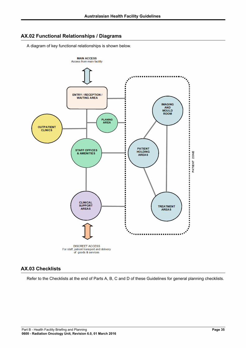
The Radiation Oncology Unit has many components. The main entry / reception / waiting area will streamvisitors to various areas including outpatient clinics, imaging and mould room and treatment areas. Theplanning areas will be close-by imaging areas to support work flows.
Australasian Health Facility Guidelines
Part B - Health Facility Briefing and Planning Page 150600 - Radiation Oncology Unit, Revision 6.0, 01 March 2016
03 DESIGN
03.01 Accessibility
EXTERNAL
The building both internally and externally must be accessible and non-threatening.
Level, undercover access is required for outpatients and inpatients in wheelchairs, trolleys and beds. Adiscreet, separate entry should be provided for patients arriving on a trolley or bed so they are not pushedthrough general public areas.
Disabled parking spots and patient drop-off and pickup points should be located close to the entry to theRadiation Oncology Unit or integrated cancer centre. Ready access should be provided from the public carpark to minimise stress for patients attending on a daily basis. If located on a hospital campus, access isrequired for deliveries (e.g. supplies) and waste removal.
After-hours access for urgent radiation therapy cases must be easy for inpatients and external (ambulance)patients. Delivery and replacement of large heavy equipment will be required.
INTERNAL
Internal circulation routes in all patient areas should allow for the efficient movement of wheelchairs andbeds. Treatment and planning areas should not be used as thoroughfares. Wherever possible, a separationbetween patient circulation and staff / material circulation within the Unit is preferred, especially wherepatients are transferred on beds.
Some access routes and circulation systems, particularly in the radiation treatment area, must allow deliverypaths for large pieces of equipment. Height, width, and floor loads must be considered in the design of theseaccess routes.
03.02 Parking
The following areas will be required:
• undercover patient parking adjacent to main entry for patients with minimal mobility;
• ambulance access; and
• parking area for volunteer drivers.
Patients attending the service as outpatients may do so on consecutive days and/or for up to eight weeksand may require a space on a short term basis or for up to five hours. Patients are often adversely affectedby the rigours of the treatment and the provision of subsidised or dedicated ‘user friendly’ parking facilitiesreduces the associated stress in attending the Unit. When planning new services, health services shouldideally have a process in place to ensure access to affordable parking, close-by the Unit, for patients whovisit daily for treatment.
For staff parking, refer to Part C: Section 790, Safety and Security Precautions for further information.
03.03 Disaster Planning
Refer to Part B: Section 80 General Requirements for further information.
Australasian Health Facility Guidelines
Part B - Health Facility Briefing and Planning Page 160600 - Radiation Oncology Unit, Revision 6.0, 01 March 2016
03.04 Infection Control
For further information, refer to:
• Part D: Infection Prevention and Control; and
• jurisdictional policies.
03.05 Environmental Considerations
TOXIC WASTE
Considerations include:
• safe handling and air exchanges for chemicals in the mould room etc.;
• provision of effective extraction systems to areas such as the mould room / appliance fabricationroom;
• drainage systems designed to meet the requirements of the relevant sewerage authority andhealth department; and
• safe storage and disposal of irradiated material.
ACOUSTICS
The appliance fabrication area of the mould room should be acoustically treated so noise associated withthis activity is minimised.
All examination, consultation rooms and offices will be acoustically treated to protect patient privacy. Wherean MRI is included, appropriate noise controls will be needed.
Modulators can be located within a bunker but a separate room is often provided in a location adjacent to thebunker to reduce heat and noise.
INTERIOR DÉCOR
The environment should have a comfortable and welcoming appearance while not compromising clinicalpractice or safety. Treatment areas such as the simulator room and ’bunkers’ should be decorated in amanner that is calming while providing positive distractions during treatment.
Visual distraction is becoming the norm in rooms such as bunkers. This may include ceiling effects includinga backlit photo mural or very large LED screens for images to be displayed on. Patients can often customisethe images.
PATIENT PRIVACY
Waiting areas for patients who have changed into a gown should be located so they are not observed bymembers of the public. Patients also require privacy to discuss billing and private health related concernswith staff.
Consideration of the movement of patients through the department should be considered to maximiseprivacy where possible.
NATURAL LIGHT
As much natural light as possible should be provided, especially into public spaces, waiting areas and thosetreatment areas that patients and staff occupy for long periods of time.
Australasian Health Facility Guidelines
Part B - Health Facility Briefing and Planning Page 170600 - Radiation Oncology Unit, Revision 6.0, 01 March 2016
03.06 Space Standards and Components
ERGONOMICS
The design and construction of Radiation Oncology Units should protect patients, visitors and maintenanceand other staff from avoidable risks of injury and/or radiation hazard.
The height, depth and design of desks in the radiation treatment area need to take into account the constantup and down nature of the tasks undertaken and the distance to the wall of the emergency stop button.
Many staff within Radiation Oncology Unit use dual screens to plan radiation treatment requirements. Referto Part C: Section 730, Human Engineering - Access and Mobility for more details.
HUMAN ENGINEERING
The design should permit effective, appropriate safe and dignified use by all people, including those withdisabilities. Also refer to Part C: Design for Access, Mobility, OHS and Security, Space Standards andDimensions for information.
ACCESS AND MOBILITY
Where appropriate, design must comply with AS 1428 (Set) 2010 Design for access and mobility Set (SAIGlobal).
Also refer to Part C: Design for Access, Mobility, OHS and Security, Space Standards and Dimensions forinformation.
DOORS AND CORRIDORS
Doors and corridors must be wide enough to accommodate large items of equipment and enable calibrationequipment and trolleys / beds to pass through with ease. Special consideration needs to be given to anybariatric bed that may travel from inpatient units of the hospital inpatient units.
Within the mould room / appliance fabrication areas, the number of doors between shop areas should beminimised to facilitate the movement of equipment. Double doors will be provided to all workshop areas.
The need for neutron doors to the maze will depend on overall design of the maze. Ideally the space willbe designed such that this is not required. Refer to Part C: Design for Access, Mobility, OHS and Security,Space Standards and Dimensions for information.
03.07 Safety and Security
GENERAL
Safety and security involves people and policies as well as physical aspects. Security of the facility must beaddressed at each stage of the planning and design process. A safety audit via a risk analysis of potentialhazards should be undertaken during the design process. Security may include:
• emergency ’stop’ buttons in treatment bunkers and control rooms;
• 'last man out’ systems;
• access control to rooms storing high cost equipment;
• radioactive source security as provided for brachytherapy services;
• fixed and personal duress alarms; and
• controlled staff access after-hours.
The Radiation Oncology Unit should only be accessible to authorised persons and must be locked and analarm activated once the area is vacated after hours. Care should be taken with wayfinding and signage todiscourage accidental entry to these areas.
Australasian Health Facility Guidelines
Part B - Health Facility Briefing and Planning Page 180600 - Radiation Oncology Unit, Revision 6.0, 01 March 2016
RADIATION SAFETY
The Australian Radiation Protection and Nuclear Safety Agency (ARPANSA) publish Codes of Practice orStandards detailing requirements for radiation safety. Refer to the Radiation Protection Series (Website)(ARPANSA, 2015) including at least the following:
• Fundamentals for Protection Against Ionising Radiation (F-1), Radiation Protection Series(ARPANSA, 2014);http://www.arpansa.gov.au/pubs/rps/rpsF-1.pdf
• National Directory for Radiation Protection (RPS 6) (ARPANSA, 2014);http://www.arpansa.gov.au/pubs/rps/rps6_6.pdf
• Code of Practice for Radiation Protection in the Medical Applications of Ionizing Radiation(RPS-14), Radiation Protection Series (ARPANSA, 2008);
• Safety Guide for Radiation Protection in Radiotherapy, Radiation Protection Series (ARPANSA,2008); and
• Code of Practice for the Security of Radioactive Sources (RPS-11), Radiation Protection Series(ARPANSA, 2007).
Each jurisdiction has legislation in place to regulate and control radiative substances, radioactive sourcesand related equipment. Refer to:
• Radiation Safety Act 1975 - 1999 (Act No 44 of 1975) (Government of Western Australia, 2004);
• NSW Radiation Control Act 1990 (amended 2002) (NSW Government, 2002) and the RadiationControl Regulation 2003 (under the Radiation Control Act 1990) (NSW Government, 2003)administered by Environment Protection Authority;
• Radiation Protection Act (Northern Territory Government, 2009), as in force at 2 July 2012;
• Queensland Radiation Safety Act 1999 (Queensland Government, 2013) and the QueenslandRadiation Safety Regulation 2010 (Queensland Government, 2014);
• South Australia, Radiation Protection and Control Act 1982 (Government of South Australia,2012);
• Tasmania, Radiation Protection Act, 2005 (Tasmanian Government, 2005); and
• Victoria Radiation Regulations 2007 (S.R. No. 89/2007), Radiation Act 2005 (State Governmentof Victoria, 2009) and Radiation Amendment Act 2013 (State Government of Victoria, 2013).
03.08 Finishes
WALL PROTECTION
The wall surfaces in the Unit areas should be washable. Refer to Part C: Design for Access, Mobility, OHSand Security, Space Standards and Dimensions.
FLOOR FINISHES
Non-slip flooring is essential for all work areas. The floor surface in clinical areas should be impervious, easyto clean, sealed with coving at the edges and have adequate drainage. Refer to Part C: Design for Access,Mobility, OHS and Security, Space Standards and Dimensions.
CEILING FINISHES
Refer to Part C: Design for Access, Mobility, OHS and Security, Space Standards and Dimensions.
Australasian Health Facility Guidelines
Part B - Health Facility Briefing and Planning Page 190600 - Radiation Oncology Unit, Revision 6.0, 01 March 2016
03.09 Fixtures, Fittings & Equipment
GENERAL
Room Data and Room Layout Sheets in the AusHFG define fixtures, fittings and equipment (FFE).
Refer to the Standard Components - Room Data Sheets (RDS) and Room Layout Sheets (RLS),Australasian Health Facility Guidelines (AHIA, 2015) and:
• Part C: Section 710, Space Standards and Dimensions; and
• Part F: Section 680 Furniture Fittings and Equipment.
EQUIPMENT - GENERAL
All items of equipment will need to be itemised and dimensions of larger items obtained during the designphase to ensure:
• equipment is housed to enable its operation and maintenance. In particular, room sizes andspecifications for linear accelerator and electronic cabinet rooms should accommodate theequipment manufacturer's recommendations, as space requirements vary from one machine toanother and one manufacturer to another. Equipment requiring services such as water, air andspecial power must be noted and project engineers advised;
• cable length between parts of major equipment should be checked and adequate in-floor conduitspace provided;
• doors are sized to allow passage of equipment;
• heat loads are estimated and catered for; and
• weight loads are estimated and checked structurally.
Space allocations should be sufficient to accommodate a range of vendor equipment as replacement isrequired every 10 to 15 years.
Adequate space must also be provided for the maintenance of major equipment. Note that electronic controlcabinets are bulky and need special access to three sides.
LINEAR ACCELERATOR MACHINES
Ideally, linear accelerator machines should be ‘paired’ within a Unit. This ensures that treatment cancontinue to be delivered should a machine malfunction. This paired machine would deliver the same beamenergy and quality.
Linear accelerator rooms (bunkers) require radiation protection that will include concrete walls, floors andceiling to a specified thickness. The radiation protection needs of the Unit shall be assessed by a certifiedphysicist or radiation safety consultant in accordance with National standards and legislative and regulatoryrequirements for each jurisdiction.
Reference should be made to local radiation licensing requirements and regulations.
Note that the schedule of accommodation indicates the bunker size including the maze rather than theactual treatment room which would be in the order of 150m2. This is to ensure that sufficient ‘footprint’ isallowed during early planning stages.
03.10 Building Service Requirements
FUTURE PROOFING
Selected services may seek to future-proof facilities to anticipate new or emerging technologies. Forexample, shielding for primary beams other than in sentinel directions may be required if cyber knifetechnology is being considered.
Australasian Health Facility Guidelines
Part B - Health Facility Briefing and Planning Page 200600 - Radiation Oncology Unit, Revision 6.0, 01 March 2016
Selected Units may include additional imaging modalities such as MRI and PET. Retrofitting these modalitiesis problematic (especially PET) so ideally, facility requirements should be considered in planning projectstages. In addition, extra space may be required within particular rooms to accommodate planningequipment. For further information, refer to:
• Part B: HPU 440 Medical Imaging Unit; and
• Part B, HPU 480 PET (Positron Emission Tomography) Unit.
STRUCTURAL
Radiation treatment bunkers, CT simulation rooms and brachytherapy treatment rooms need radiationprotection built into the facility. Bunkers need special construction to ensure they meet radiation safetyrequirements.
The flooring for a Radiation Oncology Unit will be designed to meet the load requirements for equipment andpatient care.
Ceiling mounted equipment should have properly designed rigid support structures located above thefinished ceiling sufficient to support heavy ceiling-mounted equipment such as frames of data monitors. Alay-in type of ceiling should be considered for ease of installation, service, and remodelling.
A minimum three metre ceiling height is required in procedure rooms, with a minimum one metre spaceabove for heating, ventilating and air conditioning systems.
The flooring for a Radiation Oncology Unit shall be adequate to meet the load requirements for equipment,patient and personnel.
COMMUNICATIONS AND INFORMATION SYSTEMS
The infrastructure for the following should be considered for the present and future expansion:
• voice / data systems;
• telephone and video conferencing capacity;
• duress call - fixed and personal (if required);
• CCTV monitoring systems of entry points;
• infrastructure for PACS (usually a separate system from the medical imaging PACS), electronichealth records and ROIS;
• large bandwidths to support the movement of images from hub and spoke locations; patient /nurse and emergency call systems (that should be consistent with existing systems);
• the use of Citrix systems allows for planning activities to be undertaken at workstationsthroughout the Unit;
• dedicated, air-conditioned rooms for servers, with capacity to accommodate current and futurecapacity;
• alarm systems - drug fridges, medical gases, entries etc. that register in an area manned 24hours per day; and
• patient viewing cameras, treatment delivery computers and intercoms to allow the radiationtherapist to monitor and communicate with the patient during treatment when the patient is alonein the treatment room.
ELECTRICAL SERVICES
Sufficient power should be provided for current need and future expansion of services.
An uninterruptible power supply (UPS) and an emergency back-up system should be available for highpriority equipment and illumination.
Cable ducts and/or conduits should be provided in the floors, walls and ceilings as required for specialisedequipment.
Australasian Health Facility Guidelines
Part B - Health Facility Briefing and Planning Page 210600 - Radiation Oncology Unit, Revision 6.0, 01 March 2016
There should be a maximum distance of 7.5 metres for the cable run between the simulator and thegenerator. Minimal distances are preferable to minimise the degradation of cable operation. Cable runs inthe radiation treatment control area need careful planning.
MECHANICAL SERVICES
Appropriate air exchanges and exhausts for chemicals in the appliance workroom.
Sufficient air-conditioning capacity and compressed air in radiation treatment and CT rooms; access forfuture expansion of service.
Appropriate air-handling systems in computer equipment rooms.
General air conditioning needs to cool equipment but not blow over partially undressed patients on beds.
To maintain a high level of staff concentration and to minimise the possibility of accidents, the temperature ofthe unit should be maintained within a comfortable range not exceeding 25°C.
Smoke / heat detectors in radiation treatment and simulator rooms must be of the type not sensitive toradiation (i.e. photoelectric) and require special consideration.
MEDICAL GASES
Oxygen and suction will be required in all bunkers, simulation, treatment and patient bed bays. Nitrousoxide, medical air and scavenging may additionally be required in rooms where general anaesthesia may beadministered.
RADIATION PROTECTION
Linear accelerator rooms require additional radiation protection that will include concrete walls, floors andceiling to a specified thickness. Primary beam protection may be enhanced by the use of steel plate inserts.The radiation protection needs of the Unit shall be assessed by a certified physicist or consulting radiationexpert to ensure compliance with the requirements of jurisdictional authorities.
This assessment is to specify the type, location and amount of protection to be installed in accordance withfinal approved department layout and equipment selection. The radiation protection requirements shall beincorporated into the final plans and specifications.
LIGHTING
Lighting in the Radiation Oncology Unit will need to be of various types and will be dependent on the task.The main lighting requirements are:
• even distribution of luminance throughout the non-working areas;
• walls that do not show reflections of luminaires, particularly at eye-height of staff when working;
• fully dimmable lighting in bunkers, simulator areas, planning areas and office areas wheremedical staff undertake planning activities;
• special three level lighting in radiation treatment bunkers; and
• lasers for patient positioning in bunkers and CT simulator rooms with high level luminanceavailable for maintenance and repairs.
HYDRAULIC SERVICES
The trade waste plumbing and drainage system must be designed to meet the requirements of the relevantsewerage authority and the health departments. Information of the quality of chemicals to be used /discharged must be provided by the client to the hydraulics engineer.
Australasian Health Facility Guidelines
Part B - Health Facility Briefing and Planning Page 220600 - Radiation Oncology Unit, Revision 6.0, 01 March 2016
04 COMPONENTS OF THE UNIT
04.01 Standard Components
Rooms / spaces are defined as:
• standard components (SC) which refer to rooms / spaces for which room data sheets, roomlayout sheets (drawings) and textual description have been developed;
• standard components – derived rooms are rooms, based on a SC but they vary in size. In theseinstances, the standard component will form the broad room ‘brief’ and room size and contentswill be scaled to meet the service requirement; and
• non-standard components which are unique rooms that are usually service-specific and notcommon.
The standard component types are listed in the attached Schedule of Accommodation.
The current Standard Components can be found at: www.healthfacilityguidelines.com.au/standard-components
04.02 Non-Standard Components
RESOURCE ROOM
Description and Function
This room provides a location for the storage of and access to resources related to cancer etc. Resourcesmay also be accessed by computer so patient access is facilitated. This area may also provide a smallmeeting table so visitors can discuss issues with staff or volunteers.
Location and Relationships
Centrally located in the entry / reception / waiting area so that all visitors can access.
Considerations
May be provided in a central, shared location if provided as part of an integrated cancer centre.
VOLUNTEERS WORKROOM
Description and Function
A base for volunteers who may work in the Unit, should volunteers be part of the service model. This basemay provide a worktable, storage and lockers.
Location and Relationships
Location may depend on roles undertaken by volunteers. For example, if volunteers assist with wayfindingand provide support within the resource room, then a location near the entry would be ideal.
Considerations
Requirements may change depending on the roles undertaken by volunteers.
IMAGE REVIEW ROOM
Description and Function
A room for staff to review treatment verification images off-line. The space is also used to conduct weeklychart checks and any associated image trend analysis.
Pre-treatment plan review beams-eye view and plan information updates.

Australasian Health Facility Guidelines
Part B - Health Facility Briefing and Planning Page 230600 - Radiation Oncology Unit, Revision 6.0, 01 March 2016
Location and Relationships
This room should be collocated with treatment bunkers and nearby to facilitate staff movement betweentasks. A minimum of one room per two bunkers is recommended.
Considerations
Image review rooms should be considered as separate to control rooms where the linear acceleratoroperators should be working in a distraction-free environment. Image review rooms are not for equipmentstorage for patient devices and other linear accelerator equipment which requires a separate room.Equipment storage can also be shared with another linear accelerator at the same rate of one per twobunkers.
APPLIANCE FITTING ROOM
Description and Function
Where patients are measured for immobilisation devices, masks etc.
Location and Relationships
Direct access from the corridor and into the Workroom. Away from other patient areas due to possible noiseand fumes.
Considerations
This space could be combined with clean moulding room especially for two machine service. Patient privacyneeded and the doorway screened. Bed / trolley access is needed. FF&E will include:
• hand basin;
• plinth;
• benches and cupboards;
• plaster dust extraction system and plaster trap;
• fume extraction system;
• large sink and plater trap;
• water bath (built-in or free-standing);
• heavy dusty stainless steel benching;
• shelving and cupboards;
• instruments-drill, hot wire cutter, vacuum former;
• alloy melting pot;
• block cutters (a divergent block hot wire device that mimics the set-up of the linear accelerators isrequired as well as a non-divergent hot wire cutter for electron cut-outs etc.).
• surge protection for electrical equipment;
• RCD protection for staff especially where water bath are in use;
• dust and fume extraction;
• acoustic containment; and
• storage space for low melting point allow shielding plates, compensator mounts.
MOULDING ROOM – CLEAN
Description and Function
Used for the manufacture of immobilisation devices. Storage space is required for the large volumes ofmaterial used to create the appliances.
Australasian Health Facility Guidelines
Part B - Health Facility Briefing and Planning Page 240600 - Radiation Oncology Unit, Revision 6.0, 01 March 2016
While the shell forming for head and neck patients is predominantly thermoplastic based - there still may bepatients that require plaster impressions and appliance room specific consult and mark-up.
Location and Relationships
Direct access from the fitting room but away from other patient areas due to possible noise and fumes.
Considerations
If this room is combined with the appliance fitting room then equipment listed below should not beduplicated. Surge protection for electrical equipment. RCD protection for staff especially where water bathsare in use.
The moulding room requires air extraction for the molten metal used to fabricate photons and electronshielding. Bulky foam cutters for personalised stabilisation products and/or vacuum formers may be requiredto manufacture custom masks.
Acoustic containment. FF&E will include:
• plaster dust extraction system and plaster trap;
• fume extraction cabinet;
• large sink and plaster trap;
• water bath (built-in or free-standing);
• heavy duty stainless steel benching;
• shelving and cupboards;
• instruments - drill, hot wire cutter, vacuum former;
• alloy pot; and
• block cutter.
Possible use of 3D printing technology.
WORKSHOP - DIRTY
Location and Relationships
Direct access from the moulding room but away from other patient areas due to possible noise and fumes.
PLANNING CT / SIMULATOR ROOM
Description and Function
A planning CT is typically a wide bore CT scanner with a specialised flat, indexed couch top. There may bea conventional 2D simulator (plain x-ray) but if this equipment is used there will still need to be an adjoiningCT Room or ready access to a CT. It is expected however that modern radiation therapy units will install CTscanners. A simulator must have image intensifier and CT inter-working capability.
A CT simulator combines the functionality of a conventional CT with features and image processing anddisplay tools of a three-dimensional radiation treatment planning (3D) system (TPS).
A diagnostic C-arm mobile unit may be used for similar purposes in the planning and verification of highdose rate brachytherapy if in an operating theatre otherwise the CT scanner will be used.
Fan noise from various computer systems may create noise making it difficult to converse with patients.The amount of noise varies greatly depending on which equipment is used. A large cupboard with floor toceiling access to house x-ray generator and reconstruction computers discreetly within the CT room may berequired. The cupboard should have separate air flow for cooling needs.
Location and Relationships
Adjacent to the control room.
Ready access to change cubicles, sub-waiting and patient toilets.
Australasian Health Facility Guidelines
Part B - Health Facility Briefing and Planning Page 250600 - Radiation Oncology Unit, Revision 6.0, 01 March 2016
Ready access to a resuscitation trolley (where intravenous contrast is administered).
Near the moulding room where stabilisation appliances / masks may be manufactured.
Considerations
• space for a bed to enter, turn and be placed along either side of the simulator;
• lead glass viewing window to the control room;
• radiation screening to standards;
• temperature and humidity control to manufacturer’s specifications;
• dimmable lighting controls;
• emergency ’stop’ button;
• oxygen and suction on medical services panel;
• emergency / nurse call buttons;
• CCTV camera and intercom system - patient to control room;
• hand basin;
• benches;
• wall and ceiling mounted x-ray laser lights (that require a steel plate mounted to the building studfixed at the floor and ceiling to ensure stability when mounted); and
• x-ray transformer.
CT-SIMULATOR CONTROL ROOM
Description and Function
Control area for the simulator.
Location and Relationships
Directly adjacent to the simulator room.
Considerations
FF&E will include:
• CT control console and computer or simulator control panel;
• virtual simulation workstation;
• PACS viewing monitors and x-ray viewing panels for review of mammograms and x-rays ofpatients from rural areas;
• emergency ‘stop’ button;
• patient viewing monitor and microphone; and
• work benches.
PLANNING WORKROOM
Description and Function
The area used by the radiation therapists who work individually using computer terminals with dual screensto review plans and produce radiation dosage profiles.
Location and Relationships
Ready access to the planning modalities including CT.
Easy access to the computer server data storage room for retrieval of archived data.
Australasian Health Facility Guidelines
Part B - Health Facility Briefing and Planning Page 260600 - Radiation Oncology Unit, Revision 6.0, 01 March 2016
Considerations
Specialised FF&E will include:
• work benches sized to suit the planning computers with dual screens;
• planning computers – one per staff member;
• printer; and
• PACS terminals at a ratio of one per TPS workstation.
MEDICAL PHYSICS LABORATORY
Sufficient space for computers and a work area to carry out dosimetry measurements, dosimetry equipmentQA and ultrasound and LDR brachytherapy QA.
Location and Relationships
Ready access to the bunkers.
Considerations
Sealed vinyl floor, laminated bench tops.
Hands-free telephone.
FF&E will include:
• workbenches;
• light boxes; and
• office furniture.
Note that IntraBeam dosimetry measurements require a shielded space. Several QA procedures mayhappen at one time, with one or more using radioactive sources. There must be a dedicated radioactivesource handling area, including a fume hood extraction system separate from rest of the laboratory thatcomplies with radiation safety regulations.
WORKSHOPS – ELECTRICAL AND MECHANICAL
Description and Function
Maintenance of electrical equipment divided into ‘clean’ and ‘dirty’ zones.
Location and Relationships
Part of the medical physics area.
Considerations
Light-coloured, antistatic flooring.
Electrostatic earthing throughout the area.
Hands-free telephone.
FF&E will include:
• compressed air outlet;
• benches - general and for electronic work in a clean work area;
• sink;
• peg board;
• mobile fume extraction unit;
• drill and lathe in a 'dirty' work area; and
• general office furniture.
Australasian Health Facility Guidelines
Part B - Health Facility Briefing and Planning Page 270600 - Radiation Oncology Unit, Revision 6.0, 01 March 2016
PHYSICS STORE
Description and Function
This room will house very expensive equipment and instruments for use by the physicists in the checkingand calibrating of the linacs, including the water phantom machine, approximately 1m x 1m and 1.8m high.
Location and Relationships
Ready access to the physics laboratory.
Easy access to a deep sink in the cleaner’s room for filling and emptying of the water tank.
Considerations
Access for large items of equipment including manoeuvring the water phantom trolley.
Safe for radioactive materials.
Cable storage and heavy duty shelving for numerous phantoms.
LINEAR ACCELLERATOR TREATMENT ROOM (BUNKER)
Description and Function
Treatment rooms or bunkers are the rooms in which EBRT irradiation occurs. They require a maze-likecorridor at the entrance of the room for radiation protection.
The maze, entrance and entry to the treatment room must allow access for the treatment machine, serviceequipment, hospital beds and gantry frames.
Linear accelerators with 18 MV photon beams may require additional shielding at the maze entrance (i.e.neutron door); however, particular attention should be given to the bunker and maze design in an attempt toavoid the use of a maze shielding door.
Location and Relationships
Immediately adjacent to the control area so that access can be monitored.
The treatment rooms should be located with ready access to patient amenities (change cubicles, sub-waiting, and toilets), treatment planning and support areas including patient accessory storage and utilityrooms.
Considerations
Layouts shall be designed to prevent radioactive particles from escaping. Openings into the room, includingdoors, ductwork, vents and electrical raceways and conduits shall be baffled to prevent direct exposure toother areas of the facility.
Services requirements including electrical, hydraulics, and air-conditioning will be according to theequipment manufacturer's specifications.
Provide special cable access to the treatment rooms for physics measurements.
Linear accelerators need special air exchanges and the floor needs protection when machines are installed.
FF&E will include:
• linear accelerator;
• oxygen and suction on medical services panel;
• emergency 'stop' switch;
• multiple PTZ capable CCTV cameras;
• hand basin;
• benches and storage cupboards for patient machine accessories;
• laser lights for positioning;
• treatment set-up information viewing such as large LCD TV screens;
Australasian Health Facility Guidelines
Part B - Health Facility Briefing and Planning Page 280600 - Radiation Oncology Unit, Revision 6.0, 01 March 2016
• monitors and audio equipment for patient contact;
• ceiling art (fixed or projected) and music systems for patient distraction;
• ’last man out’ interlock;
• fixed duress;
• a significant number of power outlets; and
• nurse call system, including emergency call.
CONTROL ROOM – LINEAR ACCELERATOR
Description and Function
Radiation therapists will perform all control and patient monitoring functions in the control room.
Patient radiation treatment records and planning images may be displayed in the control room area for eachtreatment unit throughout the course of the therapy. Patient viewing cameras, treatment delivery computersand intercoms allow the radiation therapist to monitor and communicate with the patient during treatmentwhen the patient is alone in the treatment room.
Location and Relationships
Direct access to treatment bunker.
Considerations
Cable trays must be easily removable for access by maintenance staff.
FF&E will include:
emergency stop switch;
intercom;
patient viewing monitors;
portal imaging computers;
workstation for image and chart viewing, access to the scheduling system, and space to store treatmentrecords (if not electronic);
linear accelerator control console;
PACS monitor; and
benches / shelving units to suit equipment.
BRACHYTHERAPY ROOM
Description and Function
A radioactive source is delivered internally through a tube or applicators implanted or inserted duringsurgery. The radiation source is inserted manually or, more commonly, performed by a remote after loadingmachine.
In centres where LDR brachytherapy seed implantation is performed, the room shall be of sufficient size andequipped as an operating room.
Location and Relationships
Adjacent to:
• induction bay;
• scrub room;
• recovery bay;
• seed implant store and loading room;
Australasian Health Facility Guidelines
Part B - Health Facility Briefing and Planning Page 290600 - Radiation Oncology Unit, Revision 6.0, 01 March 2016
• other radiation treatment rooms;
• exit buy; and
• sterile store.
Considerations
Radiation safety of radioactive materials.
Access to oxygen, suction, medical air, nitrous oxide and scavenging needed.
Controlled access to brachytherapy facility and radioactive materials.
ORTHOVOLTAGE ROOM
Description and Function
A specialised room use to treat superficial skin cancers. The treatment uses low to medium energy radiationto treat cancer. Radiation shielding is required.
Location and Relationships
Immediately adjacent to the Control Area so that access can be monitored.
Considerations
FF&E includes:
• a control room - orthovoltage should be attached to the room;
• ‘beam on’ warning lights will be required at the room entrance;
• an interlocked door linked to the beam on is required and should be shielded in lieu of needing atreatment room maze;
• a cooling unit will need to be accommodated in the facility. This may be in the treatment room butdue to associated noise it is recommended it be planned to be housed outside the treatment roombut nearby. Consider distance to plan room;
• the room will accommodate a superficial orthovoltage unit. A cupboard with exhaust will berequired within the room to accommodate the generator;
• custom storage for treatment cones and treatment shield is required;
• medical services panel with oxygen and suction; and
• nurse call system including emergency call.
Australasian Health Facility Guidelines
Part B - Health Facility Briefing and Planning Page 300600 - Radiation Oncology Unit, Revision 6.0, 01 March 2016
AX APPENDICES
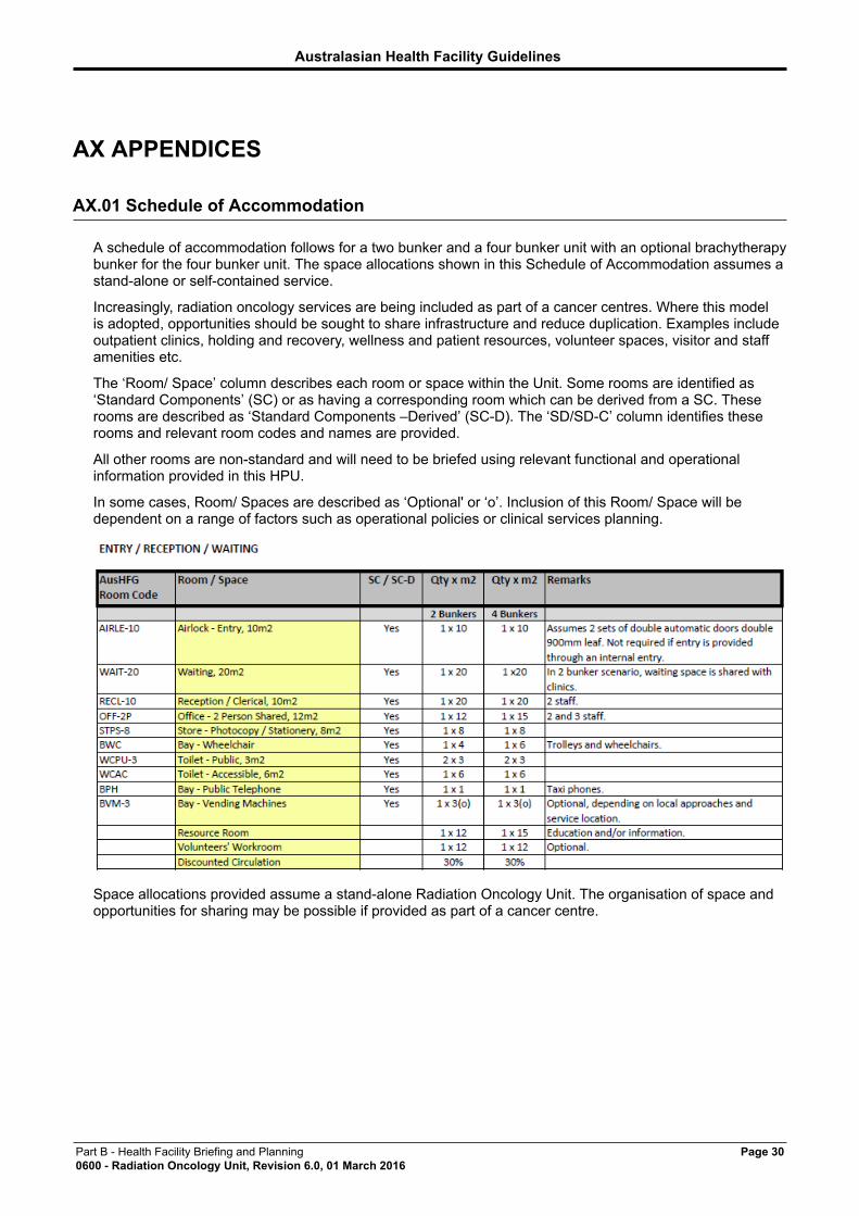
AX.01 Schedule of Accommodation
A schedule of accommodation follows for a two bunker and a four bunker unit with an optional brachytherapybunker for the four bunker unit. The space allocations shown in this Schedule of Accommodation assumes astand-alone or self-contained service.
Increasingly, radiation oncology services are being included as part of a cancer centres. Where this modelis adopted, opportunities should be sought to share infrastructure and reduce duplication. Examples includeoutpatient clinics, holding and recovery, wellness and patient resources, volunteer spaces, visitor and staffamenities etc.
The ‘Room/ Space’ column describes each room or space within the Unit. Some rooms are identified as‘Standard Components’ (SC) or as having a corresponding room which can be derived from a SC. Theserooms are described as ‘Standard Components –Derived’ (SC-D). The ‘SD/SD-C’ column identifies theserooms and relevant room codes and names are provided.
All other rooms are non-standard and will need to be briefed using relevant functional and operationalinformation provided in this HPU.
In some cases, Room/ Spaces are described as ‘Optional' or ‘o’. Inclusion of this Room/ Space will bedependent on a range of factors such as operational policies or clinical services planning.
Space allocations provided assume a stand-alone Radiation Oncology Unit. The organisation of space andopportunities for sharing may be possible if provided as part of a cancer centre.
Australasian Health Facility Guidelines
Part B - Health Facility Briefing and Planning Page 310600 - Radiation Oncology Unit, Revision 6.0, 01 March 2016
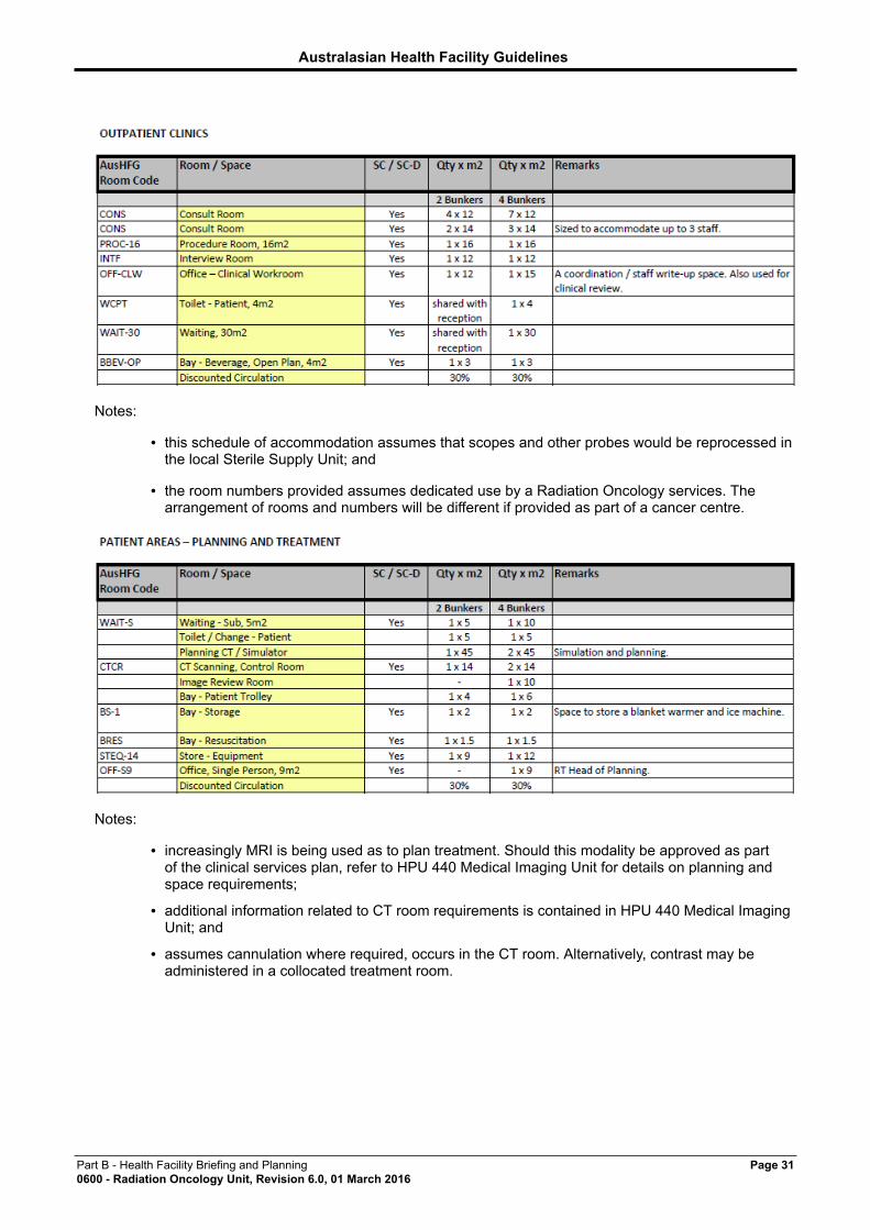
Notes:
• this schedule of accommodation assumes that scopes and other probes would be reprocessed inthe local Sterile Supply Unit; and
• the room numbers provided assumes dedicated use by a Radiation Oncology services. Thearrangement of rooms and numbers will be different if provided as part of a cancer centre.
Notes:
• increasingly MRI is being used as to plan treatment. Should this modality be approved as partof the clinical services plan, refer to HPU 440 Medical Imaging Unit for details on planning andspace requirements;
• additional information related to CT room requirements is contained in HPU 440 Medical ImagingUnit; and
• assumes cannulation where required, occurs in the CT room. Alternatively, contrast may beadministered in a collocated treatment room.
Australasian Health Facility Guidelines
Part B - Health Facility Briefing and Planning Page 320600 - Radiation Oncology Unit, Revision 6.0, 01 March 2016
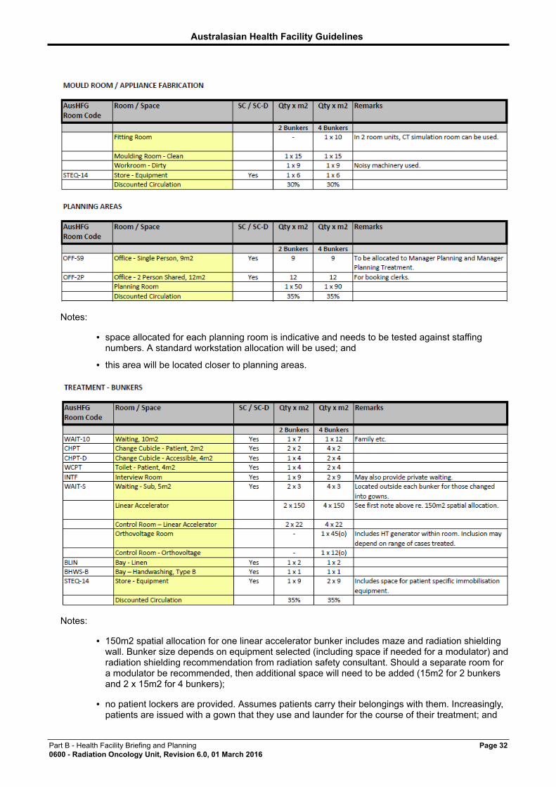
Notes:
• space allocated for each planning room is indicative and needs to be tested against staffingnumbers. A standard workstation allocation will be used; and
• this area will be located closer to planning areas.
Notes:
• 150m2 spatial allocation for one linear accelerator bunker includes maze and radiation shieldingwall. Bunker size depends on equipment selected (including space if needed for a modulator) andradiation shielding recommendation from radiation safety consultant. Should a separate room fora modulator be recommended, then additional space will need to be added (15m2 for 2 bunkersand 2 x 15m2 for 4 bunkers);
• no patient lockers are provided. Assumes patients carry their belongings with them. Increasingly,patients are issued with a gown that they use and launder for the course of their treatment; and
Australasian Health Facility Guidelines
Part B - Health Facility Briefing and Planning Page 330600 - Radiation Oncology Unit, Revision 6.0, 01 March 2016
• where children are treated, a separate waiting space should be provided.
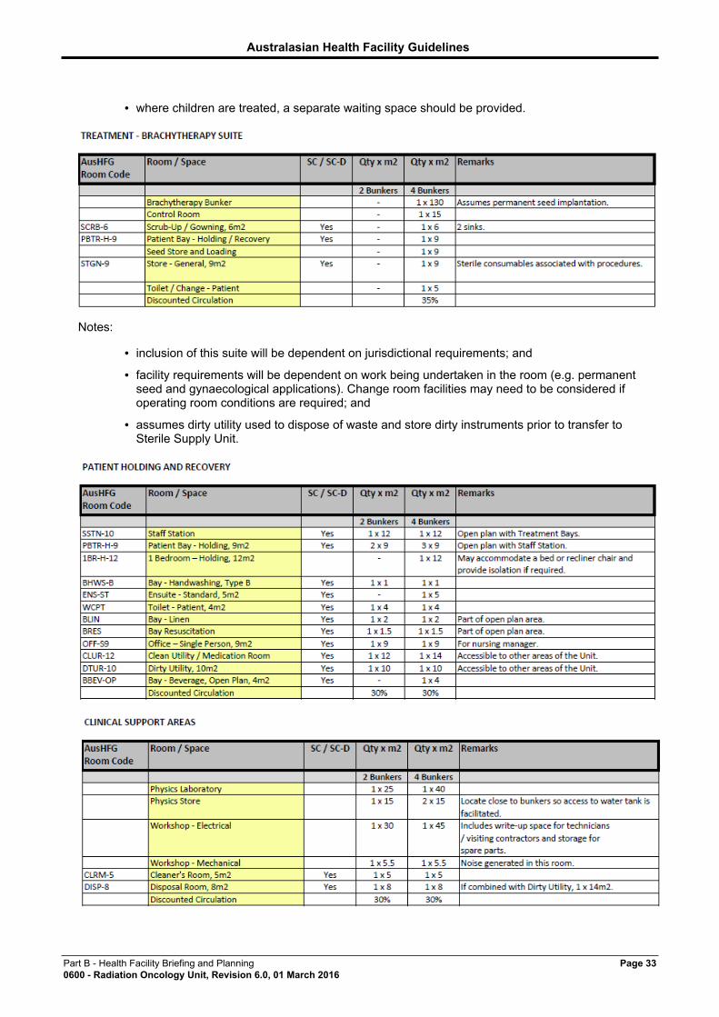
Notes:
• inclusion of this suite will be dependent on jurisdictional requirements; and
• facility requirements will be dependent on work being undertaken in the room (e.g. permanentseed and gynaecological applications). Change room facilities may need to be considered ifoperating room conditions are required; and
• assumes dirty utility used to dispose of waste and store dirty instruments prior to transfer toSterile Supply Unit.
Australasian Health Facility Guidelines
Part B - Health Facility Briefing and Planning Page 340600 - Radiation Oncology Unit, Revision 6.0, 01 March 2016
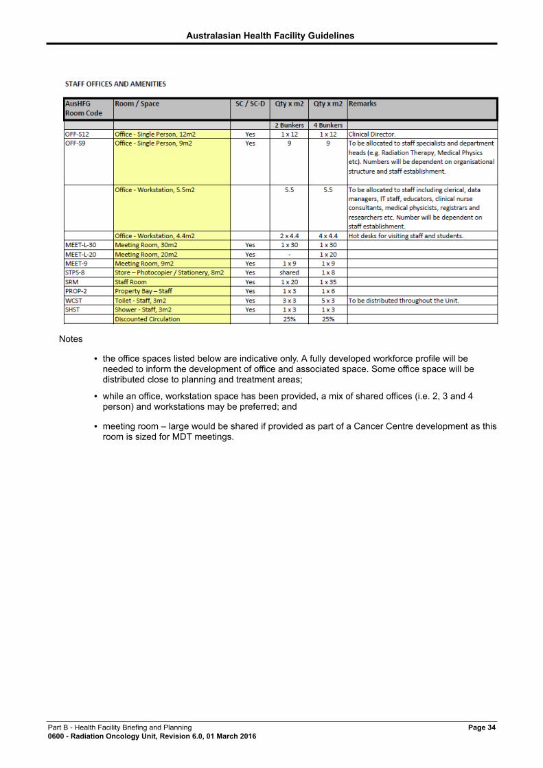
Notes
• the office spaces listed below are indicative only. A fully developed workforce profile will beneeded to inform the development of office and associated space. Some office space will bedistributed close to planning and treatment areas;
• while an office, workstation space has been provided, a mix of shared offices (i.e. 2, 3 and 4person) and workstations may be preferred; and
• meeting room – large would be shared if provided as part of a Cancer Centre development as thisroom is sized for MDT meetings.
Australasian Health Facility Guidelines
Part B - Health Facility Briefing and Planning Page 350600 - Radiation Oncology Unit, Revision 6.0, 01 March 2016
AX.02 Functional Relationships / Diagrams
A diagram of key functional relationships is shown below.
AX.03 Checklists
Refer to the Checklists at the end of Parts A, B, C and D of these Guidelines for general planning checklists.
Australasian Health Facility Guidelines
Part B - Health Facility Briefing and Planning Page 360600 - Radiation Oncology Unit, Revision 6.0, 01 March 2016
AX.04 References
• AHIA, 2010, Part C: Design for Access, Mobility, OHS and Security, Australasian HealthFacility Guidelines (AHIA, 2010), Australasian Health Facility Guidelines, Australasian HealthInfrastructure Alliance (AHIA), Sydney, NSW.
• AHIA, 2010, AusHFG Part B: Section 90, Standard Components, Australasian Health FacilityGuidelines, Australasian Health Infrastructure Alliance (AHIA), Sydney, NSW.
• AHIA, 2010, Part B: Section 80 General Requirements, Australasian Health Facility Guidelines(AHIA, 2010), Australasian Health Facility Guidelines, Australasian Health Infrastructure Alliance(AHIA), Sydney NSW.
• AHIA, 2015, Part D: Infection Prevention and Control, Australasian Health Facility Guidelines(AHIA, 2015), Australasian Health Facility Guidelines, Australasian Health Infrastructure Alliance(AHIA), Sydney, NSW.
• AHIA, 2010, Part C: Section 730, Human Engineering, Australasian Health Facility Guidelines(AHIA, 2010), Australasian Health Facility Guidelines, Australasian Health Infrastructure Alliance(AHIA), Sydney, NSW.
• AHIA, 2010, Part C: Section 710, Space Standards and Dimensions, Australasian HealthFacility Guidelines (AHIA, 2010), Australasian Health Facility Guidelines, Australasian HealthInfrastructure Alliance (AHIA), Sydney, NSW.
• AHIA, 2015, Part C: Section 790, Safety and Security Precautions, Australasian Health FacilityGuidelines (AHIA, 2015), Australasian Health Facility Guidelines, AHIA, Sydney, NSW.
• AHIA, 2010, Part F: Section 680 Furniture Fittings and Equipment, Australasian HealthFacility Guidelines (AHIA, 2010), Australasian Health Facility Guidelines, Australasian HealthInfrastructure Alliance (AHIA), Sydney, NSW.
• AHIA, 2013, Part B: HPU 440 Medical Imaging Unit, Australasian Health Facility Guidelines(AHIA, 2013), Australasian Health Facility Guidelines, Australasian Health Infrastructure Alliance(AHIA), Sydney NSW.
• AHIA, 2015, Standard Components - Room Data Sheets (RDS) and Room Layout Sheets (RLS),Australasian Health Facility Guidelines (AHIA, 2015), Australasian Health Facility Guidelines,AHIA, North Sydney, NSW.
• AHIA, 2015, Part B: HPU 600 Radiation Oncology Unit, Australasian Health Facility Guidelines(AHIA, 2015), Australasian Health Facility Guidelines, Australasian Health Infrastructure Alliance,Sydney, NSW.
• AHIA, 2010, Part B, HPU 480 PET (Positron Emission Tomography) Unit, Australasian HealthFacility Guidelines (AHIA, 2010), Australasian Health Facility Guidelines, Australasian HealthInfrastructure Alliance (AHIA), Sydney, NSW.
• ARPANSA, 2014, Fundamentals for Protection Against Ionising Radiation (F-1), RadiationProtection Series (ARPANSA, 2014), Radiation Protection Series, no. F-1, Australian RadiationProtection and Nuclear Safety Agency (ARPANSA), Sydney, NSW.
• ARPANSA, 2014, National Directory for Radiation Protection (RPS 6) (ARPANSA, 2014), NationalDirectory for Radiation Protection, Australian Radiation Protection and Nuclear Safety Agency(ARPANSA), Sydney, NSW.
• ARPANSA, 2008, Code of Practice for Radiation Protection in the Medical Applications of IonizingRadiation (RPS-14), Radiation Protection Series (ARPANSA, 2008), Radiation Protection Series,Australian Radiation Protection and Nuclear Safety Agency (ARPANSA), Barton, ACT.
• ARPANSA, 2008, Safety Guide for Radiation Protection in Radiotherapy, Radiation ProtectionSeries (ARPANSA, 2008), Radiation Protection Series, Australian Radiation Protection andNuclear Safety Agency (ARPANSA), Barton, ACT.
Australasian Health Facility Guidelines
Part B - Health Facility Briefing and Planning Page 370600 - Radiation Oncology Unit, Revision 6.0, 01 March 2016
• ARPANSA, 2007, Code of Practice for the Security of Radioactive Sources (RPS-11), RadiationProtection Series (ARPANSA, 2007), Radiation Protection Series, Australian Radiation Protectionand Nuclear Safety Agency (ARPANSA), Barton, ACT.
• ARPANSA, 2015, Radiation Protection Series (Website) (ARPANSA, 2015), Australian RadiationProtection and Nuclear Safety Agency (ARPANSA), Barton ACT.
• Government of South Australia, 2012, South Australia, Radiation Protection and Control Act 1982(Government of South Australia, 2012), Government of South Australia, Adelaide, SA.
• Government of Western Australia, 2004, Radiation Safety Act 1975 - 1999 (Act No 44 of 1975)(Government of Western Australia, 2004), Government of Western Australia, Perth, WA.
• Northern Territory Government, 2009, Radiation Protection Act (Northern Territory Government,2009), Northern Territory Government, Darwin, NT.
• NSW Government, 2002, NSW Radiation Control Act 1990 (amended 2002) (NSW Government,2002), NSW Government, Sydney.
• NSW Government, 2003, Radiation Control Regulation 2003 (under the Radiation Control Act1990) (NSW Government, 2003), NSW Government, Sydney.
• Queensland Government, 2013, Queensland Radiation Safety Act 1999 (QueenslandGovernment, 2013), Queensland Government, Brisbane, Qld.
• Queensland Government, 2014, Queensland Radiation Safety Regulation 2010 (QueenslandGovernment, 2014), Queensland Government, Brisbane, Qld.
• RANZCR, 2011, Guidelines for Medical and Dosimetry Record Storage (RANZCR, 2011), RoyalAustralian and New Zealand College of Radiologists (RANZCR), Sydney, NSW.
• Standards Australia, 2010, AS 1428 (Set) 2010 Design for access and mobility Set (SAI Global),Standards Australia, Sydney, NSW.
• State Government of Victoria, 2009, Victoria Radiation Regulations 2007 (S.R. No. 89/2007),Radiation Act 2005 (State Government of Victoria, 2009), Radiation Act 2005, State Governmentof Victoria, Melbourne, Vic.
• State Government of Victoria, 2013, Radiation Amendment Act 2013 (State Government ofVictoria, 2013), State Government of Victoria, Melbourne, Vic.
• Tasmanian Government, 2005, Tasmania, Radiation Protection Act, 2005 (TasmanianGovernment, 2005), Tasmanian Government, Hobart, Tas.
• Tripartite Committee in Radiation Oncology, 2011, Radiation Oncology Practice Standards(Tripartite Committee in Radiation Oncology, 2011), The Royal Australian and New ZealandCollege of Radiologists (RANZCR), Sydney, NSW.
• Tripartite Committee on Radiotherapy, 2011, Radiation Oncology Practice StandardsSupplementary Guide (Tripartite Committee on Radiotherapy, 2011), Royal Australian and NewZealand College of Radiologists (RANZCR), Sydney, NSW.
• Tripartite Committee on Radiotherapy, 2013, Radiation Oncology Practice Standards for NewZealand (Tripartite Committee on Radiotherapy, 2013), Royal Australian and New ZealandCollege of Radiologists (RANZCR), Sydney, NSW.
• Tripartite Committee on Radiotherapy, 2013, 2013, Radiation Oncology Practice Standards,Supplementary Guide New Zealand (Tripartite Committee on Radiotherapy, 2013, 2013), RoyalAustralian and New Zealand College of Radiologists (RANZCR), Sydney, NSW.
AX.05 Further Reading
• Adams P, Hardwick J, Embree V, Sinclair S, Conn B, Bishop J. Literature Review: Models ofCancer Services for Rural and Remote Communities. Sydney: Cancer Institute NSW, March2009.
Australasian Health Facility Guidelines
Part B - Health Facility Briefing and Planning Page 380600 - Radiation Oncology Unit, Revision 6.0, 01 March 2016
• Australian / New Zealand Standards 3200.2.1, 1999, Medical electrical equipment – Particularrequirements for safety – Electron accelerators in the range of 1 MeV to 50 MeV
• Australian / New Zealand Standards 3200.2.11, 1999, Medical electrical equipment – Particularrequirements for safety –Gamma beam therapy equipment
• Australian / New Zealand Standards 3200.2.44, 2005, Medical electrical equipment – Particularrequirements for safety – X-ray equipment for computed tomography
• Australian / New Zealand Standards 3200.2.29, 2000, Medical electrical equipment – Particularrequirements for safety – Radiotherapy simulators
• Australian / New Zealand Standards 3200.2.8, 1994, Medical electrical equipment – Particularrequirements for safety – Therapeutic X-ray generators
• Australian / New Zealand Standards 3200.2.8, 1994/Amdt 1:1999, Approval and test specification- Medical electrical equipment – Particular requirements for safety – Therapeutic X-ray generators
• Australian / New Zealand Standards 3200.2.32, 1994, Approval and test specification - Medicalelectrical equipment – Particular requirements for safety – Associated equipment of X-rayequipment
• Australian / New Zealand Standards 3200.2.9, 1997, Approval and test specification – Medicalelectrical equipment – Particular requirements for safety – Patient contact dosemeters used inradiotherapy with electrically connected detectors
• Australian / New Zealand Standards 3200.2.43, 2002, Medical electrical equipment – Particularrequirements for safety – X-ray equipment for interventional procedures (IEC60601-2-43:2000,MOD)
• Australian / New Zealand Standards 3200.1.3, 1996, Approval and test specification - Medicalelectrical equipment – General requirements for safety – Collateral Standard: Requirements forradiation protection in diagnostic X-ray equipment
• Australian / New Zealand Standards 3200.1.1, 1995, Approval and test specification - Medicalelectrical equipment – General requirements for safety – Collateral Standard: Safety requirementsfor medical electrical systems
• Australian / New Zealand Standards 3200.1.1, 1995/Amdt 1:1997, Approval and test specification- Medical electrical equipment – General requirements for safety – Collateral Standard: Safetyrequirements for medical electrical systems
• Australian / New Zealand Standards 3200.1.2, 2005, Medical electrical equipment, Generalrequirements for safety – Collateral standard: Electromagnetic compatibility – Requirements andtests
• Australian / New Zealand Standards 3200.1.0, 1998, Medical electrical equipment – Generalrequirements for safety – Parent Standard
• Australian / New Zealand Standards 4434.2, 1996, Medical electrical equipment, Medical electronaccelerators – Periodic function performance testing
• Australian / New Zealand Standards 4434.1, 1996, Medical electrical equipment, Medical electronaccelerators – Functional performance characteristics
• Australian / New Zealand Standards 4213.1, 1994, Radiotherapy stimulators – Functionalperformance characteristics
• Australian / New Zealand Standards 4213.2, 1994, Radiotherapy stimulators – Guidelines forfunctional performance characteristics
• Australian / New Zealand Standards 3824, 1998, Guidelines for radiotherapy treatment roomsdesign
• Australian / New Zealand Standards 4513, 1995, Medical electrical equipment – Fundamentalaspects of safety Standards
Australasian Health Facility Guidelines
Part B - Health Facility Briefing and Planning Page 390600 - Radiation Oncology Unit, Revision 6.0, 01 March 2016
• Australian / New Zealand Standards 4580, 1999, Medical electrical equipment – Digital imagingand communications in medicine (DICOM) – Radiotherapy objects
• Australian / New Zealand Standards 4537, 1999, Medical electrical equipment – Dosimeters withionization chambers as used in radiotherapy
• Australian / New Zealand Standards 4495, 1997, Radiotherapy equipment – Coordinates,movements and scales
• Australian / New Zealand Standards 2900.10, 2002, Quantities and units – Space and time
• Australian / New Zealand Standards 4358, 1996, Medical diagnostics X-ray equipment –Radiation conditions for use in the determination of characteristics
• Australian / New Zealand Standards 2982.1, 1997, Laboratory design and construction: Part 1:General requirements
• Australian / New Zealand Standards 2243.4, 1998, Safety in laboratories – lionizing radiations
• Australian / New Zealand Standards 1319, 1994, Safety signs for the occupational environment
• Australian / New Zealand Standards 4543.1, 1999, Protective devices against diagnosticsmedical X-radiation – Determination of attenuation properties of material
• NSW Cancer Plan 2011–15 Cancer Institute NSW, Sydney, November 2010
• Northern Territory Cancer Plan 2013 – 2016 Northern Territory Department of Health, 2013
• Queensland Statewide Cancer Treatment Services Plan 2010 – 17, Queensland Health,December 2009
• Cancer Care Statewide Health Service Strategy 2014, Department of Health, QueenslandGovernment
• Radiotherapy Services in NSW – Strategic Plan to 2016. NSW Department of Health, Sydney,June 2010.
• Statewide Cancer Control Plan 2011 – 2015, Government of South Australia, February 2011
• The Royal Australian and New Zealand College of Radiologists, Planning for the Best: TripartiteNational Strategic Plan for Radiation Oncology 2012-2022, version 1, June 2012
• Victoria's Cancer Action Plan 2008-2011, Department of Health, Victoria, December 2008
• Victorian Paediatric Integrated Cancer Service Strategy: Strategic Plan 2013-2016
• WA Cancer Plan 2012-2017. Perth: WA Cancer & Palliative Care Network, Department of Health,Western Australia 2011.
AX.06 Brachytherapy
BRACHYTHERAPY
Brachytherapy (also called internal radiation therapy) involves placement of radioactive material directlyinside the body. It allows a higher total dose of radiation to be administered to treat a smaller area and in ashorter time than is possible with EBT.
Brachytherapy may be used to treat cancers throughout the body, including the prostate, femalereproductive organs, head and neck and gallbladder.
Brachytherapy may be either temporary or permanent, low dose rate (LDR) or high dose rate (HDR).
TEMPORARY BRACHYTHERAPY
In temporary brachytherapy, the radioactive material is placed inside or near a tumour for a specific amountof time and then withdrawn. It can be administered at a low-dose rate (LDR) or high-dose rate (HDR).A delivery device, such as a catheter, needle, or applicator is placed into the tumour using fluoroscopy,
Australasian Health Facility Guidelines
Part B - Health Facility Briefing and Planning Page 400600 - Radiation Oncology Unit, Revision 6.0, 01 March 2016
ultrasound or CT to help position it. The physician may insert the radioactive material at the same timemanually through the delivery device and later remove the material and delivery device
Alternatively, the patient may be moved to a hospital room where the delivery device is connected to aremote-controlled machine (after-loader), which pushes the radioactive material to the tumour site. After aspecified amount of time, the radioactive material is withdrawn back into the machine and disconnected fromthe delivery device. The delivery device is then removed from the patient.
PERMANENT BRACHYTHERAPY
Also called seed implantation, permanent LDR brachytherapy involves placing radioactive seeds or pelletsin or near the tumour and leaving them there permanently. After several weeks or months, the radioactivitylevel of the implants eventually diminishes to nothing. The seeds then remain in the body with no lastingeffect on the patient.
HIGH DOSE RATE (HDR) BRACHYTHERAPY
High-dose rate (HDR) brachytherapy is usually an outpatient procedure. A specified dose of radiation isdelivered via a remote-controlled machine to the tumour in a short burst, lasting only a few minutes. Thismay be repeated several times in a day before the delivery device is removed and the patient returns home.Patients may receive up to 12 separate HDR brachytherapy treatments over one or more weeks.
LOW DOSE RATE (LDR) BRACHYTHERAPY
In a non-permanent LDR brachytherapy procedure, the patient is treated with radiation delivered at acontinuous rate over several hours or days. This treatment may be delivered using a manually or remotelyafter-loaded implant. A patient receiving LDR brachytherapy stays overnight at the hospital so the deliverydevice can remain in place throughout the treatment period.