Embed Size (px)
Citation preview
Right ventricle hypertrophy Regurgitation
Normal blood vessel
Pulmonary hypertension
Pulmonary arterial hypertension (PAH) is a life-threatening disease that currently has no cure. The
purpose of the pulmonary arteries is to carry de-oxygenated blood from the right side of the heart to the lungs for oxygenation. However, in PAH patients the pulmonary arteries undergo a range of destructive vascular changes, including cell proliferation and pathological remodelling. The damage inflicted on the pulmonary arteries consequently causes these blood vessels to become much narrower, leading to an increase in blood pressure. Eventually, this can cause right-sided heart failure and death.
A hallmark of PAH is the formation of plexiform lesions, which are typically found close to the branching points in the small pulmonary arterioles. Plexiform lesions are complex vascular formations that comprise dysfunctional endothelial cells (ECs). These abnormal ECs actively divide and are apoptosis resistant (controlled cell death). This means that plexiform lesions essentially obstruct the
blood vessels and, thus, may contribute to the development of hypertension.
However, relatively little is known about the molecular mechanisms underpinning the formation of PAH-associated plexiform lesions. This motivated Dr Predescu and her team to focus their research efforts on furthering our understanding of PAH. Previous research conducted by Dr Predescu and her colleagues showed that the deficiency in this multi-modular protein called intersectin-1s (ITSN-1s) resulted in EC apoptotic death and was then followed by alterations in the endothelial phenotype which led to hyperproliferation and emergence of apoptotic-resistant and hyperproliferative ECs. Since these are key traits of PAH, the team decided to further investigate the potential role of ITSN-1s in PAH.
CLEAVAGE OF INTERSECTIN-1SITSN-1s belongs to the family of adaptor proteins. These proteins mediate interactions between cell surface receptors and downstream signals in signal transduction pathways, leading to signalling specificity. As a result, adaptor proteins regulate many essential biological processes including endocytosis, proliferation, cytoskeletal organisation and cell differentiation. Significantly, the ITSN proteins couple endocytosis to nuclear events by shuttling between cytoplasm and nucleus.
Interestingly, the team discovered that inflammatory reactions associated with PAH affect the function of ITSN-1s. Inflammation attracts CD8 T-cells (a type of white blood cell) which secrete
The molecular basis of pulmonary arterial hypertension
Dr Sanda Predescu from Rush University focuses on investigating the molecular mechanisms underpinning pulmonary arterial hypertension (PAH). This lethal disease is characterised by vascular remodelling and destructive plexiform lesions. This causes narrowing of the blood vessels, increasing blood pressure and ultimately leading to heart failure. The team has discovered that the N-terminal fragment of the protein called intersectin-1s (ITSN-1s), result of granzyme B cleavage, can activate molecular pathways which increase endothelial cell (EC) proliferation and vascular remodelling. By understanding the molecular basis of PAH, Dr Predescu aims to develop novel therapies to treat this devastating disease.
Health & Medicine ︱ Dr Sanda Predescu
granzyme B (GrB), a cytotoxic serine protease that induces apoptosis. Research conducted by Dr Predescu and her team indicated that ITSN-1s is a substrate for GrB. GrB cleaves ITSN-1s at a specific, well-conserved site (IDQD271GK) on its N-terminal side. This results in a decreased expression of full-length ITSN-1s and two biologically active cleavage products – the N-terminal fragment (EHITSN) and the C-terminal fragment (SH3A-EITSN). The team showed this experimentally by treating mice with lipopolysaccharide (LPS), which is a bacterial toxin that induces a strong inflammatory response, resulting in increased GrB release. These mice had reduced full-length ITSN-1s expression and a 28 kDa protein fragment which corresponds to the molecular weight of the N-terminal GrB cleavage product (EHITSN) of ITSN-1s. To confirm these results, the team conducted analyses on lung tissue of PAH mice models. Immunohistochemistry studies were performed on the lung tissue and the results showed a decrease in full-length ITSN-1s and the presence of the two protein fragments, supporting the previous findings.
Additionally, the team showed that this N-terminal fragment of ITSN-1s has EC proliferative potential. EC growth was significantly higher in cells transfected with the EHITSN compared to controls. In fact, the results indicated a 50% increase in the number of EHITSN-expressing ECs compared with controls.
HOW DOES EHITSN CAUSE EC PROLIFERATION? The N-terminal fragment of ITSN-1s increases EC proliferation via activation of the p38 MAPK pathway. This is a fundamental cell signalling pathway that is involved in a wide variety of cellular processes, including proliferation, inflammation, apoptosis and cell division. P38 MAPK activation results in the activation of Elk-1, a transcription factor. This binds to the promoter of the c-Fos gene, resulting in over-expression of the c-Fos protein, which facilitates EC proliferation. Results showed that ECs transfected with EHITSN exhibited 50% higher Elk-1 activation in comparison to the control. These cells also showed
a 4.8-fold increase in c-Fos expression, in comparison to the control cells. Furthermore, to confirm these results, the team showed that treatment with a selective p38 MAPK inhibitor significantly reduces EC proliferation.
PAH SYMPTOMS IN ITSN-1S DEFICIENT MICE By using ITSN-1s deficient mice, Dr Predescu and her team further investigated the effects of ITSN-1 deficiency on the formation of plexiform lesions and PAH. Gene silencing was used to disrupt the ITSN gene in mice, resulting
in a reduced expression of ITSN-1s. This was achieved using small interfering RNA (siRNA). This is a double-stranded piece of RNA that is complementary to the ITSN mRNA. Essentially, the siRNA binds to the ITSN mRNA, promoting its degradation. This means that the ITSN mRNA cannot be translated and as a result there is a reduced production of the ITSN protein.
Dr Predescu and her colleagues then treated the ITSN-1s deficient mice with the EHITSN for 20 days. Results from immunohistochemistry and histological
In pulmonary arterial hypertension patients, the pulmonary arteries undergo a range of destructive vascular changes.
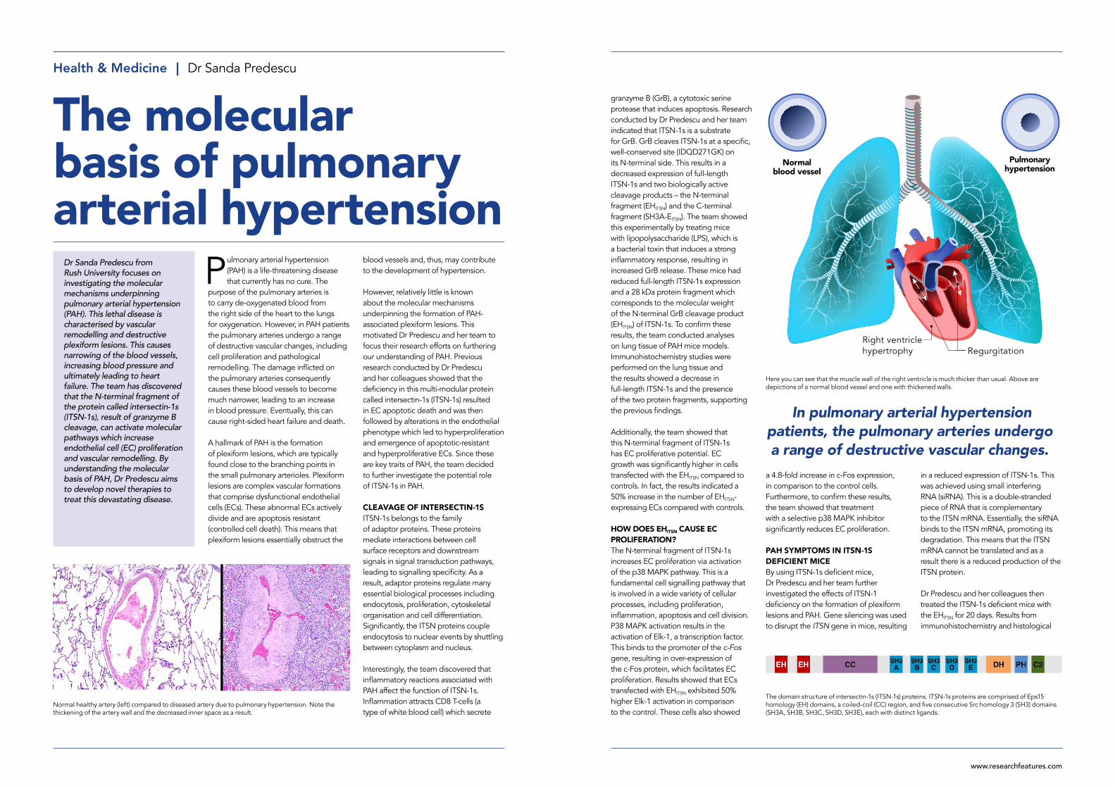
The domain structure of intersectin-1s (ITSN-1s) proteins. ITSN-1s proteins are comprised of Eps15 homology (EH) domains, a coiled-coil (CC) region, and five consecutive Src homology 3 (SH3) domains (SH3A, SH3B, SH3C, SH3D, SH3E), each with distinct ligands.
Here you can see that the muscle wall of the right ventricle is much thicker than usual. Above are depictions of a normal blood vessel and one with thickened walls.
Normal healthy artery (left) compared to diseased artery due to pulmonary hypertension. Note the thickening of the artery wall and the decreased inner space as a result.
www.researchfeatures.com
Dr Predescu’s research could potentially lead to novel therapies to treat PAH. Currently, PAH is incurable and treatments are limited, only delaying progression of the disease. Therapies include: i) drugs that enhance vasodilation of the pulmonary arteries to reduce blood pressure; ii) diuretics which reduce fluid levels in the body, lowering blood pressure; iii) atrial septostomy – a surgical procedure which improves blood flow between the chambers of the heart; and iv) lung transplant – this is sometimes the only option when PAH cases are extremely severe. Clearly there is a need for more effective therapies. Dr Predescu and her team have proposed the use of small molecules that could inhibit the cleavage of ITSN-1s by GrB. Another option could be reducing the proliferative effect of the N-terminal fragment. However, much more research is needed to explore these options.
ECs and smooth muscle cells. The cell-cell crosstalk is a biological process in which bio-active proteins synthetised by one cell type (ECs) are released and influence the proliferative and phenotypic shape of another cell type (smooth muscle cells). Dr Predescu and her colleagues also conducted transcriptional and protein assays of the lung tissue. Results showed that p38 MAPK activated Elk-1 transcription, which caused increased expression of the c-Fos gene. Therefore, these results support the findings of their previous study.
POTENTIAL THERAPIES Overall, Dr Predescu’s ground-breaking, innovative research has improved our knowledge of the molecular basis underpinning PAH. The protein fragments resulting from ITSN-1s cleavage by GrB have a significant role in increasing cell proliferation by activating the p38 MAPK signalling pathway, which in turn, activates the expression of c-Fos – a cell growth stimulator.
studies showed that in these conditions harmful vascular remodelling occurred, resulting in plexiform lesions which obstructed the lumen of the pulmonary arteries. Additionally, the team observed a two-fold increase in proliferative ECs. The team also analysed the collagen composition of the lesions and found that it was very similar to the plexiform lesions found in PAH patients. The team also discovered that right ventricular systolic pressure was elevated and right heart hypertrophy occurred, consistent with pulmonary hypertension. Furthermore, there was a 1.35-fold increase in the smooth muscle cell proliferation, suggesting a level of cross talk between
The results indicated a 50% increase in the number of EHITSN-expressing
endothelial cells compared with controls.
Representative hematoxylin and eosin (H&E) staining of paraffin-embedded lung tissue section demonstrates the formation of complex plexiform-like lesions in the EHITSN transduced ITSN-1s deficient mouse.
Detail
Research ObjectivesDr Predescu and her team recently developed a mouse model of pulmonary arterial hypertension / plexiform arteriopathy.
Sanda Predescu, PhD ProfessorRush UniversityDepartment of Internal Medicine, Division of Pulmonary and Critical Care Medicine1735 W. Harrison St.Suite 1535Chicago, IL 60612USA
Bio Dr Sanda Predescu is professor in the Department of Internal Medicine at Rush University Medical Center in Chicago. She has a PhD degree in Molecular Pathology from the University of California San Diego. Her current research is focused on endothelial cell dysfunction in the pathology of several lung diseases, including pulmonary arterial hypertension.
FundingNIH/NHLBI
Collaborators• Dan Predescu, MD • Monal Patel, PhD • Brandon Carman, MS
ReferencesJeganathan, N., Predescu, D. and Predescu, S., 2017. Intersectin-1s deficiency in pulmonary pathogenesis. Respiratory research, 18(1), p.168.
Patel, M., Predescu, D., Bardita, C., Chen, J., Jeganathan, N., Pritchard, M., DiBartolo, S., Machado, R. and Predescu, S., 2017. Modulation of intersectin-1s lung expression induces obliterative remodeling and severe plexiform arteriopathy in the murine pulmonary vascular bed. The American journal of pathology, 187(3), pp.528-542.
Patel, M., Predescu, D., Tandon, R., Bardita, C., Pogoriler, J., Bhorade, S., Wang, M., Comhair, S., Ryan-Hemnes, A., Chen, J. and Machado, R., 2013. A novel p38 mitogen-activated protein kinase/Elk-1 transcription factor-dependent molecular mechanism underlying abnormal endothelial cell proliferation in plexogenic pulmonary arterial hypertension. Journal of Biological Chemistry, 288(36), pp.25701-25716.
Roland R. (2016) Pulmonary Arterial Hypertension (PAH): Understanding Treatment Options. Healthline. Available at https://www.healthline.com/health/pulmonary-arterial-hypertension-treatments#medications (Accessed 01-07-2018)
Personal Response
How do the products of the cleaved protein ITSN-1s affect pulmonary arterial hypertension?
The interplay between the two biologically active ITSN-1s fragments has more subtle cellular effects by interfering with the uptake and transport (endocytosis, transcytosis) of small and large molecules up to 100 nm diameter. In PAH settings, to compensate for the deficient vesicular trafficking via clathrin-coated vesicles and caveolae, prolonged ITSN-1s deficiency and the presence of the two biologically active fragments cause the increased occurrence/trafficking of the alternative endocytic structures, repressed under normal conditions. By doing so, they contribute to the endocytic activity of dysfunctional ECs, their proliferation and overgrowth, and hence, the development and progression of the disease.
Red blood cells carry oxygen around the body. Their circulation is essential to the correct functioning of the human body.
Behind the ResearchSanda Predescu, PhD Dan Predescu, MD
E: [email protected] T: +1 (312) 563-2437 W: www.rushu.rush.edu/faculty/sanda-predescu-phd
www.researchfeatures.comwww.researchfeatures.com