Embed Size (px)
Citation preview
Health Occupations
Special Senses
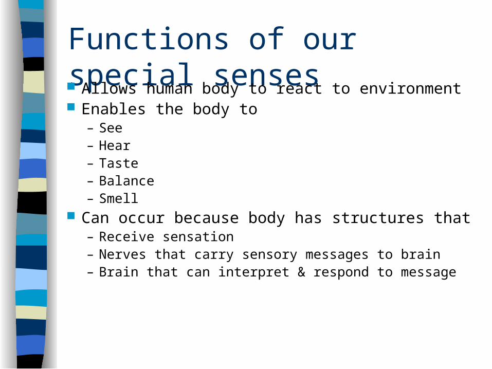
Functions of our special senses Allows human body to react to environment Enables the body to
– See– Hear– Taste– Balance– Smell
Can occur because body has structures that– Receive sensation– Nerves that carry sensory messages to brain– Brain that can interpret & respond to message
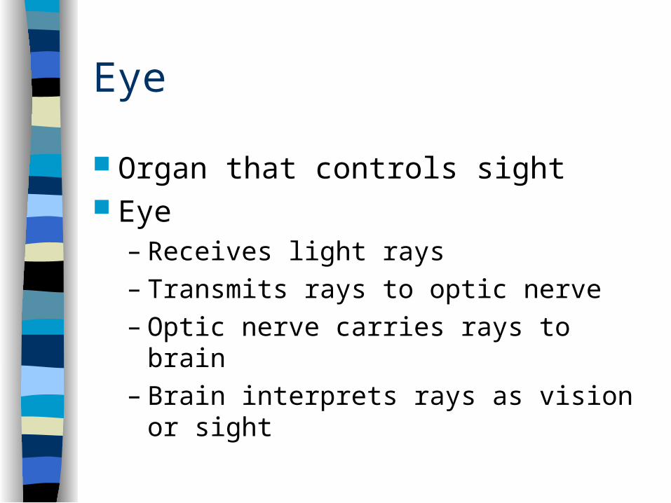
Eye
Organ that controls sight Eye
– Receives light rays– Transmits rays to optic nerve– Optic nerve carries rays to brain– Brain interprets rays as vision or sight

Protection of eye Bony socket of skull
– Partially enclosed Eye lids & lashes
– Help keep out dirt & pathogens Lacrimal glands
– Produce tears which moisten & cleanse eye– Tears flow across eye, drain through lacrimal duct into nasal
cavity Conjunctiva
– Mucous membrane that protects eye– Lines eyelids & covers front of eye– Provides protection & lubrication
3 main layers of eye
Sclera– Outermost layer– Tough connective tissue– White of eye– Maintains eye shape– Extrinsic muscles
• Responsible for moving eye in socket• Attach to outside of sclera
– Cornea• Circular transparent part on front of sclera• Allows light rays to enter eye
3 main layers of eye
Choroid coat– Middle layer of eye– Interlaced with many blood vessels that nourish
eye– Pupil
• Hole in front of choroid coat• Allows light rays in
– Iris• Colored part of eye• Muscle that controls the size of the pupil & regulates
amount of light entering eye
3 main layers of eye
Retina– Innermost layer– Made of many layers of nerve cells that transmit
light to the optic nerve– 2 types of cells in retina
• Cones – Used for light vision– Sensitive to color– Located in depression on back of retina (FOVEA
CENTRALIS) which is the area of sharpest vision
• Rods– Used mainly for dim or dark vision
Other special structures
Lens– Circular structure behind pupil– Suspended in place by ligaments– Refracts (bends) light rays so that the rays
will focus on the retina
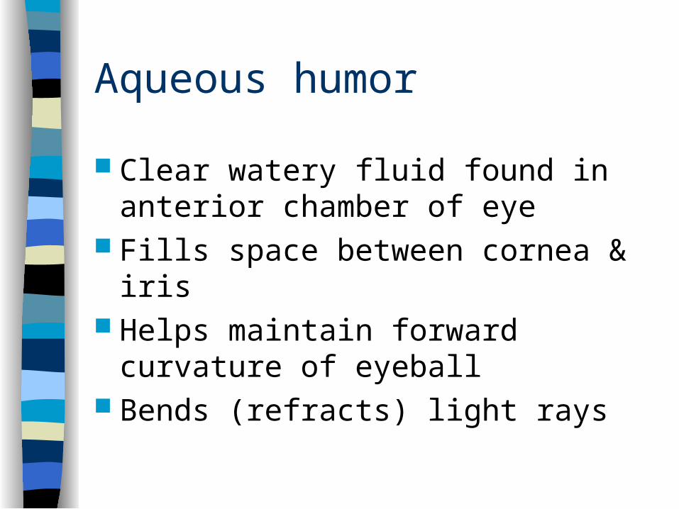
Aqueous humor
Clear watery fluid found in anterior chamber of eye
Fills space between cornea & iris Helps maintain forward curvature of
eyeball Bends (refracts) light rays

Vitreous humor
Jelly like substance found in posterior chamber of eye
Fills area behind lens Helps maintain eyeball shape Also bends or refracts light rays
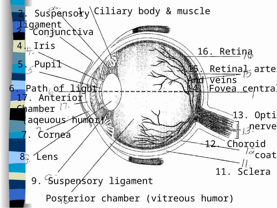
1. Ciliary body & muscle2. Suspensoryligament3. Conjunctiva
4. Iris
5. Pupil
6. Path of light
7. Cornea
8. Lens
9. Suspensory ligament
Posterior chamber (vitreous humor)
11. Sclera
12. Choroid coat
13. Optic nerve
14. Fovea centralis
15. Retinal arteriesAnd veins
16. Retina
17. AnteriorChamber (aqeuous humor)
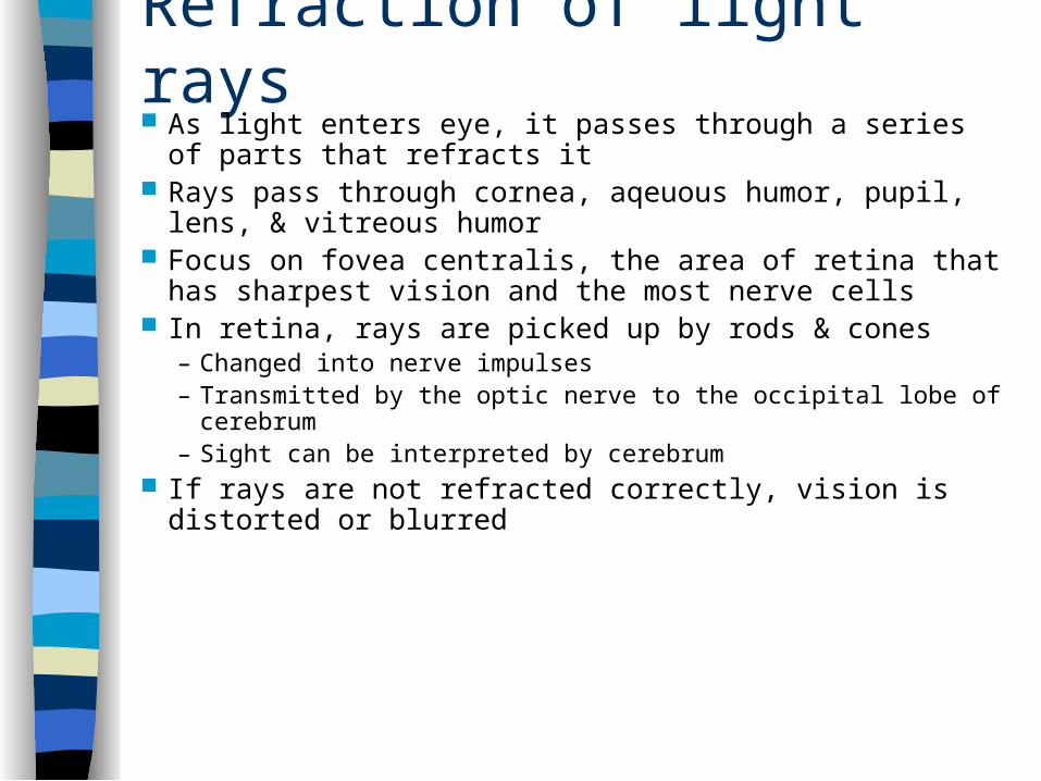
Refraction of light rays As light enters eye, it passes through a series of parts
that refracts it Rays pass through cornea, aqeuous humor, pupil, lens, &
vitreous humor Focus on fovea centralis, the area of retina that has
sharpest vision and the most nerve cells In retina, rays are picked up by rods & cones
– Changed into nerve impulses– Transmitted by the optic nerve to the occipital lobe of cerebrum– Sight can be interpreted by cerebrum
If rays are not refracted correctly, vision is distorted or blurred
Abnormal eye conditions
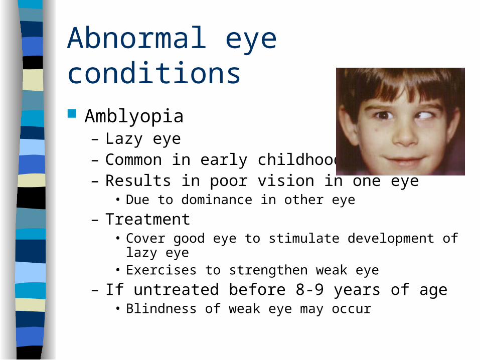
Amblyopia– Lazy eye– Common in early childhood– Results in poor vision in one eye
• Due to dominance in other eye
– Treatment• Cover good eye to stimulate development of lazy eye• Exercises to strengthen weak eye
– If untreated before 8-9 years of age• Blindness of weak eye may occur
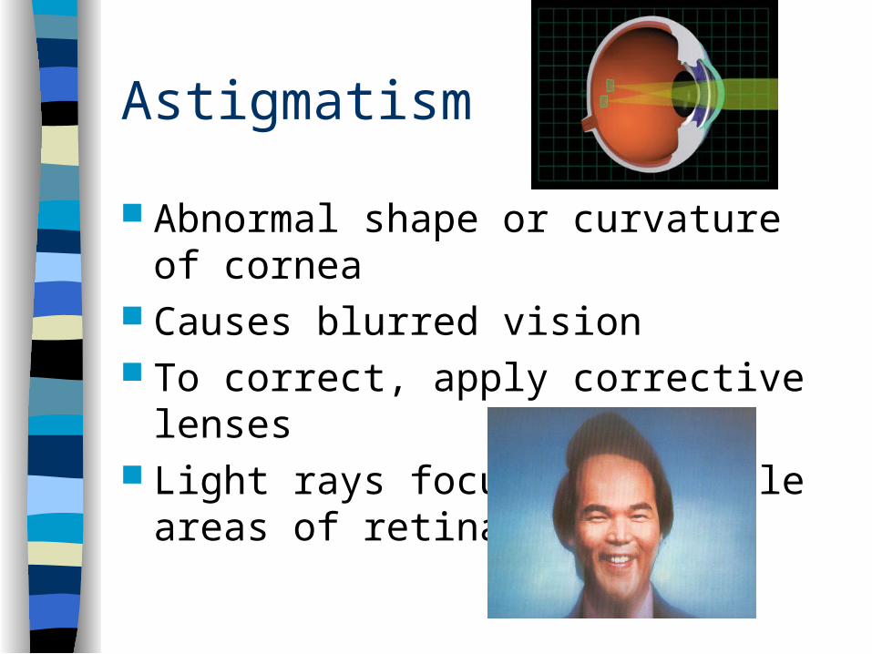
Astigmatism
Abnormal shape or curvature of cornea Causes blurred vision To correct, apply corrective lenses Light rays focus on multiple areas of
retina

Cataracts
Normally clear lens becomes cloudy or opaque
Occurs gradually, usually result of age May be caused by trauma Symptoms
– Blurred vision– Halos around lights– Gradual vision loss– Milky white pupil in late stages
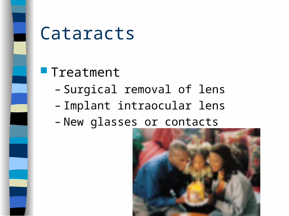
Cataracts
Treatment– Surgical removal of lens– Implant intraocular lens– New glasses or contacts
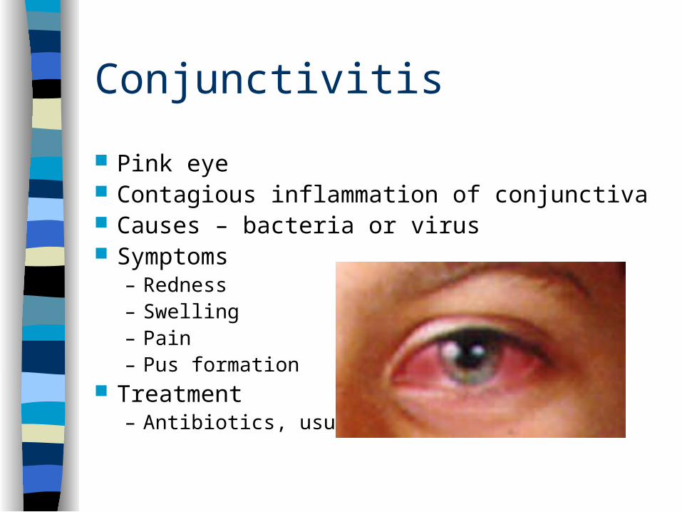
Conjunctivitis
Pink eye Contagious inflammation of conjunctiva Causes – bacteria or virus Symptoms
– Redness– Swelling– Pain– Pus formation
Treatment– Antibiotics, usually ointment

Glaucoma Results from increased intraocular pressure Caused by excess aqueous humor in anterior chamber of
eye Common after age 40 Leading cause of blindness Tonometer – instrument used to measure IOP, used
during every eye exam Symptoms
– Loss of peripheral vision– Halos around lights– Limited night vision– Mild aching
Glaucoma treatment
Controlled with meds – Decrease amount of aqueous humor– Improve drainage of aqueous humor
Surgery to create an opening for the flow of aqueous humor
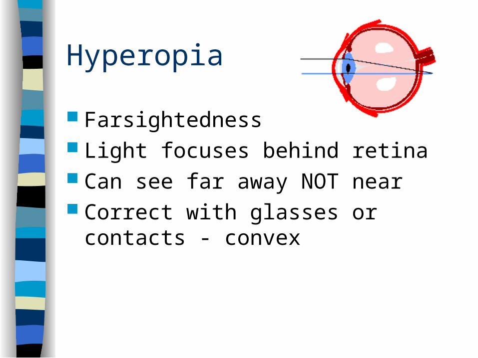
Hyperopia
Farsightedness Light focuses behind retina Can see far away NOT near Correct with glasses or contacts -
convex
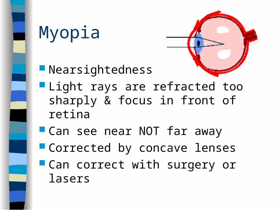
Myopia
Nearsightedness Light rays are refracted too sharply &
focus in front of retina Can see near NOT far away Corrected by concave lenses Can correct with surgery or lasers
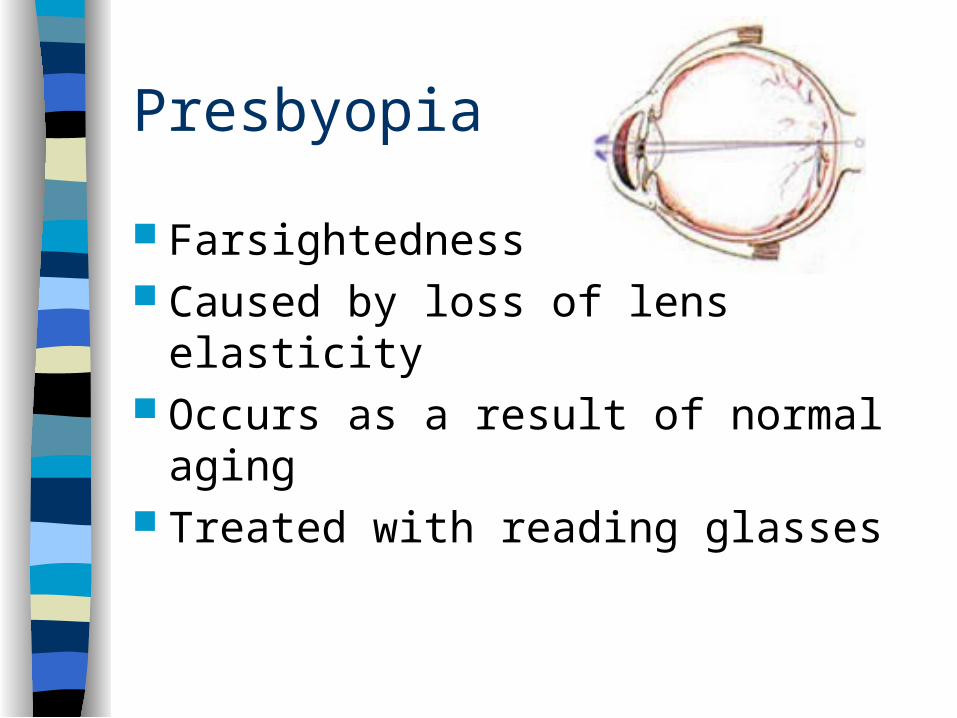
Presbyopia
Farsightedness Caused by loss of lens elasticity Occurs as a result of normal aging Treated with reading glasses
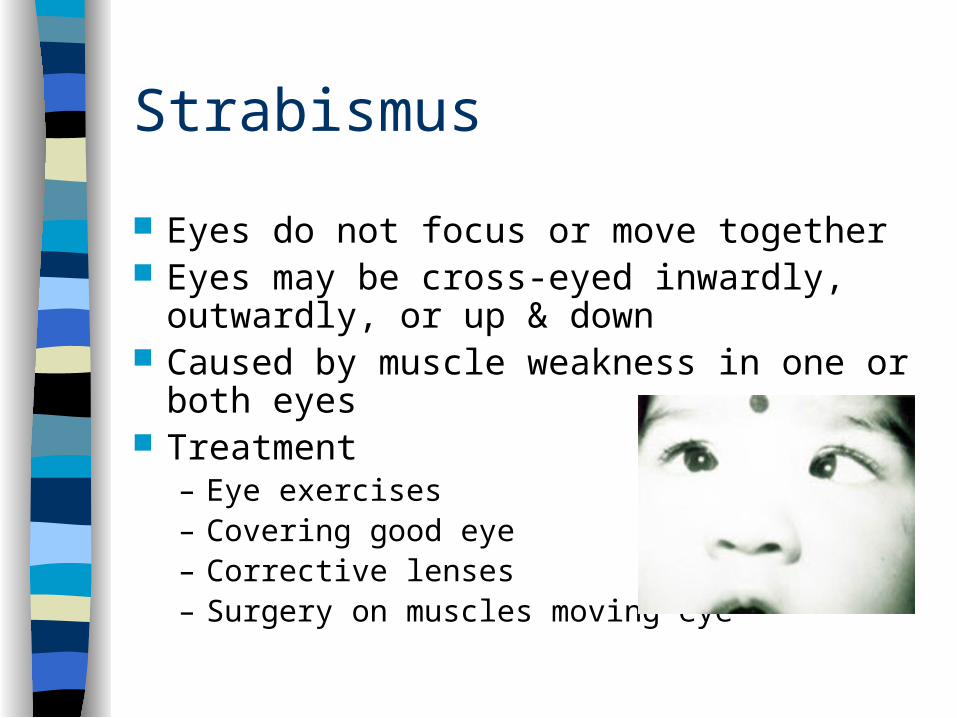
Strabismus
Eyes do not focus or move together Eyes may be cross-eyed inwardly, outwardly,
or up & down Caused by muscle weakness in one or both
eyes Treatment
– Eye exercises– Covering good eye– Corrective lenses– Surgery on muscles moving eye
Ear
Organ that controls hearing & balance 3 main sections – outer, middle, inner Hearing
– Transmits impulses from sound waves to auditory nerve (vestibulocochlear)
– Auditory nerve carries impulses to brain– Brain interprets as hearing
Outer ear Auricle or pinna
– Visible part of ear– Elastic cartilage covered by skin– Leads to canal called the external auditory meatus and auditory
canal Auditory canal
– Contains special ceruminous glands producing cerumin (wax) for protection
– Sound waves travel through this to reach tympanic membrane Tympanic membrane
– Separates external & middle ear– Vibrates when sound hits it– Transmits sound waves to middle ear
Middle Ear Small space or cavity in temporal bone Contains 3 small bones (ossicles)
– Malleus– Incus– Stapes
Bones connected & transmit sound waves from tympanic membrane to inner ear
Eustachion tube– Tube connecting middle ear to pharynx that allows air to
enter the middle ear– Helps to equalize pressure on both sides of tympanic
membrane
Inner ear Most complex part of ear Oval window
– Membrane separating inner & middle ear Vestibule
– First section acting as entrance to 2 other parts of inner ear Cochlea
– Snail shaped – Contains delicate hair like cells that make up ORGAN OF
CORTI– Receives sound waves & transmits impulses to auditory
nerve
Inner ear
Semicircular canals– Contain liquid & hair like cells– Bend when liquids move with head & body
movements– Impulses sent to cerebellum to help
maintain balance & equilibrium
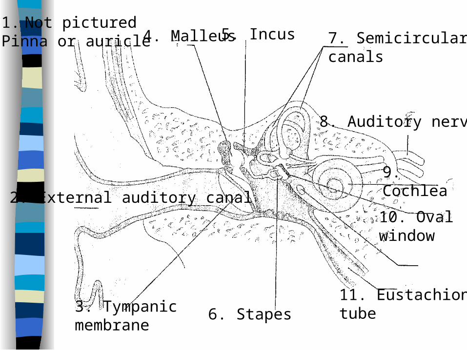
2. External auditory canal
1. Not pictured Pinna or auricle
3. Tympanic membrane
4. Malleus 5. Incus
6. Stapes
7. Semicircularcanals
8. Auditory nerve
9. Cochlea
10. Ovalwindow
11. Eustachiontube
Abnormal conditions
Hearing loss – 2 types– Conduction
• Caused by sound waves not being conducted to inner ear• Causes
– Cerumen plug– Foreign body obstruction – Otosclerosis– Infection– Ruptured tympanic membrane
• Treatment– Eliminate cause– Surgery– Hearing aids
Sensory hearing loss
Caused by damage to inner ear or auditory nerve
Usually can NOT be corrected Cochlear implants can improve severe
hearing loss
Meniere’s disease Collection of fluid in labyrinth of the inner ear &
degeneration of hair cells in cochlea & vestibule Symptoms
– Severe vertigo or dizziness– Tinnitus (ringing in ears)– N&V– Loss of balance & tendency to fall
Treatment– Drugs to reduce fluids & antihistamines– Drainage of fluid– Surgery to destroy cochlea in severe cases (causes
permanent deafness)
Otitis externa
Inflammation of external auditory canal Variety of causes
– Swimmer’s ear– Inserting foreign objects (bobby pins, fingernails,
cotton swabs)
Treatment– Antibiotics– Warm moist compresses– Pain meds
Otitis media Inflammation or infection of middle ear Causes – bacteria or virus Frequently follows sore throat because organisms
can enter ear through Eustachion tubes Susceptible –
– Infants & young children– Eustachion tube is angled differently than in adults &
secretions from nose & throat accumulate in middle ear– This causes inflammatory response that causes tube to
swell shut
Otitis media
Symptoms– Severe pain– Fever– Vertigo– Dizziness– N & V– Buildup of fluid in middle ear
Treatment– Antibiotics & pain meds– Myringotomy & tubes
Otosclerosis
Stapes becomes immobile & causes conductive hearing loss
Symptoms– Gradual hearing loss– Tinnitus– vertigo
Treatment– Surgical removal of stapes– Insertion of artificial stapes
Taste
Dependent on taste receptors located on tongue– Tongue is mass of muscle tissue with projections
called papillae– Papillae contain taste buds which are stimulated by
flavors & moistened by saliva Four main tastes
– Sweet & salty – tip of tongue– Sour – sides of tongue– Bitter – back of tongue
Influenced by smell
Smell
Nose is organ Determined by olfactory receptors in upper
part of nasal cavity Impulses from receptors are carried to brain
by olfactory nerve Sense of smell closely related to taste
– Smell much more sensitive– Human nose can detect over 6,000 different
smells
Skin & general senses
General sense receptors for pressure, heat, cold, touch, & pain located throughout body in skin & connective tissue
Each receptor perceives only 1 type of sense Messages from receptors allow human body
to respond to environment Helps body to react to conditions that could
cause injury