Embed Size (px)
Citation preview
1
Comenius University in Bratislava
Jessenius Faculty of Medicine in Martin
Department of Anatomy
HEART AND RESPIRATORY SYSTEM
STUDY GUIDE
Desanka Výbohová
Gabriela Hešková
Yvetta Mellová
Martin 2019
2
Authors:
Doc. MUDr. Desanka Výbohová, PhD.
MUDr. Gabriela Hešková, PhD.
Doc. MUDr. Yvetta Mellová, CSc.
Authors themselves are responsible for the content and English of the chapters.
Reviewers:
Prof. MUDr. Marian Adamkov, DrSc.
MUDr. Zuzana Lazarová, PhD.
ISBN 978-80-8187-065-1
EAN 9788081870651
3
TABLE OF CONTENT
Preface.................................................................................................................................6
HEART................................................................................................................................ 7
Position of the heart...............................................................................................................7
Relations of the heart...........................................................................................................10
External features of the heart...............................................................................................13
Pericardium..........................................................................................................................18
Cardiac wall.........................................................................................................................22
Cardiac skeleton...................................................................................................................23
Valves of the heart................................................................................................................25
Cavities of the heart..............................................................................................................29
Right atrium..........................................................................................................................29
Right ventricle.......................................................................................................................31
Left atrium.............................................................................................................................33
Left ventricle.........................................................................................................................34
Vessel of the heart.................................................................................................................36
Right coronary artery.............................................................................................................37
Left coronary artery...............................................................................................................39
Veins of the heart...................................................................................................................42
Lymphatic drainage of the heart............................................................................................44
Conduction system of the heart..............................................................................................45
Nerves of the heart.................................................................................................................46
Introduction to the blood circulation......................................................................................48
Foetal circulation....................................................................................................................49
4
RESPIRATORY SYSTEM ................................................................................................. 53
Nose ....................................................................................................................................... 54
EXTERNAL NOSE ............................................................................................................. 54
Vessels and nerves of external nose ....................................................................................... 55
NASAL CAVITY ................................................................................................................. 56
Vessels and nerves of nasal cavity ......................................................................................... 59
PARANASAL SINUSES ..................................................................................................... 61
Vessels and nerves of paranasal sinuses ................................................................................ 62
LARYNX .............................................................................................................................. 63
Anatomical position and relations of larynx to neighbouring structures and organs ............ 63
Laryngeal cartilages ............................................................................................................... 63
Laryngeal joints ..................................................................................................................... 66
Laryngeal membranes and ligaments .................................................................................... 67
Laryngeal muscles .................................................................................................................. 69
Laryngeal inlet ........................................................................................................................ 73
Laryngeal cavity ..................................................................................................................... 74
Vessels and nerves of larynx ................................................................................................. 77
TRACHEOBRONCHIAL TREE, TRACHEA, BRONCHI AND BRONCHIOLES ....78
TRACHEA ............................................................................................................................ 79
Anatomical position and relations of trachea to neighbouring structures and organs .......... 79
Composition of trachea ......................................................................................................... 80
Vessels and nerves of trachea ............................................................................................... 81
MAIN BRONCHI (PRIMARY, PRINCIPAL) ................................................................ 83
Composition of main bronchi ................................................................................................ 83
Vessels and nerves of main bronchi ...................................................................................... 83
LUNGS, PLEURA AND MEDIASTINUM ..................................................................... 84
LUNGS ................................................................................................................................. 84
Groos anatomy of lungs .........................................................................................................84
Root and hilum of lungs ........................................................................................................88
Intrapulmonary part of the air-conducting zone, bronchopulmonary segment .....................89
Composition of intrapulmonary bronchi and bronchioles .....................................................89
Relations, grooves and impressions of lungs ........................................................................ 92
Projection of lungs onto skeleton .......................................................................................... 95
5
Vessels and nerves of lungs ................................................................................................... 95
PLEURA ............................................................................................................................... 97
Visceral (pulmonary) pleura .................................................................................................. 97
Parietal pleura ........................................................................................................................ 97
Pleural recesses ...................................................................................................................... 98
Projection of pleura onto skeleton ......................................................................................... 98
Vessels and nerves of pleura .................................................................................................. 99
MEDIASTINUM ................................................................................................................ 101
REVIEW QUESTIONS .................................................................................................... 103
REFERENCES .................................................................................................................. 123
6
PREFACE
This study guide has been prepared for medical students as a basic educational material
for the anatomy practicals and anatomy exam.
The first chapter provides introduction to the cardiovascular system, the heart and basic
description of the circulations.
The second chapter deals with the organs of respiratory system.
The text is fulfilled by the photographs of formalin fixed cadaveric preparates used
during the anatomy practicals in our department, however, it is necessary combine the study
with using of anatomy atlases (Sobotta´s, Gray´s, Gilroy ́ s or Netter´s atlas of human anatomy).
Several figures of joints and ligaments are retrieved from public domein of Gray´s Anatomy of
human Body (1918) and Servier Medical Art.
The last part of the study guide contains a set of review questions which can help the
students to check their knowledge.
We hope you will find this study guide useful material for your individual study
and review.
Authors
7
Heart (Cor)
The heart is a hollow muscular organ consisting of four chambers: the right atrium and
ventricle, and the left atrium and vetricle. The shape of the heart is usually described as conical
or pyramidal and the size of the heart is generally comparable with a fist of a respective human.
The average weight of the heart is 230 – 340 g or 0.40 – 0.45% of the total body weight.
The myocardium rhythmically contracts and relaxes and thus pumps the blood throught
the heart.
The heart works as two pumps. The left part of the heart pumps the oxygenated blood
to the systemic circulation. The right part pumps the deoxygenated blood to the small (lung)
circulation.
The period when the chamber of the heart is relaxed and filling with a blood is called a
diastole. Contraction of the heart chamber when the blood is ejecting from the chamber is called
a systole.
The right atrium of the heart receives the deoxygenated blood of the body. This blood
continues into the right ventricle. From the right ventricle the blood is ejected into the
pulmonary arteries to reach the lungs where the deoxygenated blood is oxygenating.
The oxygenated blood from the lungs is transported via the pulmonary veins into the
left atrium. The blood from the left atrium inflows to the left ventricle and during the contraction
(systole) of the left ventricle it is ejected to the aorta and from the aorta via the arteries to the
whole body.
Position of the heart
The heart enclosed in pericardium lies in the thoracic cavity - in the mediastinum (the
space between the pleural cavities). According the anatomical subdivision of mediastinum, the
heart is situated in lower middle mediastinum. However, clinicians usually use easier clinical
subdivision of mediastinum and according to this clinical classification the heart is in anterior
mediastinum. As for the position of the heart to the median plane, one third of the heart is
situated on the right side and two thirds on the left side.
8
The axis of the heart (imaginary line passing from the base of the heart to the apex of
the heart) directs anteriorly, inferiorly and to the left.
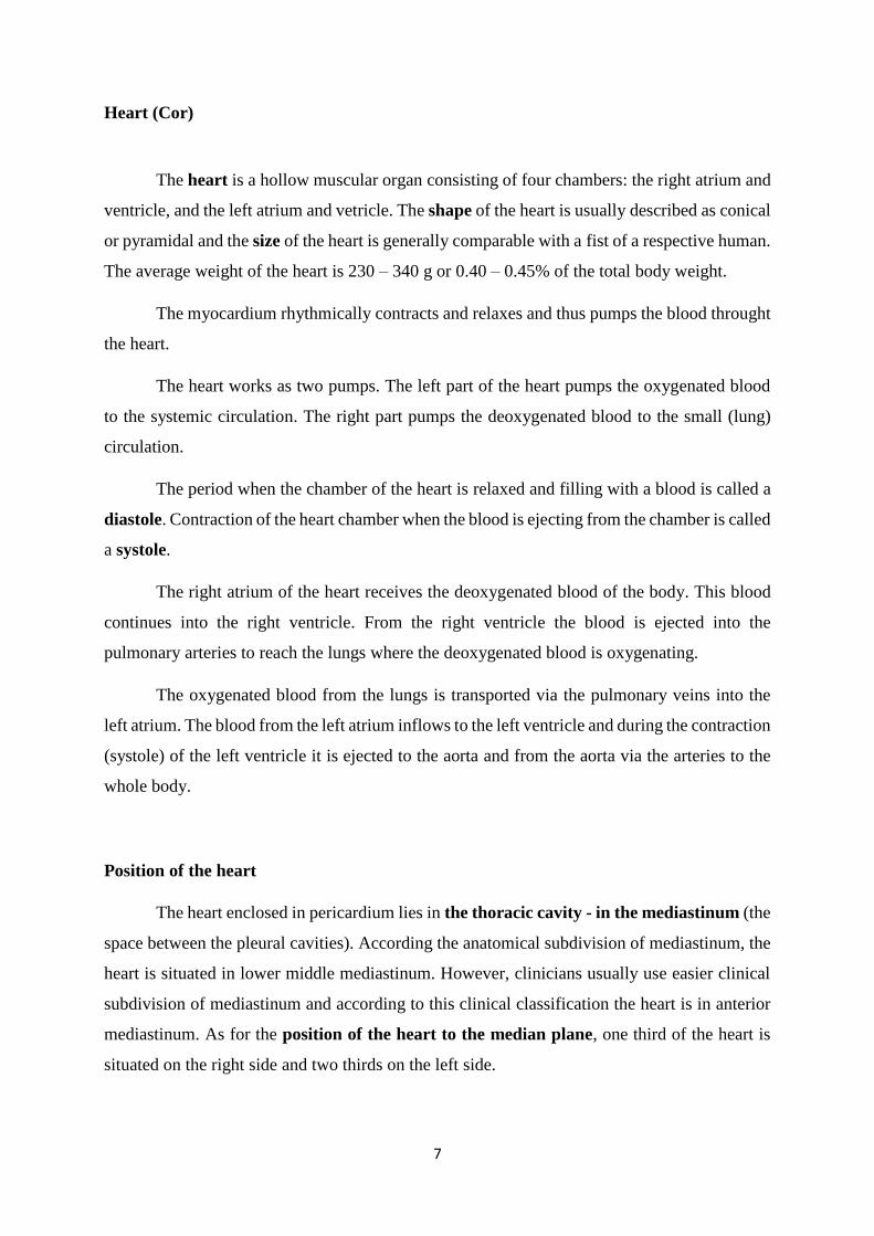
Fig. 1
Heart in pericardium in the thorax
Anterior view
Dissection of formalin-fixed cadaver
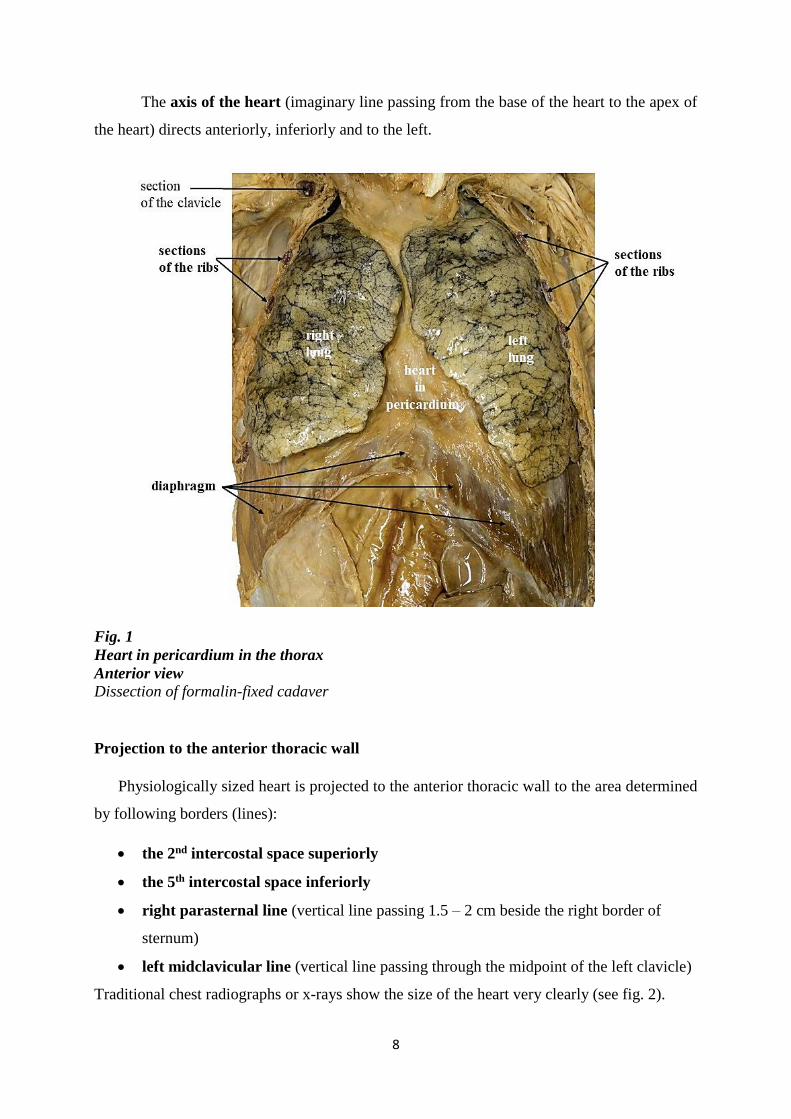
Projection to the anterior thoracic wall
Physiologically sized heart is projected to the anterior thoracic wall to the area determined
by following borders (lines):
the 2nd intercostal space superiorly
the 5th intercostal space inferiorly
right parasternal line (vertical line passing 1.5 – 2 cm beside the right border of
sternum)
left midclavicular line (vertical line passing through the midpoint of the left clavicle)
Traditional chest radiographs or x-rays show the size of the heart very clearly (see fig. 2).
9
Fig. 2
Chest radiograph
of physiologically
sized heart.
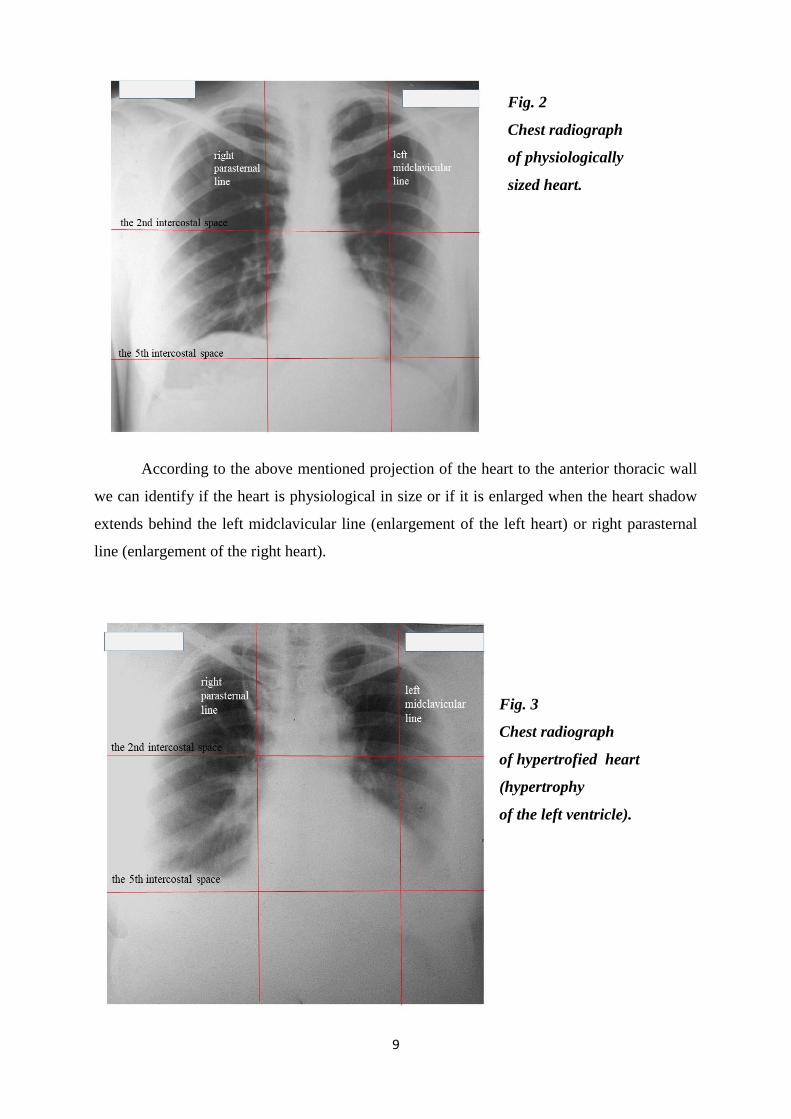
According to the above mentioned projection of the heart to the anterior thoracic wall
we can identify if the heart is physiological in size or if it is enlarged when the heart shadow
extends behind the left midclavicular line (enlargement of the left heart) or right parasternal
line (enlargement of the right heart).
Fig. 3
Chest radiograph
of hypertrofied heart
(hypertrophy
of the left ventricle).
10
Enlargement of the heart can be caused by the hypertrophy and/or the dilation of the
heart. The left side of the heart mainly the left ventricle is the most often enlarged from the
arterial hypertension (high arterial blood pressure). The left ventricle have to pump the blood
against higher resistence in arterial circulation thus it becomes hypertrophic. The enlargement
of the right ventricle usually results from the pulmonary hypertension that is caused by
pulmonary diseases. The acute enlargement of the right ventricle (dilation of the right ventricle)
usually results from pulmonary embolia. Chronic hypertrophy of the righ ventricle can be
caused e.g. by pulmonary fibrosis or bronchial athma. Certain pathological conditions can lead
to the enormous enlargment of the heart termed cardiomegaly or „cor bovinum“, when the
weight of the heart can increase up to 1 kg. However, the enlarged heart with the weight above
500g is in high risk of myocardial ischemia because usual blood flow in the coronary arteries
is insufficient for such enlarged mass of myocardium.
Interestingly, the hearts of the athlets can show „physiological enlargement of the left
ventricle“. So called „athlet's heart“ is a result of the endurance excercise training leading
to the physiologic growth caused by both hypertrophy and neoformation of cardiomyocytes and
concomitant angiogenesis.
Relations of the heart
Each physician should have a perfect knowledge as for the relations of the organs. It is
important as for the understanding how the pathological processes (tumour, inflamation) can
spread from affected organ to the other surronding ones „per continuitatem“ through
surrounding tissue (not by the blood or lymph).
The heart is related to the following organs:
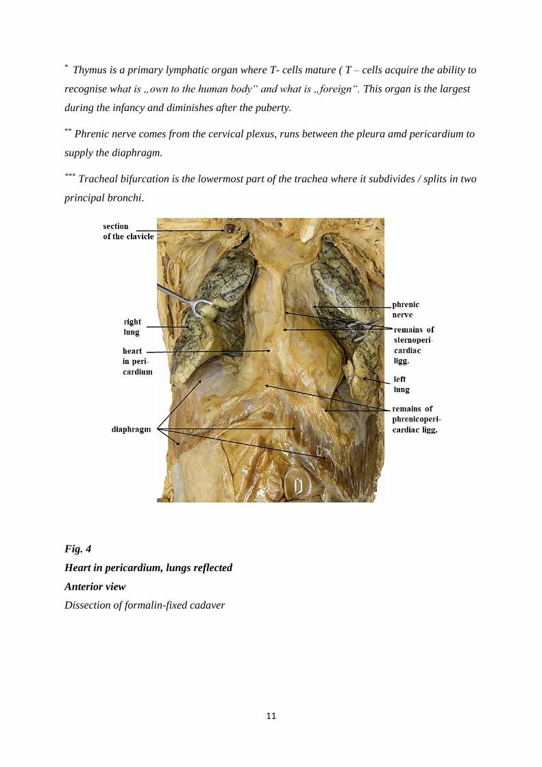
• Anteriorly, in front of the heart, there is thymus* or its remnants, sternum and ribs.
• Laterally, by the sides, there are lungs (inside the pleural cavities) and
phrenic nerves**
• Posteriorly, behind the heart, there is trachea, tracheal bifurcation***, principal
bronchi, oesophagus and thoracic aorta.
• Inferiorly, below the heart, there is the diaphragm and below it the liver and
stomach.
11
* Thymus is a primary lymphatic organ where T- cells mature ( T – cells acquire the ability to
recognise what is „own to the human body“ and what is „foreign“. This organ is the largest
during the infancy and diminishes after the puberty.
** Phrenic nerve comes from the cervical plexus, runs between the pleura amd pericardium to
supply the diaphragm.
*** Tracheal bifurcation is the lowermost part of the trachea where it subdivides / splits in two
principal bronchi.
Fig. 4
Heart in pericardium, lungs reflected
Anterior view
Dissection of formalin-fixed cadaver
12
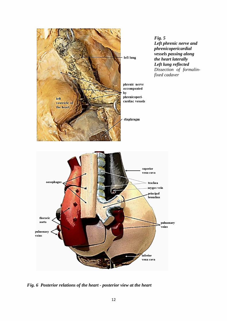
Fig. 5
Left phrenic nerve and
phrenicopericardial
vessels passing along
the heart laterally
Left lung reflected
Dissection of formalin-
fixed cadaver
Fig. 6 Posterior relations of the heart - posterior view at the heart
13
Cardiac massage
Rhytmic compressing of the chest also causes the compression of the ventricles and thus
the blood is ejected into the great vessels. This procedure can provide some blood flow to the
brain and the other organs to reduce the ischemic injuries and postpone the metabolic
deterioration.
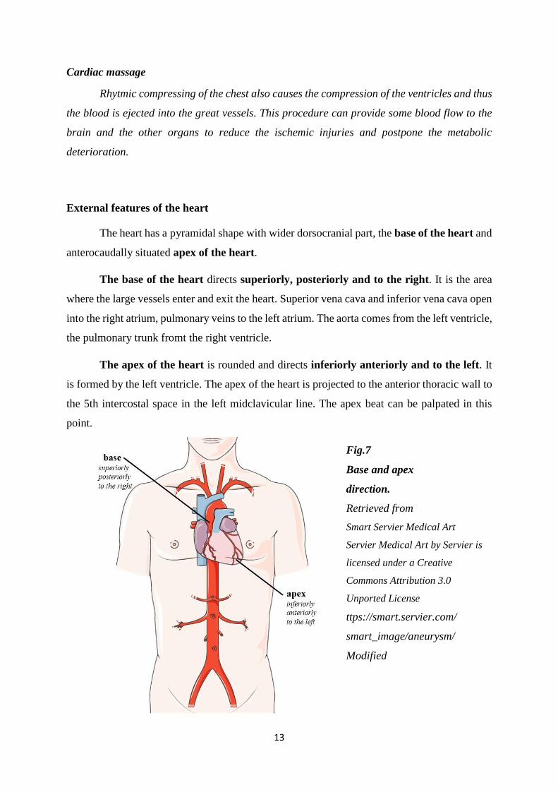
External features of the heart
The heart has a pyramidal shape with wider dorsocranial part, the base of the heart and
anterocaudally situated apex of the heart.
The base of the heart directs superiorly, posteriorly and to the right. It is the area
where the large vessels enter and exit the heart. Superior vena cava and inferior vena cava open
into the right atrium, pulmonary veins to the left atrium. The aorta comes from the left ventricle,
the pulmonary trunk fromt the right ventricle.
The apex of the heart is rounded and directs inferiorly anteriorly and to the left. It
is formed by the left ventricle. The apex of the heart is projected to the anterior thoracic wall to
the 5th intercostal space in the left midclavicular line. The apex beat can be palpated in this
point.
Fig.7
Base and apex
direction.
Retrieved from
Smart Servier Medical Art
Servier Medical Art by Servier is
licensed under a Creative
Commons Attribution 3.0
Unported License
ttps://smart.servier.com/
smart_image/aneurysm/
Modified
14
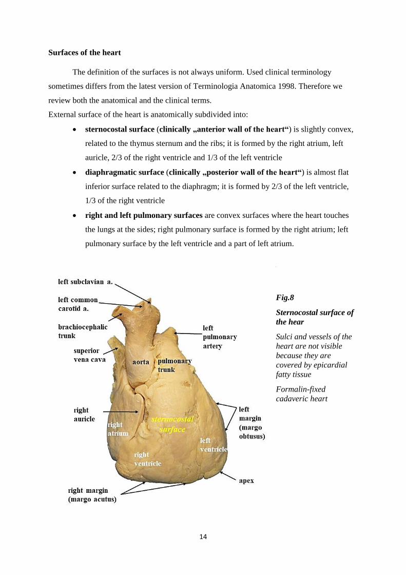
Surfaces of the heart
The definition of the surfaces is not always uniform. Used clinical terminology
sometimes differs from the latest version of Terminologia Anatomica 1998. Therefore we
review both the anatomical and the clinical terms.
External surface of the heart is anatomically subdivided into:
sternocostal surface (clinically „anterior wall of the heart“) is slightly convex,
related to the thymus sternum and the ribs; it is formed by the right atrium, left
auricle, 2/3 of the right ventricle and 1/3 of the left ventricle
diaphragmatic surface (clinically „posterior wall of the heart“) is almost flat
inferior surface related to the diaphragm; it is formed by 2/3 of the left ventricle,
1/3 of the right ventricle
right and left pulmonary surfaces are convex surfaces where the heart touches
the lungs at the sides; right pulmonary surface is formed by the right atrium; left
pulmonary surface by the left ventricle and a part of left atrium.
F
Fig.8
Sternocostal surface of
the hear
Sulci and vessels of the
heart are not visible
because they are
covered by epicardial
fatty tissue
Formalin-fixed
cadaveric heart
15
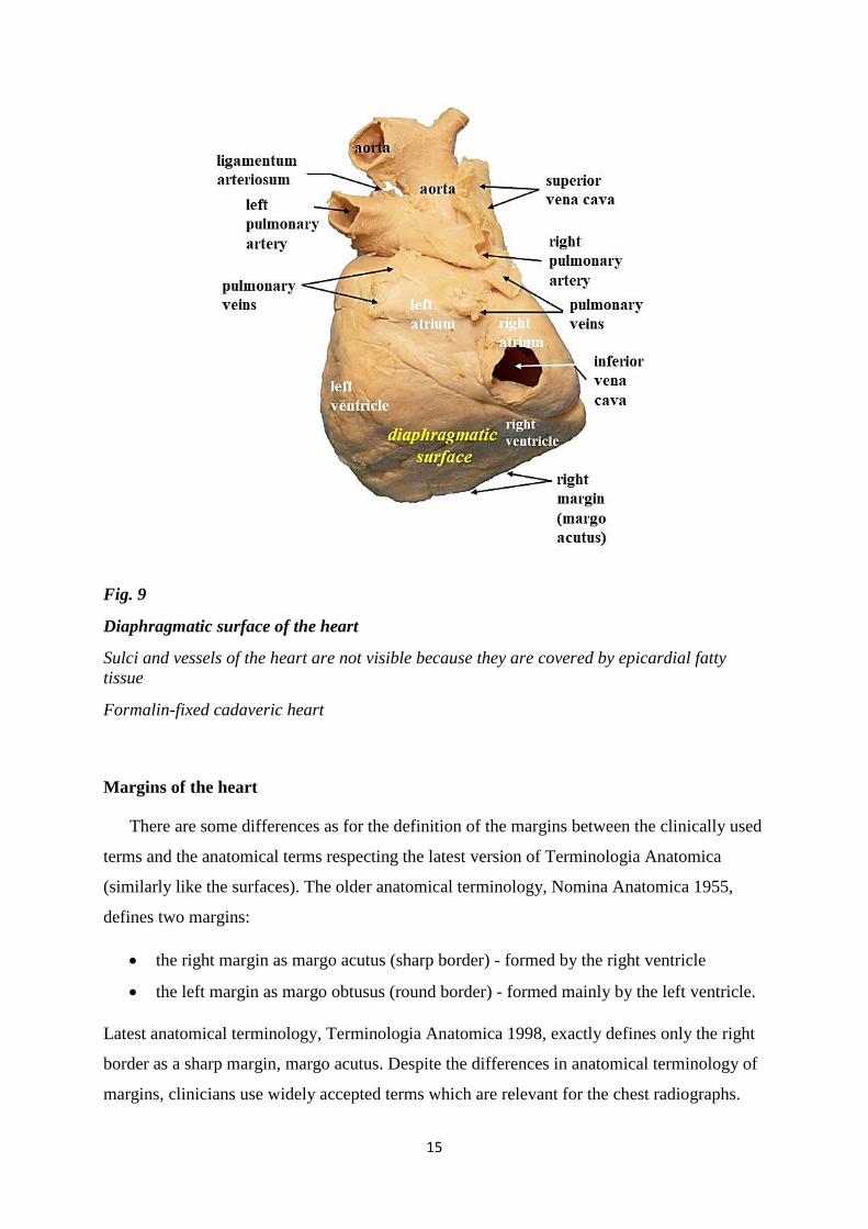
Fig. 9
Diaphragmatic surface of the heart
Sulci and vessels of the heart are not visible because they are covered by epicardial fatty
tissue
Formalin-fixed cadaveric heart
Margins of the heart
There are some differences as for the definition of the margins between the clinically used
terms and the anatomical terms respecting the latest version of Terminologia Anatomica
(similarly like the surfaces). The older anatomical terminology, Nomina Anatomica 1955,
defines two margins:
the right margin as margo acutus (sharp border) - formed by the right ventricle
the left margin as margo obtusus (round border) - formed mainly by the left ventricle.
Latest anatomical terminology, Terminologia Anatomica 1998, exactly defines only the right
border as a sharp margin, margo acutus. Despite the differences in anatomical terminology of
margins, clinicians use widely accepted terms which are relevant for the chest radiographs.
16
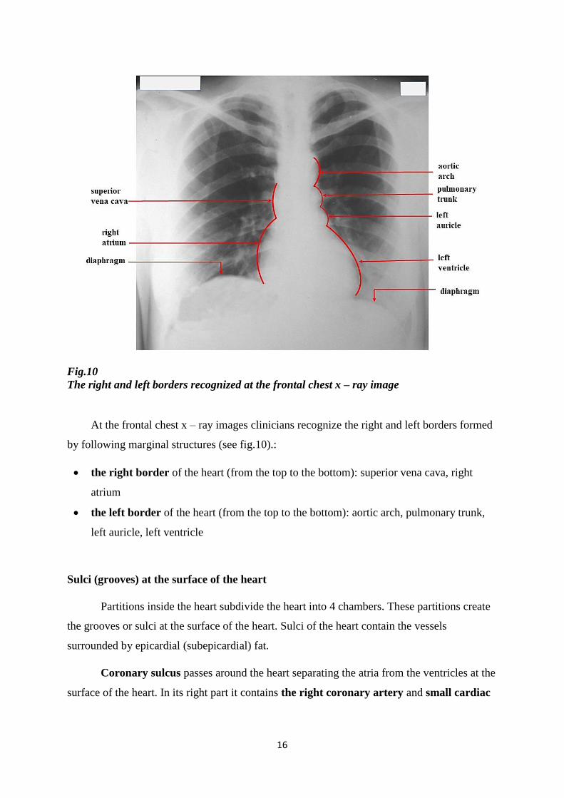
Fig.10
The right and left borders recognized at the frontal chest x – ray image
At the frontal chest x – ray images clinicians recognize the right and left borders formed
by following marginal structures (see fig.10).:
the right border of the heart (from the top to the bottom): superior vena cava, right
atrium
the left border of the heart (from the top to the bottom): aortic arch, pulmonary trunk,
left auricle, left ventricle
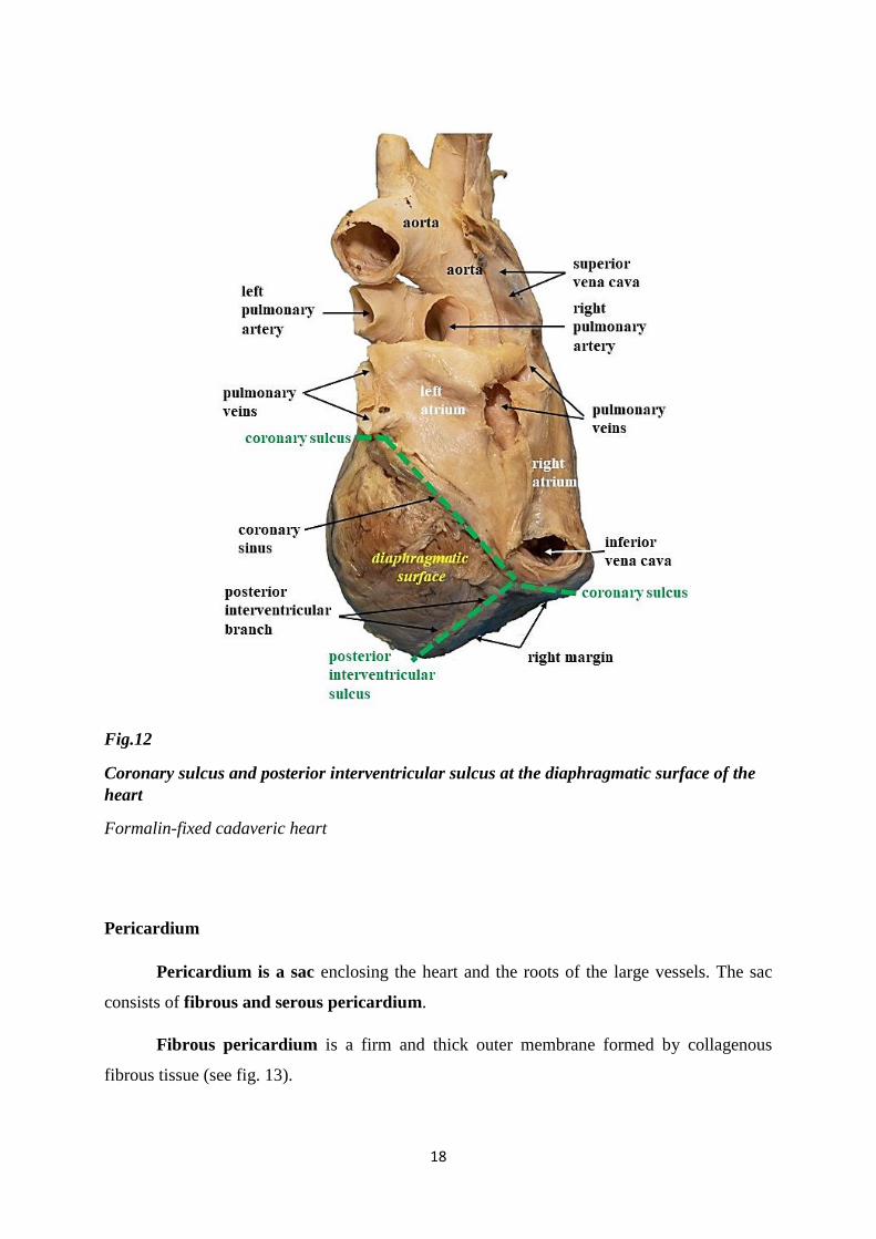
Sulci (grooves) at the surface of the heart
Partitions inside the heart subdivide the heart into 4 chambers. These partitions create
the grooves or sulci at the surface of the heart. Sulci of the heart contain the vessels
surrounded by epicardial (subepicardial) fat.
Coronary sulcus passes around the heart separating the atria from the ventricles at the
surface of the heart. In its right part it contains the right coronary artery and small cardiac
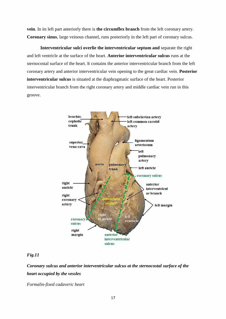
17
vein. In its left part anteriorly there is the circumflex branch from the left coronary artery.
Coronary sinus, large veinous channel, runs posteriorly in the left part of coronary sulcus.
Interventricular sulci overlie the interventricular septum and separate the right
and left ventricle at the surface of the heart. Anterior interventricular sulcus runs at the
sternocostal surface of the heart. It contains the anterior interventricular branch from the left
coronary artery and anterior interventricular vein opening to the great cardiac vein. Posterior
interventricular sulcus is situated at the diaphragmatic surface of the heart. Posterior
interventricular branch from the right coronary artery and middle cardiac vein run in this
groove.
Fig.11
Coronary sulcus and anterior interventricular sulcus at the sternocostal surface of the
heart occupied by the vessles
Formalin-fixed cadaveric heart
18
Fig.12
Coronary sulcus and posterior interventricular sulcus at the diaphragmatic surface of the
heart
Formalin-fixed cadaveric heart
Pericardium
Pericardium is a sac enclosing the heart and the roots of the large vessels. The sac
consists of fibrous and serous pericardium.
Fibrous pericardium is a firm and thick outer membrane formed by collagenous
fibrous tissue (see fig. 13).
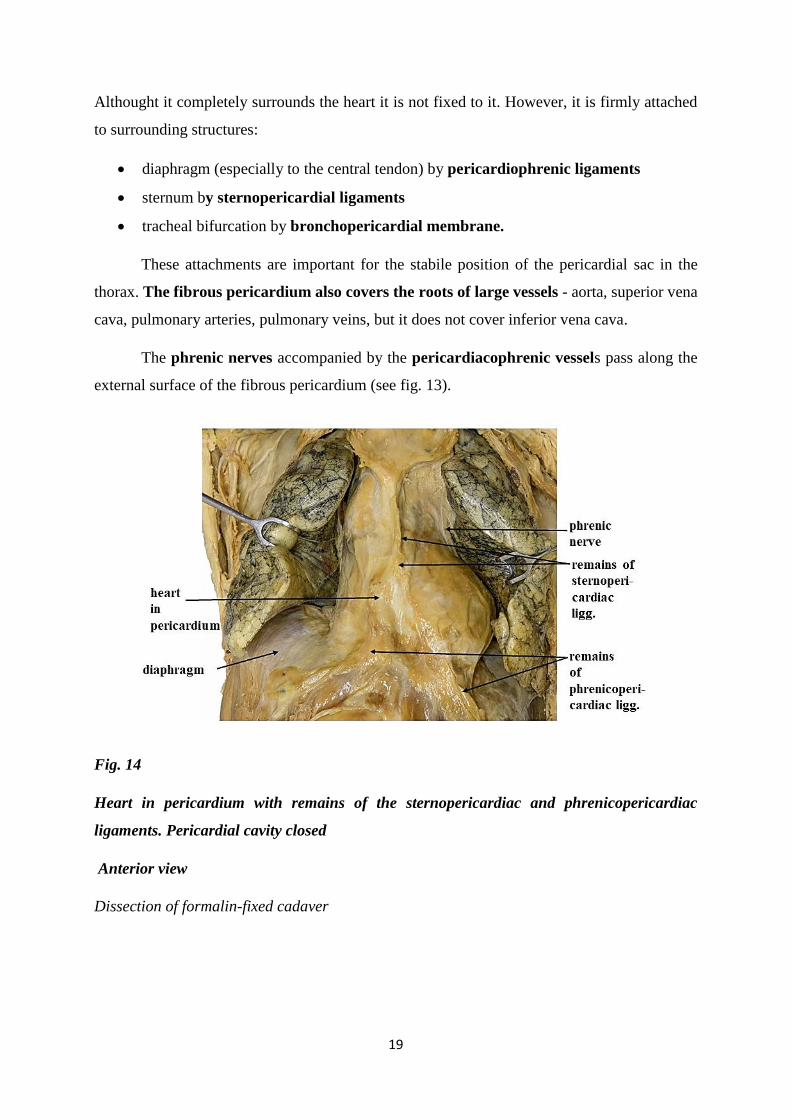
19
Althought it completely surrounds the heart it is not fixed to it. However, it is firmly attached
to surrounding structures:
diaphragm (especially to the central tendon) by pericardiophrenic ligaments
sternum by sternopericardial ligaments
tracheal bifurcation by bronchopericardial membrane.
These attachments are important for the stabile position of the pericardial sac in the
thorax. The fibrous pericardium also covers the roots of large vessels - aorta, superior vena
cava, pulmonary arteries, pulmonary veins, but it does not cover inferior vena cava.
The phrenic nerves accompanied by the pericardiacophrenic vessels pass along the
external surface of the fibrous pericardium (see fig. 13).
Fig. 14
Heart in pericardium with remains of the sternopericardiac and phrenicopericardiac
ligaments. Pericardial cavity closed
Anterior view
Dissection of formalin-fixed cadaver
20
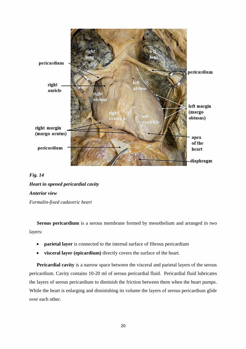
Fig. 14
Heart in opened pericardial cavity
Anterior view
Formalin-fixed cadaveric heart
Serous pericardium is a serous membrane formed by mesothelium and arranged in two
layers:
parietal layer is connected to the internal surface of fibrous pericardium
visceral layer (epicardium) directly covers the surface of the heart.
Pericardial cavity is a narrow space between the visceral and parietal layers of the serous
pericardium. Cavity contains 10-20 ml of serous pericardial fluid. Pericardial fluid lubricates
the layers of serous pericardium to diminish the friction between them when the heart pumps.
While the heart is enlarging and diminishing its volume the layers of serous pericardium glide
over each other.
21
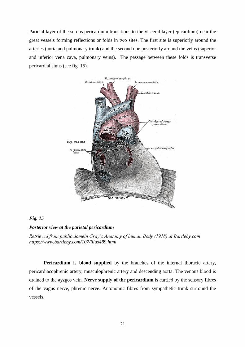
Parietal layer of the serous pericardium transitions to the visceral layer (epicardium) near the
great vessels forming reflections or folds in two sites. The first site is superiorly around the
arteries (aorta and pulmonary trunk) and the second one posteriorly around the veins (superior
and inferior vena cava, pulmonary veins). The passage between these folds is transverse
pericardial sinus (see fig. 15).
Fig. 15
Posterior view at the parietal pericardium
Retrieved from public domein Gray´s Anatomy of human Body (1918) at Bartleby.com
https://www.bartleby.com/107/illus489.html
Pericardium is blood supplied by the branches of the internal thoracic artery,
pericardiacophrenic artery, musculophrenic artery and descending aorta. The venous blood is
drained to the ayzgos vein. Nerve supply of the pericardium is carried by the sensory fibres
of the vagus nerve, phrenic nerve. Autonomic fibres from sympathetic trunk surround the
vessels.
22
Cardiac tamponade
Pericardium has no elasticity it is tough. Therefore any excessive accumulation of the
fluid inside the pericardial cavity (e.g. blood, inflamation derived fluids) can compress the heart
and reduce the ability of the heart expansion (limited volume of the blood the heart can
receives) and thus minimize the cardiac output. Compression of the heart or „cardiac
tamponade“ is severe and potentially lethal complication.
Cardiac wall
Cardiac wall consists of 3 layers: epicardium, myocardium and endocardium.
Epicardium (visceral layer of the serous pericardium) is the outermost layer of cardiac
wall. It is formed by a single layer of epithelium (mesothelium). Subepicardial (subserous)
areolar tissue connects the epicardium to myocardium. Cardiac vessels and nerves pass
immediately below the epicardium within the subepicardial tissue.
Myocardium is involuntary striated muscle consisting of cardiomyocytes. It forms the
thickest part of cardiac wall. Myocardium of the atria is usually created by 3 layers, myocardium
of the ventricles 3 layers. Myocardium of the atria is thinner than myocardium of the ventricles.
Because of the higher blood pressure in the systemic circulation than pulmonary circulation,
the myocardium of the left ventricle is approximately 3 times thicker than myocardium in the
right ventricle. Conducting system of the heart is formed by specialized cardiomyocytes.
Endocardium is the innermost layer of the cardiac wall formed by the endothelial cells
and connective tissue. It also forms the cardiac valves. Endocardium is firmly connected to
myocardium.
23
Cardiac skeleton
Cardiac skeleton is situated between the atria and the ventricles of the heart. Externally,
at the surface of the heart it is projected to the level of coronary sulcus. It is a framework formed
by the dense collagenous connective tissue.
Cardiac skeleton consists of 4 fibrous rings (anuli fibrosi) and 2 trigones. Cardiac
valves are anchored to the fibrous rings of cardiac skeleton: right and left atrioventricular valves
to the right and left fibrous rings, aortic and pulmonary trunk valves to the aortic and
pulmonary trunk ring.
Atrioventricular fibrous rings are situated posteriorly - behind the aortic ring. In front of
the aortic ring there is the ring for the valve of pulmonary trunk (pulmonary ring).
Right and left fibrous trigones are wedged between the fibrous rings. In the area between
the right and left fibrous rings and behind the aortic ring there is the right fibrous trigone. It
is connected with the membranous part of interventricular septum and with the interatrial and
atrioventricular septum. The atrioventricular bundle (bundle of Hiss) traverses the right fibrous
trigone. The left fibrous trigone is wedged between the aortic ring and the left fibrous ring.
Cardiac skeleton has 3 important functions:
• it provides the attachment (stabilization) of the heart valves and also prevents their
orifices from the overdistention (keep the caliber of the orifice constant)
• it provides the attachment for the myocardium
• it electrically isolates the myocardium of the atria from the myocardium of the
ventricles (the only atrioventricular bundle (Hiss bundle) passing through the right
fibrous trigone provides a physiological electrical linkage between the atria and
ventricles (see page 46).
24
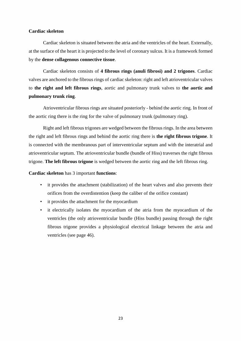
Valves of the heart
Heart valves are duplicatures of the endocardium anchored to the rings of the cardiac
skeleton. In physiological conditions they allow only unidirectional blood flow.
Fig.16
Valves of the heart
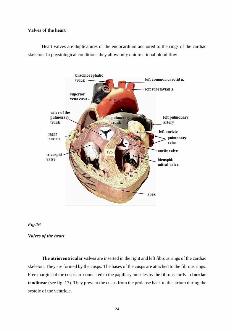
The atrioventricular valves are inserted in the right and left fibrous rings of the cardiac
skeleton. They are formed by the cusps. The bases of the cusps are attached to the fibrous rings.
Free margins of the cusps are connected to the papillary muscles by the fibrous cords – chordae
tendineae (see fig. 17). They prevent the cusps from the prolapse back to the atrium during the
systole of the ventricle.
25
Fig. 17
Chordae tendineae attached to the papillary muscle
Formalin-fixed cadaveric heart
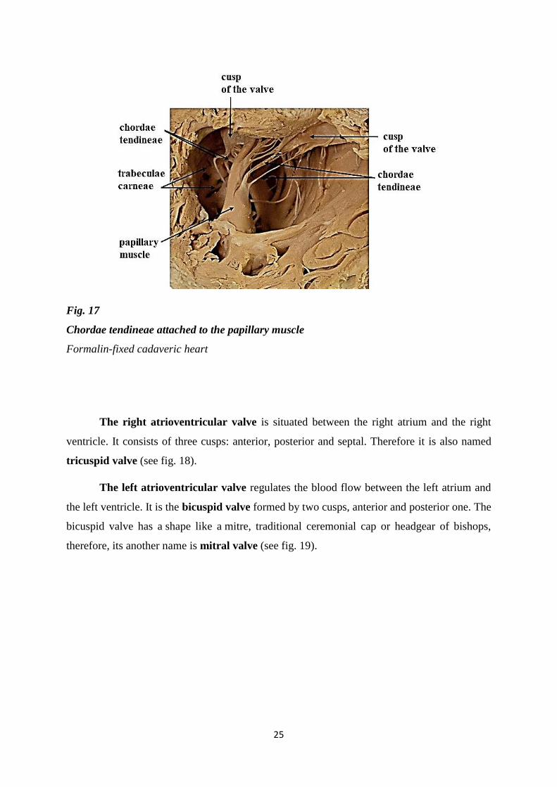
The right atrioventricular valve is situated between the right atrium and the right
ventricle. It consists of three cusps: anterior, posterior and septal. Therefore it is also named
tricuspid valve (see fig. 18).
The left atrioventricular valve regulates the blood flow between the left atrium and
the left ventricle. It is the bicuspid valve formed by two cusps, anterior and posterior one. The
bicuspid valve has a shape like a mitre, traditional ceremonial cap or headgear of bishops,
therefore, its another name is mitral valve (see fig. 19).
26
Fig. 18
Tricuspid valve
View from the right
ventricle
Formalin-fixed cadaveric
heart
Fig.19
Bicuspid (mitral) valve
View from the left
ventricle
Formalin-fixed
cadaveric heart
27
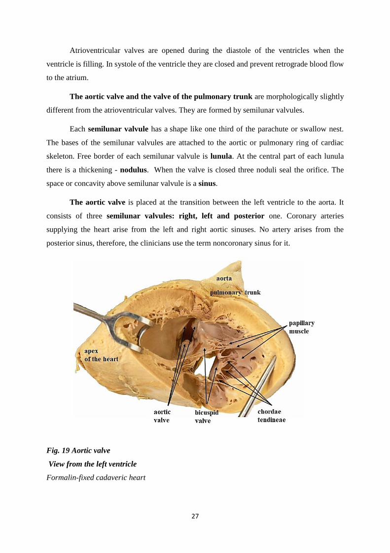
Atrioventricular valves are opened during the diastole of the ventricles when the
ventricle is filling. In systole of the ventricle they are closed and prevent retrograde blood flow
to the atrium.
The aortic valve and the valve of the pulmonary trunk are morphologically slightly
different from the atrioventricular valves. They are formed by semilunar valvules.
Each semilunar valvule has a shape like one third of the parachute or swallow nest.
The bases of the semilunar valvules are attached to the aortic or pulmonary ring of cardiac
skeleton. Free border of each semilunar valvule is lunula. At the central part of each lunula
there is a thickening - nodulus. When the valve is closed three noduli seal the orifice. The
space or concavity above semilunar valvule is a sinus.
The aortic valve is placed at the transition between the left ventricle to the aorta. It
consists of three semilunar valvules: right, left and posterior one. Coronary arteries
supplying the heart arise from the left and right aortic sinuses. No artery arises from the
posterior sinus, therefore, the clinicians use the term noncoronary sinus for it.
Fig. 19 Aortic valve
View from the left ventricle
Formalin-fixed cadaveric heart
28
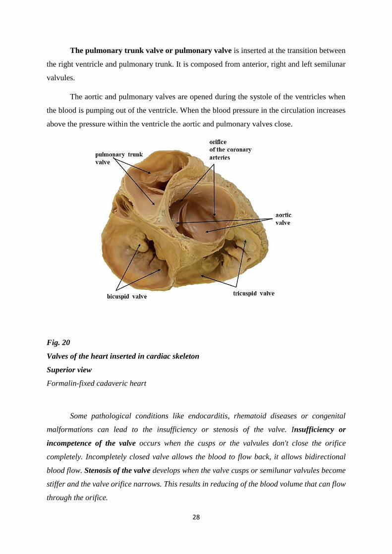
The pulmonary trunk valve or pulmonary valve is inserted at the transition between
the right ventricle and pulmonary trunk. It is composed from anterior, right and left semilunar
valvules.
The aortic and pulmonary valves are opened during the systole of the ventricles when
the blood is pumping out of the ventricle. When the blood pressure in the circulation increases
above the pressure within the ventricle the aortic and pulmonary valves close.
Fig. 20
Valves of the heart inserted in cardiac skeleton
Superior view
Formalin-fixed cadaveric heart
Some pathological conditions like endocarditis, rhematoid diseases or congenital
malformations can lead to the insufficiency or stenosis of the valve. Insufficiency or
incompetence of the valve occurs when the cusps or the valvules don't close the orifice
completely. Incompletely closed valve allows the blood to flow back, it allows bidirectional
blood flow. Stenosis of the valve develops when the valve cusps or semilunar valvules become
stiffer and the valve orifice narrows. This results in reducing of the blood volume that can flow
through the orifice.
29
The auscultation of the heart
The physiological heart sounds (1st and 2nd hear sounds or “lab dab”) are generated by:
the closure of the atrioventricular valves - 1st heart sound or “lab”
the closure of the semilunar valves -2nd heart sound or “dab”
When the valves are damaged (stenotic or insufficient) typical pathological heart murmurs can
be identified. The anatomical position of the heart valves doesn't correspond with the
auscultation points because the heart sounds and murmurs are transmitted via the blood
stream.
Cavities of the heart
The heart consists of four chambers: right atrium, right ventricle, left atrium and left ventricle.
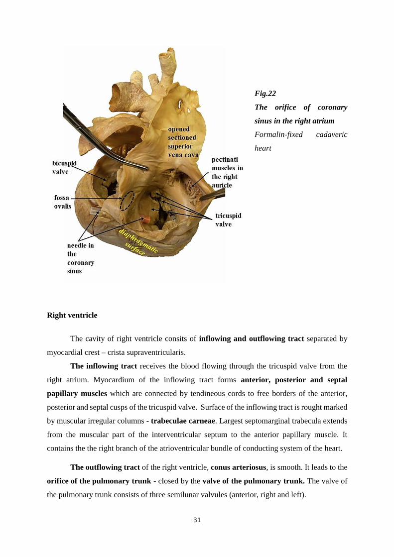
Right atrium
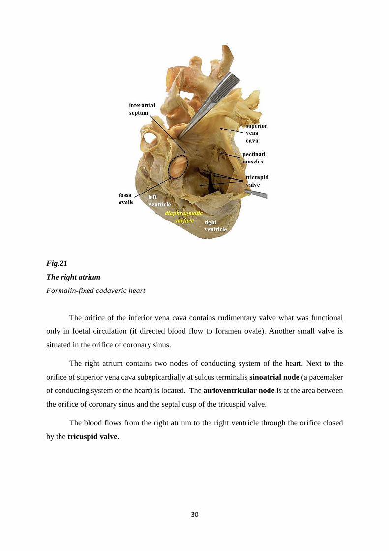
The right atrium receives the deoxygenated venous blood from the systemic
circulation via the superior and inferior vena cava and the venous blood from the cardiac
walls via the coronary sinus.
Orifices (openings) of the superior vena cava and inferior vena cava are situated at the
dorsal wall of the right atrium called sinus of venae cavae (in Latin sinus venarum cavarum).
Surface of this atrial sinus is smooth. Between the orifices of superior and inferior vena cava
there is an internal crest, crista terminalis. At the same position but at the external surface of
the right atrium there is sulcus terminalis. Crista terminalis separates the sinus of venae cavae
(that is posteriorly) from proper atrium (that is anteriorly). The ear – shaped outpouching part
of the atrium is the auricle (pinna). The walls of the proper atrium and especially the auricle
have uneven surface containing pectinate muscles (in Latin musculi pectinati). The orifice
(opening) of coronary sinus is situated between the orifice of inferior vena cava and the right
atrioventricular orifice.
The interatrial septum (septal wall) shows a depression, fossa ovalis bordered by
limbus fossae ovalis. In foetal circulation it was the true opening between the right and left
atrium - foramen ovale.
30
Fig.21
The right atrium
Formalin-fixed cadaveric heart
The orifice of the inferior vena cava contains rudimentary valve what was functional
only in foetal circulation (it directed blood flow to foramen ovale). Another small valve is
situated in the orifice of coronary sinus.
The right atrium contains two nodes of conducting system of the heart. Next to the
orifice of superior vena cava subepicardially at sulcus terminalis sinoatrial node (a pacemaker
of conducting system of the heart) is located. The atrioventricular node is at the area between
the orifice of coronary sinus and the septal cusp of the tricuspid valve.
The blood flows from the right atrium to the right ventricle through the orifice closed
by the tricuspid valve.
31
Fig.22
The orifice of coronary
sinus in the right atrium
Formalin-fixed cadaveric
heart
Right ventricle
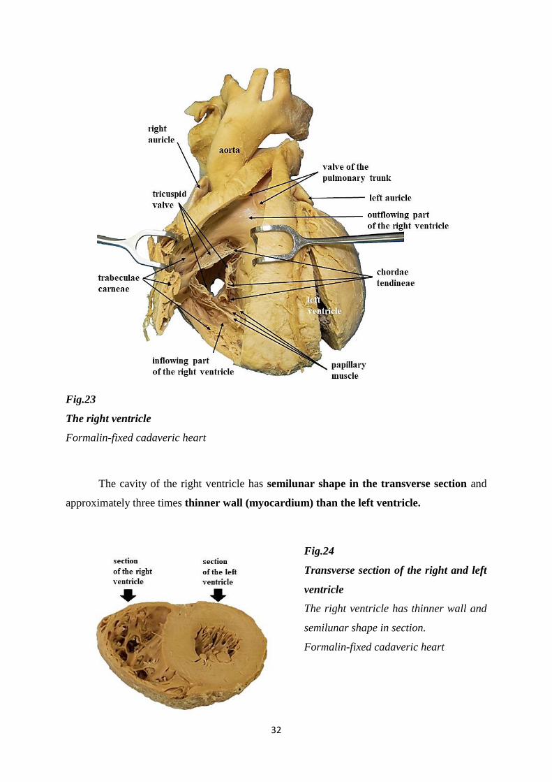
The cavity of right ventricle consits of inflowing and outflowing tract separated by
myocardial crest – crista supraventricularis.
The inflowing tract receives the blood flowing through the tricuspid valve from the
right atrium. Myocardium of the inflowing tract forms anterior, posterior and septal
papillary muscles which are connected by tendineous cords to free borders of the anterior,
posterior and septal cusps of the tricuspid valve. Surface of the inflowing tract is rought marked
by muscular irregular columns - trabeculae carneae. Largest septomarginal trabecula extends
from the muscular part of the interventricular septum to the anterior papillary muscle. It
contains the the right branch of the atrioventricular bundle of conducting system of the heart.
The outflowing tract of the right ventricle, conus arteriosus, is smooth. It leads to the
orifice of the pulmonary trunk - closed by the valve of the pulmonary trunk. The valve of
the pulmonary trunk consists of three semilunar valvules (anterior, right and left).
32
Fig.23
The right ventricle
Formalin-fixed cadaveric heart
The cavity of the right ventricle has semilunar shape in the transverse section and
approximately three times thinner wall (myocardium) than the left ventricle.
Fig.24
Transverse section of the right and left
ventricle
The right ventricle has thinner wall and
semilunar shape in section.
Formalin-fixed cadaveric heart
33
Left atrium
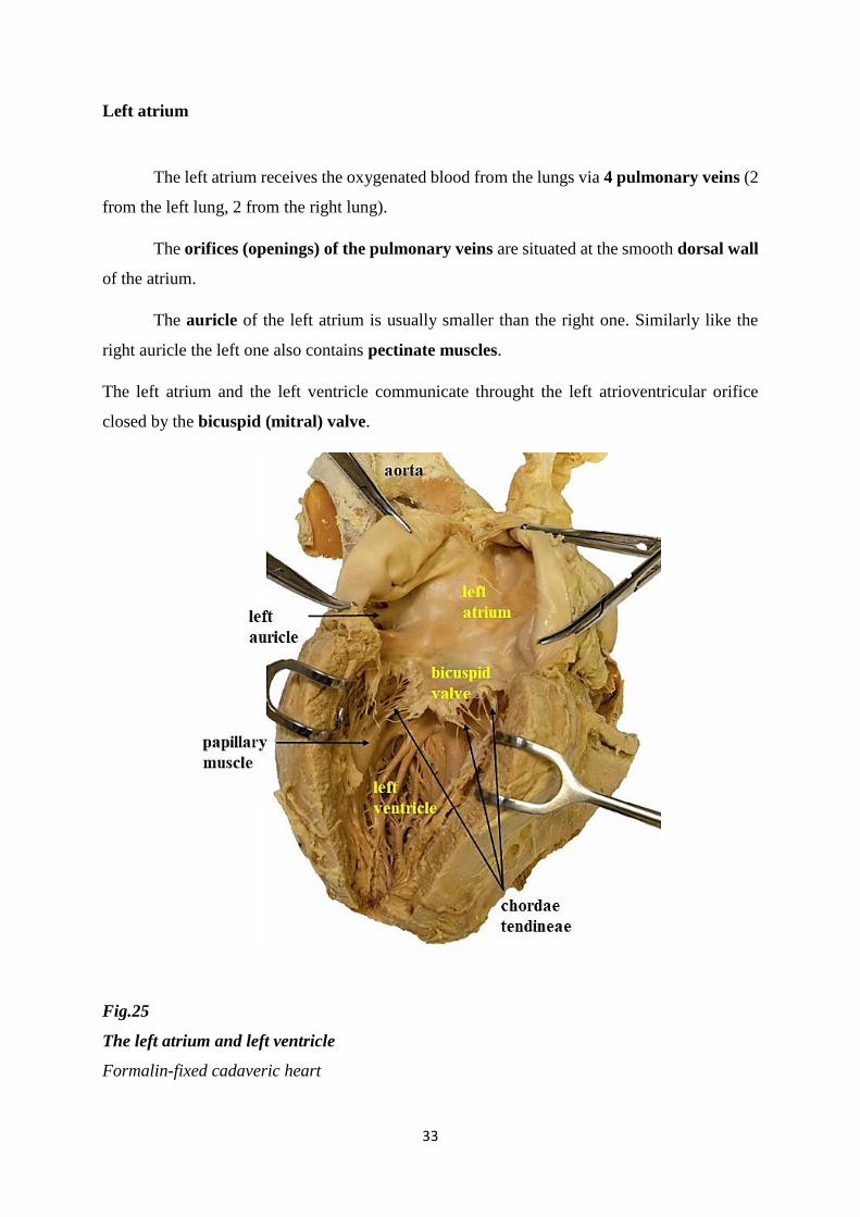
The left atrium receives the oxygenated blood from the lungs via 4 pulmonary veins (2
from the left lung, 2 from the right lung).
The orifices (openings) of the pulmonary veins are situated at the smooth dorsal wall
of the atrium.
The auricle of the left atrium is usually smaller than the right one. Similarly like the
right auricle the left one also contains pectinate muscles.
The left atrium and the left ventricle communicate throught the left atrioventricular orifice
closed by the bicuspid (mitral) valve.
Fig.25
The left atrium and left ventricle
Formalin-fixed cadaveric heart
34
Left ventricle
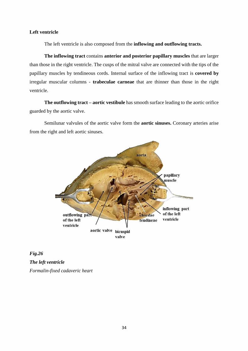
The left ventricle is also composed from the inflowing and outflowing tracts.
The inflowing tract contains anterior and posterior papillary muscles that are larger
than those in the right ventricle. The cusps of the mitral valve are connected with the tips of the
papillary muscles by tendineous cords. Internal surface of the inflowing tract is covered by
irregular muscular columns - trabeculae carneae that are thinner than those in the right
ventricle.
The outflowing tract – aortic vestibule has smooth surface leading to the aortic orifice
guarded by the aortic valve.
Semilunar valvules of the aortic valve form the aortic sinuses. Coronary arteries arise
from the right and left aortic sinuses.
Fig.26
The left ventricle
Formalin-fixed cadaveric heart
35
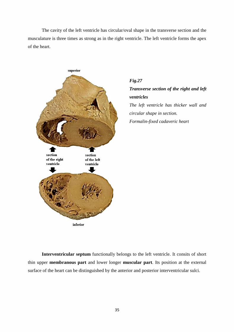
The cavity of the left ventricle has circular/oval shape in the transverse section and the
musculature is three times as strong as in the right ventricle. The left ventricle forms the apex
of the heart.
Fig.27
Transverse section of the right and left
ventricles
The left ventricle has thicker wall and
circular shape in section.
Formalin-fixed cadaveric heart
Interventricular septum functionally belongs to the left ventricle. It consits of short
thin upper membranous part and lower longer muscular part. Its position at the external
surface of the heart can be distinguished by the anterior and posterior interventricular sulci.
36
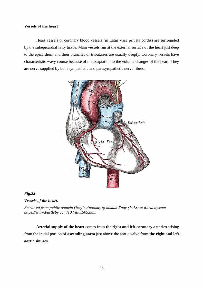
Vessels of the heart
Heart vessels or coronary blood vessels (in Latin Vasa privata cordis) are surrounded
by the subepicardial fatty tissue. Main vessels run at the external surface of the heart just deep
to the epicardium and their branches or tributaries are usually deeply. Coronary vessels have
characteristic wavy course because of the adaptation to the volume changes of the heart. They
are nerve supplied by both sympathetic and parasympathetic nerve fibres.
Fig.28
Vessels of the heart.
Retrieved from public domein Gray´s Anatomy of human Body (1918) at Bartleby.com
https://www.bartleby.com/107/illus505.html
Arterial supply of the heart comes from the right and left coronary arteries arising
from the initial portion of ascending aorta just above the aortic valve from the right and left
aortic sinuses.
37
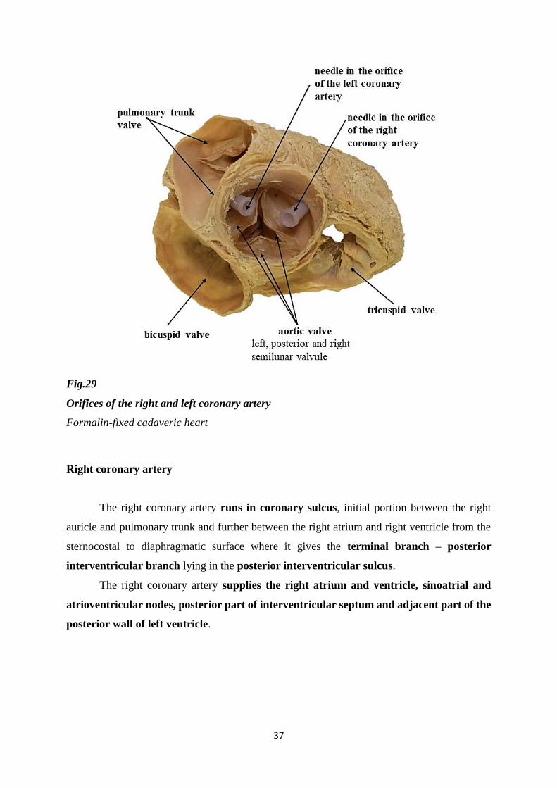
Fig.29
Orifices of the right and left coronary artery
Formalin-fixed cadaveric heart
Right coronary artery
The right coronary artery runs in coronary sulcus, initial portion between the right
auricle and pulmonary trunk and further between the right atrium and right ventricle from the
sternocostal to diaphragmatic surface where it gives the terminal branch – posterior
interventricular branch lying in the posterior interventricular sulcus.
The right coronary artery supplies the right atrium and ventricle, sinoatrial and
atrioventricular nodes, posterior part of interventricular septum and adjacent part of the
posterior wall of left ventricle.
38
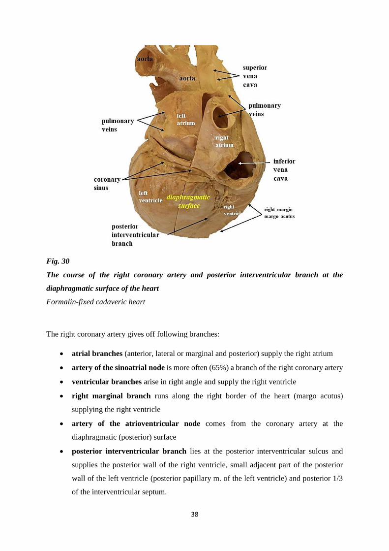
Fig. 30
The course of the right coronary artery and posterior interventricular branch at the
diaphragmatic surface of the heart
Formalin-fixed cadaveric heart
The right coronary artery gives off following branches:
atrial branches (anterior, lateral or marginal and posterior) supply the right atrium
artery of the sinoatrial node is more often (65%) a branch of the right coronary artery
ventricular branches arise in right angle and supply the right ventricle
right marginal branch runs along the right border of the heart (margo acutus)
supplying the right ventricle
artery of the atrioventricular node comes from the coronary artery at the
diaphragmatic (posterior) surface
posterior interventricular branch lies at the posterior interventricular sulcus and
supplies the posterior wall of the right ventricle, small adjacent part of the posterior
wall of the left ventricle (posterior papillary m. of the left ventricle) and posterior 1/3
of the interventricular septum.
39
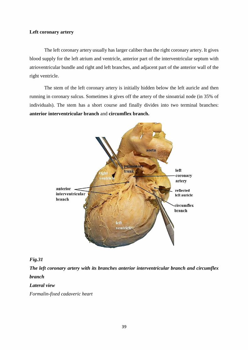
Left coronary artery
The left coronary artery usually has larger caliber than the right coronary artery. It gives
blood supply for the left atrium and ventricle, anterior part of the interventricular septum with
atrioventricular bundle and right and left branches, and adjacent part of the anterior wall of the
right ventricle.
The stem of the left coronary artery is initially hidden below the left auricle and then
running in coronary sulcus. Sometimes it gives off the artery of the sinoatrial node (in 35% of
individuals). The stem has a short course and finally divides into two terminal branches:
anterior interventricular branch and circumflex branch.
Fig.31
The left coronary artery with its branches anterior interventricular branch and circumflex
branch
Lateral view
Formalin-fixed cadaveric heart

40
Anterior interventricular branch runs in the anterior interventricular sulcus to the apex
of the heart. During its course it gives these branches:
anterior ventricular branches
- left anterior ventricular branches or diagonal branches (1-2) that supply
anterior wall of the left ventricle
- right anterior ventricular branches that supply adjacent part of the anterior wall
of the right ventricle (anterior papillary m.)
septal branches that supply anterior 2/3 of the interventricular septum including the
atrioventricular bundle and right and left branches.
Fig.32
The anterior interventricular branch and the great cardiac vein
Formalin-fixed cadaveric heart
Circumflex branch passes in the left part of the coronary sulcus giving following branches:
atrial branches
left marginal branch running along the left margin (margo obtusus)
anterior and posterior ventricular branches for the left ventricle.
41
The interventricular septum, anterior papillary muscle in the right ventricle and posterior
papillary muscle in the left ventricle recieves blood from the branches of both the right coronary
artery and the left coronary artery.
Coronary arterial system shows a variability especially at the diaphragmatic surface of the
heart. Above - mentioned coronary distribution is the most common occured type.
Clinicians classify the types of the coronary distribution according the „dominance of the
coronary artery“. The dominant coronary artery is that one which gives rise to the posterior
interventricular branch. The righ coronary artery is dominant in 67% of persons, the left
coronary artery is dominant in 15%. Sometimes (in 18%) so called codominant type of coronary
distribution can be seen when both the right and left coronary arteries give the branches
running along the posterior interventricular sulcus.
Sometimes, coronary arteries and their branches have alternative names in the clinical
practise:
the stem of the left coronary artery is called the left main stem vessel
anterior interventricular branch is called left descending artery (LDA)
posterior interventricular branch is called posterior descending artery (PDA).
During the fetal development there are numerous anastomoses between the right and left
coronary arteries, however, they are diminished by the first year. In adulthood the degree of
anastomoses and collateral circulation is usually insignifficant as for the acute ischemia
(during the acute coronary obstruction anastomoses or collateral pathways cannot provide
sufficient blood supply) but they can be effective in pathological conditions with slow
progression.
Coronary artery disease is characterized by the atherosclerotic plaque formation in the
vascular wall. The plaque reduces the lumen diameter or it can be ruptured forming the
thrombus that can cause acute occlusion of the coronary artery resulting in myocardial
infarction. Visualization of the coronary arteries is possible using the various ragiological
techniques. Patients with serious high grade stenosis usually undergo invasive radiologic
cardiovascular techniques or coronary artery bypass grafting. As the grafts are used saphenous
veins or internal thoracic arteries.
42
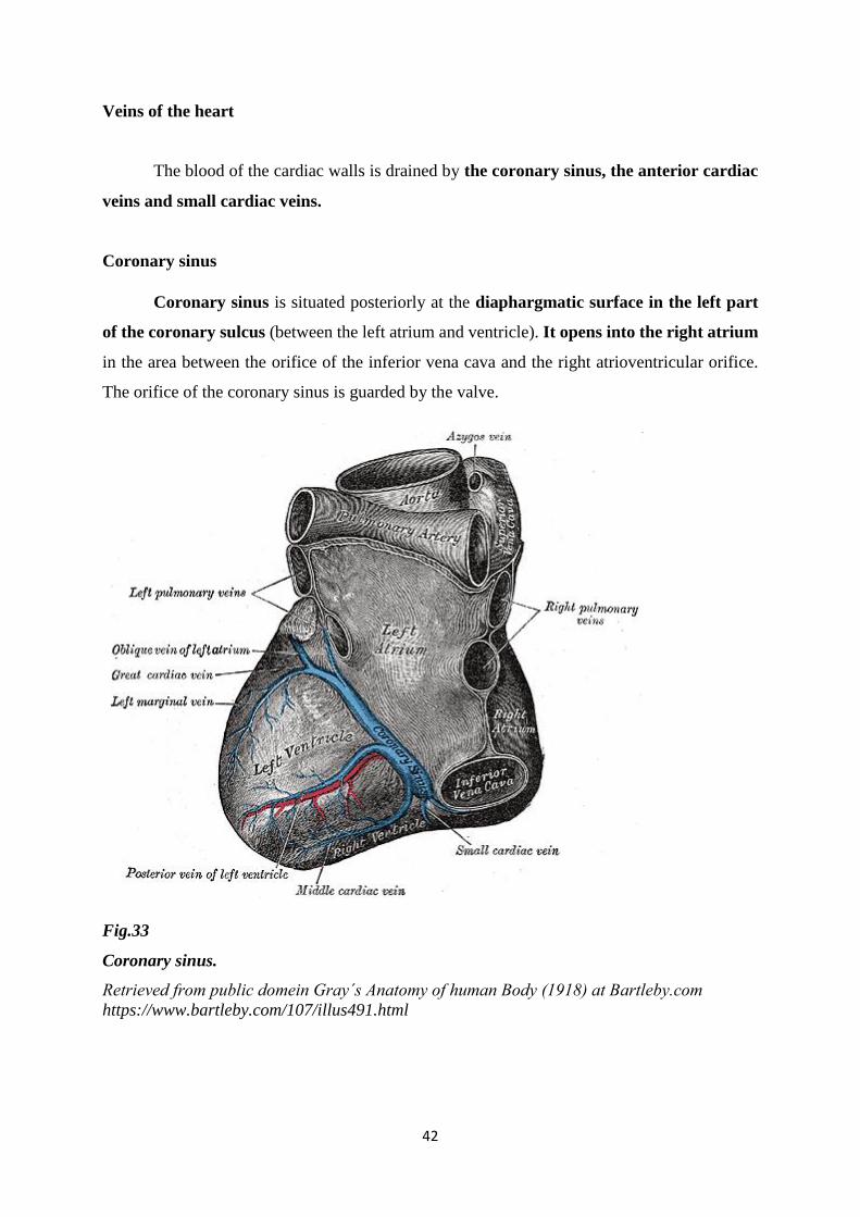
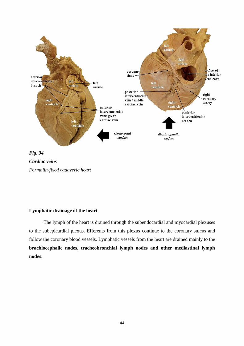
Veins of the heart
The blood of the cardiac walls is drained by the coronary sinus, the anterior cardiac
veins and small cardiac veins.
Coronary sinus
Coronary sinus is situated posteriorly at the diaphargmatic surface in the left part
of the coronary sulcus (between the left atrium and ventricle). It opens into the right atrium
in the area between the orifice of the inferior vena cava and the right atrioventricular orifice.
The orifice of the coronary sinus is guarded by the valve.
Fig.33
Coronary sinus.
Retrieved from public domein Gray´s Anatomy of human Body (1918) at Bartleby.com
https://www.bartleby.com/107/illus491.html
43
Coronary sinus receives the blood from these veins:
• great cardiac vein
- it begins at the apex of the heart, ascends in the anterior interventricular groove
accompanied by the anterior interventricular branch of the left coronary artery; then
it turns to the left, passes in the coronary sulcus and enters the coronary sinus at the
left side;
- it drains the venous blood from the parts of the heart that are supplied by the left
coronary artery
• middle cardiac vein
- it ascends in the posterior interventricular sulcus accompanied by posterior
interventricular branch from the right coronary artery and finally opens into the
coronary sinus;
- it receives the blood from the posterior aspect of both ventricles and interventricular
septum
• small cardiac vein
- it runs in the right part of the coronary sulcus and opens into the coronary sinus at
the right side;
- it drains posterior part of the right atrium and ventricle and from the margo acutus
via the right marginal vein (sometimes it opens directly to the right atrium)
• posterior vein of the left ventricle takes the blood from the diaphragmatic surface of
the left ventricle; it opens directly into the coronary sinus or to the great cardiac vein.
Anterior cardiac veins (2-5) drain the anterior wall of the right ventricle and open directly
into the right atrium independently of the coronary sinus.
The smallest cardiac veins or Thebesian veins are vessels with the diameter less than 2mm.
They open to all chambers of the heart, mainly to the right atrium and ventricle, rarely to the
left atrium and ventricle.
44
Fig. 34
Cardiac veins
Formalin-fixed cadaveric heart
Lymphatic drainage of the heart
The lymph of the heart is drained through the subendocardial and myocardial plexuses
to the subepicardial plexus. Efferents from this plexus continue to the coronary sulcus and
follow the coronary blood vessels. Lymphatic vessels from the heart are drained mainly to the
brachiocephalic nodes, tracheobronchial lymph nodes and other mediastinal lymph
nodes.
45
Conduction system of the heart
Conduction system of the heart works as an initiator and coordinator of spontaneous
contraction. It consists of specialised cardiac muscle cells that are morphologically different
from normal working cardiac cells. Cardiac conduction system is composed from several
components: sinoatrial node, atrioventricular node, atrioventricular bundle, right and left
Tawara´s branches and subendocardial Purkinje fibres.
Sinuatrial node (Keith-Flack node)
In physiological condition, sinuatrial node is the „pacemaker“ of the heart. Sinuatrial
node is elliptical in shape, 10-20 mm long. It is situated subepicardially in the wall of the right
atrium at the upper part of crista terminalis next to the superior vena cava orifice. The excitation
generated by the sinuatrial node spreads across the atrium by myogenic conduction to the
atrioventricular node.
Atrioventricular node (Aschoff - Tawara node)
Atrioventricular node is placed at the right atrium just between the orifice of the
coronary sinus and the right atrioventricular orifice.
Atrioventricular bandle (bundle of His), right and left branches (Tawara´s branches),
subendocardial branches (Purkinje fibres)
The signal from the atrioventicular node is conducted by the atrioventricular bundle.
This bundle traverses the right fibrous trigone of the cardiac skeleton and passes along the
membranous part of the interventricular septum. At the border between the membranous and
muscular part of the interventricular septum the atrioventricular bundle divides into the right
and left branches that continue inferiorly along the right and left side of the muscular part of
the interventricular septum. Right and left branches ramify into the subendocardial branches
(Purkinje fibres) continuing to the right and left ventricle, respectively.
Nodes, bundle and main branches are separated from working myocardium by the
sheaths of connective tissue. Only terminal parts, subendocardial branches, have numerous
functional contacts with working myocardial cells.
46
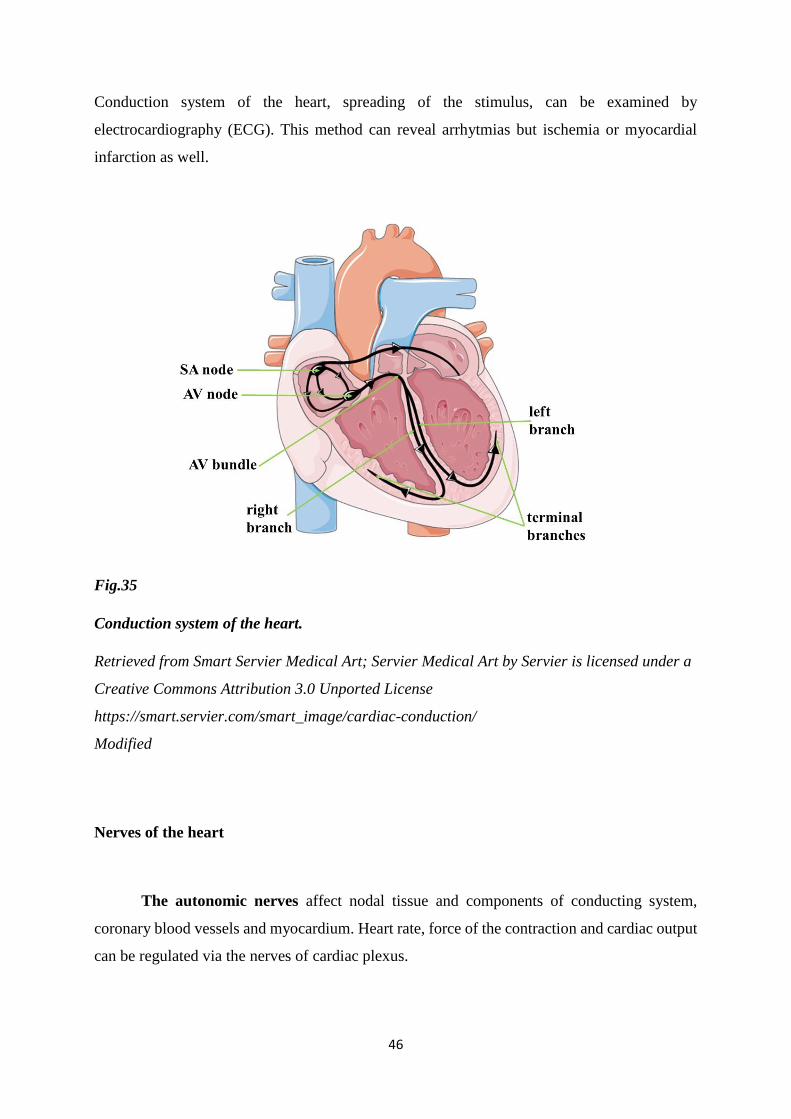
Conduction system of the heart, spreading of the stimulus, can be examined by
electrocardiography (ECG). This method can reveal arrhytmias but ischemia or myocardial
infarction as well.
Fig.35
Conduction system of the heart.
Retrieved from Smart Servier Medical Art; Servier Medical Art by Servier is licensed under a
Creative Commons Attribution 3.0 Unported License
https://smart.servier.com/smart_image/cardiac-conduction/
Modified
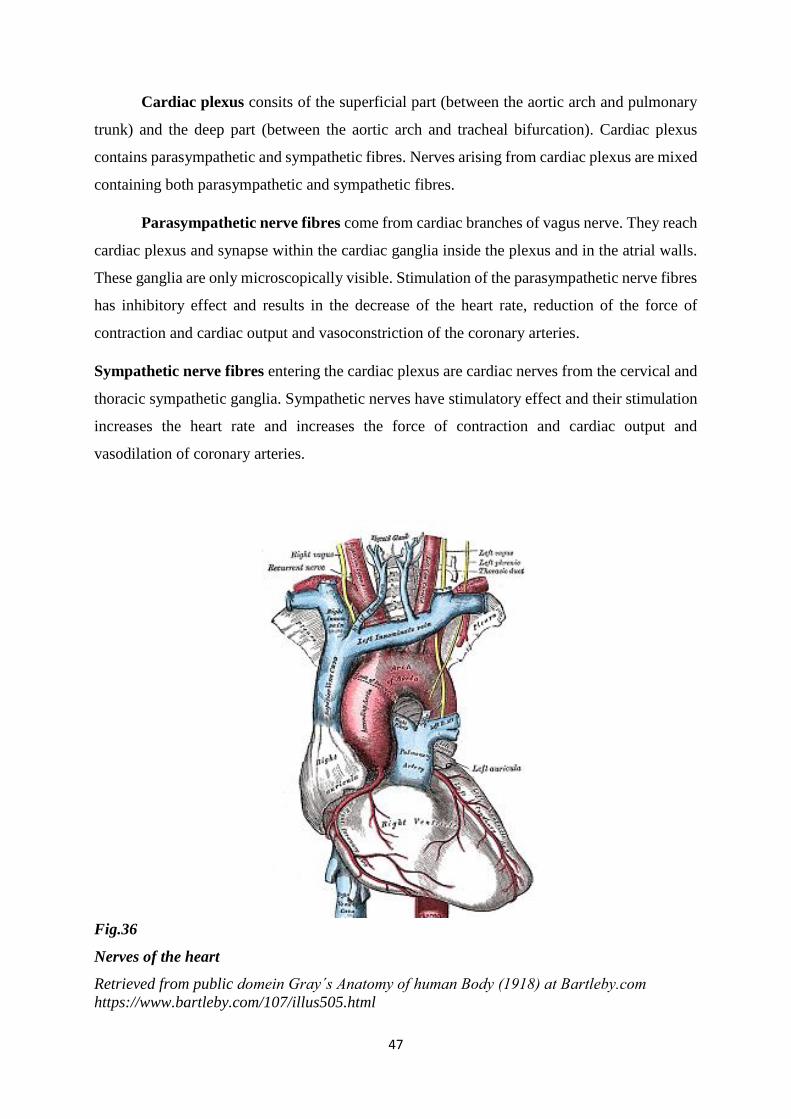
Nerves of the heart
The autonomic nerves affect nodal tissue and components of conducting system,
coronary blood vessels and myocardium. Heart rate, force of the contraction and cardiac output
can be regulated via the nerves of cardiac plexus.
47
Cardiac plexus consits of the superficial part (between the aortic arch and pulmonary
trunk) and the deep part (between the aortic arch and tracheal bifurcation). Cardiac plexus
contains parasympathetic and sympathetic fibres. Nerves arising from cardiac plexus are mixed
containing both parasympathetic and sympathetic fibres.
Parasympathetic nerve fibres come from cardiac branches of vagus nerve. They reach
cardiac plexus and synapse within the cardiac ganglia inside the plexus and in the atrial walls.
These ganglia are only microscopically visible. Stimulation of the parasympathetic nerve fibres
has inhibitory effect and results in the decrease of the heart rate, reduction of the force of
contraction and cardiac output and vasoconstriction of the coronary arteries.
Sympathetic nerve fibres entering the cardiac plexus are cardiac nerves from the cervical and
thoracic sympathetic ganglia. Sympathetic nerves have stimulatory effect and their stimulation
increases the heart rate and increases the force of contraction and cardiac output and
vasodilation of coronary arteries.
Fig.36
Nerves of the heart
Retrieved from public domein Gray´s Anatomy of human Body (1918) at Bartleby.com
https://www.bartleby.com/107/illus505.html
48
Introduction to the blood circulation
Arteries carry the blood from the heart to the whole body.
Veins return the blood from the periphery to the heart.
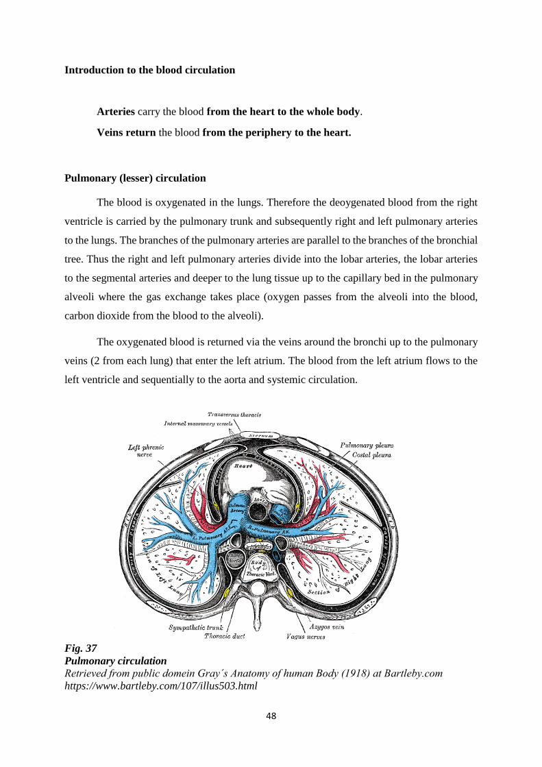
Pulmonary (lesser) circulation
The blood is oxygenated in the lungs. Therefore the deoygenated blood from the right
ventricle is carried by the pulmonary trunk and subsequently right and left pulmonary arteries
to the lungs. The branches of the pulmonary arteries are parallel to the branches of the bronchial
tree. Thus the right and left pulmonary arteries divide into the lobar arteries, the lobar arteries
to the segmental arteries and deeper to the lung tissue up to the capillary bed in the pulmonary
alveoli where the gas exchange takes place (oxygen passes from the alveoli into the blood,
carbon dioxide from the blood to the alveoli).
The oxygenated blood is returned via the veins around the bronchi up to the pulmonary
veins (2 from each lung) that enter the left atrium. The blood from the left atrium flows to the
left ventricle and sequentially to the aorta and systemic circulation.
Fig. 37
Pulmonary circulation
Retrieved from public domein Gray´s Anatomy of human Body (1918) at Bartleby.com
https://www.bartleby.com/107/illus503.html
49
Systemic or greater circulation
The oxygenated blood from the left ventricle outflows through the aorta and via its
branches to the arteries, arteriols up to the capillary beds inside the organs in the whole body.
Microcirculatory bed (arteriols, capillaries and venules) allows the oxygen and
nutrients delivery, and carbon dioxide and waste removal. Via the venules and veins the
deoxygeneated blood is returned to the superior vena cava (from the head, neck, upper limb and
the thorax) and inferior vena cava (from the lower limbs and the abdomen). Both superior and
inferior vena cava open into the right atrium. The blood from the right atrium continues to the
right ventricle. The pulmonary trunk carries the blood to the right and left pulmonary arteries
and to the lungs.
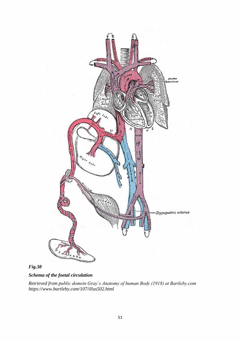
Foetal circulation
Foetal circulation shows characteristic differences from the postnatal circulation. These
differences result from the fact that the blood in fetus in not oxygeneated in the lungs and it is
not detoxified in the liver. Both these functions are provided by placenta that serves as the organ
for the foetal nutrition, excretion and gas exchange.
Foetal circulation differs from the adult circulation by the presence of 3 major vascular
shunts. In foetus the function of the lungs and the liver is carried by placenta and following
shunts divert the blood from them:
• ductus venosus (Arantii) is connection between the umbilical vein and inferior vena
cava and it diverts the blood from passing through the liver (because the capillary bed
of the liver is immature with high resistance and the blood is detoxified in placenta)
• foramen ovale is the opening between the right and left atrium and it allows the blood
that enters the right atrium to go around or to bypass the lungs (because there is no gas
exchange in the lungs, they are full of amniotic fluid and the blood has already been
oxygenated in placenta)
• ductus arteriosus (Botalli) is connection between the pulmonary trunk and the aortic
arch after arising large branches and it allows to divert the blood from the lungs (because
the blood has already been oxygenated in placenta)
50
The oxygeneted blood from the placenta is taken by the umbilical vein to the foetus.
Umbilical vein passes within the umbilical cord, traverses through the navel and then in the
lower part of the falciforme ligament to the liver. Majority of the oxygenated blood from the
umbilical vein passes to the inferior vena cava through ductus venosus. This shunt allows to
divert the blood from the liver to the inferior vena cava without passing through the capillary
bed in the liver. Only a small volume of the blood flow from the umbilical vein enters the portal
vein and passes through the liver to the hepatic veins and to the inferior vena cava. The inferior
vena cava also receives the the blood from ductus venosus, hepatic veins and deoxygenated
blood from the lower limbs and abdominal wall. This mixed blood in inferior vena cava entering
the right atrium has slightely lower oxygenation than the blood in umbililical vein (cca 67%
oxygen saturation in inferior vena cava vs 80% oxygen saturation in umbilical vein).
The blood from the inferior vena cava is directed by the valve towards the foramen ovale
and to the left atrium.
Majority of the blood in the left atrium inflows from the right atrium through foramen
ovale. Only limited volume comes from the pulmonary veins, therefore, the oxygen saturation
is slightly lower (65%) than in the right atrium.
The blood from the left atrium continues to the left ventricle and through the aorta to
the whole body, however, majority of the blood flow is distributed to the branches of ascending
aorta and the aortic arch (supplying the heart, head, neck and upper limbs), and minority to the
descending aorta.
Superior vena cava takes deoxygenated blood from the upper limbs, head and neck and
opens into the right atrium. This blood flows into the right ventricle with minimal interfusion
(mixture) with the blood coming from the inferior vena cava.
The blood exits the right ventricle via the pulmonary trunk. Only limited volume (cca
10%) of the blood in pulmonary trunk continues to the lungs via the right and left pulmonary
arteries. Most of the volume bypasses the lungs through ductus arteriosus connecting the
pulmonary trunk and the aortic arch after the large branches arising. The blood from the aortic
arch mixes with the lesser oxygenated blood from ductus arteriosus. Thus the descending aorta
contains lesser oxygenated blood (55%) than the ascending aorta and aortic arch (65%).
Descending aorta supplies the abdominal and pelvic organs and lower limbs. Two
umbilical arteries take the deoxygenated blood from the feotal body to placenta.
51
Fig.38
Schema of the foetal circulation
Retrieved from public domein Gray´s Anatomy of human Body (1918) at Bartleby.com https://www.bartleby.com/107/illus502.html
52
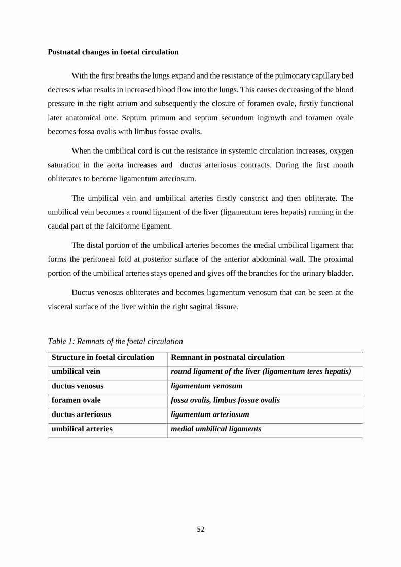
Postnatal changes in foetal circulation
With the first breaths the lungs expand and the resistance of the pulmonary capillary bed
decreses what results in increased blood flow into the lungs. This causes decreasing of the blood
pressure in the right atrium and subsequently the closure of foramen ovale, firstly functional
later anatomical one. Septum primum and septum secundum ingrowth and foramen ovale
becomes fossa ovalis with limbus fossae ovalis.
When the umbilical cord is cut the resistance in systemic circulation increases, oxygen
saturation in the aorta increases and ductus arteriosus contracts. During the first month
obliterates to become ligamentum arteriosum.
The umbilical vein and umbilical arteries firstly constrict and then obliterate. The
umbilical vein becomes a round ligament of the liver (ligamentum teres hepatis) running in the
caudal part of the falciforme ligament.
The distal portion of the umbilical arteries becomes the medial umbilical ligament that
forms the peritoneal fold at posterior surface of the anterior abdominal wall. The proximal
portion of the umbilical arteries stays opened and gives off the branches for the urinary bladder.
Ductus venosus obliterates and becomes ligamentum venosum that can be seen at the
visceral surface of the liver within the right sagittal fissure.
Table 1: Remnats of the foetal circulation
Structure in foetal circulation Remnant in postnatal circulation
umbilical vein round ligament of the liver (ligamentum teres hepatis)
ductus venosus ligamentum venosum
foramen ovale fossa ovalis, limbus fossae ovalis
ductus arteriosus ligamentum arteriosum
umbilical arteries medial umbilical ligaments
53
RESPIRATORY SYSTEM
Main functions of respiratory system are to obtain oxygen from external environment
and supply it to cells, and to remove carbon dioxide produced by cellular metabolism from
body. Respiratory system is also responsible for speech and phonation, and olfaction.
In accordance with main function of respiratory system, it can be divided into two parts:
air-conducting zone and respiratory zone. Air-conducting zone consists of nasal cavities,
pharynx, larynx, trachea, bronchii and bronchioles. It conveys, moistens, warms and cleans air
during breathing. Respiratory zone is located within lungs parenchyma and it consists of
respiratory bronchioles, alveolar ducts, alveolar sacs and alveoli. Gas exchange occurs in
alveoli of lungs that are enveloped by capillaries.
Structurally, respiratory system is divided into upper and lower respiratory airways and
into major organs of respiration.
Upper respiratory airway:
Nose – external nose and nasal cavity
Paranasal sinuses
*Pharynx
Lower respiratory airway:
Larynx
Trachea and bronchi (tracheobronchial tree)
Organs of respiration:
Lungs associated with pleura and pleural cavity
*Pharynx is situated behind nasal and oral cavities and behind larynx. It communicates with
them through choanae, oropharyngeal isthmus and laryngeal inlet. The most upper part of
larynx is called nasopharynx, its middle part is oropharynx and lowerpart is laryngopharynx.
Functionally, pharynx has roles in both respiratory and alimentary systems. It is common
pathway for air and food. It transmits air from nasal cavity to larynx and it permits passage of
swallowed solids and liquids from oral cavity into oesophagus. These two passages unite in
oropharynx and both are separated in laryngopharynx.
54
NOSE
Nose is the entry point for inspired air and the first of the structures which forms the
respiratory system. Nose has several functions. The main function is to warm, cool or humidify
the inspired air. Nose forms a protective barier and filters out pathogens, dust and other
particles. It has reflex functions. Nose protects the lower respiratory airway and it also plays
a role as an organ responsible for olfaction and the vocal resonance. Nose consists of external
nose and nasal cavity. The nasal cavity communicates with all paranasal sinuses, including
the ethmoidal air cells and the frontal, sphenoidal and maxillary sinuses.
EXTERNAL NOSE
External nose is prominent part of the face which is pyramid-shaped. It has a free tip –
apex of the nose. Its upper part located between two orbits is root of the nose. Root forms the
border between the nose and forehead. Dorsum of the nose is rounded part between the apex
and root. On the sides of external nose there are alae of the nose (wings). Alae of the nose are
movable and they form lateral border of two oval openings – nares. Nares are situated at the
beginning of the nasal cavity.
External nose is composed of bones, cartilages and connective tissue and it is covered
by the muscles and skin. Its upper one third portion is based on the skeletal framework therefore
is more vulnerable and frequently is closely involved with the midfacial fracture. Its lower two
thirds are cartilaginous, more elastic and less susceptible to injury.
Bony framework of external nose is made up by the nasal bones, frontal processes of
maxillae and nasal part of the frontal bone.
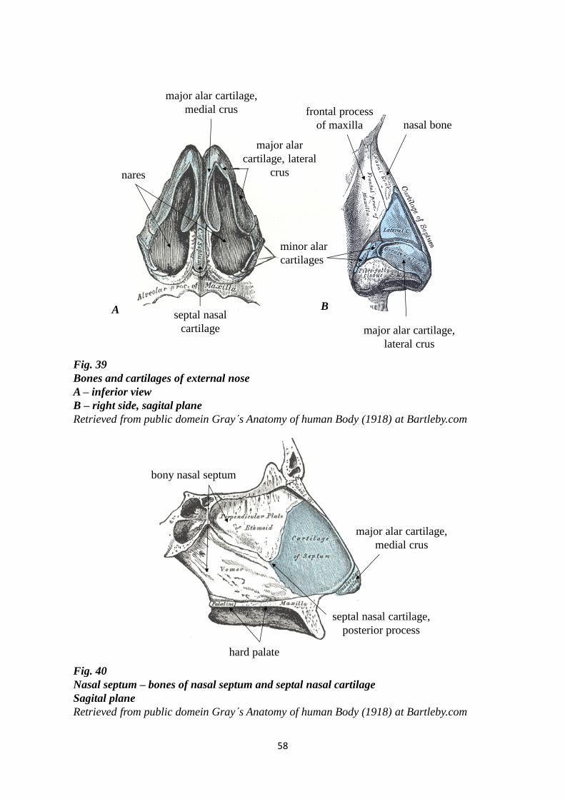
Cartilaginous framework is formed by septal nasal cartilage and alar cartilages
embedded within connective tissue of alae of the nose.
Septal nasal cartilage is larger independent piece of cartilage in the nasal septum. It
forms ventral portion of the nasal septum and its upper border creates dorsum of the
nose. Posterior process of the septal nasal cartilage is situated between vomer and
perpendicular plate of the ethmoid bone. Lateral processes of this cartilage contribute
to formation of lateral nasal walls.
Major alar cartilages are paired cartilages around the nares. They contribute to form
apex of the nose.
55
Major alar cartilage is thin, flexible and hook-shaped cartilage surrounding nares. It is
formed by medial and lateral crus. Medial crus is loosely connected with the
corresponding portion of the opposite cartilage. Both medial crura are attached to the
nasal septum. Lateral crus runs laterally around nares.
Minor alar cartilages are individual small cartilaginous plates of ala of the nose located
posterior to major alar cartilage.
Accessory alar cartilages are smaller pieces of cartilage that are occasionally found
between lateral process of septal nasal cartilage and major alar cartilage.
Muscles of the external nose form a distinct subgroup within the muscles of facial
expression. This subgroup includes procerus, nasalis, depressor septi nasi muscles. Some of the
muscles, e.g. orbicularis oris, levator labii superioris alaequae nasi are in more than one
subgroup. There are also muscles of the mouth.
Skin of the external nose is thin especially over the root and dorsum of the nose where
is loosely attached to the underlying structures. Skin over the apex and ala of the nose is firmly
adherent to cartilaginous base and contains numerous sebaceous glands.
Vessels and nerves of external nose
o Arterial supply to ala of the nose comes from lateral nasal artery and to root of the nose
from angular artery – both arteries are branches of facial artery. Root and dorsum of the
external nose are also supplied by dorsal nasal artery which is branch of ophthalmic
artery. There are the anastomoses between angular and dorsal nasal arteries.
o Venous blood drains primarily to facial vein. Limited venous blood is drained to
superior ophthalmic vein.
o Lymphatic vessels drain into submandibular lymph nodes.
o Nerves of general sensation are basically derived from ophthalmic and maxillary nerves
– both nerves are branches of trigeminal nerve.
Root and dorsum of the external nose are nerve supplied by supratrochlear nerve, which
originates from frontal nerve, and by external nasal rami, which originate from
nasociliary nerve. Frontal and nasociliary nerves are branches of ophthalmic nerve.
Ala of the nose is nerve supplied by infraorbital nerve, which comes from maxillary
nerve.
Muscles of the nose are nerve supplied by branches of facial nerve.
56
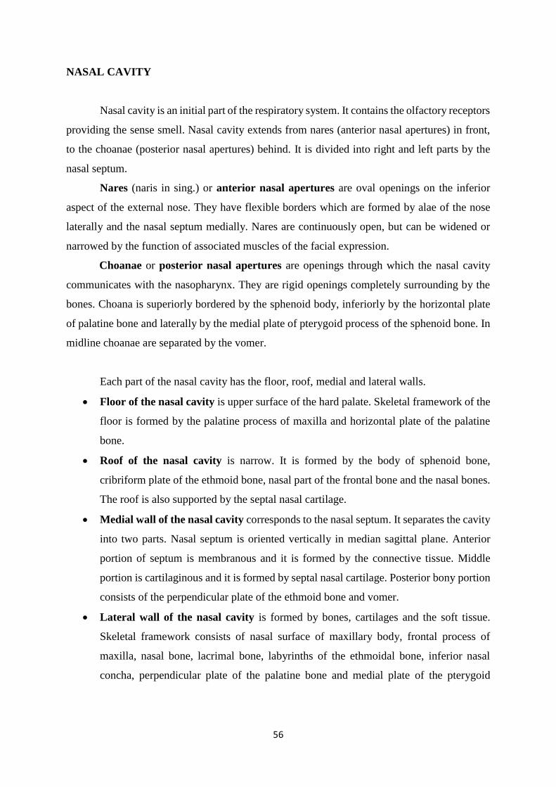
NASAL CAVITY
Nasal cavity is an initial part of the respiratory system. It contains the olfactory receptors
providing the sense smell. Nasal cavity extends from nares (anterior nasal apertures) in front,
to the choanae (posterior nasal apertures) behind. It is divided into right and left parts by the
nasal septum.
Nares (naris in sing.) or anterior nasal apertures are oval openings on the inferior
aspect of the external nose. They have flexible borders which are formed by alae of the nose
laterally and the nasal septum medially. Nares are continuously open, but can be widened or
narrowed by the function of associated muscles of the facial expression.
Choanae or posterior nasal apertures are openings through which the nasal cavity
communicates with the nasopharynx. They are rigid openings completely surrounding by the
bones. Choana is superiorly bordered by the sphenoid body, inferiorly by the horizontal plate
of palatine bone and laterally by the medial plate of pterygoid process of the sphenoid bone. In
midline choanae are separated by the vomer.
Each part of the nasal cavity has the floor, roof, medial and lateral walls.
Floor of the nasal cavity is upper surface of the hard palate. Skeletal framework of the
floor is formed by the palatine process of maxilla and horizontal plate of the palatine
bone.
Roof of the nasal cavity is narrow. It is formed by the body of sphenoid bone,
cribriform plate of the ethmoid bone, nasal part of the frontal bone and the nasal bones.
The roof is also supported by the septal nasal cartilage.
Medial wall of the nasal cavity corresponds to the nasal septum. It separates the cavity
into two parts. Nasal septum is oriented vertically in median sagittal plane. Anterior
portion of septum is membranous and it is formed by the connective tissue. Middle
portion is cartilaginous and it is formed by septal nasal cartilage. Posterior bony portion
consists of the perpendicular plate of the ethmoid bone and vomer.
Lateral wall of the nasal cavity is formed by bones, cartilages and the soft tissue.
Skeletal framework consists of nasal surface of maxillary body, frontal process of
maxilla, nasal bone, lacrimal bone, labyrinths of the ethmoidal bone, inferior nasal
concha, perpendicular plate of the palatine bone and medial plate of the pterygoid
57
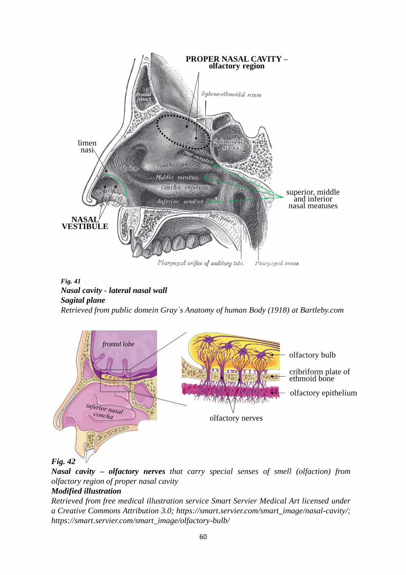
processes of the sphenoid bone. The lateral nasal wall is also supported by the lateral
process of the septal nasal cartilage and alar cartilages.
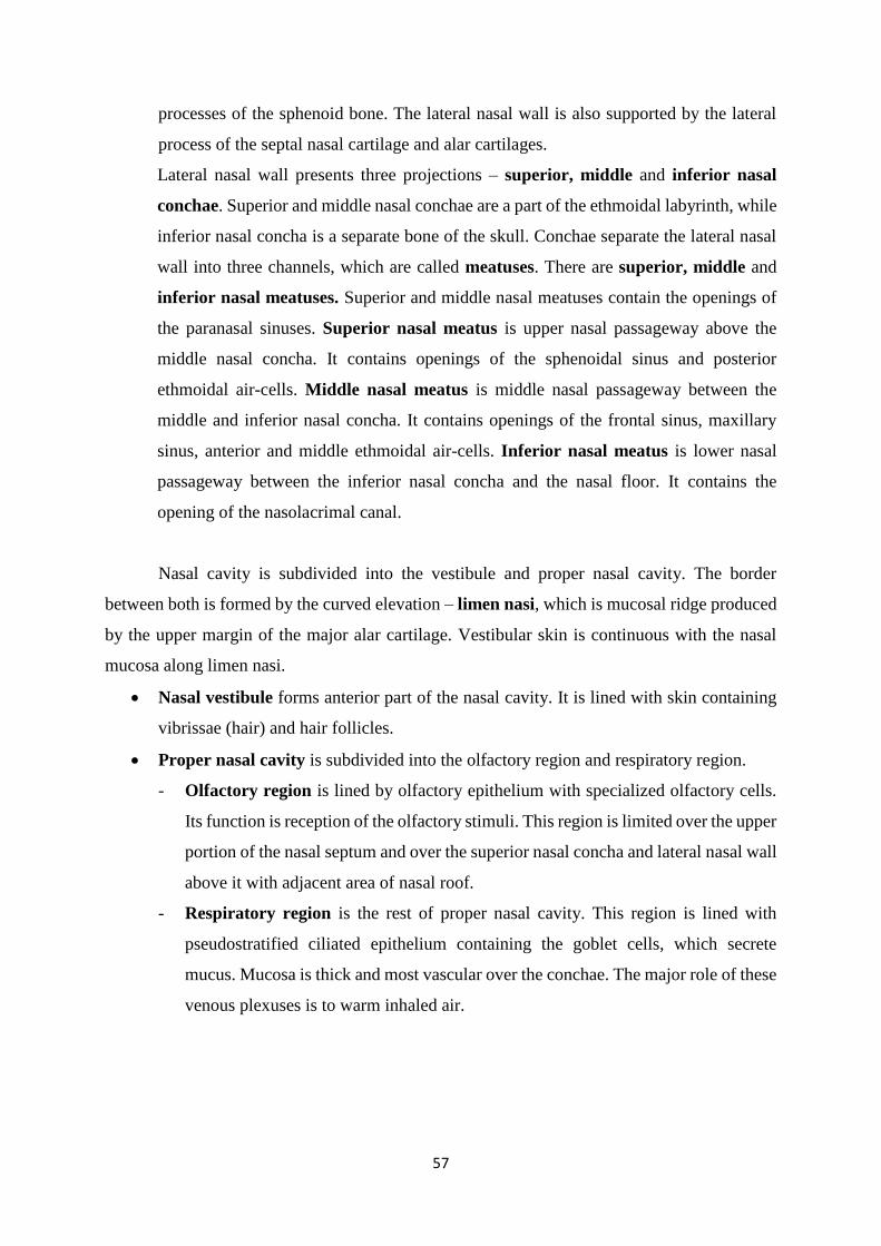
Lateral nasal wall presents three projections – superior, middle and inferior nasal
conchae. Superior and middle nasal conchae are a part of the ethmoidal labyrinth, while
inferior nasal concha is a separate bone of the skull. Conchae separate the lateral nasal
wall into three channels, which are called meatuses. There are superior, middle and
inferior nasal meatuses. Superior and middle nasal meatuses contain the openings of
the paranasal sinuses. Superior nasal meatus is upper nasal passageway above the
middle nasal concha. It contains openings of the sphenoidal sinus and posterior
ethmoidal air-cells. Middle nasal meatus is middle nasal passageway between the
middle and inferior nasal concha. It contains openings of the frontal sinus, maxillary
sinus, anterior and middle ethmoidal air-cells. Inferior nasal meatus is lower nasal
passageway between the inferior nasal concha and the nasal floor. It contains the
opening of the nasolacrimal canal.
Nasal cavity is subdivided into the vestibule and proper nasal cavity. The border
between both is formed by the curved elevation – limen nasi, which is mucosal ridge produced
by the upper margin of the major alar cartilage. Vestibular skin is continuous with the nasal
mucosa along limen nasi.
Nasal vestibule forms anterior part of the nasal cavity. It is lined with skin containing
vibrissae (hair) and hair follicles.
Proper nasal cavity is subdivided into the olfactory region and respiratory region.
- Olfactory region is lined by olfactory epithelium with specialized olfactory cells.
Its function is reception of the olfactory stimuli. This region is limited over the upper
portion of the nasal septum and over the superior nasal concha and lateral nasal wall
above it with adjacent area of nasal roof.
- Respiratory region is the rest of proper nasal cavity. This region is lined with
pseudostratified ciliated epithelium containing the goblet cells, which secrete
mucus. Mucosa is thick and most vascular over the conchae. The major role of these
venous plexuses is to warm inhaled air.
58
Fig. 39
Bones and cartilages of external nose
A – inferior view
B – right side, sagital plane
Retrieved from public domein Gray s Anatomy of human Body (1918) at Bartleby.com
Fig. 40
Nasal septum – bones of nasal septum and septal nasal cartilage
Sagital plane
Retrieved from public domein Gray s Anatomy of human Body (1918) at Bartleby.com
septal nasal cartilage,
posterior process
major alar cartilage,
medial crus
minor alar
cartilages
major alar
cartilage, lateral
crus
major alar cartilage,
lateral crus
septal nasal
cartilage
major alar cartilage,
medial crus
hard palate
bony nasal septum
nares
nasal bone
frontal process
of maxilla
A B
59
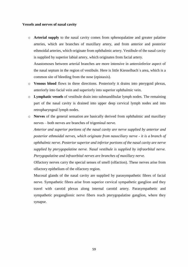
Vessels and nerves of nasal cavity
o Arterial supply to the nasal cavity comes from sphenopalatine and greater palatine
arteries, which are branches of maxillary artery, and from anterior and posterior
ethmoidal arteries, which originate from ophthalmic artery. Vestibule of the nasal cavity
is supplied by superior labial artery, which originates from facial artery.
Anastomoses between arterial branches are more intensive in anteroinferior aspect of
the nasal septum in the region of vestibule. Here is little Kiesselbach᾽s area, which is a
common site of bleeding from the nose (epistaxis).
o Venous blood flows in three directions. Posteriorly it drains into pterygoid plexus,
anteriorly into facial vein and superiorly into superior ophthalmic vein.
o Lymphatic vessels of vestibule drain into submandibular lymph nodes. The remaining
part of the nasal cavity is drained into upper deep cervical lymph nodes and into
retropharyngeal lymph nodes.
o Nerves of the general sensation are basically derived from ophthalmic and maxillary
nerves – both nerves are branches of trigeminal nerve.
Anterior and superior portions of the nasal cavity are nerve supplied by anterior and
posterior ethmoidal nerves, which originate from nasociliary nerve - it is a branch of
ophthalmic nerve. Posterior superior and inferior portions of the nasal cavity are nerve
supplied by pterygopalatine nerve. Nasal vestibule is supplied by infraorbital nerve.
Pterygopalatine and infraorbital nerves are branches of maxillary nerve.
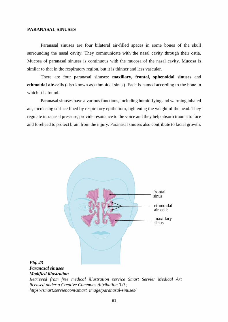
Olfactory nerves carry the special senses of smell (olfaction). These nerves arise from
olfactory epithelium of the olfactory region.
Mucosal glands of the nasal cavity are supplied by parasympathetic fibres of facial
nerve. Sympathetic fibres arise from superior cervical sympathetic ganglion and they
travel with carotid plexus along internal carotid artery. Parasympathetic and
sympathetic preganglionic nerve fibers reach pterygopalatine ganglion, where they
synapse.
60
limen nasi
PROPER NASAL CAVITY –olfactory region
NASAL VESTIBULE
Fig. 41
Nasal cavity - lateral nasal wall
Sagital plane
Retrieved from public domein Gray s Anatomy of human Body (1918) at Bartleby.com
superior, middleand inferior
nasal meatuses
Fig. 42
Nasal cavity – olfactory nerves that carry special senses of smell (olfaction) from
olfactory region of proper nasal cavity
Modified illustration
Retrieved from free medical illustration service Smart Servier Medical Art licensed under
a Creative Commons Attribution 3.0; https://smart.servier.com/smart_image/nasal-cavity/;
https://smart.servier.com/smart_image/olfactory-bulb/
olfactory nerves
frontal lobe
cribriform plate ofethmoid bone
olfactory bulb
olfactory epithelium
61
frontalsinus
ethmoidal air-cells
Fig. 43
Paranasal sinuses
Modified illustration
Retrieved from free medical illustration service Smart Servier Medical Art
licensed under a Creative Commons Attribution 3.0 ;
https://smart.servier.com/smart_image/paranasal-sinuses/
maxillary sinus
PARANASAL SINUSES
Paranasal sinuses are four bilateral air-filled spaces in some bones of the skull
surrounding the nasal cavity. They communicate with the nasal cavity through their ostia.
Mucosa of paranasal sinuses is continuous with the mucosa of the nasal cavity. Mucosa is
similar to that in the respiratory region, but it is thinner and less vascular.
There are four paranasal sinuses: maxillary, frontal, sphenoidal sinuses and
ethmoidal air-cells (also known as ethmoidal sinus). Each is named according to the bone in
which it is found.
Paranasal sinuses have a various functions, including humidifying and warming inhaled
air, increasing surface lined by respiratory epithelium, lightening the weight of the head. They
regulate intranasal pressure, provide resonance to the voice and they help absorb trauma to face
and forehead to protect brain from the injury. Paranasal sinuses also contribute to facial growth.
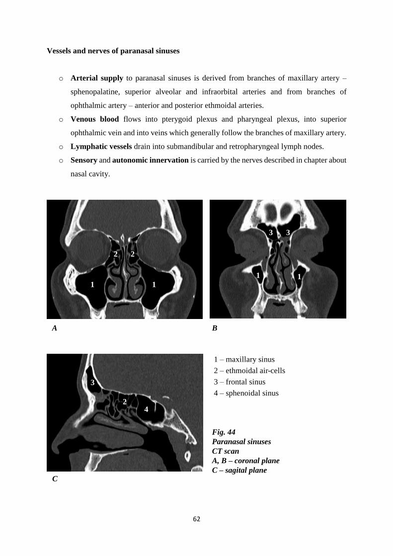
62
1 – maxillary sinus
2 – ethmoidal air-cells
3 – frontal sinus
4 – sphenoidal sinus
Fig. 44
Paranasal sinuses
CT scan
A, B – coronal plane
C – sagital plane
A
C
B
42
3
2 2
1111
3 3
Vessels and nerves of paranasal sinuses
o Arterial supply to paranasal sinuses is derived from branches of maxillary artery –
sphenopalatine, superior alveolar and infraorbital arteries and from branches of
ophthalmic artery – anterior and posterior ethmoidal arteries.
o Venous blood flows into pterygoid plexus and pharyngeal plexus, into superior
ophthalmic vein and into veins which generally follow the branches of maxillary artery.
o Lymphatic vessels drain into submandibular and retropharyngeal lymph nodes.
o Sensory and autonomic innervation is carried by the nerves described in chapter about
nasal cavity.
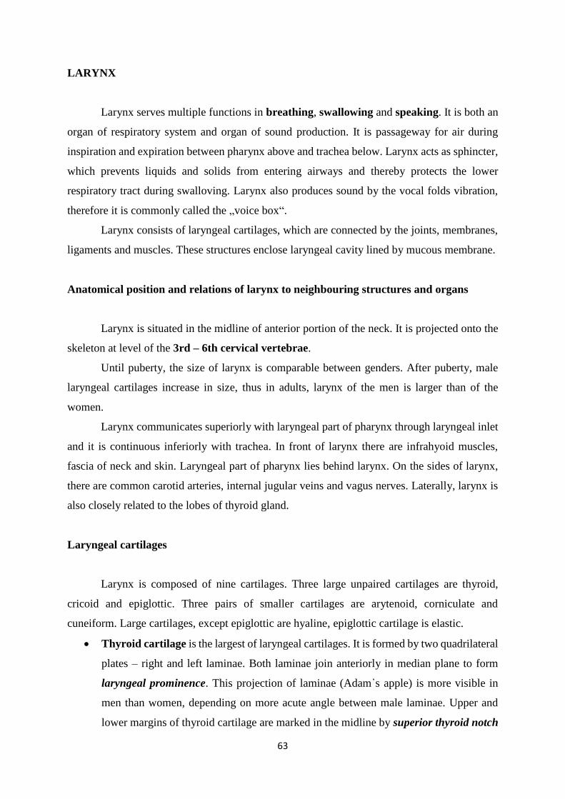
63
LARYNX
Larynx serves multiple functions in breathing, swallowing and speaking. It is both an
organ of respiratory system and organ of sound production. It is passageway for air during
inspiration and expiration between pharynx above and trachea below. Larynx acts as sphincter,
which prevents liquids and solids from entering airways and thereby protects the lower
respiratory tract during swalloving. Larynx also produces sound by the vocal folds vibration,
therefore it is commonly called the „voice box“.
Larynx consists of laryngeal cartilages, which are connected by the joints, membranes,
ligaments and muscles. These structures enclose laryngeal cavity lined by mucous membrane.
Anatomical position and relations of larynx to neighbouring structures and organs
Larynx is situated in the midline of anterior portion of the neck. It is projected onto the
skeleton at level of the 3rd – 6th cervical vertebrae.
Until puberty, the size of larynx is comparable between genders. After puberty, male
laryngeal cartilages increase in size, thus in adults, larynx of the men is larger than of the
women.
Larynx communicates superiorly with laryngeal part of pharynx through laryngeal inlet
and it is continuous inferiorly with trachea. In front of larynx there are infrahyoid muscles,
fascia of neck and skin. Laryngeal part of pharynx lies behind larynx. On the sides of larynx,
there are common carotid arteries, internal jugular veins and vagus nerves. Laterally, larynx is
also closely related to the lobes of thyroid gland.
Laryngeal cartilages
Larynx is composed of nine cartilages. Three large unpaired cartilages are thyroid,
cricoid and epiglottic. Three pairs of smaller cartilages are arytenoid, corniculate and
cuneiform. Large cartilages, except epiglottic are hyaline, epiglottic cartilage is elastic.
Thyroid cartilage is the largest of laryngeal cartilages. It is formed by two quadrilateral
plates – right and left laminae. Both laminae join anteriorly in median plane to form
laryngeal prominence. This projection of laminae (Adam᾽s apple) is more visible in
men than women, depending on more acute angle between male laminae. Upper and
lower margins of thyroid cartilage are marked in the midline by superior thyroid notch
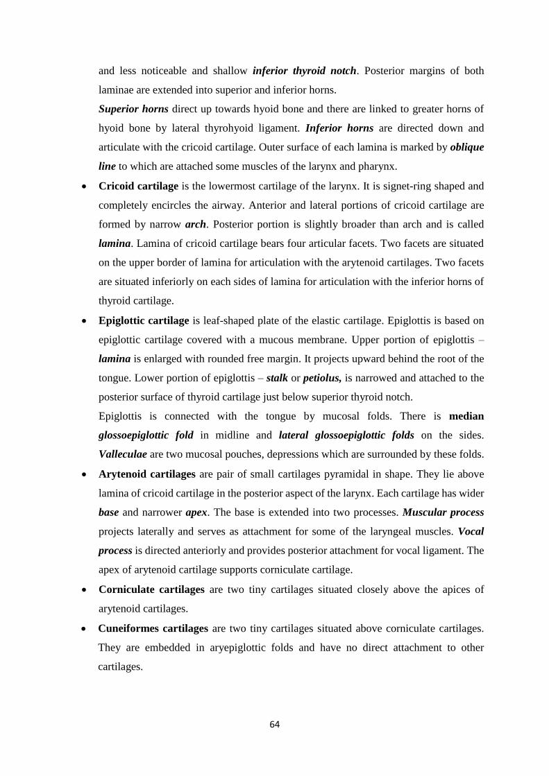
64
and less noticeable and shallow inferior thyroid notch. Posterior margins of both
laminae are extended into superior and inferior horns.
Superior horns direct up towards hyoid bone and there are linked to greater horns of
hyoid bone by lateral thyrohyoid ligament. Inferior horns are directed down and
articulate with the cricoid cartilage. Outer surface of each lamina is marked by oblique
line to which are attached some muscles of the larynx and pharynx.
Cricoid cartilage is the lowermost cartilage of the larynx. It is signet-ring shaped and
completely encircles the airway. Anterior and lateral portions of cricoid cartilage are
formed by narrow arch. Posterior portion is slightly broader than arch and is called
lamina. Lamina of cricoid cartilage bears four articular facets. Two facets are situated
on the upper border of lamina for articulation with the arytenoid cartilages. Two facets
are situated inferiorly on each sides of lamina for articulation with the inferior horns of
thyroid cartilage.
Epiglottic cartilage is leaf-shaped plate of the elastic cartilage. Epiglottis is based on
epiglottic cartilage covered with a mucous membrane. Upper portion of epiglottis –
lamina is enlarged with rounded free margin. It projects upward behind the root of the
tongue. Lower portion of epiglottis – stalk or petiolus, is narrowed and attached to the
posterior surface of thyroid cartilage just below superior thyroid notch.
Epiglottis is connected with the tongue by mucosal folds. There is median
glossoepiglottic fold in midline and lateral glossoepiglottic folds on the sides.
Valleculae are two mucosal pouches, depressions which are surrounded by these folds.
Arytenoid cartilages are pair of small cartilages pyramidal in shape. They lie above
lamina of cricoid cartilage in the posterior aspect of the larynx. Each cartilage has wider
base and narrower apex. The base is extended into two processes. Muscular process
projects laterally and serves as attachment for some of the laryngeal muscles. Vocal
process is directed anteriorly and provides posterior attachment for vocal ligament. The
apex of arytenoid cartilage supports corniculate cartilage.
Corniculate cartilages are two tiny cartilages situated closely above the apices of
arytenoid cartilages.
Cuneiformes cartilages are two tiny cartilages situated above corniculate cartilages.
They are embedded in aryepiglottic folds and have no direct attachment to other
cartilages.
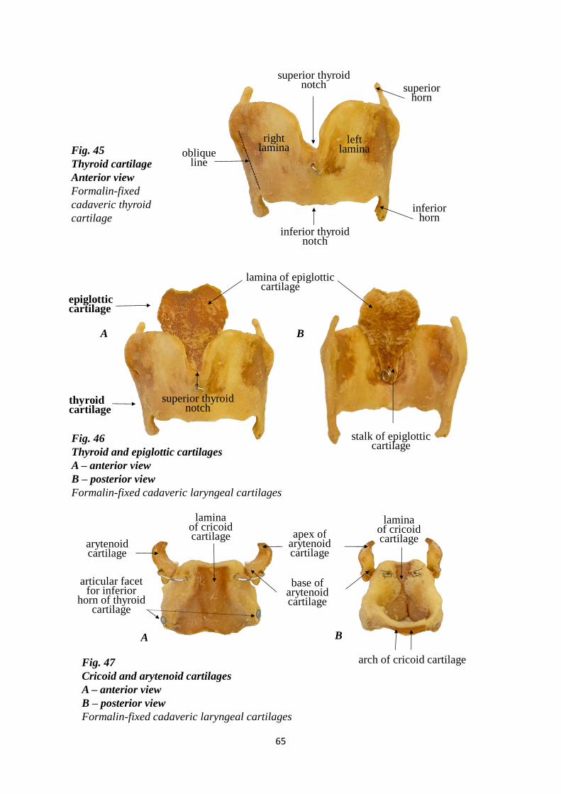
65
epiglottic cartilage
thyroid cartilage
laminaof cricoidcartilage
superior horn
inferior thyroid notch
rightlamina
superior thyroid notch
inferiorhorn
left laminaoblique
line
Fig. 46
Thyroid and epiglottic cartilages
A – anterior view
B – posterior view
Formalin-fixed cadaveric laryngeal cartilages
A B
stalk of epiglottic cartilage
lamina of epiglottic cartilage
arch of cricoid cartilage
Fig. 45
Thyroid cartilage
Anterior view
Formalin-fixed
cadaveric thyroid
cartilage
Fig. 47
Cricoid and arytenoid cartilages
A – anterior view
B – posterior view
Formalin-fixed cadaveric laryngeal cartilages
A B
base of arytenoid cartilage
arytenoidcartilage
lamina of cricoid cartilage apex of
arytenoid cartilage
superior thyroid notch
articular facetfor inferior
horn of thyroidcartilage
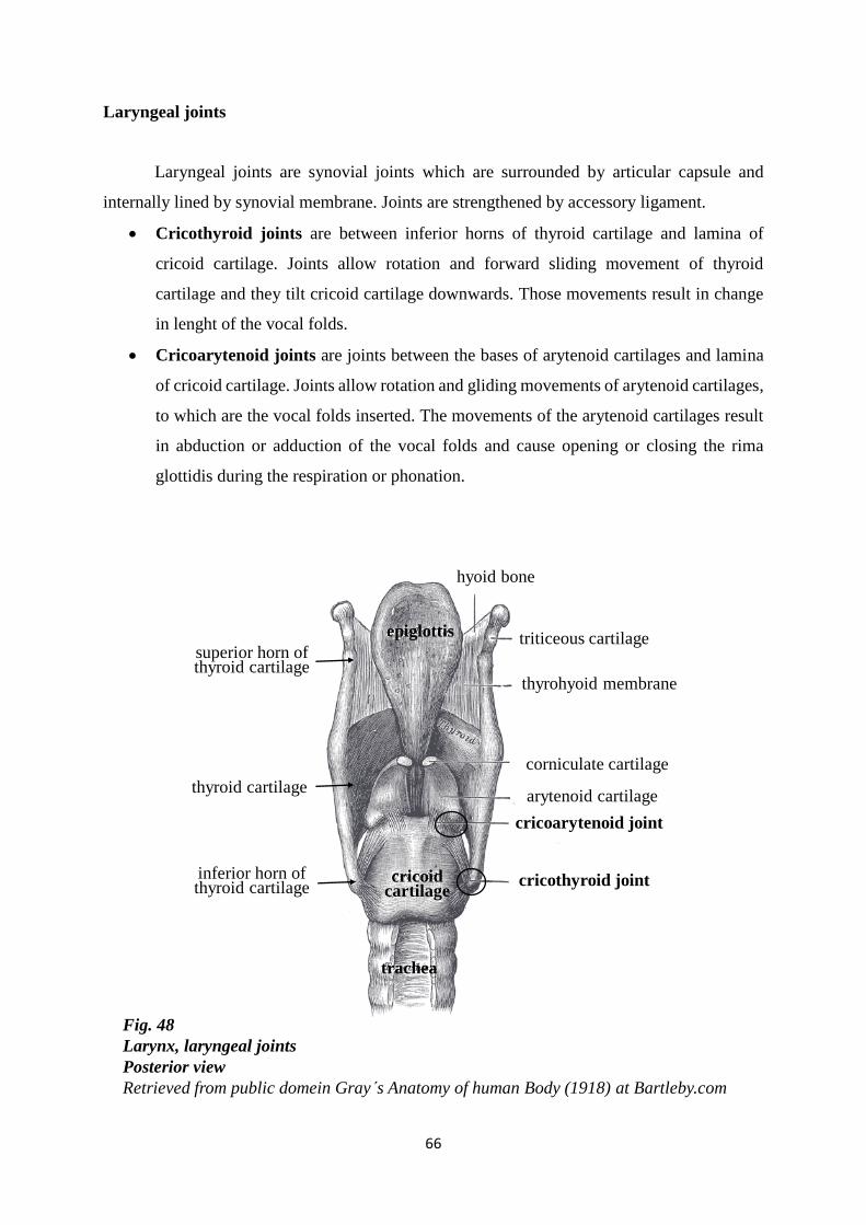
66
Fig. 48
Larynx, laryngeal joints
Posterior view
Retrieved from public domein Gray s Anatomy of human Body (1918) at Bartleby.com
cricothyroid joint
cricoarytenoid joint
thyrohyoid membrane
corniculate cartilage
arytenoid cartilage
triticeous cartilage
cricoid cartilage
trachea
epiglottis
hyoid bone
inferior horn ofthyroid cartilage
superior horn ofthyroid cartilage
thyroid cartilage
Laryngeal joints
Laryngeal joints are synovial joints which are surrounded by articular capsule and
internally lined by synovial membrane. Joints are strengthened by accessory ligament.
Cricothyroid joints are between inferior horns of thyroid cartilage and lamina of
cricoid cartilage. Joints allow rotation and forward sliding movement of thyroid
cartilage and they tilt cricoid cartilage downwards. Those movements result in change
in lenght of the vocal folds.
Cricoarytenoid joints are joints between the bases of arytenoid cartilages and lamina
of cricoid cartilage. Joints allow rotation and gliding movements of arytenoid cartilages,
to which are the vocal folds inserted. The movements of the arytenoid cartilages result
in abduction or adduction of the vocal folds and cause opening or closing the rima
glottidis during the respiration or phonation.
67
Laryngeal membranes and ligaments
Larynx is composed of several membranes and ligaments. They are subdivided to
extrinsic and intrinsic groups.
Extrinsic membrane and ligaments are elastic or fibroelastic bands that connect
larynx to external neighbouring structures.
Thyrohyoid membrane connects the upper margin of thyroid cartilage below to the
hyoid bone above. In the midline and laterally the membrane is thickened to form
median thyrohyoid ligament and lateral thyrohyoid ligaments. Median thyrohyoid
ligament is extended between superior thyroid notch and body of the hyoid bone. Lateral
thyrohyoid ligaments link together superior horns of thyroid cartilage and greater horns
of the hyoid bone. On each side thyrohyoid membrane is perforated by superior
laryngeal vessels and nerves.
Cricotracheal ligament connects cricoid cartilage above with the trachea below.
Hyoepiglottic ligament extends in midline between the body of the hyoid bone and
anterior surface of the epiglottis.
Intrinsic membranes and ligaments – fibroelastic membrane of the larynx, connect
individual laryngeal cartilages together and lie beneath the laryngeal mucosa. Fibroelastic
membranes are formed by upper quadrangular membrane and lower cricothyroid ligament. On
each side, these two parts are interrupted by a gap between vestibular ligaments above and vocal
ligaments below.
Quadrangular membrane on each side extends between lateral margin of epiglottis
and anterolateral surface of arytenoid cartilage. Its free upper margin is covered by
mucosa to form aryepiglottic fold. Its free lower margin is thickened and forms
vestibular ligament. Vestibular ligament is covered loosely by mucosa to form
vestibular fold. Vestibular fold extends between thyroid and arytenoid cartilages and
lies superior to vocal fold.
Vestibular folds have no role in sound production, therefore they are also called false
vocal cords.
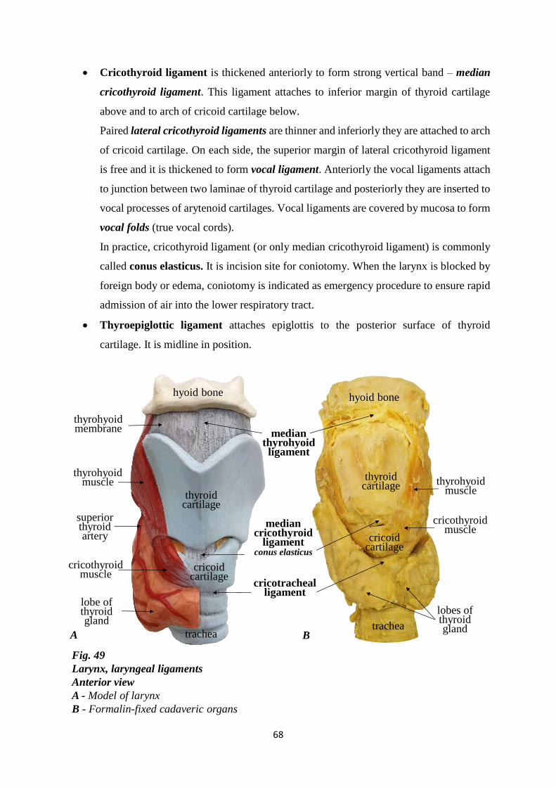
68
Fig. 49
Larynx, laryngeal ligaments
Anterior view
A - Model of larynx
B - Formalin-fixed cadaveric organs
thyrohyoid membrane
thyroid cartilage
cricoid cartilage
trachea
lobe of thyroid gland
lobes of thyroid gland
thyroid cartilage
cricoid cartilage
hyoid bonehyoid bone
median thyrohyoid ligament
trachea
cricothyroid muscle
cricotrachealligament
superior thyroid artery
thyrohyoid muscle
cricothyroid muscle
thyrohyoid muscle
mediancricothyroid
ligamentconus elasticus
A B
Cricothyroid ligament is thickened anteriorly to form strong vertical band – median
cricothyroid ligament. This ligament attaches to inferior margin of thyroid cartilage
above and to arch of cricoid cartilage below.
Paired lateral cricothyroid ligaments are thinner and inferiorly they are attached to arch
of cricoid cartilage. On each side, the superior margin of lateral cricothyroid ligament
is free and it is thickened to form vocal ligament. Anteriorly the vocal ligaments attach
to junction between two laminae of thyroid cartilage and posteriorly they are inserted to
vocal processes of arytenoid cartilages. Vocal ligaments are covered by mucosa to form
vocal folds (true vocal cords).
In practice, cricothyroid ligament (or only median cricothyroid ligament) is commonly
called conus elasticus. It is incision site for coniotomy. When the larynx is blocked by
foreign body or edema, coniotomy is indicated as emergency procedure to ensure rapid
admission of air into the lower respiratory tract.
Thyroepiglottic ligament attaches epiglottis to the posterior surface of thyroid
cartilage. It is midline in position.
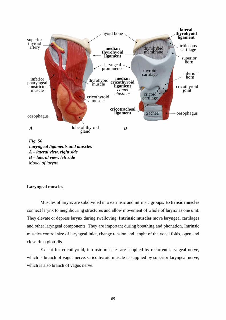
69
Fig. 50
Laryngeal ligaments and muscles
A – lateral view, right side
B – lateral view, left side
Model of larynx
trachea oesophagus
cricothyroid joint
superiorhorn
cricoid cartilage
laryngeal prominence
inferiorhorn
superior thyroid artery
thyrohyoid muscle
cricothyroid muscle
lobe of thyroidgland
inferior pharyngeal constrictor
muscle
oesophagus
thyrohyoid membrane
median thyrohyoid ligament
lateral thyrohyoid ligament
A B
cricotracheal ligament
mediancricothyroid
ligamentconus
elasticus
thyroidcartilage
hyoid bone
triticeouscartilage
Laryngeal muscles
Muscles of larynx are subdivided into extrinsic and intrinsic groups. Extrinsic muscles
connect larynx to neighbouring structures and allow movement of whole of larynx as one unit.
They elevate or depress larynx during swalloving. Intrinsic muscles move laryngeal cartilages
and other laryngeal components. They are important during breathing and phonation. Intrinsic
muscles control size of laryngeal inlet, change tension and lenght of the vocal folds, open and
close rima glottidis.
Except for cricothyroid, intrinsic muscles are supplied by recurrent laryngeal nerve,
which is branch of vagus nerve. Cricothyroid muscle is supplied by superior laryngeal nerve,
which is also branch of vagus nerve.
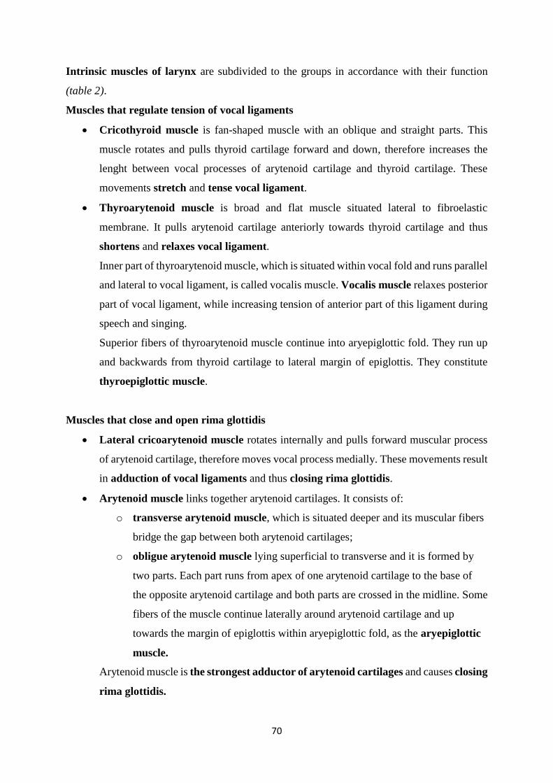
70
Intrinsic muscles of larynx are subdivided to the groups in accordance with their function
(table 2).
Muscles that regulate tension of vocal ligaments
Cricothyroid muscle is fan-shaped muscle with an oblique and straight parts. This
muscle rotates and pulls thyroid cartilage forward and down, therefore increases the
lenght between vocal processes of arytenoid cartilage and thyroid cartilage. These
movements stretch and tense vocal ligament.
Thyroarytenoid muscle is broad and flat muscle situated lateral to fibroelastic
membrane. It pulls arytenoid cartilage anteriorly towards thyroid cartilage and thus
shortens and relaxes vocal ligament.
Inner part of thyroarytenoid muscle, which is situated within vocal fold and runs parallel
and lateral to vocal ligament, is called vocalis muscle. Vocalis muscle relaxes posterior
part of vocal ligament, while increasing tension of anterior part of this ligament during
speech and singing.
Superior fibers of thyroarytenoid muscle continue into aryepiglottic fold. They run up
and backwards from thyroid cartilage to lateral margin of epiglottis. They constitute
thyroepiglottic muscle.
Muscles that close and open rima glottidis
Lateral cricoarytenoid muscle rotates internally and pulls forward muscular process
of arytenoid cartilage, therefore moves vocal process medially. These movements result
in adduction of vocal ligaments and thus closing rima glottidis.
Arytenoid muscle links together arytenoid cartilages. It consists of:
o transverse arytenoid muscle, which is situated deeper and its muscular fibers
bridge the gap between both arytenoid cartilages;
o obligue arytenoid muscle lying superficial to transverse and it is formed by
two parts. Each part runs from apex of one arytenoid cartilage to the base of
the opposite arytenoid cartilage and both parts are crossed in the midline. Some
fibers of the muscle continue laterally around arytenoid cartilage and up
towards the margin of epiglottis within aryepiglottic fold, as the aryepiglottic
muscle.
Arytenoid muscle is the strongest adductor of arytenoid cartilages and causes closing
rima glottidis.
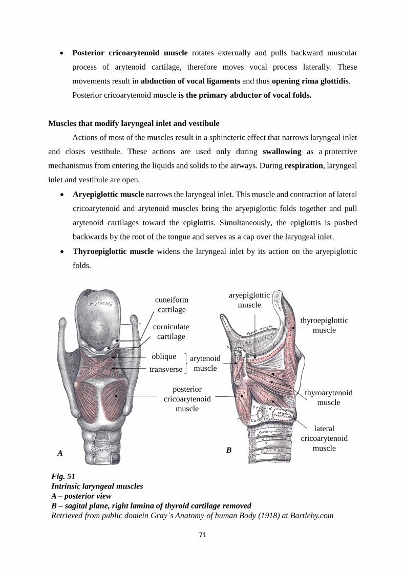
71
Fig. 51
Intrinsic laryngeal muscles
A – posterior view
B – sagital plane, right lamina of thyroid cartilage removed
Retrieved from public domein Gray s Anatomy of human Body (1918) at Bartleby.com
posterior
cricoarytenoid
muscle
transverse
oblique arytenoid
muscle
lateral
cricoarytenoid
muscle
cuneiform
cartilage
corniculate
cartilage
thyroarytenoid
muscle
aryepiglottic
muscle
thyroepiglottic
muscle
A B
Posterior cricoarytenoid muscle rotates externally and pulls backward muscular
process of arytenoid cartilage, therefore moves vocal process laterally. These
movements result in abduction of vocal ligaments and thus opening rima glottidis.
Posterior cricoarytenoid muscle is the primary abductor of vocal folds.
Muscles that modify laryngeal inlet and vestibule
Actions of most of the muscles result in a sphincteric effect that narrows laryngeal inlet
and closes vestibule. These actions are used only during swallowing as a protective
mechanismus from entering the liquids and solids to the airways. During respiration, laryngeal
inlet and vestibule are open.
Aryepiglottic muscle narrows the laryngeal inlet. This muscle and contraction of lateral
cricoarytenoid and arytenoid muscles bring the aryepiglottic folds together and pull
arytenoid cartilages toward the epiglottis. Simultaneously, the epiglottis is pushed
backwards by the root of the tongue and serves as a cap over the laryngeal inlet.
Thyroepiglottic muscle widens the laryngeal inlet by its action on the aryepiglottic
folds.
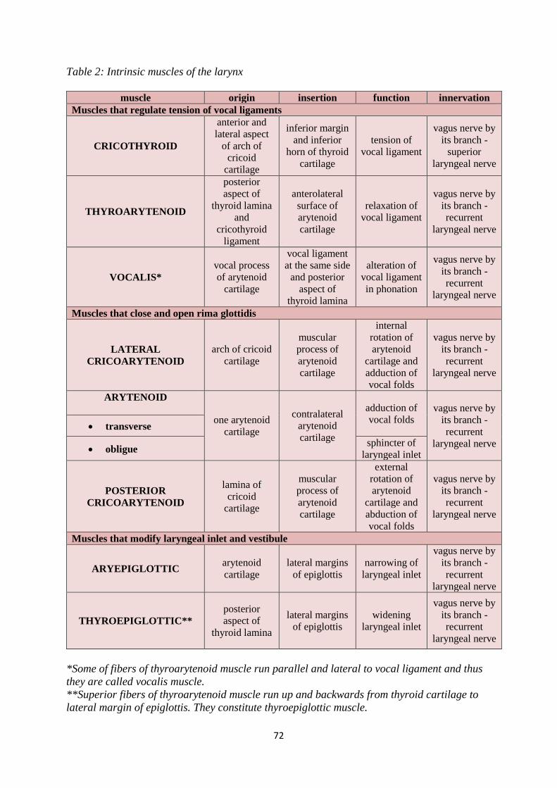
72
Table 2: Intrinsic muscles of the larynx
muscle origin insertion function innervation
Muscles that regulate tension of vocal ligaments
CRICOTHYROID
anterior and
lateral aspect
of arch of
cricoid
cartilage
inferior margin
and inferior
horn of thyroid
cartilage
tension of
vocal ligament
vagus nerve by
its branch -
superior
laryngeal nerve
THYROARYTENOID
posterior
aspect of
thyroid lamina
and
cricothyroid
ligament
anterolateral
surface of
arytenoid
cartilage
relaxation of
vocal ligament
vagus nerve by
its branch -
recurrent
laryngeal nerve
VOCALIS*
vocal process
of arytenoid
cartilage
vocal ligament
at the same side
and posterior
aspect of
thyroid lamina
alteration of
vocal ligament
in phonation
vagus nerve by
its branch -
recurrent
laryngeal nerve
Muscles that close and open rima glottidis
LATERAL
CRICOARYTENOID
arch of cricoid
cartilage
muscular
process of
arytenoid
cartilage
internal
rotation of
arytenoid
cartilage and
adduction of
vocal folds
vagus nerve by
its branch -
recurrent
laryngeal nerve
ARYTENOID
one arytenoid
cartilage
contralateral
arytenoid
cartilage
adduction of
vocal folds vagus nerve by
its branch -
recurrent
laryngeal nerve
transverse
obligue sphincter of
laryngeal inlet
POSTERIOR
CRICOARYTENOID
lamina of
cricoid
cartilage
muscular
process of
arytenoid
cartilage
external
rotation of
arytenoid
cartilage and
abduction of
vocal folds
vagus nerve by
its branch -
recurrent
laryngeal nerve
Muscles that modify laryngeal inlet and vestibule
ARYEPIGLOTTIC arytenoid
cartilage
lateral margins
of epiglottis
narrowing of
laryngeal inlet
vagus nerve by
its branch -
recurrent
laryngeal nerve
THYROEPIGLOTTIC**
posterior
aspect of
thyroid lamina
lateral margins
of epiglottis
widening
laryngeal inlet
vagus nerve by
its branch -
recurrent
laryngeal nerve
*Some of fibers of thyroarytenoid muscle run parallel and lateral to vocal ligament and thus
they are called vocalis muscle.
**Superior fibers of thyroarytenoid muscle run up and backwards from thyroid cartilage to
lateral margin of epiglottis. They constitute thyroepiglottic muscle.
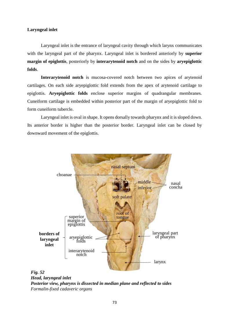
73
soft palate
borders of
laryngeal
inlet
choanae
larynx
inferior
middle
nasal septum
root of tongue
Fig. 52
Head, laryngeal inlet
Posterior view, pharynx is dissected in median plane and reflected to sides
Formalin-fixed cadaveric organs
superior margin of epiglottis
interarytenoid notch
aryepiglottic folds
laryngeal part of pharynx
nasal concha
Laryngeal inlet
Laryngeal inlet is the entrance of laryngeal cavity through which larynx communicates
with the laryngeal part of the pharynx. Laryngeal inlet is bordered anteriorly by superior
margin of epiglottis, posteriorly by interarytenoid notch and on the sides by aryepiglottic
folds.
Interarytenoid notch is mucosa-covered notch between two apices of arytenoid
cartilages. On each side aryepiglottic fold extends from the apex of arytenoid cartilage to
epiglottis. Aryepiglottic folds enclose superior margins of quadrangular membranes.
Cuneiform cartilage is embedded within posterior part of the margin of aryepiglottic fold to
form cuneiform tubercle.
Laryngeal inlet is oval in shape. It opens dorsally towards pharynx and it is sloped down.
Its anterior border is higher than the posterior border. Laryngeal inlet can be closed by
downward movement of the epiglottis.
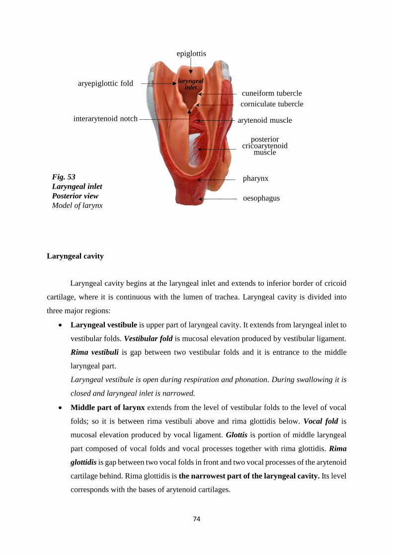
74
epiglottis
Fig. 53
Laryngeal inlet
Posterior view
Model of larynx
interarytenoid notch
aryepiglottic fold
cuneiform tubercle
corniculate tubercle
arytenoid muscle
posterior cricoarytenoid
muscle
pharynx
oesophagus
laryngeal inlet
Laryngeal cavity
Laryngeal cavity begins at the laryngeal inlet and extends to inferior border of cricoid
cartilage, where it is continuous with the lumen of trachea. Laryngeal cavity is divided into
three major regions:
Laryngeal vestibule is upper part of laryngeal cavity. It extends from laryngeal inlet to
vestibular folds. Vestibular fold is mucosal elevation produced by vestibular ligament.
Rima vestibuli is gap between two vestibular folds and it is entrance to the middle
laryngeal part.
Laryngeal vestibule is open during respiration and phonation. During swallowing it is
closed and laryngeal inlet is narrowed.
Middle part of larynx extends from the level of vestibular folds to the level of vocal
folds; so it is between rima vestibuli above and rima glottidis below. Vocal fold is
mucosal elevation produced by vocal ligament. Glottis is portion of middle laryngeal
part composed of vocal folds and vocal processes together with rima glottidis. Rima
glottidis is gap between two vocal folds in front and two vocal processes of the arytenoid
cartilage behind. Rima glottidis is the narrowest part of the laryngeal cavity. Its level
corresponds with the bases of arytenoid cartilages.
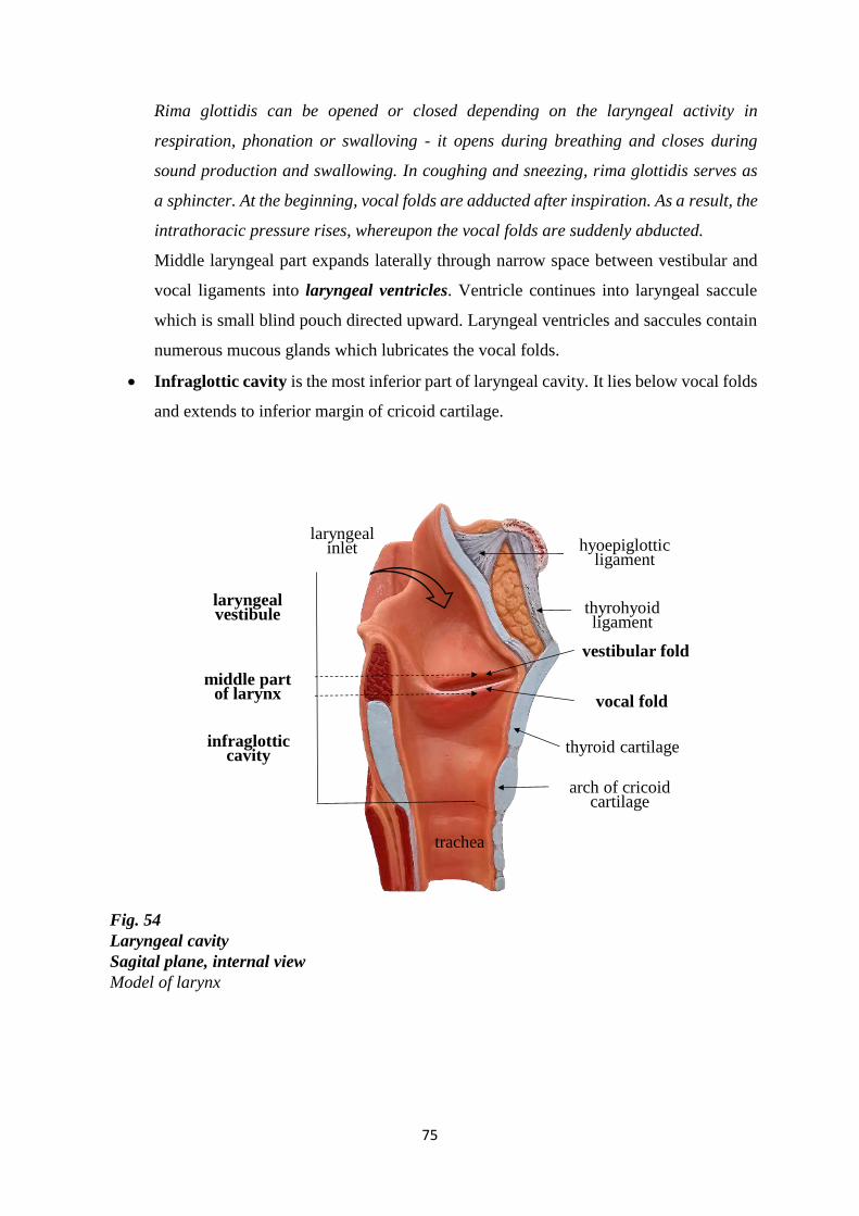
75
Fig. 54
Laryngeal cavity
Sagital plane, internal view
Model of larynx
vocal fold
vestibular fold
arch of cricoid cartilage
trachea
thyrohyoid ligament
infraglottic cavity
hyoepiglottic ligament
thyroid cartilage
middle part of larynx
laryngeal inlet
laryngeal vestibule
Rima glottidis can be opened or closed depending on the laryngeal activity in
respiration, phonation or swalloving - it opens during breathing and closes during
sound production and swallowing. In coughing and sneezing, rima glottidis serves as
a sphincter. At the beginning, vocal folds are adducted after inspiration. As a result, the
intrathoracic pressure rises, whereupon the vocal folds are suddenly abducted.
Middle laryngeal part expands laterally through narrow space between vestibular and
vocal ligaments into laryngeal ventricles. Ventricle continues into laryngeal saccule
which is small blind pouch directed upward. Laryngeal ventricles and saccules contain
numerous mucous glands which lubricates the vocal folds.
Infraglottic cavity is the most inferior part of laryngeal cavity. It lies below vocal folds
and extends to inferior margin of cricoid cartilage.
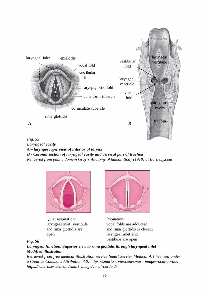
76
Fig. 55
Laryngeal cavity
A – laryngoscopic view of interior of larynx
B - Coronal section of laryngeal cavity and cervical part of trachea
Retrieved from public domein Gray s Anatomy of human Body (1918) at Bartleby.com
B
Fig. 56
Laryngeal function. Superior view to rima glottidis through laryngeal inlet
Modified illustration
Retrieved from free medical illustration service Smart Servier Medical Art licensed under
a Creative Commons Attribution 3.0; https://smart.servier.com/smart_image/vocal-cords/;
https://smart.servier.com/smart_image/vocal-cords-2/
A
rima glottidis
cuneiform tubercle
corniculate tubercle
aryepiglottic fold
vocal fold
vestibular
fold
laryngeal inlet epiglottisvestibular
fold
vocal
fold
laryngeal
vestibule
infraglottic
cavity
laryngeal
ventricle
trachea
Quiet respiration:
laryngeal inlet, vestibule
and rima glottidis are
open
Phonation:
vocal folds are adducted
and rima glottidis is closed;
laryngeal inlet and
vestibule are open
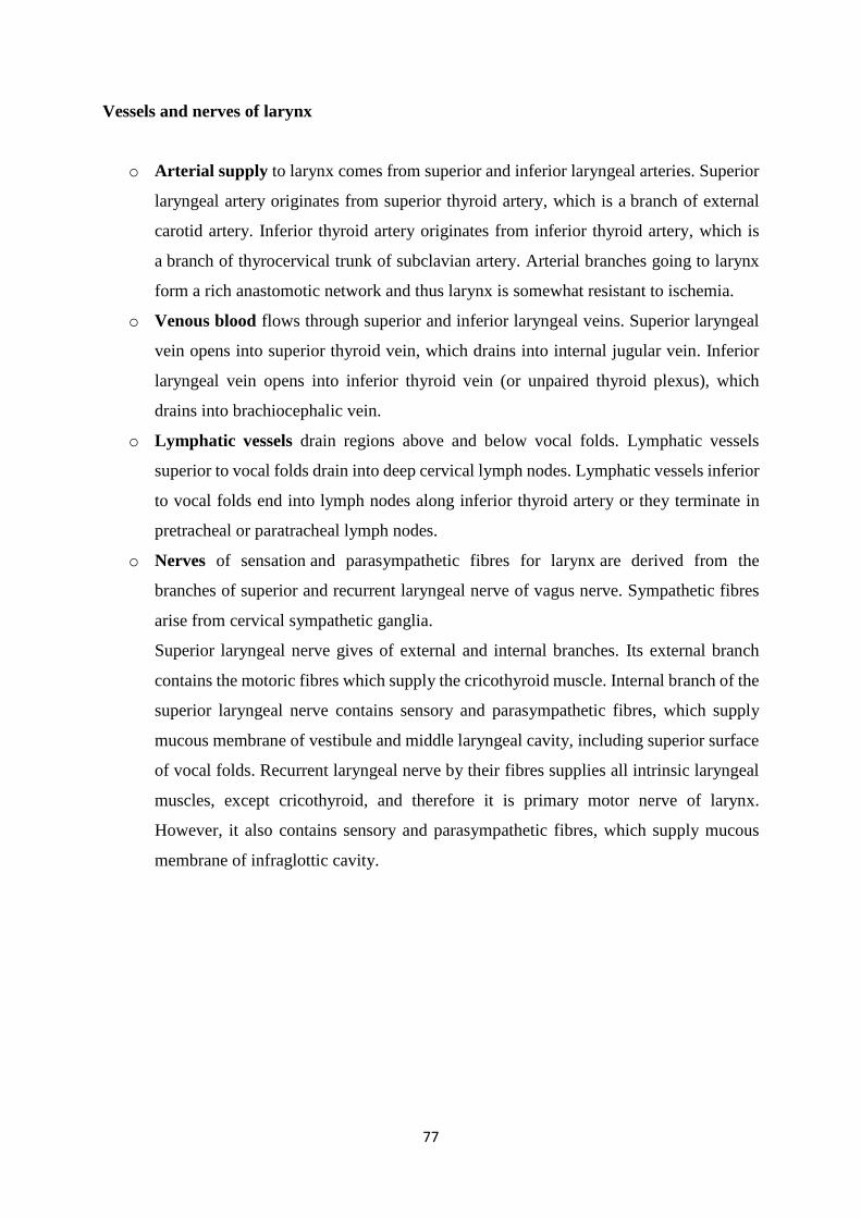
77
Vessels and nerves of larynx
o Arterial supply to larynx comes from superior and inferior laryngeal arteries. Superior
laryngeal artery originates from superior thyroid artery, which is a branch of external
carotid artery. Inferior thyroid artery originates from inferior thyroid artery, which is
a branch of thyrocervical trunk of subclavian artery. Arterial branches going to larynx
form a rich anastomotic network and thus larynx is somewhat resistant to ischemia.
o Venous blood flows through superior and inferior laryngeal veins. Superior laryngeal
vein opens into superior thyroid vein, which drains into internal jugular vein. Inferior
laryngeal vein opens into inferior thyroid vein (or unpaired thyroid plexus), which
drains into brachiocephalic vein.
o Lymphatic vessels drain regions above and below vocal folds. Lymphatic vessels
superior to vocal folds drain into deep cervical lymph nodes. Lymphatic vessels inferior
to vocal folds end into lymph nodes along inferior thyroid artery or they terminate in
pretracheal or paratracheal lymph nodes.
o Nerves of sensation and parasympathetic fibres for larynx are derived from the
branches of superior and recurrent laryngeal nerve of vagus nerve. Sympathetic fibres
arise from cervical sympathetic ganglia.
Superior laryngeal nerve gives of external and internal branches. Its external branch
contains the motoric fibres which supply the cricothyroid muscle. Internal branch of the
superior laryngeal nerve contains sensory and parasympathetic fibres, which supply
mucous membrane of vestibule and middle laryngeal cavity, including superior surface
of vocal folds. Recurrent laryngeal nerve by their fibres supplies all intrinsic laryngeal
muscles, except cricothyroid, and therefore it is primary motor nerve of larynx.
However, it also contains sensory and parasympathetic fibres, which supply mucous
membrane of infraglottic cavity.
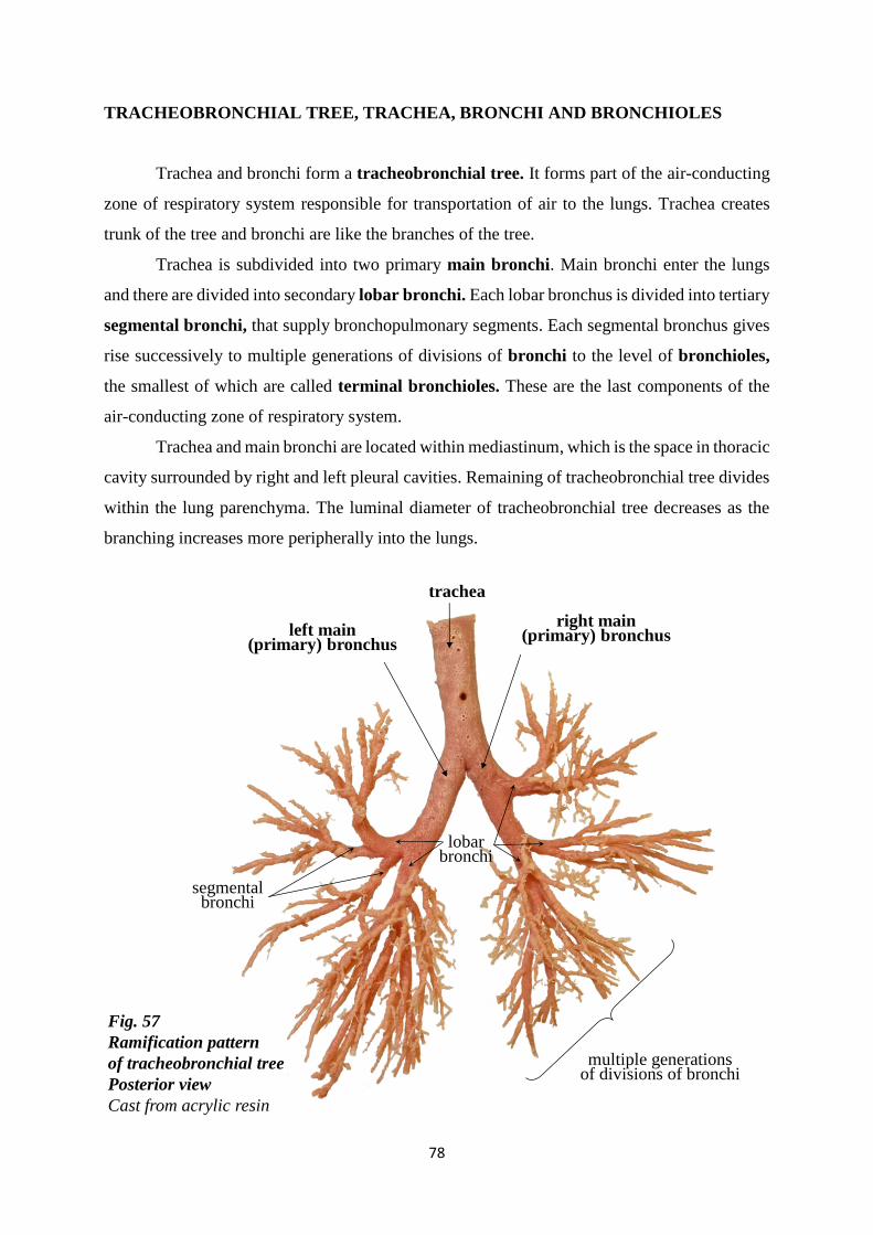
78
Fig. 57
Ramification pattern
of tracheobronchial tree
Posterior view
Cast from acrylic resin
trachea
right main (primary) bronchusleft main
(primary) bronchus
lobar bronchi
segmental bronchi
multiple generationsof divisions of bronchi
TRACHEOBRONCHIAL TREE, TRACHEA, BRONCHI AND BRONCHIOLES
Trachea and bronchi form a tracheobronchial tree. It forms part of the air-conducting
zone of respiratory system responsible for transportation of air to the lungs. Trachea creates
trunk of the tree and bronchi are like the branches of the tree.
Trachea is subdivided into two primary main bronchi. Main bronchi enter the lungs
and there are divided into secondary lobar bronchi. Each lobar bronchus is divided into tertiary
segmental bronchi, that supply bronchopulmonary segments. Each segmental bronchus gives
rise successively to multiple generations of divisions of bronchi to the level of bronchioles,
the smallest of which are called terminal bronchioles. These are the last components of the
air-conducting zone of respiratory system.
Trachea and main bronchi are located within mediastinum, which is the space in thoracic
cavity surrounded by right and left pleural cavities. Remaining of tracheobronchial tree divides
within the lung parenchyma. The luminal diameter of tracheobronchial tree decreases as the
branching increases more peripherally into the lungs.
79
TRACHEA
Trachea is cartilaginous and membranous tube at the beginning of the tracheobronchial
tree, allowing passage of air into lungs. It is flexible, distensible organ of continuously varying
size and shape. Trachea widens and lengthens slightly with each breath in, returning to its
resting size with each breath out. In the adult its lenght is 10 – 13 cm with diameter 1.5 – 2
cm.
Anatomical position and relations of trachea to neighbouring structures and organs
Trachea is continuation of larynx. Its uppermost portion is located at the lower border
of cricoid cartilage at the level of CVI in the neck. It enters thoracic cavity and descends within
mediastinum, where it bifurcates into right and left main bronchus, one for each lung. Its lower
end – tracheal bifurcation lies at the level of TIV – TV in the chest.
Upper, cervical part of trachea is in midline of the neck and it may be palpated just
above jugular notch. Thoracic part of trachea is located within mediastinum and it inclines
slightly to the right. The superior thoracic aperture (thoracic inlet) forms a border between
cervical and thoracic part of trachea.
In front of the cervical part of trachea there are isthmus of thyroid gland, inferior
thyroid veins, jugular arch, infrahyoid muscles, fasciae and skin of neck. Oesophagus runs
behind trachea and it is located between trachea and vertebral column. On the sides, there are
the lobes of thyroid gland, vagus nerves and recurrent laryngeal nerves, common carotid arteries
and internal juguar veins.
In front of the thoracic part of the trachea, there are sternum, remnant of thymus, heart
in pericardium, aortic arch, brachiocephalic and left common carotid arteries, inferior thyroid
and brachiocephalic veins. Trachea descends anterior to oesophagus. On the right side, there
are right lung and pleura, right brachiocephalic vein, superior vena cava, azygos vein and right
vagus nerve. On the left side, there are left lung and pleura, aortic arch, left subclavian artery
and left laryngeal recurrent nerve.
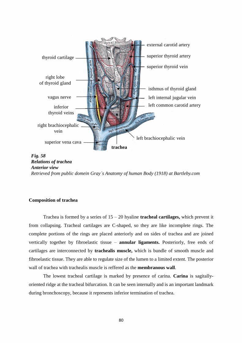
80
Retrieved
from public
domein
Gray s
Anatomy of
human
Body
(1918) at
Bartleby.co
m
Retriev
ed
from
public
domei
n
Gray s
Anato
my of
human
Body
(1918)
at
Bartle
by.comFig. 58
Relations of trachea
Anterior view
Retrieved from public domein Gray s Anatomy of human Body (1918) at Bartleby.com
right brachiocephalic
vein
right lobe
of thyroid gland
vagus nerve
superior vena cava
left internal jugular vein
external carotid artery
superior thyroid vein
superior thyroid artery
left common carotid artery
left brachiocephalic vein
inferior
thyroid veins
isthmus of thyroid gland
trachea
thyroid cartilage
Composition of trachea
Trachea is formed by a series of 15 – 20 hyaline tracheal cartilages, which prevent it
from collapsing. Tracheal cartilages are C-shaped, so they are like incomplete rings. The
complete portions of the rings are placed anteriorly and on sides of trachea and are joined
vertically together by fibroelastic tissue – annular ligaments. Posteriorly, free ends of
cartilages are interconnected by trachealis muscle, which is bundle of smooth muscle and
fibroelastic tissue. They are able to regulate size of the lumen to a limited extent. The posterior
wall of trachea with trachealis muscle is reffered as the membranous wall.
The lowest tracheal cartilage is marked by presence of carina. Carina is sagitally-
oriented ridge at the tracheal bifurcation. It can be seen internally and is an important landmark
during bronchoscopy, because it represents inferior termination of trachea.
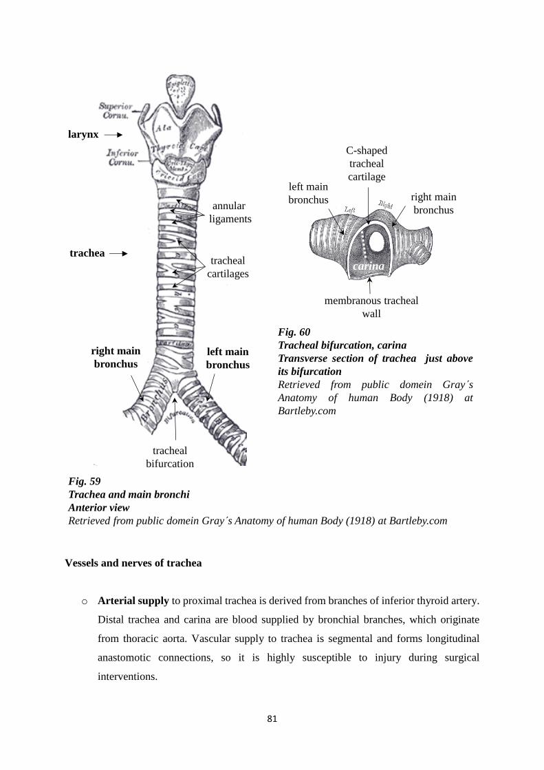
81
C-shaped
tracheal
cartilage
membranous tracheal
wall
left main
bronchus
carina
right main
bronchus
Fig. 60
Tracheal bifurcation, carina
Transverse section of trachea just above
its bifurcation
Retrieved from public domein Gray s
Anatomy of human Body (1918) at
Bartleby.com
right main
bronchus
left main
bronchus
tracheal
cartilages
annular
ligaments
trachea
larynx
tracheal
bifurcation
Fig. 59
Trachea and main bronchi
Anterior view
Retrieved from public domein Gray s Anatomy of human Body (1918) at Bartleby.com
Vessels and nerves of trachea
o Arterial supply to proximal trachea is derived from branches of inferior thyroid artery.
Distal trachea and carina are blood supplied by bronchial branches, which originate
from thoracic aorta. Vascular supply to trachea is segmental and forms longitudinal
anastomotic connections, so it is highly susceptible to injury during surgical
interventions.
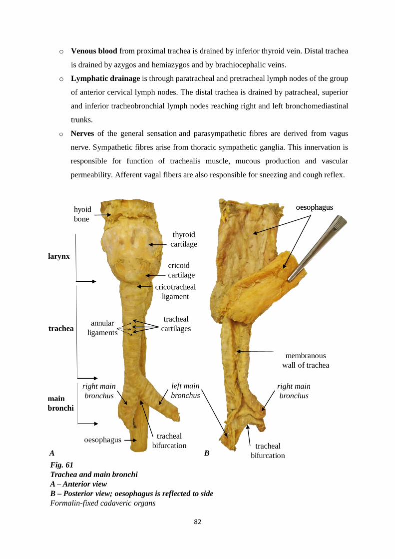
82
Fig. 61
Trachea and main bronchi
A – Anterior view
B – Posterior view; oesophagus is reflected to side
Formalin-fixed cadaveric organs
BA
larynx
hyoid
bone
left main
bronchusmain
bronchi
tracheaannular
ligaments
cricoid
cartilage
tracheal
cartilages
thyroid
cartilage
cricotracheal
ligament
oesophagus
tracheal
bifurcation
tracheal
bifurcation
right main
bronchus
membranous
wall of trachea
right main
bronchus
oesophagus
oesophagus
o Venous blood from proximal trachea is drained by inferior thyroid vein. Distal trachea
is drained by azygos and hemiazygos and by brachiocephalic veins.
o Lymphatic drainage is through paratracheal and pretracheal lymph nodes of the group
of anterior cervical lymph nodes. The distal trachea is drained by patracheal, superior
and inferior tracheobronchial lymph nodes reaching right and left bronchomediastinal
trunks.
o Nerves of the general sensation and parasympathetic fibres are derived from vagus
nerve. Sympathetic fibres arise from thoracic sympathetic ganglia. This innervation is
responsible for function of trachealis muscle, mucous production and vascular
permeability. Afferent vagal fibers are also responsible for sneezing and cough reflex.
83
MAIN BRONCHI (PRIMARY, PRINCIPAL)
The bronchial structures begin at the level of the tracheal bifurcation (or at the level of
the sternal angle), where the trachea is subdivided into two primary (principal) main bronchi.
At the bifurcation, two main bronchi are not equally divided.
Right main bronchus has wider diameter and is shorter than the left one. Its lenght
varies between 2 and 2.5 cm and its luminal diameter is about 1.5 cm. It has vertical orientation
being almost in direct line with trachea and it passes directly to hilum of the lung. Therefore,
inhaled foreign bodies tend to enter more frequently on the right side than on the left. Azygos
vein arches over right main bronchus. Both main bronchi are related to pulmonary artery. Right
main bronchus gives off a branch for superior lobe. This branch arises above the level of
pulmonary artery, and therefore named eparterial bronchus.
Left main bronchus has smaller diameter and is longer than right one. It is
approximately 4 – 6 cm long with luminal diameter about 1.1 cm. It arises at more oblique angle
and has more horizontal orientation. Left main bronchus passes inferolaterally, inferior to aortic
arch and anterior to oesophagus and thoracic aorta, to reach hilum of the lung. Left main
bronchus passes below the level of pulmonary artery before it divides, and hence all its branches
are hyparterial.
Composition of main bronchi
Composition of main bronchus is similar to that of trachea. Its wall is formed by C-
shaped cartilages and posteriorly by membranous wall, which consists mainly of the smooth
muscles and connective tissue with elastic fibres.
Vessels and nerves of main bronchi
o Arterial supply to bronchi comes from bronchial branches, which originate from
thoracic aorta. Bronchial branches accompany divisions of bronchial tree.
o Venous blood from bronchial veins drains into azygos vein on the right side and into
accessory hemiazygos vein on the left side.
o Lymphatic drainage is through tracheobronchial lymph nodes reaching right and left
bronchomediastinal trunks.
84
o Nerves of the general sensation and parasympathetic fibres are derived from vagus
nerve. Sympathetic fibres arise from the thoracic sympathetic ganglia.
LUNGS, PLEURA AND MEDIASTINUM
Lungs are vital organs of respiration, which are responsible for exchange of oxygen and
carbon dioxid between air we breathe and blood. The exchange of gases occurs between alveoli
of lungs and blood capillaries that surround them.
Lungs are situated within thoracic cavity. Each lung is surrounded by pleural cavity,
which is formed by visceral and parietal pleura. Two pleural cavities are situated on either side
of mediastinum. Each lung is attached to organs of mediastinum by a root that is formed by
airway, blood and lymph vessels and nerves.
LUNGS
Healthy lungs of living people are soft, porous, light organs with spongy consistence
and, because they contain air, they float in water and crepitates when handled. Their surfaces
are smooth and shining. At birth the lungs are pinkish color. In adulthood, the color of lungs
depends on several factors. Commonly, they are mottled with dark gray or bluish patches caused
by inhalation of atmospheric dust and by deposition of carbon particles.
Groos anatomy of lungs
Lungs are not equal in size. The right lung is a slightly larger and heavier than the left
and it is also shorter (the right dome of the diaphragm being higher) and wider (heart bulging
more to the left).
Lobes and fissures of lungs
Right lung is divided into superior, middle and inferior lobes. Middle lobe is the
smallest wedge-shaped lobe present only in the right lung. Lobes are freely movable against
each other and they are separated by fissures. Lobes of the right lung are separated by oblique
and horizontal fissures. Oblique fissure separates inferior lobe from middle and superior lobes.
85
Its direction is more vertical. Horizontal fissure separates superior lobe from middle lobe. It
runs horizontally, approximately along the level of the fourth intercostal space.
Left lung has superior and inferior lobes, which are separated by oblique fissure. The
superior lobe lies above and in front of this fissure. Inferior lobe, the larger of two, is situated
below and behind oblique fissure. Anterior margin of superior lobe is notched because of heart᾽s
projection and it forms cardiac notch. Projection of superior lobe of left lung that lies between
cardiac notch and oblique fissure is tongue-like process know as lingula.
Each lung is conical in shape and has an apex, a base, three borders and surfaces.
The apex of lung is blunt rounded superior end. It projects above the first rib into the
neck. Apex is covered by the cervical pleura.
The base of lung is concave. It is oriented inferiorly and overlies the dome of
diaphragm. Diaphragm separates right lung from right lobe of liver and left lung from
left hepatic lobe, gastric fundus and spleen.
The surfaces of lung – smooth and extensive convex costal surface is related to the
inner surfaces of ribs, costal cartilages and intercostal spaces. It is contact with costal
pleura. Medial surface is concave. It has two parts - mediastinal and vertebral.
Mediastinal part (mediastinal surface) is larger and it is situated more anteriorly than
the vertebral part. It is related to mediastinum with heart and other mediastinal
structures. Mediastinal surface includes hilum, where bronchi, vessels and nerves enter
and leave lung to form root. Vertebral part is oriented posteriorly and it is in contact
with thoracic vertebrae and intervertebral discs. Diaphragmatic surface forms the base
of lung. It sits on diaphragm and it is also concave. The concavity is deeper on right
lung because of the higher position of right dome of diaphragm which overlies liver.
The borders of lung – thin and sharp anterior border is situated at the junction of
costal and mediastinal surfaces. It overlaps heart. Anterior border of right lung is almost
vertical whereas anterior border on left lung is marked by cardiac notch, a deep notch
consequent to deviation of the apex of heart to the left side. Inferior border is sharp
and separates diaphragmatic surface from costal and mediastinal surfaces. Posterior
border separates costal surface from mediastinal surface. It is broad and rounded and
lies beside vertebral column.
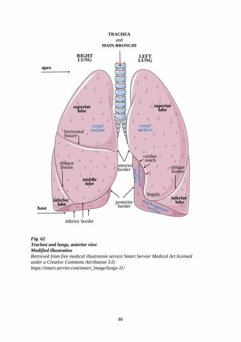
86
RIGHT LUNG
superior lobe
middlelobe
anterior border
horizontal fissure
cardiacnotch
costal surface
oblique fissure
Fig. 62
Trachea and lungs, anterior view
Modified illustration
Retrieved from free medical illustration service Smart Servier Medical Art licensed
under a Creative Commons Attribution 3.0;
https://smart.servier.com/smart_image/lungs-11/
LEFT LUNG
superior lobe
inferiorlobe
inferiorlobeposterior
border
inferior border
oblique fissure
costal surface
apex
base
TRACHEA
and MAIN BRONCHI
lingula
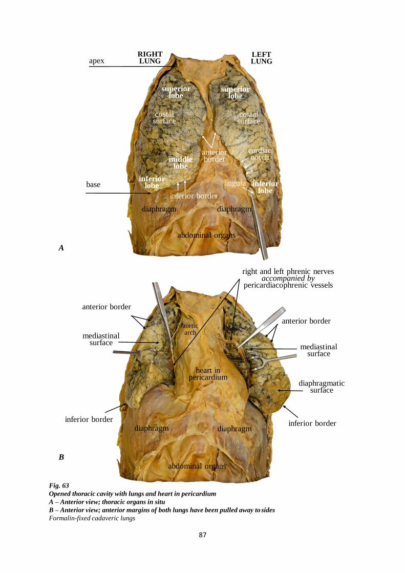
87
RIGHT LUNG
anterior border
cardiacnotch
anterior border
Fig. 63
Opened thoracic cavity with lungs and heart in pericardium
A – Anterior view; thoracic organs in situ
B – Anterior view; anterior margins of both lungs have been pulled away to sides
Formalin-fixed cadaveric lungs
LEFT LUNG
inferior border
inferior border
anterior border
apex
base
diaphragmdiaphragm
costal surface
costal surface
diaphragmdiaphragm
lingula
abdominal organs
inferior border
heart in pericardium
abdominal organs
A
B
superior lobe
inferior lobe
superior lobe
middle lobe
inferior lobe
right and left phrenic nervesaccompanied by
pericardiacophrenic vessels
aorticarch
mediastinal surface
diaphragmatic surface
mediastinal surface
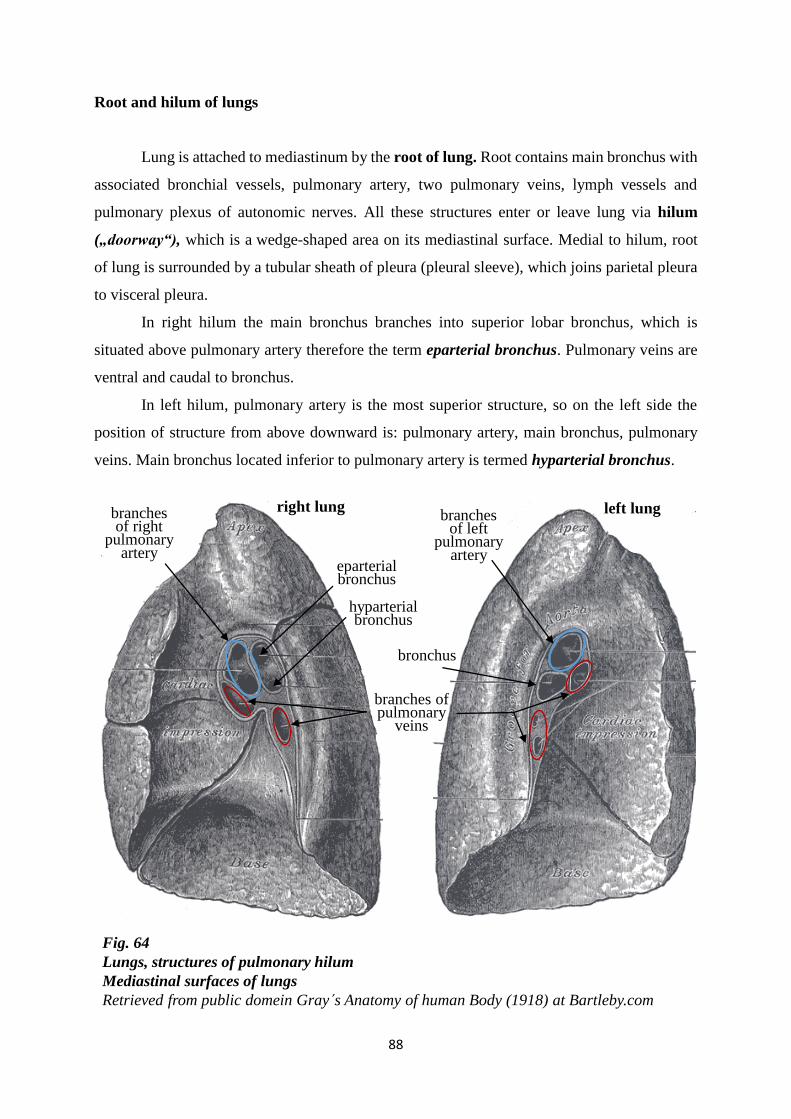
88
Fig. 64
Lungs, structures of pulmonary hilum
Mediastinal surfaces of lungs
Retrieved from public domein Gray s Anatomy of human Body (1918) at Bartleby.com
right lung
eparterial bronchus
hyparterial bronchus
branches of pulmonary
veins
bronchus
branches of right
pulmonary artery
branches of left
pulmonary artery
left lung
Root and hilum of lungs
Lung is attached to mediastinum by the root of lung. Root contains main bronchus with
associated bronchial vessels, pulmonary artery, two pulmonary veins, lymph vessels and
pulmonary plexus of autonomic nerves. All these structures enter or leave lung via hilum
(„doorway“), which is a wedge-shaped area on its mediastinal surface. Medial to hilum, root
of lung is surrounded by a tubular sheath of pleura (pleural sleeve), which joins parietal pleura
to visceral pleura.
In right hilum the main bronchus branches into superior lobar bronchus, which is
situated above pulmonary artery therefore the term eparterial bronchus. Pulmonary veins are
ventral and caudal to bronchus.
In left hilum, pulmonary artery is the most superior structure, so on the left side the
position of structure from above downward is: pulmonary artery, main bronchus, pulmonary
veins. Main bronchus located inferior to pulmonary artery is termed hyparterial bronchus.
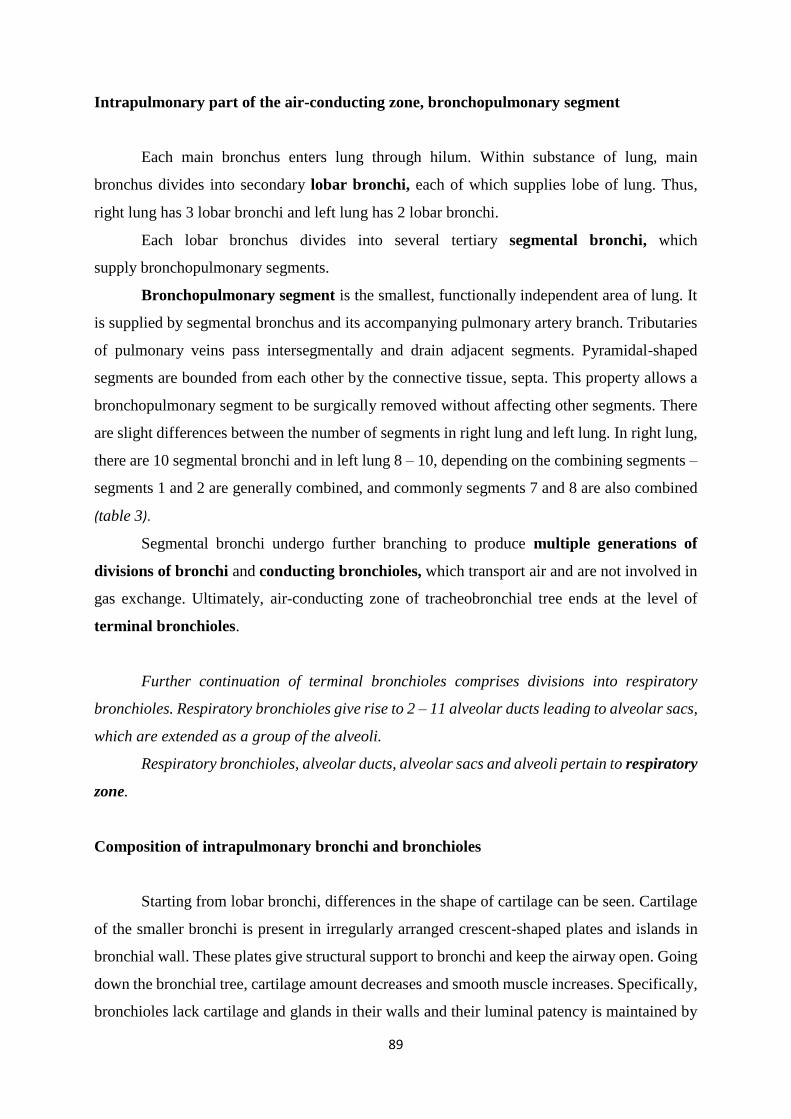
89
Intrapulmonary part of the air-conducting zone, bronchopulmonary segment
Each main bronchus enters lung through hilum. Within substance of lung, main
bronchus divides into secondary lobar bronchi, each of which supplies lobe of lung. Thus,
right lung has 3 lobar bronchi and left lung has 2 lobar bronchi.
Each lobar bronchus divides into several tertiary segmental bronchi, which
supply bronchopulmonary segments.
Bronchopulmonary segment is the smallest, functionally independent area of lung. It
is supplied by segmental bronchus and its accompanying pulmonary artery branch. Tributaries
of pulmonary veins pass intersegmentally and drain adjacent segments. Pyramidal-shaped
segments are bounded from each other by the connective tissue, septa. This property allows a
bronchopulmonary segment to be surgically removed without affecting other segments. There
are slight differences between the number of segments in right lung and left lung. In right lung,
there are 10 segmental bronchi and in left lung 8 – 10, depending on the combining segments –
segments 1 and 2 are generally combined, and commonly segments 7 and 8 are also combined
(table 3).
Segmental bronchi undergo further branching to produce multiple generations of
divisions of bronchi and conducting bronchioles, which transport air and are not involved in
gas exchange. Ultimately, air-conducting zone of tracheobronchial tree ends at the level of
terminal bronchioles.
Further continuation of terminal bronchioles comprises divisions into respiratory
bronchioles. Respiratory bronchioles give rise to 2 – 11 alveolar ducts leading to alveolar sacs,
which are extended as a group of the alveoli.
Respiratory bronchioles, alveolar ducts, alveolar sacs and alveoli pertain to respiratory
zone.
Composition of intrapulmonary bronchi and bronchioles
Starting from lobar bronchi, differences in the shape of cartilage can be seen. Cartilage
of the smaller bronchi is present in irregularly arranged crescent-shaped plates and islands in
bronchial wall. These plates give structural support to bronchi and keep the airway open. Going
down the bronchial tree, cartilage amount decreases and smooth muscle increases. Specifically,
bronchioles lack cartilage and glands in their walls and their luminal patency is maintained by
90
Fig. 65
Schematic longitudinal section of respiratory zone with respiratory bronchiole
Retrieved from public domein Gray s Anatomy of human Body (1918) at Bartleby.com
r. b. - respiratory bronchiole
al. d. - alveolar duct
at. - atria
a. s. - alveolar sac
a. - alveolus
p. a. - pulmonary artery
p. v. - pulmonary vein
l. - lymphatic
l.n. - lymphatic node
the smooth muscle and elastic fibers. Smooth muscle is also important for control of air flow
through contraction and dilation of the airway.
Table 3: Lobes and bronchopulmonary segments of lungs
Right lung Left lung
lobe segment lobe segment
superior
apical (S I )
superior
apical (S I) apicoposterior
(S I & II) * posterior (S II) posterior (S II)
anterior (S III) anterior (S III)
middle lateral (S IV) superior lingular (S IV)
medial (S V) inferior lingular (S V)
inferior
superior (S VI)
inferior
superior (S VI)
medial basal (S VII) medial basal
(S VII) anteriomedial
basal
(S VII & VIII)** anterior basal (S VIII) anterior basal
(S VIII)
lateral basal (S IX) lateral basal (S IX)
posterior basal (S X) posterior basal (S X)
typical combination of * apical and posterior segments into apicoposterior segment and
**medial basal and anterior basal segments into anteriomedial basal segment
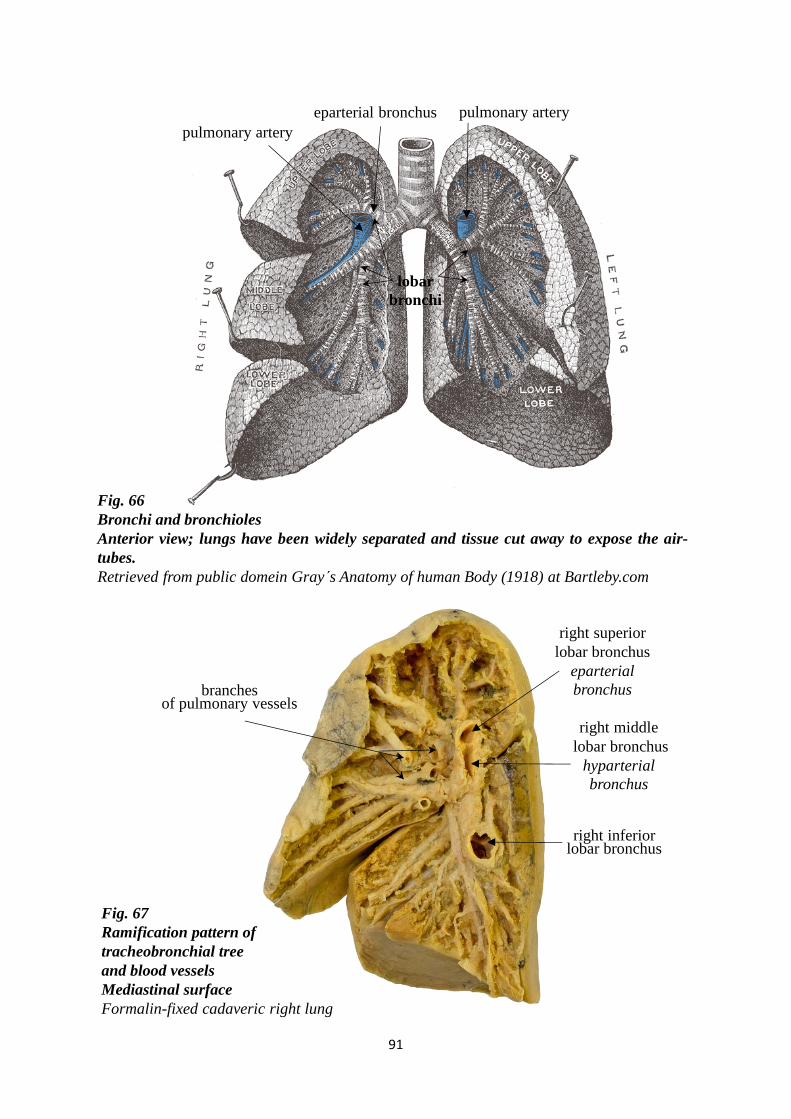
91
Fig. 67
Ramification pattern of
tracheobronchial tree
and blood vessels
Mediastinal surface
Formalin-fixed cadaveric right lung
Fig. 66
Bronchi and bronchioles
Anterior view; lungs have been widely separated and tissue cut away to expose the air-
tubes.
Retrieved from public domein Gray s Anatomy of human Body (1918) at Bartleby.com
eparterial bronchus
lobar
bronchi
pulmonary artery
pulmonary artery
right superior
lobar bronchus
eparterial
bronchus
right inferior lobar bronchus
branches of pulmonary vessels
right middle
lobar bronchus
hyparterial
bronchus
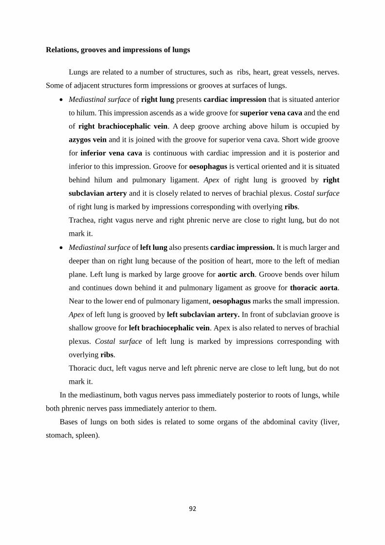
92
Relations, grooves and impressions of lungs
Lungs are related to a number of structures, such as ribs, heart, great vessels, nerves.
Some of adjacent structures form impressions or grooves at surfaces of lungs.
Mediastinal surface of right lung presents cardiac impression that is situated anterior
to hilum. This impression ascends as a wide groove for superior vena cava and the end
of right brachiocephalic vein. A deep groove arching above hilum is occupied by
azygos vein and it is joined with the groove for superior vena cava. Short wide groove
for inferior vena cava is continuous with cardiac impression and it is posterior and
inferior to this impression. Groove for oesophagus is vertical oriented and it is situated
behind hilum and pulmonary ligament. Apex of right lung is grooved by right
subclavian artery and it is closely related to nerves of brachial plexus. Costal surface
of right lung is marked by impressions corresponding with overlying ribs.
Trachea, right vagus nerve and right phrenic nerve are close to right lung, but do not
mark it.
Mediastinal surface of left lung also presents cardiac impression. It is much larger and
deeper than on right lung because of the position of heart, more to the left of median
plane. Left lung is marked by large groove for aortic arch. Groove bends over hilum
and continues down behind it and pulmonary ligament as groove for thoracic aorta.
Near to the lower end of pulmonary ligament, oesophagus marks the small impression.
Apex of left lung is grooved by left subclavian artery. In front of subclavian groove is
shallow groove for left brachiocephalic vein. Apex is also related to nerves of brachial
plexus. Costal surface of left lung is marked by impressions corresponding with
overlying ribs.
Thoracic duct, left vagus nerve and left phrenic nerve are close to left lung, but do not
mark it.
In the mediastinum, both vagus nerves pass immediately posterior to roots of lungs, while
both phrenic nerves pass immediately anterior to them.
Bases of lungs on both sides is related to some organs of the abdominal cavity (liver,
stomach, spleen).
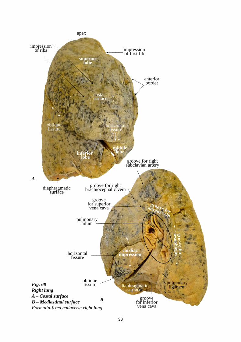
93
Fig. 68
Right lung
A – Costal surface
B – Mediastinal surface
Formalin-fixed cadaveric right lung
apex
superior lobe
horizontal fissure
inferiorlobe
oblique fissure
middle lobe
cardiac impression
B
A
costal surface
diaphragmatic surface
impression of ribs
anterior border
groove for right brachiocephalic vein
pulmonary hilum
groove for right subclavian artery
pulmonary ligament
impression of first fib
groove for superior vena cava
groove for inferior vena cava
horizontal fissure
obliquefissure diaphragmatic
surface
94
superior lobe
cardiac notch
apex
inferior lobe
lingula
Fig. 69
Left lung
A – Costal surface
B – Mediastinal surface
Formalin-fixed cadaveric left lungB
A
cardiac impression
groove for left brachiocephalic vein
groove for left subclavian artery
impression of oesophagus
groove for aortic arch
obliquefissure
diaphragmatic surface
oblique fissure
costal surface
impressions of ribs
impression of first fib
anterrior border
inferior border
pulmonary hilum
pulmonary ligament
anterior border
lingula
95
Projection of lungs onto skeleton
Apex of lung extends upward through superior thoracic aperture into neck, posterior to
sternocleidomastoid muscle. Its highest point is approx. 2 cm above the level of medial third of
clavicle or 3 – 4 cm above the first costal cartilage.
Lower boundary of lungs depends on diaphragmatic excursion. This means, lungs are
positioned lower during inspiration and higher during expiration. In resting expiratory position,
inferior border of lungs is projected onto skeleton in midclavicular line at the level of the 6th
rib, in midaxillary line at the level of the 8th rib and in paravertebral line at the level of the
11th rib.
Vessels and nerves of lungs
o Blood supply of lungs includes two types of circulation - pulmonary and bronchial.
Both circulation are connected by shunts.
Pulmonary circulation is responsible for gas exchange. It transports deoxygenated
blood (low in oxygen) to lungs to absorb oxygen and release carbon dioxide. The
oxygenated blood (oxygen-rich) then flows back to heart.
Deoxygenated blood is carried from right ventricle by pulmonary trunk and, after
division of this trunk, by two pulmonary arteries into lungs. Branches of pulmonary
arteries within lungs accompany bronchi and end in capillary networks in alveoli.
Oxygenated blood is drained by veins. Smaller veins within lungs are intersegmental
in location so they run independently of arteries and bronchi. They then flow into
larger and larger vessels and finally into pulmonary veins. Pulmonary veins begin
at hilum of lung and they pass through root of lung accompanied by pulmonary
arteries and main bronchi. Pulmonary veins, usually four in number, enter left
atrium.
Bronchial circulation is a part of systemic circulation and it forms the nutritive
vascular system of pulmonary tissues. It includes bronchial arteries and veins.
The left bronchial arteries originate directly from thoracic aorta. A single right
bronchial artery originates from the third posterior intercostal artery, however it
may also arise directly from aorta. Bronchial artery's origin varies from person to
person. Bronchial arteries supply oxygenated blood to the non-respiratory
conducting tissues of lungs (bronchial walls and glands, walls of the large vessels)
96
and visceral pleura. Bronchial arteries accompany the bronchial tree and they end
at the level of respiratory bronchioles. The distal-most branches of bronchial arteries
anastomose with branches of pulmonary arteries. Bronchial veins drain into azygos
vein on the right side and to accessory hemiazygos vein on the left side or into left
superior intercostal vein. Bronchial veins drain only part of the blood supplied to
lungs by bronchial arteries. Blood that remains, is carried away by pulmonary veins.
o Lymph vessels form two plexuses. Superficial or subpleural plexus lies beneath the
visceral pleura and drains lung tissue and visceral pleura. Deep or septal (periarterial or
peribronchial) plexus travels along bronchi. Both lymph vessel systems run together at
hilum where lymph enters bronchopulmonary nodes. Further outflow of lymph from
lungs is carried out by superior and inferior tracheobronchial lymph nodes and then into
paratracheal lymph nodes or directly into bronchomediastinal lymph trunks.
Right lung drains primarily into lymph nodes on the right side and finally through right
bronchomediastinal lymph trunk into right lymphatic duct (trunk).
Superior lobe of left lung drains primarily through corresponding nodes of the left side
and finally through left bronchomediastinal lymph trunk into thoracic duct. In many
cases lymph from inferior lobe of left lung is drained to right tracheobronchial nodes
and then continues to follow the right-side pathway.
o Lungs are nerve supplied by two pulmonary plexuses. They contain parasympathetic,
sympathetic and visceral sensory fibres. Parasympathetic and visceral sensory fibres are
derived from vagus nerve. Parasympathetic nervous system stimulates secretion from
bronchial glands and contraction of bronchial smooth muscle resulting in narrowing of
bronchi (bronchoconstriction) and it leads to vasodilation of pulmonary vessels.
Sympathetic fibres arise from inferior cervical ganglion and from the 1st – 4th thoracic
sympathetic ganglia. Sympathetic nervous system stimulates relaxation of bronchial
smooth muscle resulting to expansion of bronchi (bronchodilatation) and it leads to
vasoconstriction of pulmonary vessels.
97
PLEURA
Pleurae are thin, glistening, smooth serous membranes that surround lungs. Based on
the location, each pleura is divided into two parts – visceral and parietal. Visceral (pulmonary)
pleura covers entire surface of lung. Parietal pleura covers the corresponding half of thoracic
wall, diaphragm and mediastinum. Potencial space between visceral and parietal pleura is
known as pleural cavity. This space is filled by small amount of pleural serous fluid. This fluid
lubricates surfaces of parietal and visceral pleura allowing them to glide smoothly during
respiration. It also establishes adhesion between two layers and maintains negative pressure
within pleural space.
Two pleural cavities containing lungs are completely separated by mediastinum so
there is no communication between them.
Visceral (pulmonary) pleura
Visceral pleura adheres to and completely covers lung, except pulmonary hilum. It is
firmly attached to the outer surfaces of lung and it also passes into interlobar fissures.
Visceral pleura is continuous with parietal pleura at hilum and along the root of lung.
Below the root of lung visceral and parietal pleura continue as a double layer of pleura which
is called pulmonary ligament.
Parietal pleura
Parietal pleura is attached through endothoracic fascia to the inner surface of thoracic
wall and in addition, it covers diaphragm and mediastinum. It also separates pleural cavity from
mediastinum. It is thicker than the visceral pleura and contains pain receptors. Parietal pleura
is highly sensitive to pain, while visceral pleura is not, due to its lack of sensory innervation.
Parietal pleura is also sensitive to temperature, touch and pressure.
Parietal pleura is named according to region in which it lies on, or the surface it covers.
Costal part of parietal pleura (costal pleura) covers the internal surfaces of ribs,
intercostal muscles, membranes and sternum. It is separated from these structures by
endothoracic fascia.
98
Mediastinal part of parietal pleura (mediastinal pleura) covers mediastinal
surface of lung and forms lateral boundary of mediastinum. At root of lung,
mediastinal pleura turns laterally and forms sleeve-like covering for the structures of
root and here it is continuous with visceral pleura.
Diaphragmatic part of parietal pleura (diaphragmatic pleura) covers superior
surface of its own half of diaphragm except the central tendon. Diaphragmatic pleura
is connected with diaphragm by phrenicopleural fascia.
Cervical pleura (dome of pleura or pleural cupola) covers the apex of lung and
forms a cup-like dome over it. Cervical pleura extends up into neck. It is a superior
continuation of costal and mediastinal pleurae. This region is only protected by soft
tissue, and thus is vulnerable to injury.
Pleural recesses
Lungs do not completely fill pleural cavities in their anterior and postero-inferior areas
during quiet respiration. Pleural cavity is slightly larger than lung. This results in recesses –
reserve spaces between two parts of parietal pleura, which become opposed. Lung expands into
this recess during forced inspiration; however, the recess never fills completely. During
expiration, recess contains no lung tissue, only pleural fluid. Costodiaphragmatic recess is the
lowest area of pleural cavity. It is located at the junction of costal and diaphragmatic pleura.
These two pleurae facing each other below the lower border of lung. Costodiaphragmatic recess
occurs in each pleural cavity. It is the largest and clinically most important, as a place where
patological fluid can collect in standing position. On both sides, costodiaphragmatic recess is
separated from posterior surface of superior pole of kidney by diaphragm.
Anteriorly, costomediastinal recess is located behind sternum at the junction of costal and
mediastinal pleura in each pleural cavity.
Projection of pleura onto skeleton
Cervical pleura (pleural cupola) forms the uppermost part of pleural cavity. It is filled
by the apex of lung so cervical pleura has identical boundary to the apex. It extends approx. 2
cm above the level of medial third of clavicle or 3 – 4 cm above the first costal cartilage.
Inferior border of pleural cavities lies lower than inferior border of lungs. The distance
between two borders corresponds to costodiaphragmatic recess. Inferior boundary of pleural
99
cavities is projected onto skeleton in midclavicular line at the level of the 7th rib, in
midaxillary line at the level of the 10th rib and in paravertebral line at the level of the 12th
rib.
Vessels and nerves of pleura
Visceral pleura forms an integral part of lung, therefore it is supplied by vessels and nerves
of lungs. Although visceral pleura is nerve supplied by visceral afferent fibres, common
sensation such as pain and touch is generally not elicited from this tissue.
Parietal pleura is supplied by vessels and nerves that supply thoracic wall.
o Arterial supply to costal pleura comes from posterior intercostal arteries which
originate from thoracic aorta and from branches of internal thoracic artery which
originates from subclavian artery. Cervical pleura is supplied by the branches of
subclavian artery. Diaphragmatic pleura is supplied by superior phrenic artery which
originates from thoracic aorta and by intercostal arteries.
o Venous blood is drained by veins accompany arteries.
o Lymph vessels from parietal pleura drain into lymph nodes of thoracic wall –
intercostal, parasternal, mediastinal and phrenic. A few lymphatic vessels from cervical
pleura drain into axillary lymph nodes.
o Parietal pleura is nerve supplied by somatic afferent fibres. Nerves of parietal pleura
are derived mainly from intercostal and phrenic nerves. Costal pleura and peripheral
part of diaphragmatic pleura are supplied by intercostal nerves. Mediastinal pleura and
central part of diaphragmatic pleura are supplied by phrenic nerve. Cervical pleura is
supplied by branches of nerves of brachial plexus and by phrenic nerve.
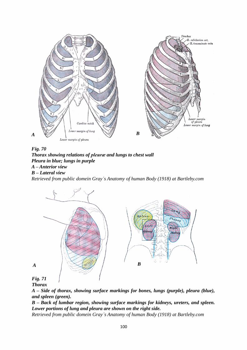
100
Fig. 71
Thorax
A – Side of thorax, showing surface markings for bones, lungs (purple), pleura (blue),
and spleen (green).
B – Back of lumbar region, showing surface markings for kidneys, ureters, and spleen.
Lower portions of lung and pleura are shown on the right side.
Retrieved from public domein Gray s Anatomy of human Body (1918) at Bartleby.com
A B
Fig. 70
Thorax showing relations of pleuræ and lungs to chest wall
Pleura in blue; lungs in purple
A – Anterior view
B – Lateral view
Retrieved from public domein Gray s Anatomy of human Body (1918) at Bartleby.com
A B
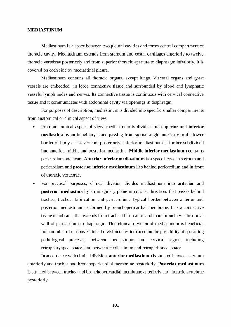
101
MEDIASTINUM
Mediastinum is a space between two pleural cavities and forms central compartment of
thoracic cavity. Mediastinum extends from sternum and costal cartilages anteriorly to twelve
thoracic vertebrae posteriorly and from superior thoracic aperture to diaphragm inferiorly. It is
covered on each side by mediastinal pleura.
Mediastinum contains all thoracic organs, except lungs. Visceral organs and great
vessels are embedded in loose connective tissue and surrounded by blood and lymphatic
vessels, lymph nodes and nerves. Its connective tissue is continuous with cervical connective
tissue and it communicates with abdominal cavity via openings in diaphragm.
For purposes of description, mediastinum is divided into specific smaller compartments
from anatomical or clinical aspect of view.
From anatomical aspect of view, mediastinum is divided into superior and inferior
mediastina by an imaginary plane passing from sternal angle anteriorly to the lower
border of body of T4 vertebra posteriorly. Inferior mediastinum is further subdivided
into anterior, middle and posterior mediastina. Middle inferior mediastinum contains
pericardium and heart. Anterior inferior mediastinum is a space between sternum and
pericardium and posterior inferior mediastinum lies behind pericardium and in front
of thoracic vertebrae.
For practical purposes, clinical division divides mediastinum into anterior and
posterior mediastina by an imaginary plane in coronal direction, that passes behind
trachea, tracheal bifurcation and pericardium. Typical border between anterior and
posterior mediastinum is formed by bronchopericardial membrane. It is a connective
tissue membrane, that extends from tracheal bifurcation and main bronchi via the dorsal
wall of pericardium to diaphragm. This clinical division of mediastinum is beneficial
for a number of reasons. Clinical division takes into account the possibility of spreading
pathological processes between mediastinum and cervical region, including
retropharyngeal space, and between mediastinum and retroperitoneal space.
In accordance with clinical division, anterior mediastinum is situated between sternum
anteriorly and trachea and bronchopericardial membrane posteriorly. Posterior mediastinum
is situated between trachea and bronchopericardial membrane anteriorly and thoracic vertebrae
posteriorly.
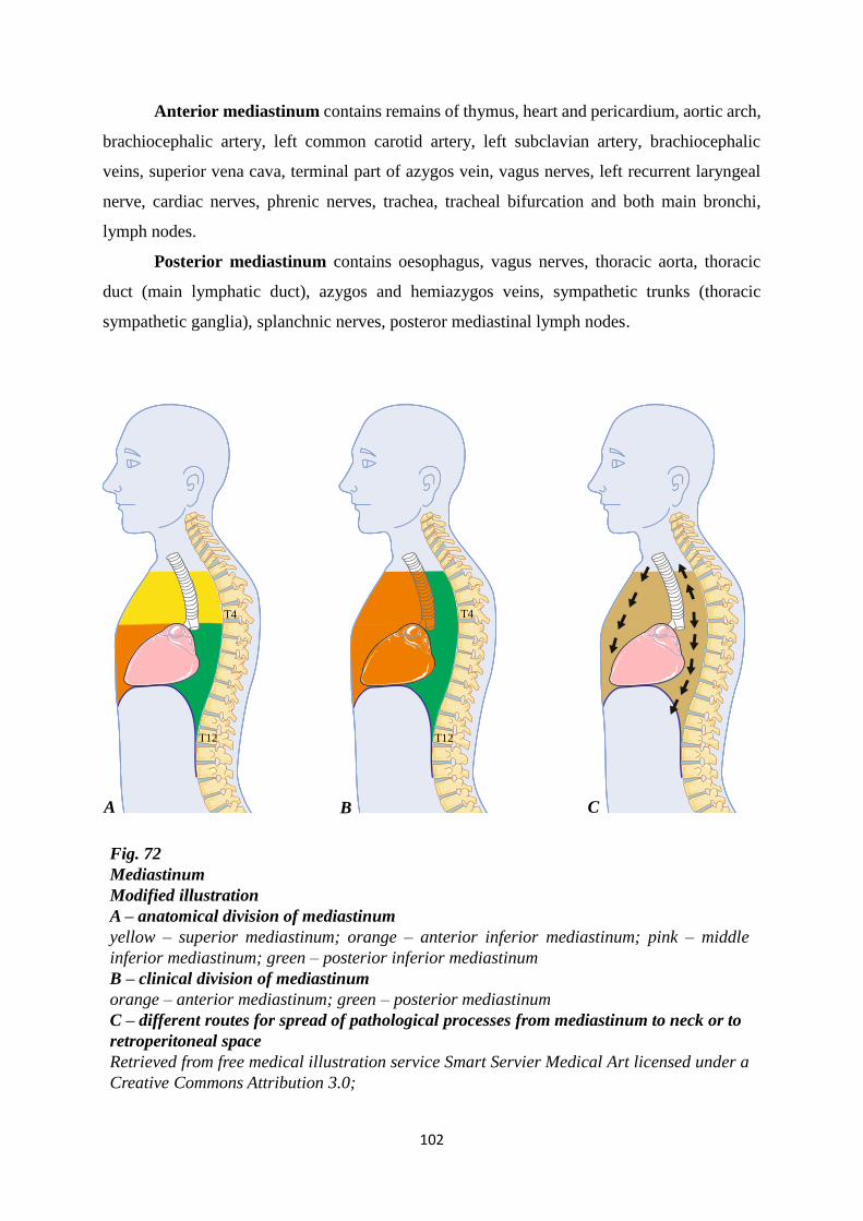
102
Fig. 72
Mediastinum
Modified illustration
A – anatomical division of mediastinum
yellow – superior mediastinum; orange – anterior inferior mediastinum; pink – middle
inferior mediastinum; green – posterior inferior mediastinum
B – clinical division of mediastinum
orange – anterior mediastinum; green – posterior mediastinum
C – different routes for spread of pathological processes from mediastinum to neck or to
retroperitoneal space
Retrieved from free medical illustration service Smart Servier Medical Art licensed under a
Creative Commons Attribution 3.0;
A B C
T4 T4
T12 T12
Anterior mediastinum contains remains of thymus, heart and pericardium, aortic arch,
brachiocephalic artery, left common carotid artery, left subclavian artery, brachiocephalic
veins, superior vena cava, terminal part of azygos vein, vagus nerves, left recurrent laryngeal
nerve, cardiac nerves, phrenic nerves, trachea, tracheal bifurcation and both main bronchi,
lymph nodes.
Posterior mediastinum contains oesophagus, vagus nerves, thoracic aorta, thoracic
duct (main lymphatic duct), azygos and hemiazygos veins, sympathetic trunks (thoracic
sympathetic ganglia), splanchnic nerves, posteror mediastinal lymph nodes.
103
REVIEW QUESTIONS
HEART
Position of the heart:
a/ the heart is situated in posterior mediastinum
b/ the apex of the heart directs forward, downward, to the left
c/ the heart is located between the 2nd and 7th intercostal space
d/ the heart extends to the midclavicular line on the right
e/ the heart extends to the parasternal line on the left
Position of the heart:
a/ the heart is situated in anterior mediastinum (according the clinical subdivision of mediastinum)
b/ it extends to the midclavicular line on the left
c/ it extends to the parasternal line on the right
d/ apex of the heart lies in the point of cross-section of the left midclavicular line and the 2nd rib
e/ 2/3 of the heart are situated on the right side from the median plane
Relations of the heart:
a/ the phrenic nerves are behind the heart
b/ the thymus is in front of the heart
c/ the lungs are on the sides of the heart
d/ the thoracic aorta is behind the heart
e/ the thoracic duct is in front of the heart
Relations of the heart:
a/ oesophagus is behind the heart
b/ phrenic nerves run laterally at both sides of the heart
c/ ascending aorta is behind the heart
d/ thoracic part of trachea is in front of the heart
e/ principal bronchi are behind the heart
External description of the heart:
a/ the sternocostal surface is the anterior wall „clinically“
b/ margo acutus is the left margin
c/ the anterior interventricular sulcus is situated at the diaphragmatic surface
d/ the coronary sulcus separates the atria from the ventricles
e/ the diaphragmatic surface is formed mostly by the left and right atrium
The external description of the heart:
a/ the anterior interventricular sulcus contains small cardiac vein
b/ the coronary sulcus separates the atria from the ventricles
c/ the sternocostal surface is formed mostly by the left atrium and left ventricle
d/ the left auricle forms the diaphragmatic surface
e/ the right ventricle forms margo obtusus
104
External description of the heart:
a/ the diaphragmatic surface is convex
b/ left ventricle forms margo obtusus
c/ anterior interventricular sulcus contains the terminal branch of the right coronary artery
d/ the base of the heart directs superiorly, posteriorly, to the right
e/ small part of diaphragmatic surface is formed by right and left auricle
Surfaces of the heart:
a/ sternocostal surface of the heart is flat
b/ diaphragmatic surface of the heart is convex
c/ coronary sulcus passes at the diaphragmatic surface of the heart
d/ coronary sulcus passes at the diaphragmatic surface of the heart
e/ anterior interventricular sulcus can be visible at the diapragmatic surface of the heart
Margins of the heart:
a/ margo acutus is formed by the left ventricle
b/ margo obtusus is formed by the left ventricle
c/ circumflex branch runs along margo acutus
d/ margo acutus is supplied by the branches of the right coronary artery
e/ middle cardiac vein passes along margo acutus
Mark correct sentences:
a/ superior vena cave enters the right ventricle
b/ pulmonary trunk exits from the right atrium
c/ aorta exits from the left ventricle
d/ pulmonary arteries exit from the left atrium
e/ pulmonary veins enter the right ventricle
The right atrium:
a/ sinus venarum is situated at dorsal part of the right atrium
b/ sinus venarum contains pectinati muscles
c/ coronary sinus opens into the right atrium closely to the orifice of superior vena cava
d/ the right auricle contains the papillary muscles
e/ the wall of the right atrium is thinner than than the wall of the right ventricle
The right ventricle:
a/ it forms a part of both sternocostal and diaphragmatic surfaces
b/ its outflowing tract is called conus pulmonalis
c/ the wall of the inflowing tract contains pectinati mm.
d/ the right atrioventricular orifice contains the tricuspid valve
e/ the valve of the pulmonary trunk consists of three valvules: right, left and anterior
105
The right atrium and the right ventricle:
a/ the pectinati mm. are contained in the auricle of the right atrium
b/ at the dorsal wall of the right atrium there are orifices of the pulmonary veins
c/ conus arteriosus forms the inflowing tract of the right ventricle
d/ the right ventricle has semilunar shape in the transverse section
e/ the myocardium of the right ventricle is thinner than the myocardium of the left ventricle
The left ventricle:
a/ its outflowing part is called the aortic vestibule
b/ its inflowing part forms irregular muscular columns - trabeculae carneae
c/ the left atrioventricular orifice is closed by the tricuspid valve
d/ in transverse section it has semilunar shape
e/ it has thicker wall than the right ventricle
The left ventricle:
a/ it has semilunar shape in the trasverse section
b/ trabeculae carneae are irregular columns formed by the epicardium
c/ its inflowing part is conus arteriosus
d/ its outflowing part is smooth
e/ pulmonary artery exits the left ventricle
Pericardium and cardiac wall:
a/ pericardium is cartilagenous sac surrouding the heart and roots of the great vessels
b/ pericardial cavity is the narrow space between the parietal and visceral layer of serous
pericardium
c/ ligaments connect the fibrous pericardium to the diaphragm and posterior surface of sternum
d/ parietal layer of serous pericardium forms epicardium
e/ myocardium of the atria is thinner than myocardium of the ventricles
Pericardium:
a/ fibrous pericardium is attached to the large vessels
b/ fibrous pericardium is the innermost layer of pericardial sac
c/ serous pericardium consists od two layers: mediastinal and cardial
d/ visceral layer of serous pericardium is epicardium
e/ pericardial cavity is the narrow space between the fibrous and serous pericardium
The cardiac wall:
a/ the epicardium lines the inner surface of the heart
b/ two layers of serous pericardium border the pericardial cavity
c/ the myocardium is the thickest part of the cardiac wall
d/ the myocardium of the ventricles is thinner than the myocardium of the atria
e/ the cardiac skeleton separates the myocardium of the right atrium from the myocardium of the
left atrium
106
The cardiac wall:
a/ the inner surface of the cardiac chambers is lined by the endocardium
b/ the myocardium of the left ventricle is thicker than the myocardium of the right ventricle
c/ the myocardium of the atria is thicker than the myocardium of the ventricles
d/ the epicardium is a parietal layer of the serous pericardium
e/ endocardium forms the valves
The valves:
a/ the valve of the pulmonary trunk consists of anterior, posterior and septal cusps
b/ the bicuspid valve is situated between the right atrium and the right ventricle
c/ the valvules of the aortic valve form aortic sinuses
d/ the valves are formed by endocardium
e/ inside of each cusp and valvule there is a plate of cartilage
The valves:
a/ the chordae tendineae arise from the fibrous rings (anulus fibrosus) of the cardiac skeleton
b/ chordae tendineae are inserted to the papillary mm.
c/ the valve of the pulmonary trunk is formed by the right, left and anterior semilunar valvules
d/ the aortic valve consists of anterior, posterior and septal valvules
e/ the valves are formed by the epicardium
The atrioventricular valves:
a/ physiologically they allow blood to flow in only one direction
b/ they are formed by the endocardium
c/ the tricuspid valve closes the left atrioventricular orifice
d/ the base of the cusp is attached to the fibrous ring of the cardiac skeleton
e/ chordae tendineae prevent the cusps from the prolapse back to the atrium during the systole of
the ventricle
Cardiac skeleton:
a/ its components are formed by the elastic cartilage
b/ at the surface of the heart it is projected to the level of interventricular sulci
c/ it separates the myocardium of the atria from the myocardium of the ventricles
d/ fibrous ring of the aortic valve is situated in front of the fibrous ring of the pulmonary trunk
valve
e/ atrioventricular bundle runs through the left fibrous trigone of the cardiac skeleton
Conducting system of the heart:
a/ the sinuatrial node is situated in the right atrium near the orifice of superior vena cava
b/ the atrioventricular node is always a pacemaker
c/ the atrioventricular node is located near the right artioventricular orifice
d/ the sinuatrial and atrioventricular nodes are connected by the Tawara´s branches
e/ Purkinje fibers are terminal fibers of the conducting system
107
Conducting system of the heart:
a/ nodal tissue is formed by specialized nerve cells
b/ in physiologic conditions the sinuatrial node is a pacemaker
c/ the sinuatrial node is located near the orifice of the inferior vena cava
d/ the atrioventricular node is situated near the left atrioventricular orifice
e/ the atrioventricular bundle connects sinuatrial and atrioventricular node
Conducting system of the heart:
a/ the sinuatrial node is situated in the right atrium near the orifice of superior vena cava
b/ in physiological conditions the atrioventricular node is always a pacemaker
c/ the atrioventricular node is situated in the right atrium near the right artioventricular orifice
d/ nodal tissue and components of the conducting system are influenced by the nerves of cardiac
plexus
e/ parasympathetic fibres of cardiac plexus stimulate (accelarate) the heart rate
The sinuatrial node:
a/ it is situated near the orifice of superior vena cava
b/ it is connected with atrioventricular node by the atrioventricular bundle
c/ it is a pacemarker
d/ it is located in the interatrial septum
e/ it is formed by the specialised cardiac muscle cells
The blood supply of the heart:
a/ the left coronary artery supplies the sinuatrial node
b/ the right coronary artery supplies the posterior papillary m. in the right ventricle
c/ the coronary sinus opens into the right atrium
d/ the great cardiac vein runs in posterior interventricular sulcus
e/ the middle cardiac vein runs along the right margin of the heart
The blood supply of the heart:
a/ the right coronary artery supplies the anterior papillary m. of the left ventricle
b/ the left coronary artery supplies the dorsal part of the interventricular septum
c/ the great cardiac vein drains the blood from the left ventricle
d/ the middle cardiac vein runs in posterior interventricular sulcus and opens to coronary sinus
e/ the smallest cardiac veins (Thebesian veins) open into all chambers of the heart
The left coronary artery:
a/ it arises from coronary sinus
b/ the circumflex artery (branch) runs in coronary sulcus
c/ the anterior intervenricular branch is accompanied by the middle cardiac vein
d/ it supplies the posterior papillary muscle in the right ventricle
e/ it supplies the dorsal part of the interventricular septum
108
Cardiac veins:
a/ the great cardiac vein runs in posterior interventricular sulcus
b/ the middle cardiac vein runs along the left margin of the heart
c/ the small cardiac vein opens into the coronary sinus
d/ coronary sinus opens into the left ventricle
e/ small cardiac vein opens into the great cardiac vein
Vessels of the heart:
a/ coronary arteries arise from the right and left aortic sinus
b/ artery of the sinuatrial node is usually (more often) a branch of the right coronary artery
c/ margo obtusus is supplied by branches of the right coronary artery
d/ posterior papillary muscle of the left ventricle is supplied by branches
of the left coronary artery
e/ posterior papillary muscle of the left ventricle is supplied by branches
of the right coronary artery
Vessels of the heart:
a/ coronary arteries arise from coronary sinus
b/ left coronary artery supplies dorsal 1/3 of the interventricular septum
c/ circumflex branch supplies the area around margo obtusus
d/ small cardiac vein runs along the left margin of the heart
e/ the great cardiac vein runs in posterior interventricular sulcus
The right coronary artery and its branches supply:
a/ the anterior papillary m. in the left ventricle
b/ the ventral part of the interventricular septum
c/ a part of the posterior wall of the left ventricle closely to the posterior interventricular sulcus
d/ the right atrium
e/ the right auricle
The left coronary artery and its branches supply:
a/ the posterior papillary muscle in the left ventricle
b/ the anterior papillary muscle in the right ventricle
c/ the dorsal part of the interventricular septum
d/ the right auricle
e/ a part of the ventral wall of the right ventricle closely to the anterior interventricular sulcus
The foetal circulation:
a/ the umbilical vein carries the deoxygenated blood to placenta
b/ ductus arteriosus connects the pulmonary trunk and pulmonary arteries
c/ ductus venosus allows to divert the blood from the liver
d/ after the birth the umbilical artery becomes the round ligament of the liver
e/ the blood contained in ductus venosus is more oxygenated than the blood in ductus arteriosus
109
The foetal circulation:
a/ umbilical artery carries the oxygenated blood from placenta to the foetus
b/ ductus venosus directly connects the superior and inferior vena cava in the foetal circulation
c/ medial umbilical ligament is a remnant of umbilical artery
d/ foramen ovale is the opening between the left and right ventricle during the foetal development
e/ ductus arteriosus allows to divert the blood from the lungs
The foetal circulation:
a/ ductus venosus connects the umbilical vein and portal vein in the foetal circulation
b/ ductus arteriosus allows to redirect the blood from the pulmonary trunk to the aorta
c/ ascending aorta contains lesser oxygenated blood than descending aorta
d/ ductus arteriosus contains lesser oxygenated blood than ductus venosus
e/ round ligament of the liver is remnant of ductus arteriosusň
Ductus venosus:
a/ it is functional only during the foetal development
b/ after the birth it becomes the round ligament of the liver
c/ it allows to divert the blood from the liver
d/ it contains the blood which is lesser oxygenated than the blood in the superior vena cava
e/ it connects the umbilical artery and the inferior vena cava
Opened (active) questions:
Define projection of the heart to the anterior thoracic wall.
Which organs are posteriorly related to the heart ?
Which ventricle forms margo acutus?
Which sulcus separates the right and left ventricle at the diaphragmatic surface of the heart?
Which nerves run along the lateral sides of the heart?
How does the base of the heart direct?
Which surface of the heart is almost flat?
110
Name the ligaments which connect the fibrous pericardium to surrounding structures.
Which vessels open into the left atrium?
Which layer of the cardiac wall forms the valves of the heart?
Which valve is situated in the left atrioventricular orifice? Do you remember all names of this
valve?
Where is conus arteriosus situated?
Name the muscles visible at the auricles of the atria?
Which chamber of the heart has the thickest wall?
Name the membranes (and layers) which enclose the pericardial cavity.
Name the depression which can be seen at the interatrial septum.
Which shape has the right ventricle in the transverse section?
Name the fibrous strings which connect the free margines of the cusps of atrioventricular valves
to the papillary muscles.
Name the irregular muscular columns (with zigzag arrangement) visible in the ventricles?
Where (in which cardiac chamber) is the septal papillary muscle situated?
Which valve is formed by the right, left and posterior semilunar valvules?
Which structures prevent the cusps of atrioventricular valves from the prolapse to the atrium
during the systole of the ventricle?
111
Name the main branches of the left coronary artery.
Name the fibrous sac which encloses the heart.
Which vessels run in the posterior interventricular sulcus?
Which vein runs in the anterior interventricular sulcus?
Where does the small cardiac vein open (drain)?
Which parts of conducting system of the heart run through the interventricular septum?
Which nerve fibres have a stimulatory effect on the nodal tissue and compartments of the
conductive system of the heart?
Which duct connects the umbilical vein and inferior vena cava in the foetal circulation?
What (which shunt) allows to divert the blood flow from the liver?
Which duct allows to redirect the blood from the pulmonary trunk to the aorta?
What is the remnat of the umbilical vein?
What is the remnant of the foramen ovale?
What shunt connects the right and left atrium?
Which vessel carries the oxygenated blood from placenta to foetus?
Which vessels take the deoxygenated blood from foetus to placenta?
112
RESPIRATORY SYTEM
Multichoice questions:
Nasal cavity:
a/ nasal cavity anteriorly opens through nares
b/ nasal cavity posteriorly directly communicates with larynx through choanae
c/ nasal vestibule is lined by the skin
d/ middle nasal meatus is situated between the middle and superior nasal concha
e/ olfactory receptors are also contained within mucosa of the paranasal sinuses
Nasal cavity:
a/ through the choanae it directly communicates with larynx
b/ olfactory region of the nasal mucosa is limited to the inferior nasal concha nad the floor of the
nasal cavity
c/ the perpendicular plate of the ethmoid bone forms a part of the nasal septum
d/ vibrissae are situated in the nasal vestibule
e/ the floor of the nasal cavity is formed by the hard and soft palate
Nasal cavity:
a/ nasal vestibule is lined by the skin
b/ choanae are anterior openings of the nasal cavity
c/ anterior part of the nasal septum is formed by the bones, posterior part of the nasal septum is
formed by the septal cartilage
d/ mucosa in the respiratory region of the nasal cavity contains vibrissae
e/ olfactory region of the nasal mucosa is limited to superior nasal concha, opposing nasal septum
and intervening roof
Nasal cavity:
a/ through the choanae it directly communicates with larynx
b/ olfactory region of the nasal mucosa is limited to the inferior nasal concha nad the floor of the
nasal cavity
c/ the perpendicular plate of the ethmoid bone forms a part of the nasal septum
d/ vibrissae are situated in the nasal vestibule
e/ the floor of the nasal cavity is formed by the hard and soft palate
113
The roof of the bony nasal cavity is formed by:
a/ vomer
b/ body of sphenoid bone
c/ frontal bone
d/ perpendicular plate of the ethmoid bone
e/ nasal bones
Lateral wall of the bony nasal cavity is formed by:
a/ vomer
b/ body of the sphenoid bone
c/ body of the maxilla
d/ perpendicular plate of the palatine bone
e/ lacrimal bone
The floor of the bony nasal cavity is formed by:
a/ vomer
b/ body of maxilla
c/ palatine process of maxilla
d/ perpendicular plate of the palatine bone
e/ horizontal plate of the palatine bone
The bony nasal septum is formed by:
a/ vomer
b/ nasal bone
c/ body of the maxilla
d/ perpendicular plate of the palatine bone
e/ perpendicular plate of the ethmoid bone
Middle nasal meatus:
a/ it is situated above the middle nasal concha
b/ it is situated above the inferior nasal concha
c/ frontal sinus opens into this meatus
d/ maxillary sinus opens into this meatus
e/ sphenoid sinus opens into this meatus
Inferior nasal meatus:
a/ it is situated below the inferior nasal concha
b/ maxillary sinus opens into this meatus
c/ inferiorly it is bordered by the palate
d/ it is lined by the skin
e/ nasolacrimal canal opens into this space
114
Paranasal sinuses:
a/ paranasal sinuses are lined by the olfactory epithelium
b/ in physiologic condition paranasal sinuses contain cerebrospinal fluid
c/ paranasal sinuses are not fully developed at the birth
d/ maxillary sinus has the largest volume of all paranasal sinuses
e/ sphenoid sinus opens into the superior nasal meatus
Paranasal sinuses:
a/ they are lined by the olfactory epithelium
b/ in physiologic condition they contain cerebrospinal fluid
c/ sphenoid sinus opens into the superior nasal meatus
d/ frontal sinus opens into the superior nasal meatus
e/ maxillary sinus opens into the inferior nasal meatus
Paranasal sinuses:
a/ they are lined by mucosa
b/ they contain air
c/ anterior ethmoid cells open into superior nasal meatus
d/ posterior ethmoid cells open into superior nasal meatus
e/ frontal sinus opens into middle nasal meatus
Larynx:
a/ it extends from the base of the skull to the level of CIII
b/ epiglottic cartilage consists of the stalk and lamina
c/ posterior cricoarytenoid m. is the adductor of the vocal folds
d/ thyroepiglottic m. enlarges the laryngeal inlet
e/ arytenoid m. is the abductor of the vocal folds
Larynx:
a/ larynx is related to suprahyoid muscles
b/ larynx is situated behind pharynx
c/ free upper margin of conus elasticus is thickened to form vocal ligament
d/ vestibulum extends from the vocal folds to the lower border of cricoid cartilage
e/ thyroepiglotticus muscle enlarges the laryngeal inlet
Larynx:
a/ larynx is situated behind the pharynx (more posteriorly)
b/ quadrangular membrane forms the lower part of fibroelastic membrane of the larynx
c/ laryngeal vestibule extends from the laryngeal inlet to the rima vestibuli
d/ arytenoid muscle is the strongest adductor of vocal folds
e/ glottis is the lowermost part of the laryngeal cavity
115
Cartilages of the larynx:
a/ thyroid cartilage is situated below (more caudally than) cricoid cartilage
b/ inferior horns of the thyroid cartilage are shorter than superior horns of the thyroid cartilage
c/ superior horns of the thyroid cartilage articulate with the cricoid cartilage
d/ epiglottic cartilage is situated below (more caudally than) cricoid cartilage
e/ arytenoid cartilage consists of the lamina and the stalk
Cartilages and ligaments of the larynx:
a/ cricoid cartilage is situated more caudally than thyroid cartilage
b/ epiglottic cartilage consists of the stalk and lamina
c/ lamina of cricoid cartilage is situated posteriorly, arch of the of cricoid cartilage is situated
anteriorly
d/ vestibular ligament is thickened upper part of conus elasticus (cricothyroid membrane)
e/ vocal ligament extends (passes) from cricoid to arytenoid cartilage
Larynx:
a/ cricoid cartilage is formed by the arch anteriorly and lamina posteriorly
b/ epiglottic cartilage closes the laryngeal inlet as prevention against the aspiration
c/ vestibular ligament is thickened upper part of conus elasticus
d/ vocal ligament passes from the thyroid cartilage to the vocal process of arytenoid cartilage
e/ laryngeal ventricle lies above vestibular fold in laryngeal vestibule
Larynx:
a/ it extends from the base of the skull to the level of C3
b/ epiglottic cartilage consists of the stalk and lamina
c/ arytenoid cartilages join (articulate) with the thyroid cartilage
d/ inferior horns of the thyroid cartilage join (articulate) with cricoid cartilage
e/ epiglottic cartilage and hyoid bone are connected by the ligament
Relations of the trachea:
a/ oesophagus is situated behind the trachea
b/ inferior thyroid veins are situated in front of the cervical part of the trachea
c/ inferior thyroid veins are situated in front of the thoracic part of the trachea
d/ azygos vein runs on the left side of the thoracic part of the trachea
e/ tracheal bifurcation projects to the level of the second thoracic vertebra
Relations of the trachea:
a/ its cervical part is related to the laryngeal recurrent nerves
b/ its cervical part is related to the isthmus of the thyroid gland
c/ its thoracic part is related to the azygos vein on the left side
c/ its thoracic part is related to the heart
e/ both cervical and thoracic part is related to the esophagus
116
Trachea:
a/ it extends from the level of CVI to TIV
b/ trachea is connected with larynx by cricotracheal ligament
c/ trachealis muscle is voluntary striated muscle
d/ inferior thyroid veins pass in front of the trachea
e/ pharynx is situated behind the cervical part of the trachea
Left principal bronchus:
a/ it is cca 15-20cm long
b/ its lumen has cca 5cm in diameter
c/ it is related to hemiazygos vein in its course in pulmonary hilum
d/ aortic arch curves around it
e/ it is situated below the pulmonary artery in pulmonary hilum
Bronchi:
a/ right principal bronchus is cca 5-10 cm long
b/ left principal bronchus divides into the superior and inferior lobar bronchi
c/ left principal bronchus is related to azygos vein
d/ left superior lobar bronchus usually divides into 5 segmental bronchi
e/ right principal bronchus is situated below the pulmonary artery in pulmonary hilum
Bronchi:
a/ lumen of the right principal bronchus has cca 3-5cm in diameter
b/ the left principal bronchus is related to the azygos vein
c/ the right principal bronchus enters the pulmonary hilum above the pulmonary artery
d/ the left principal bronchus divides into 3 lobar bronchi
e/ segmental bronchi contains no cartilage in their walls
Principal bronchi:
a/the right principal bronchus is wider and shorter than the left one
b/ the left principal bronchus is related to the aortic arch
c/ the right principal bronchus is related to the azygos vein
d/ the left principal bronchus divides into 3 lobar bronchi
e/ the left principal bronchus is situated above the pulmonary artery in the pulmonary hilum
Lungs:
a/ superior and inferior lobes of the left lung are separated by oblique fissure
b/ superior and middle lobes of the left lung are separated by horizontal fissure
c/ anterior margin of the lung is round, posterior margin is sharp
d/ apex of the lung projects 2cm above the clavicle
e/ inferior margin of the lung projects to the level of the 8th rib in the midaxillary line
117
Lungs:
a/ apex of the lung extends 5 cm above the clavicle
b/ diaphragmatic surface of the lung is concave
c/ anterior margin of the right lung forms cardiac incisure
d/ inferior lobe of the left lung usually contains 5 segments
e/ inferior margin of the lung projects to the level of the 8th rib in the midclavicular line
Lungs:
a/ right lung consists of 3 segments
b/ cardiac incisure is visible at the anterior margin of the left lung
c/ posterior margin of the lung is sharp
d/ oblique fissure separates superior and middle lobes of the right lung
e/ inferior margin of the lungs projects to the 8th rib in the midclavicular line
Lungs:
a/ superior and inferior lobes of the left lung are separated by oblique fissure
b/ superior and middle lobes of the left lung are separated by horizontal fissure
c/ anterior margin of the lung is round, posterior margin is sharp
d/ apex of the lung projects 2cm above the clavicle
e/ inferior margin of the lung projects to the level of the 8th rib in the midaxillary line
Lungs:
a/ right lung consists of three lobes
b/ horizontal fissure separates superior and inferior lobe of the left lung
c/ apex of the lung is projected 2 cm above the clavicle
d/ anterior margin of the lung is round, posterior margin of the lung is sharp
e/ the base of the lung is related to the diaphragm
Mediastinal surface of the left lung is related to:
a/ superior vena cava
b/ oesophagus
c/ heart
d/ thoracic aorta
e/ azygos vein
Mediastinal surface of the right lung is related to:
a/ superior vena cava
b/ oesophagus
c/ heart
d/ thoracic aorta
e/ hemiazygos vein
118
Pleura:
a/ parietal pleura covers the walls of thoracic cavity and mediastinum
b/ visceral pleura bridges the fissures of the lungs (it does not enter the fissures of the lungs)
c/ parietal mediastinal pleura reflects (bends) into the visceral pleura along the root of the lung
d/ right and left pleural cavities are completely separated
e/ costomediastinal recess is the lowermost part of the pleural cavity
Pleural cavity:
a/ the lower border of the pleural cavity in the midaxillary line projects to the level of the 7th rib
b/ the lower border of the pleural cavity in the paravertebral line projects to the level of the 10th
rib
c/ cupula pleurae (the dome - the uppermost part of the cervical pleura) extends 4 cm above the
clavicle
d/ the right and the left pleural cavities are interconnected by pleural recesses
e/ costodiaphragmatic recess is the lowermost part of the pleural cavity
Pleura and mediastinum:
a/ the right and the left pleural cavities do not communicate
b/ the right and the left pleural cavities are separated by mediastinum
c/ heart is situated in the posterior mediastinum according to the clinical subdivision
d/ trachea is situated in the posterior mediastinum according to the clinical subdivision
e/ phrenic nerves are situated in the anterior mediastinum according to the clinical subdivision
Mediastinum:
a/ anteriorly it is bordered by sternum
b/ superiorly it is bordered by superior thoracic aperture
c/ the thoracic duct runs in anterior mediastinum according the clinical division
d/ heart is situated in anterior mediastinum according the clinical division
e/ the azygos vein and the hemiazygos vein are situated in posterior mediastinum according the
clinical division
Mediastinum:
a/ inferiorly it is bordered by the diaphragm
b/ posteriorly it extends to the vertebral column
c/ oesophagus is situated in the posterior mediastinum according to the clinical subdivision
d/ the azygos vein is situated in the posterior mediastinum according to the clinical subdivision
e/ splanchnic nerves are situated in the anterior mediastinum according to the clinical subdivision
Mediastinum:
a/ mediastinum is situated between the pleural cavities in the median portion of the thorax
b/ inferiorly it is bordered by diaphragm
c/ oesophagus is situated in the posterior mediastinum according to the clinical subdivision
d/ the azygos vein is situated in the posterior mediastinum according to the clinical subdivision
e/ phrenic nerves are situated in the anterior mediastinum according to the clinical subdivision
119
Which of following structures are situated in anterior mediastinum according the clinical
division :
a/ thymus
b/ oesophagus
c/ heart
d/ phrenic nerves
e/ hemiazygos vein
Which of following structures are situated in anterior mediastinum according the clinical
division :
a/ trachea
b/ brachiocephalic veins
c/ splanchnic nerves
d/ sympathetic trunk
e/ azygos vein
Which of following structures are situated in posterior mediastinum according the clinical
division :
a/ vagus nerve
b/ oesophagus
c/ thoracic duct
d/ thoracic aorta
e/ azygos vein
Which of following structures are situated in posterior mediastinum according the clinical
division :
a/ splanchnic nerves
b/ tracheal bifurcation
c/ thoracic duct
d/ aortic arch
e/ hemiazygos vein
Opened (active) questions:
Which bones form the nasal septum?
Which bones form the choanae?
Which bones form the floor of the bony nasal cavity?
Name the opening through which the nasal cavity opens into the nasopharynx.
120
Which part of the nasal cavity contains vibrissae?
Which part of the nasal cavity is lined by the skin?
What is the olfactory region of the nasal cavity?
Where is the olfactory region. Explain the most exactly.
What separates the nasal vestibule and the proper nasal cavity?
Name the structures which subdivide the nasal cavity into superior, middle and inferior nasal
meatus.
What is contained within the mucosa covering the conchae?
Which paranasal sinuses open into the superior nasal meatus?
Which paranasal sinus has usually the largest volume?
Which paranasal sinus has its opening (aperture) highly than the floor?
Determine projection of the larynx to the vertebral column.
Which cartilage closes the laryngeal inlet as prevention against aspiration?
Which cartilage of the larynx is the lowermost situated?
Which part of the laryngeal cavity is narrowest?
Which ligament connects the larynx to trachea?
121
What is the name of thickened free upper margin of conus elasticus?
Name the synovial joints situated between the laryngeal cartilages.
What is the function of posterior cricoarytenoid muscle?
Determine projection of the tracheal bifurcation to the vertebral column (vertebrae).
Which organ is situated behind the trachea in its thoracic part?
Vhich veins run in front of the thoracic part of the trachea?
Explain projection of the trachea to the vertebral column (vertebrae).
Which nerves run along thelateral sides of the crvical part of the trachea?
What forms the dorsal (posterior )wall of the trachea?
Which principal bronchus is wider?
Explain position of the right principal bronchus to the pulmonary artery in pulmonary hilum
(above, below the artery?).
How many segmental bronchi arise from the middle lobar bronchus?
What is contained in pulmonary root?
Which organs are related to the mediastinal surface of both lungs?
Which lobes are separated by the oblique fissure in the right lung?
122
Which structures enter and exit the lungs in pulmonary hilum?
How many segments there are in the inferior lobe of the left lung?
How many segments are in the left lung totally?
Which arteries are related to the mediastinal surface of the left lung?
Which surface of the lung is concave?
Which margin of which lung shows (bears) cardiac incisure?
Name the fissure which separates the superior and inferior lobe of the left lung?
Determine projection of the inferior margin of the lungs to the ribs in
midclavicular, midaxillary and paravertebral line .
Name the fissure which separates the superior and middle lobe of the right lung?
How many segments form the superior lobe of the left lung?
Which veins are related to the mediastinal surface of the right lung?
Where is projected the apex of the lung (to the position of the first rib)?
What is the lowermost part of the pleural cavity?
What forms the borders of mediastinum?
Determine projection of the inferior margin of the pleural cavity to the ribs in
midclavicular, midaxillary and paravertebral lines.
123
REFERENCES
Acland's Video Atlas of Human Anatomy. Wolters Kluwer Health, 2018.
Available online at: http://aclandanatomy.com/
Amador Ch.& Varacallo M.: Anatomy, Thorax, Bronchial. StatPearls Publishing LLC. © 2019.
Available online at: https://www.ncbi.nlm.nih.gov/books/NBK537353/
Bertaso, A. G., Bertol, D., Duncan, B. B., & Foppa, M. (2013). Epicardial fat: definition,
measurements and systematic review of main outcomes. Arquivos brasileiros de cardiologia,
101(1), e18–e28. doi:10.5935/abc.20130138
Čihák, R.: Anatomie 2. Třetí, upravené a doplněné vydání. Grada Publishing, a.s., 2013.
Čihák, R.: Anatomie 3. Třetí, upravené a doplněné vydání. Grada Publishing, a.s., 2016.
Dauber W.: Pocket Atlas of Human Anatomy. Founded by Heinz Feneis. 5th edition. Thieme,
2007.
Drake R., Vogl W. et Mitchell A.: Gray´s Anatomy for Students. Churchill Livingstone,
2nd Edition. 2010.
Drake R., Vogl W. et Mitchell A.: Gray´s basic Anatomy. Churchill Livingstone, 2012.
Drevet G., Conti M. et Deslauriers J.: Surgical anatomy of the tracheobronchial tree. Journal of
Thoracic Disease, jtd. amegroups.com, 8 (Suppl. 2): S121-S129, 2016.
Available online at: https://www.ncbi.nlm.nih.gov/pmc/articles/PMC4775257/pdf/jtd-08-S2-
S121.pdf
Dylevský I.: Funkční anatomie. Grada Publishing, a.s., 2009.
Ellison GM, Waring CD, Vicinanza C, et al. Physiological cardiac remodelling in response to
endurance exercise training: cellular and molecular mechanisms. Heart 2012;98:5-10.
doi:10.1136/heartjnl-2011-300639
Available online at: https://heart.bmj.com/content/98/1/5
Gaillard F. and Ren J. et al. : Cardiac silhouette. Radiopaedia.
Available online at: https://radiopaedia.org/articles/cardiac-silhouette
Gilroy A.M.: Anatomy An Essential Textbook. Thieme, 2013.
Gray H.: Anatomy of the Human Body. Philadelphia: Lea & Febiger, 1918.
Available online: at Bartleby.com, 2000. www.bartleby.com.
Hiat J.L., Gartner L.P.: Textbook of Head & Neck Anatomy. Fourth edition. Wolters Kluwer,
Lippincott Williams & Wilkins, 2010.
124
Hurt R. Modern cardiopulmonary resuscitation—not so new after all. J R Soc Med.
2005;98(7):327-331
Available online at: https://www.ncbi.nlm.nih.gov/pmc/articles/PMC1168923/
Jentzer J.C., Callaway C.W.: Cardiopulmonary Resuscitation and Critical Care After Cardiac
Arrestin Cardiac Intensive Care (Third Edition), 2019 in Cardiopulmonary Resuscitation and
Critical Care After Cardiac Arrest . Available online at:
https://www.sciencedirect.com/topics/medicine-and-dentistry/heart-massage
Loukas M., Tubbs R.S., Abrahams P.et al.: Gray´s Anatomy Review. Churchill Livingstone,
2009.
Moore K.L., Dalley A.F., Agur A.M.: Clinically Oriented Anatomy. Wolters Kluwer, 2018.
Mráz P. et al.: Anatómia ľudského tela 1. Slovac Academic Press, 2004.
Norton N.S.: Netter᾽s Head and neck anatomy for dentistry. Elsevier Saunders, 2nd edition, 2012.
Snell, R. S.: Clinical Anatomy by Systems. Lippincott Williams & Wilkins, 2007.
Standring S. : Gray's Anatomy: The Anatomical Basis of Clinical Practice. 40th Edition.
Churchill and Livingstone, 2008.
Waschke J., Bockers T.M. and Paulsen F.: Sobotta Anatomy Textbook. Urban &Fischer, 1st
edition, 2018.
Electronic sources:
American Heart Association. CPR &First Aid Emergency Cardiovascular CAre.
https://cpr.heart.org/en/resources/what-is-cpr
American Heart Association. Cardiopulmonary resuscitation. https://en.ppt-online.org/488336
Harvard Health Publishing. Harvard medical school.
https://www.health.harvard.edu/newsletter_article/Ask_the_doctor_What_does_an_enlarged_he
art_signify
Heart foundation. https://www.heartfoundation.org.au/your-heart/how-your-heart-works
Human embryology. http://www.embryology.ch/anglais/pcardio/umstellung02.html
ZSMU: Department of general practise-family medicine. Cardiopulmonary resuscitation.
https://en.ppt-online.org/104663
125
https://www.anatomynext.com/693-nasal-cavity/
https://www.dartmouth.edu/~humananatomy/
https://www.medilexicon.com/dictionary
https://www.ncbi.nlm.nih.gov/pmc/articles/PMC4332049/
https://radiopaedia.org/
https://www.sciencedirect.com/science/article/pii/B9780123813619000093
https://smart.servier.com/
https://www.statpearls.com/kb/viewarticle/28281/
https://teachmeanatomy.info/