Embed Size (px)
Citation preview
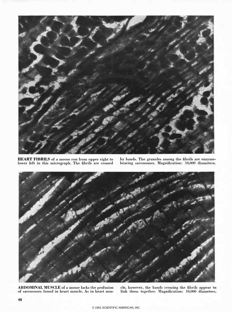
HEART FIBRILS of a mouse run from upper right to lower left in this micrograph. The fibrils are crossed
ABDOMINAL MUSCLE of a mouse lacks the profusion of sarcosomes found in heart muscle. As in heart mus-
48
by bands. The granules among the fibrils are enzymebearing sarcosomes. Magnification: 10,000 diameters.
cle, however, the bands crossing the fihrils appear to link them together. Magnification: 10,000 diameters.
© 1951 SCIENTIFIC AMERICAN, INC

HEART
MUSCLE Structure underlying function is shown by electron micrographs
HO"W does the heart beat without tiring? This momentous question
recently led Bruno Kisch, research associate in cardiology at New York's Mount Sinai Hospital, to make a fundamental studv of heart muscle with the electron microscope.
Heart muscle, like other striated muscle, is made up of slender fibers. These in turn are composed of tiny fibrils. At regular intervals the fibrils are crossed by bands. Dr. Kisch's electron micrographs of mouse heart muscle, which were made in collaboration with Joan Bardet of the Interchemical Corp., indicate that the bands connect the fibrils and bold them in place as they contract and relax.
The micrographs also show that heart muscle differs from other muscle in two important respects. The first is that the capillaries that carry blood to the heart muscle actually penetrate the muscle fibers; in other muscle the capillaries have only been observed on the surface of the fibers. The second is that among the fibrils of heart muscle are an unusually large number of small granules called sarcosomes, which in other cells are known to contain enzymes. Dr. Kisch believes that this rich network of capillaries, together with the number and location of the sarcosomes, are the principal reasons why the heart is able to beat a lifetime without resting.
CAPILLARY with two red cells penetrates fiher (bottom) . Top: a nucleus.
RELAXED heart muscle of a mouse, shown in thin section like the other tissues on these two pages, has transparent areas along the bands that cross fibrils.
CONTRACTED heart muscle lacks transparent areas along the bands crossing the fibrils. The magnification of both micrographs: 10,000 diameters.
49
© 1951 SCIENTIFIC AMERICAN, INC