Embed Size (px)
Citation preview
Heart Surgery and Dissection
1 2 3 4 5
Suitable for Key Stage:
bbsrc.ac.uk
The heart is an amazing organ that continues to beat roughly every second, every day, for your entire life. That is 100,000 beats each day, and every minute about five litres of blood are pumped out of the heart. It has to keep working non-stop to maintain the proper flow of blood around our bodies and keep us alive. Scientists are now using mathematics, computer modelling and novel imaging techniques to visualise the workings of the heart. Understanding what causes cardiovascular disease, the greatest cause of death in the UK, will lead to new cures for broken hearts. From stem cells to artificial blood, researchers are investigating new treatments, and work on biomaterials is producing improved medical implants and devices such as pacemakers.

www.bbsrc.ac.uk 2 of 11
Teacher
www.bbsrc.ac.uk
Teacher
2 of 46
Key Information
Contents02 Key information05 Recent research14 Teacher preparation15 Health and safety16 Stage 1 – External anatomy18 Stage 2 – Identification and repair of heart damage19 Stage 3 – Testing blood flow through a repaired heart20 Stage 4 – Examining the internal anatomy of a heart23 Curriculum links24 Further reading25 How the heart works29 Stage 1 – External anatomy30 Stage 2 – Identification and repair of heart damage31 Stage 3 – Testing blood flow through a repaired heart32 Stage 4 – Examining the internal anatomy of a heart33 Autopsy report form34 Heart anatomy35 Card flow36 Circulation worksheet37 Missing Words39 Wordsearch40 Crossword41 Answers44 Glossary
View online
Scan the QR Code.
www.bbsrc.ac.uk 2 of 11
Teacher
www.bbsrc.ac.uk
Teacher
3 of 46
Students will be able to:• Identify the internal and external anatomy of a heart• Dissect a heart and be able to model the techniques of a heart surgeon• Discuss heart diseases and disorders, describe how they occur, and name risk factors and
possible preventative measures.
Learning outcomes
Heart, blood, circulation, cardiovascular, anatomy, dissection, surgery, ventricle, aorta, atrium, muscle, valve, coronary, pulmonary, artery, vein, vena cava, suture, papillary, atrioventricular, mitral, oxygen, carbon dioxide
Keywords
Key Information
Age
Physiology, anatomy, cardiovascular system, exchange and transport, pathology, disease and injury
Science topics
14–18 years old
Duration
125 minutes
• Student sheets• PowerPoint presentation
Resources
www.bbsrc.ac.uk 2 of 11
Teacher
www.bbsrc.ac.uk
Teacher
4 of 46
• Sheep or lamb hearts• Rubber tubing
and syringe• Dissecting equipment – trays, pins or
cocktail sticks, forceps, blunt probes,round-ended scissors
• Masking tapeor stickers
• Marker pens• Washing up bowls or access to sinks• Curved needles• Dental floss or fishing line
• Rulers• Eye protection• Waterproof aprons• BalanceOptional• Disposable nitrile or vinyl gloves• Scalpels• Slides featuring heart muscle tissue
and cardiovascular pathology• Camera for students to record
the progress of their activity
What you will need
Students should carry out a preparatory activity to familiarise themselves with the structures of the heart. Resources such as worksheets, animations and videos can ensure students get the most from the learning session. A description of how the heart works and diagrams of the heart and circulatory system for students to label are provided.
Prior Learning
Key Information
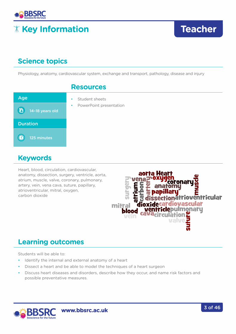
Equipment
© BBSRC

5 of 46
The Biotechnology and Biological Sciences Research Council (BBSRC) is working towards lifelong health and well-being by funding research investigating the normal ageing process. Research is being conducted to understand how the heart, blood and circulatory system work, as well as looking at technologies that could improve our health. Scientists are also looking at the effects of diet, physical activity and development on the ageing process to understand risk factors for poor health and to identify interventions that can improve well-being.
Key issues include linking changes at the molecular and cellular level to those observed at the tissue and whole organism level. A large body of evidence demonstrates that the quality and quantity of food, and dietary choice affects ageing and lifespan. There are also good data that aerobic exercise increases healthy lifespan, improves regulation of glucose metabolism and can reduce age-related deterioration of the musculoskeletal system.
From stem cells to artificial blood, researchers are investigating treatments for broken hearts and a variety of cardiovascular diseases. Work on biomaterials and tissue engineering is producing improved medical implants and devices such as pacemakers, as well as a range of substances with anti-infection properties.
Throughout this research, BBSRC encourages work that adopts the principles of the 3Rs (Replacement, Refinement and Reduction) in the use of animals, and aims to improve animal welfare.
Recent Research
Change at cellular level
© Babraham Institute
6 of 46
Can we ever mend a broken heart
Recent Research
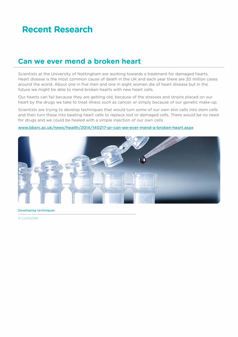
Developing techniques
© Luchschen
Scientists at the University of Nottingham are working towards a treatment for damaged hearts. Heart disease is the most common cause of death in the UK and each year there are 20 million cases around the world. About one in five men and one in eight women die of heart disease but in the future we might be able to mend broken hearts with new heart cells.
Our hearts can fail because they are getting old, because of the stresses and strains placed on our heart by the drugs we take to treat illness such as cancer, or simply because of our genetic make-up.
Scientists are trying to develop techniques that would turn some of our own skin cells into stem cells and then turn these into beating heart cells to replace lost or damaged cells. There would be no need for drugs and we could be healed with a simple injection of our own cells.
www.bbsrc.ac.uk/news/health/2014/140217-pr-can-we-ever-mend-a-broken-heart.aspx

7 of 46
The answer to high blood pressure may be in our brains
Recent Research
Blood pressure is controlled by our brains and our kidneys. Scientists are now beginning to look more closely at the brain and genes involved in the development of high blood pressure. Having high blood pressure, known as hypertension, increases the risk of stroke, heart attacks and kidney failure. You can’t ‘feel’ whether you have high blood pressure, which makes it so dangerous. Nearly one billion people around the world have hypertension.
The kidney controls blood pressure by regulating the amount of water and salt reabsorbed into the blood. High levels of salt in the blood cause the kidneys to retain water and lead to raised blood pressure. Most high blood pressure treatments target the kidneys but new research will look at ways to target the nervous system. The brain detects blood pressure using the carotid sinus, a small swelling in the carotid artery. The carotid sinus has stretch receptors that send signals to the brain when blood pressure rises. These signals go to the cardiovascular centre in the medulla and a negative feedback system sends out signals to lower heart rate and dilate blood vessels to lower the pressure.
For up to 50 per cent of patients on blood pressure tablets the treatment is ineffective and many suffer from unpleasant side effects. Researchers will explore how the genes in the brain trigger hypertension and study how ageing, exercise and a condition known as sleep apnoea affect the activity of these genes. Sleep apnoea is a blockage of airways during the night that cuts off oxygen and causes people to wake. It is associated with obesity and is often accompanied by loud snoring.
To find novel drug targets and improve current treatments, scientists will use tissue from brain banks to determine how genes are regulated in specific brain regions and how these genes interact during the development of high blood pressure. The scientists will also study how the environment may affect gene activity. In order to do this they will use a special technique that allows them to record the activity of single nerve fibres that control the diameter of arteries.

8 of 46
What causes a big heart?
Recent Research
3D Reconstruction of a section through a rats heart
© Dr Llewelyn Roderick Group
Under some conditions heart cells get larger in a process known as hypertrophy. While this may sound romantic, and is necessary for developmental growth, hypertrophy often leads to heart failure. If there is a long-term demand on the heart to pump more blood – for example during exercise or in pregnancy – the body responds by making the heart cells larger.
Conditions like high blood pressure also cause the heart to grow, but in this case increased size does not improve the heart’s pumping capacity. Instead it promotes the transition to cell death and heart failure as the heart becomes prone to irregular heartbeats – arrhythmias. Researchers have discovered how signalling processes within the heart can trigger the development of enlarged heart cells which lead to heart failure. The discovery provides new insight into the mechanisms controlling cardiac growth and the processes that cause adaptation and remodelling of heart muscle. Cardiac failure accounts for 25% of deaths in the UK and is a primary cause of death in the elderly. Understanding how these pathological changes occur in the heart, in response to disease and ageing, may reveal therapeutic targets and new approaches to the treatment of heart disease.
The research team found that a tiny molecule made of ribonucleic acid (RNA), microRNA, controls the levels of specific receptors produced in heart cells. MicroRNAs are copied from deoxyribonucleic acid (DNA) but do not contain code for protein. Rather, they control gene activity by binding to specific related sequences. It is the interactions between these microRNA molecules and the receptors that promote hypertrophic remodelling of heart muscle. The receptors are channels controlling the movement of calcium ions, which are an important ‘messenger’ inside cells, regulating heart rhythm and function. Calcium ions are the link between electrical excitation of a muscle cell and its contraction. When an electrical impulse arrives at a muscle it causes calcium to enter the cell and releases calcium from internal stores resulting in contraction of the cell. If calcium signals occur at the wrong place or time, for example due to changes in receptor regulation, this can change the heart structure – decreasing its ability to pump efficiently, or triggering irregular heartbeats.
9 of 46
Scientists discover the cause of a broken heart
Recent Research
Around 1–2% of people who are initially suspected of having a heart attack are found to have ‘broken heart syndrome’. This condition causes temporary heart failure but tests find no blockage in the coronary arteries. The condition is called Takotsubo cardiomyopathy and it affects people who suffer severe emotional stress after bereavement, often elderly women. People experience symptoms that resemble a heart attack and the heart develops a balloon-like appearance caused by the bottom of the heart not contracting properly. Most patients make a full recovery within days or weeks.
Researchers now think this ‘broken heart syndrome’ is a protective response to very high levels of adrenaline released during stress. Instead of stimulating the heart, the body responds to the adrenaline by reducing its pumping power. The same condition is sometimes seen in people who are injected with adrenaline to treat severe allergic reactions. Therefore drugs that stimulate adrenaline are likely to make the condition worse. The scientists used their animal model of the disease to investigate suitable treatments and found beneficial drugs that stimulate the heart using a different pathway to adrenaline.

10 of 46
Why some people endure exercise better than others
Recent Research
Human Heart
© Thinkstock
Exercise is essential for maintaining good health. An understanding of how elite athletes’ bodies function may help prevent elderly people developing chronic illnesses.
Researchers are studying elite athletes using non-invasive technologies, such as magnetic resonance spectroscopy (MRS). Studying how the heart, lungs and muscles work together will provide insights into why some people have higher levels of endurance than others.
Computer models will be used to improve our understanding of why exercise tolerance is limited in sedentary or elderly individuals. The findings will be used as the basis for treating patients with heart and lung conditions who have problems with exercising.
11 of 46
Recent Research
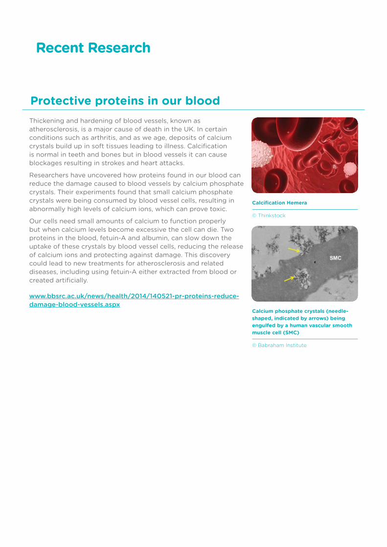
Thickening and hardening of blood vessels, known as atherosclerosis, is a major cause of death in the UK. In certain conditions such as arthritis, and as we age, deposits of calcium crystals build up in soft tissues leading to illness. Calcification is normal in teeth and bones but in blood vessels it can cause blockages resulting in strokes and heart attacks.
Researchers have uncovered how proteins found in our blood can reduce the damage caused to blood vessels by calcium phosphate crystals. Their experiments found that small calcium phosphate crystals were being consumed by blood vessel cells, resulting in abnormally high levels of calcium ions, which can prove toxic.
Our cells need small amounts of calcium to function properly but when calcium levels become excessive the cell can die. Two proteins in the blood, fetuin-A and albumin, can slow down the uptake of these crystals by blood vessel cells, reducing the release of calcium ions and protecting against damage. This discovery could lead to new treatments for atherosclerosis and related diseases, including using fetuin-A either extracted from blood or created artificially.
www.bbsrc.ac.uk/news/health/2014/140521-pr-proteins-reduce-damage-blood-vessels.aspx
Protective proteins in our blood
Calcification Hemera
© Thinkstock
Calcium phosphate crystals (needle- shaped, indicated by arrows) being engulfed by a human vascular smooth muscle cell (SMC)
© Babraham Institute
12 of 46
Every day thousands of people around the world have their lives saved or improved thanks to someone giving blood – but many are not so lucky. Blood transfusions are the only option when someone loses a lot of blood, but getting the right blood in time is not always possible. Blood donations have to be screened for disease, organised according to blood type and can only be stored for a short time using a temperature-controlled environment.
Over 100 million units of donated blood are given by people worldwide for use in hospitals each year and demand is rising, but the number of donors is decreasing. Imagine how many more lives could be saved if a long-lasting blood substitute could be found, which could easily be stored at room temperature and was available to all patients, regardless of their blood type.
Scientists at the University of Essex are hoping to overcome this challenge in a project to develop an artificial blood substitute that is a safe, long-lasting, virus-free alternative to current blood transfusions. An artificial blood substitute could save many lives by being available to all countries and being immediately accessible at the site of natural disasters or in remote inaccessible locations.
Haemoglobin is the key protein in red blood cells that carries oxygen around our bodies. The researchers aim to create an artificial haemoglobin-based oxygen carrier (HBOC) that could be used as a substitute for blood lost in surgery or trauma. Outside the protective environment of the red cell, haemoglobin can be toxic which has caused previous scientists problems in making artificial blood. So the team have developed and patented an artificial HBOC that it is detoxified by the body’s own defences.
www.bbsrc.ac.uk/news/health/2014/140610-pr-quest-for-long-lasting-blood.aspx
Recent Research
Artifical Hemera
© Thinkstock
The quest for long-lasting blood

13 of 46
Recent Research
Silk Fibres
© Oxford Silk Group
Many implantable medical devices can trigger an acute inflammatory response. Inflammation can prevent devices functioning properly and can cause stress-cracking of pacemakers or a re-narrowing of coronary arteries following a stent implant.
Inflammation can be triggered by the initial implantation injury or by the biomaterial itself. Treatment with steroids or non-steroidal anti-inflammatory drugs (NSAIDS) can lead to side effects due to a lack of sensitivity or a lack of a specific targeted site of action.
A team of scientists have developed a new method for generating model surfaces with bioactive anti-inflammatory properties. These bioactive materials also have anti-microbial activity and could be added to a variety of surgical tools, such as stents or stitches, to enable wounds to heal quickly and safely.
Researchers in Oxford are looking to the natural world to create a new breed of exotic materials for biomedicine based on silkworm and spider silk. Silk is a superb material – light, strong, and highly elastic. Scientists in the UK studying the fundamental properties of this natural substance have now formed a number of spin-off companies to develop a new generation of biomedical implants.
Silk is also biocompatible, and thus makes an ideal substance for implants used inside the human body.
Intimate knowledge of beneficial amino acid sequences and morphology from studying spider silks has led to the combination of excellent mechanical and tissue-regenerative properties. Because the silk implants do not irritate or harm the body’s cells they not only provide tough replacement surfaces but also encourage new regrowth of tissue.
Improving biomaterials
www.bbsrc.ac.uk 2 of 11
Teacher
www.bbsrc.ac.uk
Teacher
14 of 46
To complete the whole practical activity you will require two lessons or a double lesson. Stages 1 and 2 can be carried out in the first lesson and the repaired hearts refrigerated and saved until the next lesson to complete stages 3 and 4. Alternatively, the whole procedure can be carried out in a double lesson.
Order sheep or lamb hearts from a biological supply company such as Blades Biologicals Ltd, or obtain hearts from a local butcher or supermarket. All materials for dissection must be obtained from animals that have been inspected to ensure that they are fit for consumption. Sheep hearts should be stored according to the supplier’s instructions and used before the use-by date. Sheep hearts are easily obtained and allow students with religious concerns over cows or pigs to take part.
When obtaining hearts from butchers, ask for the hearts to be removed with as much of the blood vessels as possible and with no damage to the heart. In some instances there may already be damage to the heart caused by the meat inspection process and this activity is ideal for incorporating this into the dissection.
If the heart is intact, use a scalpel to create a small hole in the wall of one of the ventricles. A slit about 1.5 cm long in the right ventricle on the dorsal side is unobtrusive yet effective. Ensure the hole created passes right through the ventricle wall. Due to the thinner wall of the right ventricle it is easier to ensure that the hole has been made correctly and is easier for the students to repair.
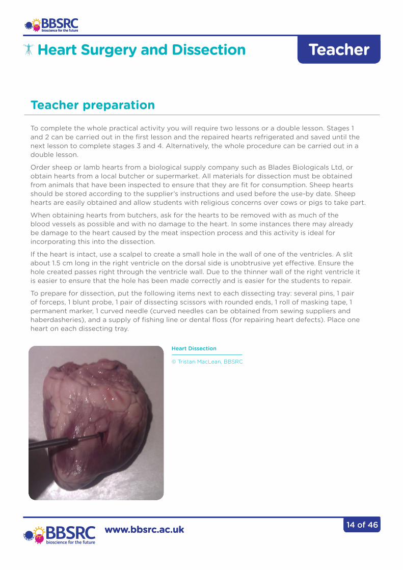
To prepare for dissection, put the following items next to each dissecting tray: several pins, 1 pair of forceps, 1 blunt probe, 1 pair of dissecting scissors with rounded ends, 1 roll of masking tape, 1 permanent marker, 1 curved needle (curved needles can be obtained from sewing suppliers and haberdasheries), and a supply of fishing line or dental floss (for repairing heart defects). Place one heart on each dissecting tray.
Heart Dissection
© Tristan MacLean, BBSRC
Heart Surgery and Dissection
Teacher preparation
www.bbsrc.ac.uk 2 of 11
Teacher
www.bbsrc.ac.uk
Teacher
15 of 46
Health and Safety
Heart Surgery and Dissection
These instructions are for guidance only. Observe Good Laboratory Practice when carrying out these activities. Wear eye protection and lab coats or disposable waterproof aprons to protect clothes. Use suitable waterproof surfaces or dissection trays for the activity. After the activity, disinfect work surfaces with 1% Virkon and wash and autoclave dissecting instruments. Consult the CLEAPSS Starter Guide to Dissection, G267 and guidance available on the CLEAPSS and Scottish Schools Education Research Centre (SSERC) web page for further details on dissections and disinfection.
Students will be handling sharp scissors (or a scalpel) in order to dissect the heart, together with pins and needles. Students should be instructed in the very careful use of each sharp implement they will be using and vigilance should be maintained during the activity to ensure students behave appropriately. If using scalpels, ensure that new blades are used for the activity. Removable blades should be disposed of in a suitable container and fixed blades should be cleaned with detergent after the activity.
Gloves can be used to simulate the heart surgery procedure but they are not essential and do not need to be sterile. Students should ensure any cuts or grazes are covered with gloves or a plaster and that they carefully and thoroughly wash their hands after handling hearts. Gloves should be disposed of immediately after use.

www.bbsrc.ac.uk 2 of 11
Teacher
www.bbsrc.ac.uk
Teacher
16 of 46
Inform the students that they will be learning about heart anatomy, blood flow through the heart, identifying damage to the heart and conducting heart surgery. Share the learning outcomes with the students and recap their prior knowledge of the heart structure and function.
Introduction
Once students have familiarised themselves with heart anatomy either online or by completing a worksheet, it is time to learn to identify the structures on a real sheep’s heart. It may help students to have the diagram of the heart to hand to help identify the orientation of the heart and to locate the regions correctly.
1. Divide students into groups of 2–3 and have them put on gloves, eye protection and aprons.
2. Provide each group with a dissecting tray, a sheep or lamb heart, pins or cocktail sticks, forceps, blunt probe, round-ended scissors, masking tape, and a marker pen.
3. Have each group create a set of seven pins with identification flags on them using masking tape or stickers and the marker pen.
Label the pins with the following: 1. Left Ventricle 2. Right Ventricle 3. Coronary blood vessel 4. Right Atrium 5. Left Atrium 6. Pulmonary Artery 7. Aorta
If the blood vessels have been retained, students may also be able to label the superior and inferior vena cava.
Stage 1 - External Anatomy
Duration
5 minutes
Duration
20 minutes
Heart Surgery and Dissection
www.bbsrc.ac.uk 2 of 11
Teacher
www.bbsrc.ac.uk
Teacher
17 of 46
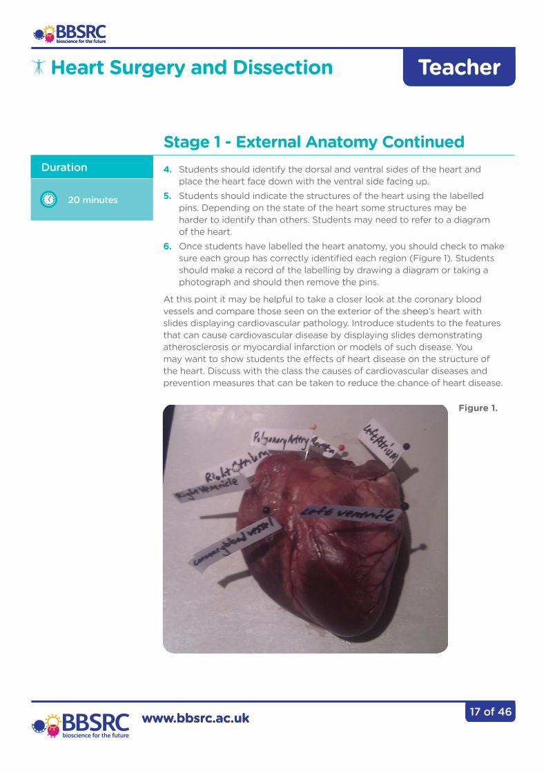
4. Students should identify the dorsal and ventral sides of the heart and place the heart face down with the ventral side facing up.5. Students should indicate the structures of the heart using the labelled pins. Depending on the state of the heart some structures may be harder to identify than others. Students may need to refer to a diagram of the heart.6. Once students have labelled the heart anatomy, you should check to make
sure each group has correctly identified each region (Figure 1). Students should make a record of the labelling by drawing a diagram or taking a photograph and should then remove the pins.
At this point it may be helpful to take a closer look at the coronary blood vessels and compare those seen on the exterior of the sheep’s heart with slides displaying cardiovascular pathology. Introduce students to the features that can cause cardiovascular disease by displaying slides demonstrating atherosclerosis or myocardial infarction or models of such disease. You may want to show students the effects of heart disease on the structure of the heart. Discuss with the class the causes of cardiovascular diseases and prevention measures that can be taken to reduce the chance of heart disease.
Stage 1 - External Anatomy ContinuedDuration
20 minutes
Figure 1.
Heart Surgery and Dissection
www.bbsrc.ac.uk 2 of 11
Teacher
www.bbsrc.ac.uk
Teacher
18 of 46
Students are to assume the roles of heart surgeons and determine where and what the damage to the heart is and then perform surgical repair of the injury.1. Provide each group with a curved needle and a length of dental floss.2. Students should examine the heart and identify the damaged region.
This should be reported to the teacher and a record made by annotating the diagram made in stage 1 or by taking a photograph.
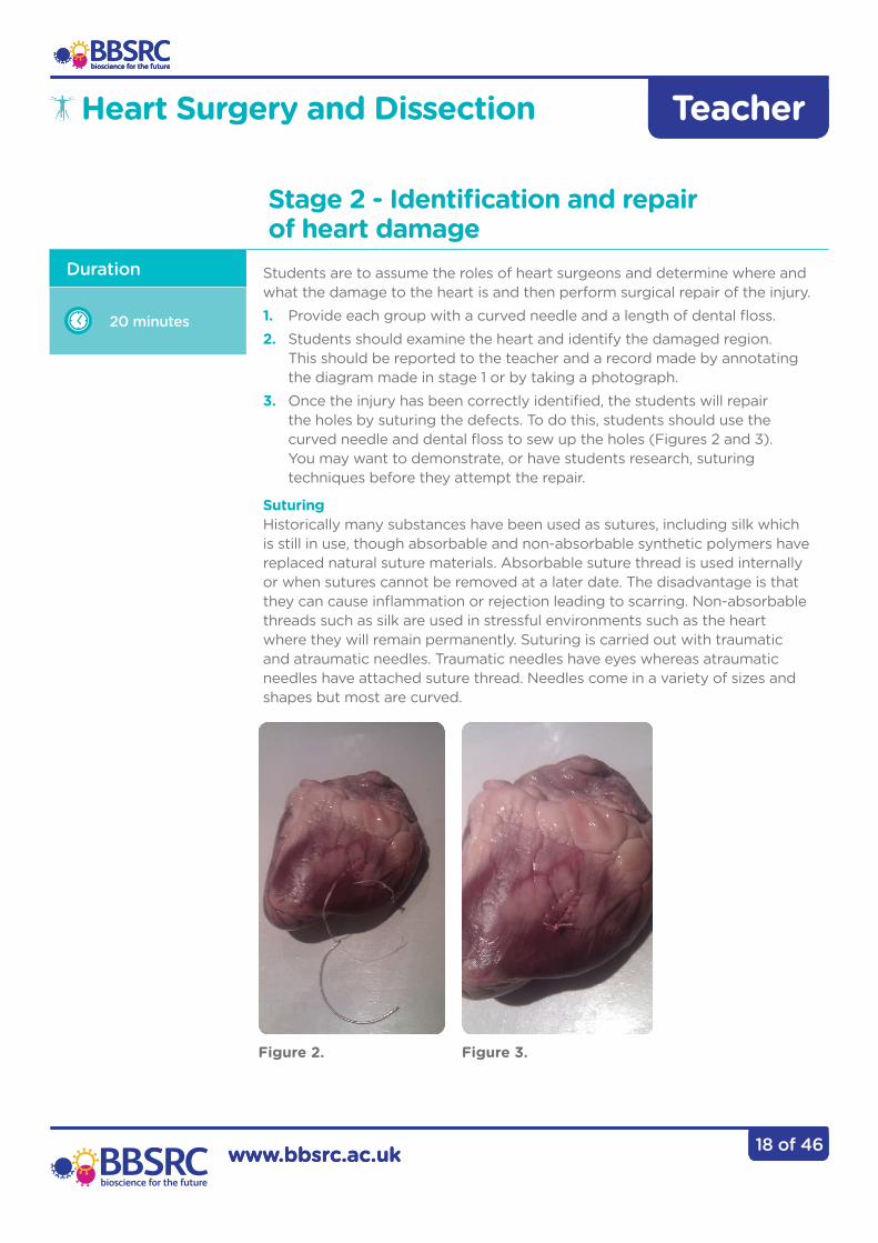
3. Once the injury has been correctly identified, the students will repair the holes by suturing the defects. To do this, students should use the curved needle and dental floss to sew up the holes (Figures 2 and 3). You may want to demonstrate, or have students research, suturing techniques before they attempt the repair.
Suturing Historically many substances have been used as sutures, including silk which is still in use, though absorbable and non-absorbable synthetic polymers have replaced natural suture materials. Absorbable suture thread is used internally or when sutures cannot be removed at a later date. The disadvantage is that they can cause inflammation or rejection leading to scarring. Non-absorbable threads such as silk are used in stressful environments such as the heart where they will remain permanently. Suturing is carried out with traumatic and atraumatic needles. Traumatic needles have eyes whereas atraumatic needles have attached suture thread. Needles come in a variety of sizes and shapes but most are curved.
Duration
20 minutes
Stage 2 - Identification and repair of heart damage
Heart Surgery and Dissection
Figure 2. Figure 3.
www.bbsrc.ac.uk 2 of 11
Teacher
www.bbsrc.ac.uk
Teacher
19 of 46
Students are to assume the roles of heart surgeons and determine where and what the damage to the heart is and then perform surgical repair of the injury.1. Demonstrate the flow of water through an intact heart. Using a syringe and rubber tubing squirt water into the veins and atria and identify the vessels the water exits from. Compare this to a diagram showing blood flow through the heart.2. Students should squirt some water into the veins to check that the repair has sealed the damage to the heart and to check that they have identified the correct blood vessels. The pulmonary vein water should exit the aorta (the larger rubbery arterial vessel) and the vena cava water should exit the pulmonary artery.
Duration
10 minutes
Stage 3 – Testing blood flow through a repaired heart
Heart Surgery and Dissection

www.bbsrc.ac.uk 2 of 11
Teacher
www.bbsrc.ac.uk
Teacher
20 of 46
Inform the students that there was a post-operative complication and the internal structure of the heart must be examined as part of the autopsy.
Students should work in pairs with one taking the role of the pathologist and the other the assistant pathologist. The pathologist will be responsible for carrying out the dissection. The assistant will be responsible for recording the appropriate information on the autopsy record sheet, providing equipment to the pathologist and taking photographs or drawing diagrams at different stages of the dissection.
• Use attached autopsy record form. • Use ruler to establish scale. • Record progress using a camera or drawings.
1. Record the weight of the heart. A typical sheep’s heart weighs about 250g. Note any discrepancy.2. Measure the width of the heart, across the ventricles at the widest part.3. Cut from the bottom to the top of the right ventricle using the scissors.
Open up the right ventricle identifying the right atrioventricular (tricuspid) valve and the attached papillary muscles (Figure 4). Use the blunt probe to carefully move tissues.
4. Measure the thickness of the right ventricle wall. Students should see whether they can observe their repair internally and identify whether the suturing caused any damage or if any of the tendons or papillary muscles were affected by the sutures.5. Repeat the cut through the left ventricle and atrium this time identifying the mitral (bicuspid) valve (Figure 5).6. Once again record the thickness of the ventricle wall.
Students should be asked to suggest a reason for the difference in size of the ventricle walls and relate it to the function of the heart.
Duration
25 minutes
Stage 4 – Examining the internal anatomy of a heart
Heart Surgery and Dissection
Figure 3. Figure 4.

www.bbsrc.ac.uk 2 of 11
Teacher
www.bbsrc.ac.uk
Teacher
21 of 46
Recap the internal and external features of the heart that were identified. Relate the anatomy of the heart to the heart structure. Discuss the pros and cons of the suturing techniques and materials with the class. You may want to discuss the difficulties in treating cardiovascular disease, treatments available and the benefits of preventative measures.
Based on the lesson: You Gotta Have Heart: Congenital Heart Defects and Heart Surgery by Rebecca Johns
Once all the measurements have been taken and the diagrams labelled, implements and tissues will need to be cleaned or discarded. The students should wrap the dissected heart in tissue paper or newspaper, and then in a double layer of plastic bags. The package should be kept in cold storage until it can be placed in an external bin on the day of refuse collection.
Water that has come into contact with either the heart or any other tissues should be poured carefully down a sink, and the sink washed out well afterwards. Be careful to pour the water very slowly to avoid creating aerosols.
Scissors and other dissecting implements should be carefully collected for appropriate disposal or washing by a member of staff. If using disposable scalpels, blades or razors they should be wrapped and placed in a stout-sided container, which may need to be autoclaved by a member of staff prior to prompt disposal. After the activity, disinfect work surfaces with 1% Virkon. Consult the CLEAPSS Starter Guide to Dissection, G267, for further information.
Ensure hands are thoroughly washed using a bactericidal hand wash, and that aprons are removed before leaving the room. Protective clothing should be appropriately disposed of or thoroughly cleaned.
Blades Biological Limited www.blades-bio.co.uk Cowden, Edenbridge, Kent, TN8 7DX tel: 01342 850 242, fax: 01342 850 924
Duration
5 minutes
Plenary
Clean up
Suppliers
Heart Surgery and Dissection
www.bbsrc.ac.uk 2 of 11
Teacher
www.bbsrc.ac.uk
Teacher
22 of 46
Curriculum Links
Transport systems in people and plants • The need for a transport system in multicellular organisms, including surface area: volume calculations• The structure and function of the heart, including control of the muscular contractions
and the working of the valves both in the heart and the vessels
B3.2 Transport systems in plants and animals B3.2.1 The blood system B3.2.2 The blood Suggested ideas for practical work to develop skills and understanding include the following:• Dissection of the heart• Use software simulations of the work of the heart and blood vessels• Observation of arteries and veins from slides• Observation of blood smears• Observation of valves in veins preventing backflow of blood using the ‘athletic’ arm/prominent vein
3.3.2 Module B2: Keeping healthy 3.5.1 Module B7: Further BiologyOpportunities for practical work• Heart dissection
Draft Key Stage 4 programme of study
AQA Biology GCSE
OCR Twenty First century science suite – GCSE Science A, Biology A

www.bbsrc.ac.uk 2 of 11
Teacher
www.bbsrc.ac.uk
Teacher
23 of 46
Curriculum Links
Module 2: Exchange and Transport The practical work outlined below may be carried out as part of skill development. Collection and presentation of qualitative (descriptive) data:• Make measurements and annotated drawings during a heart dissection.• Use a light microscope to make annotated drawings of blood vessels.
Body systems and cellsLearners… develop informed views on the moral and ethical implications of controversial biological procedures.
Topical scienceBy considering current issues of science, learners increasingly develop their understanding of scientific concepts and their capacity to form informed social, moral and ethical views. They reflect upon and critically evaluate media portrayal of scientific findings.
OCR Biology A-level
Scotland: Curriculum for Excellence – Science
www.bbsrc.ac.uk 2 of 11
Teacher
www.bbsrc.ac.uk
Teacher
24 of 46
Calcium Signalling – Getting to the Heart of the Matter
Understanding Animal Research Teachers’ Zone www.understandinganimalresearch.org.uk/teacherszone
The NC3Rs is working with the BBSRC to increase investment and awareness in tissue engineering as a means of replacing animal models. www.nc3rs.org.uk/event.asp?id=830
Carry out a heart transplant interactive animation www.pbs.org/wgbh/nova/eheart/transplantwave.html
Anatomy for Beginners. Lesson 2 Circulation (contains scenes of anatomical demonstrations and exhibits on humans and animals which are not suitable for young pupils and some viewers may find distressing).
Further reading
Heart Surgery and Dissection
25 of 46
Heart Surgery and Dissection
Key Stage 4
Age
13 years old
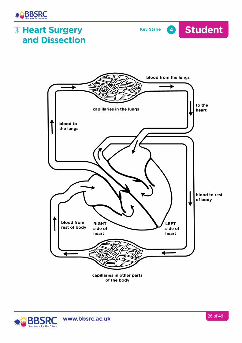
Our blood is continuously pumped around our body by the heart. There are four main chambers in the heart – the left and right atria and ventricles. The walls of the heart are made primarily of muscle and connective tissue. There is a special type of striated involuntary muscle tissue in the heart called cardiac muscle. This muscle keeps beating on average 72 times a minute for 66 years without stopping and without us having to think about it.
Valves in the heart ensure that blood flows in the correct direction. Blood carries substances, such as oxygen, nutrients and waste, around the body through the circulatory system. Blood flows away from the heart through arteries, which have thick elastic walls made with muscle. In the organs, blood flows through very narrow, thin-walled blood vessels called capillaries. One circulatory system takes the blood to the lungs to collect oxygen and then back to the heart while the other takes blood round all the other organs. The blood collects waste carbon dioxide as it passes through the capillaries. The blood returns to the heart along thin-walled veins which often have valves to prevent the blood flowing backwards. The carbon dioxide is removed from the blood in the lungs. It takes about 30 seconds for a single blood cell to travel round the body and back to the heart.
How the heart works
Cardiac – relating to the heartPulmonary – relating to the lungs
Heart Surgery and Dissection
Key Stage 4 Student
blood from the lungs
capillaries in the lungs
blood to rest of body
blood from rest of body
capillaries in other parts of the body
www.bbsrc.ac.uk 26 of 46
to the heart
blood to the lungs
RIGHTside of heart
LEFTside of heart
27 of 46
Heart Surgery and Dissection
Key Stage 4
Age
13 years old
How does the blood travel through the heart?1. Blood leaves the heart via the aorta and goes to the head and body.2. After travelling along the arteries, through the tissues and back along
the veins the blood returns to the heart along the vena cava and entersthe right atrium.
3. Blood is then pushed into the right ventricle through thetricuspid valve.
4. The right ventricle contracts and pushes blood out of the heart and intothe pulmonary artery.
5. The pulmonary valve stops blood flowing back into the right ventricle.6. The blood travels along the pulmonary artery to the lungs where it
picks up oxygen.7. Blood returns to the heart along the pulmonary vein and enters the
left atrium.8. Blood is forced into the left ventricle through the mitral valve when the
left atrium contracts.9. The left ventricle contracts and pushes blood out of the heart and
into the aorta. The aortic valve stops blood flowing back into theleft ventricle.
10. The blood then travels round the head and body again.
Continued
Amazing facts
• Your heart is about the samesize as your fist.
• The sound of your heartbeating is caused by thevalves as they openand close.
• An average adult bodycontains about five anda half litres of blood.
• All the blood vessels in thebody joined end to endwould stretch 62,000 miles(99,779 km) or two-and-a-
half times around the earth.• The heart circulates the
body’s blood supply about1,000 times each day.
• The heart pumps theequivalent of 5,000 to6,000 litres of bloodeach day.
Coronary artery – This is a large blood vessel that runs down the middle of the heart on the outside, branching across the front and back of the heart to supply fresh blood with oxygen and nutrients to the heart muscle itself.

28 of 46
Heart Surgery and Dissection
Key Stage 4
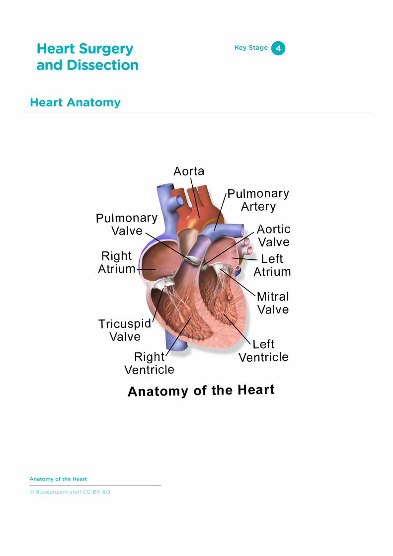
Anatomy of the Heart
© Blausen.com staff CC-BY-3.0

29 of 46
1. Work in groups of 2–32. Put on gloves, eye protection and aprons3. Ensure you have all the following items:
• dissecting tray• sheep’s heart• pins or cocktail sticks• forceps• blunt probe• scissors• masking tape or stickers• marker pen
4. Create a set of seven pins with identification flags on them using maskingtape or stickers and the marker pen, as seen in Figure 1.Label the pins with the following:1. Left Ventricle2. Right Ventricle3. Coronary blood vessel4. Right Atrium5. Left Atrium6. Pulmonary Artery7. Aorta
You may also be able to label the superior and inferior vena cava.5. Identify the dorsal and ventral sides of the heart and place the heart face
down with the ventral side facing up.6. Indicate the structures of the heart using the labelled pins. Depending
on the state of the heart some structures may be harder to identifythan others. Refer to a diagram of the heart to help you.
7. Make a record of the labelling by drawing a diagram or taking aphotograph and then remove the pins.
Stage 1 - External Anatomy Duration
20 minutes
Heart Surgery and Dissection
Key Stage 4
Equpiment.
Figure 1.
30 of 46
Heart Surgery and Dissection
Duration
20 minutes
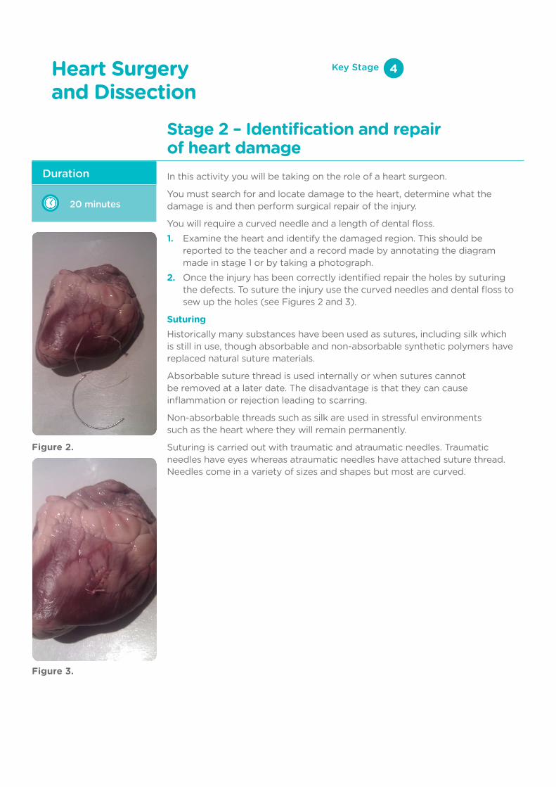
In this activity you will be taking on the role of a heart surgeon.
You must search for and locate damage to the heart, determine what the damage is and then perform surgical repair of the injury.
You will require a curved needle and a length of dental floss.1. Examine the heart and identify the damaged region. This should be
reported to the teacher and a record made by annotating the diagrammade in stage 1 or by taking a photograph.
2. Once the injury has been correctly identified repair the holes by suturingthe defects. To suture the injury use the curved needles and dental floss tosew up the holes (see Figures 2 and 3).
SuturingHistorically many substances have been used as sutures, including silk which is still in use, though absorbable and non-absorbable synthetic polymers have replaced natural suture materials.
Absorbable suture thread is used internally or when sutures cannot be removed at a later date. The disadvantage is that they can cause inflammation or rejection leading to scarring.
Non-absorbable threads such as silk are used in stressful environments such as the heart where they will remain permanently.
Suturing is carried out with traumatic and atraumatic needles. Traumatic needles have eyes whereas atraumatic needles have attached suture thread. Needles come in a variety of sizes and shapes but most are curved.
Stage 2 – Identification and repair of heart damage
Figure 2.
Figure 3.
Key Stage 4
31 of 46
Heart Surgery and Dissection
Duration
10 minutes
To test your heart surgery you will now pass water through the heart to check that the repair has sealed the damage and that there are no leaks. You will need a syringe to pass water into the heart.1. First identify the pulmonary vein and insert water into it using the syringe.2. Water should only exit the aorta (the larger rubbery arterial vessel).3. Check there isn’t water in the right-hand side of the heart.4. Now empty the heart of water and carefully add water into the vena cava.5. Water should now only exit the pulmonary artery.6. Compare your observations to a diagram showing blood flow through
the heart.
Stage 3 – Testing blood flow through a repaired heart
Key Stage 4
32 of 46
Heart Surgery and Dissection
Duration
25 minutes
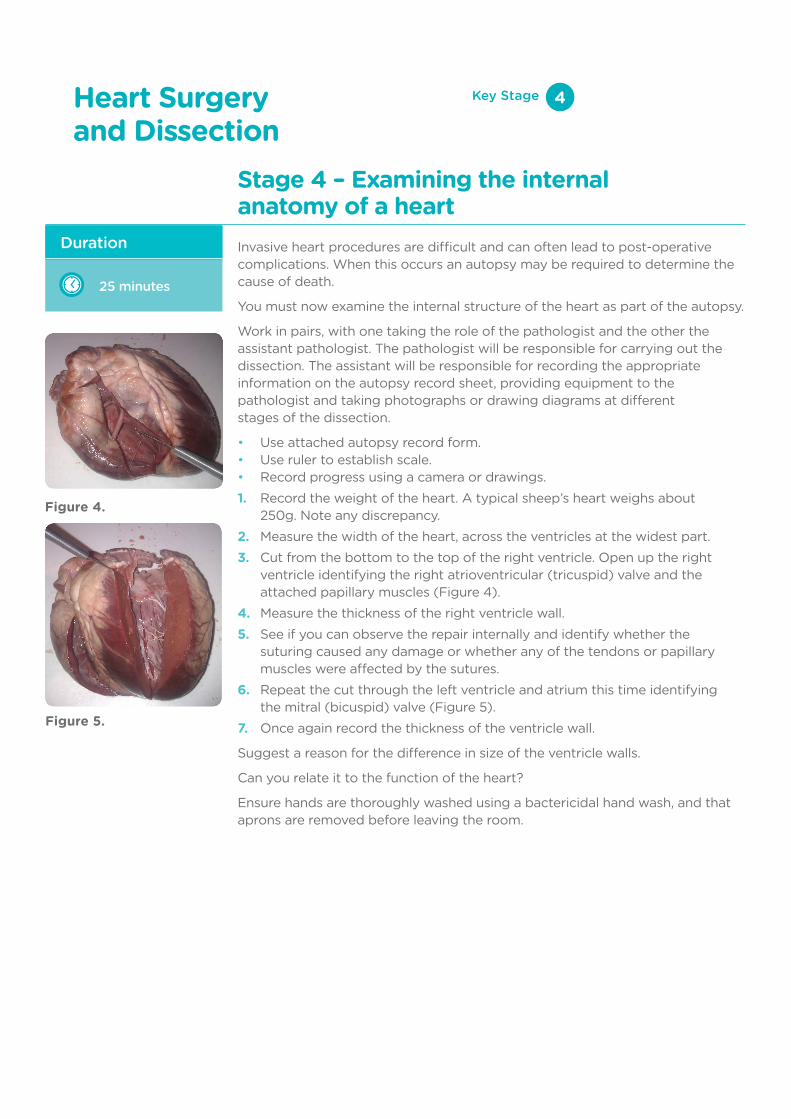
Invasive heart procedures are difficult and can often lead to post-operative complications. When this occurs an autopsy may be required to determine the cause of death.
You must now examine the internal structure of the heart as part of the autopsy.
Work in pairs, with one taking the role of the pathologist and the other the assistant pathologist. The pathologist will be responsible for carrying out the dissection. The assistant will be responsible for recording the appropriate information on the autopsy record sheet, providing equipment to the pathologist and taking photographs or drawing diagrams at different stages of the dissection.
• Use attached autopsy record form.• Use ruler to establish scale.• Record progress using a camera or drawings.1. Record the weight of the heart. A typical sheep’s heart weighs about
250g. Note any discrepancy.2. Measure the width of the heart, across the ventricles at the widest part.3. Cut from the bottom to the top of the right ventricle. Open up the right
ventricle identifying the right atrioventricular (tricuspid) valve and theattached papillary muscles (Figure 4).
4. Measure the thickness of the right ventricle wall.5. See if you can observe the repair internally and identify whether the
suturing caused any damage or whether any of the tendons or papillarymuscles were affected by the sutures.
6. Repeat the cut through the left ventricle and atrium this time identifyingthe mitral (bicuspid) valve (Figure 5).
7. Once again record the thickness of the ventricle wall.
Suggest a reason for the difference in size of the ventricle walls.
Can you relate it to the function of the heart?
Ensure hands are thoroughly washed using a bactericidal hand wash, and that aprons are removed before leaving the room.
Stage 4 – Examining the internal anatomy of a heart
Key Stage 4
Figure 4.
Figure 5.

33 of 46
Observations
With any injuries note the size, shape, pattern, location, colour, direction, depth and structures involved. Note any coronary artery disease or damaged tissues in the heart muscle characterised by an altered colour or texture:
Autopsy Report Form
Date:
Weight of heart
Location:
Width of heart (mm):
Start time:
Thickness of left ventricle wall (mm):
Finish time:
Thickness of right ventricle wall (mm):
Name of Pathologist:
Probable cause of death:
Signed:
Institution:
Key Stage 4
34 of 46
Heart Surgery and Dissection
Heart Anatomy
Key Stage 4
Anatomy of the Heart
© Blausen.com staff CC-BY-3.0

35 of 46
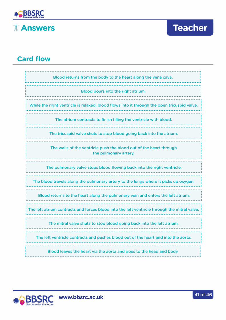
The walls of the ventricle push the blood out of the heart through the pulmonary artery.
Blood returns from the body to the heart along the vena cava.
The walls of the ventricle push the blood out of the heart through the pulmonary artery.
Blood leaves the heart via the aorta and goes to the head and body.
Blood returns to the heart along the pulmonary vein and enters the left atrium.
The mitral valve shuts to stop blood going back into the left atrium.
The left ventricle contracts and pushes blood out of the heart and into the aorta.
The left atrium contracts and forces blood into the left ventricle through the mitral valve.
The atrium contracts to finish filling the ventricle with blood.
Blood pours into the right atrium.
The tricuspid valve shuts to stop blood going back into the atrium.
The blood travels along the pulmonary artery to the lungs where it picks up oxygen.
While the right ventricle is relaxed, blood flows into it through the open tricuspid valve.
The pulmonary valve stops blood flowing back into the right ventricle.
Card Flow
Heart Surgery and Dissection
Key Stage 4
Put the cards in the correct order starting with the blood returning to the heart.
36 of 46
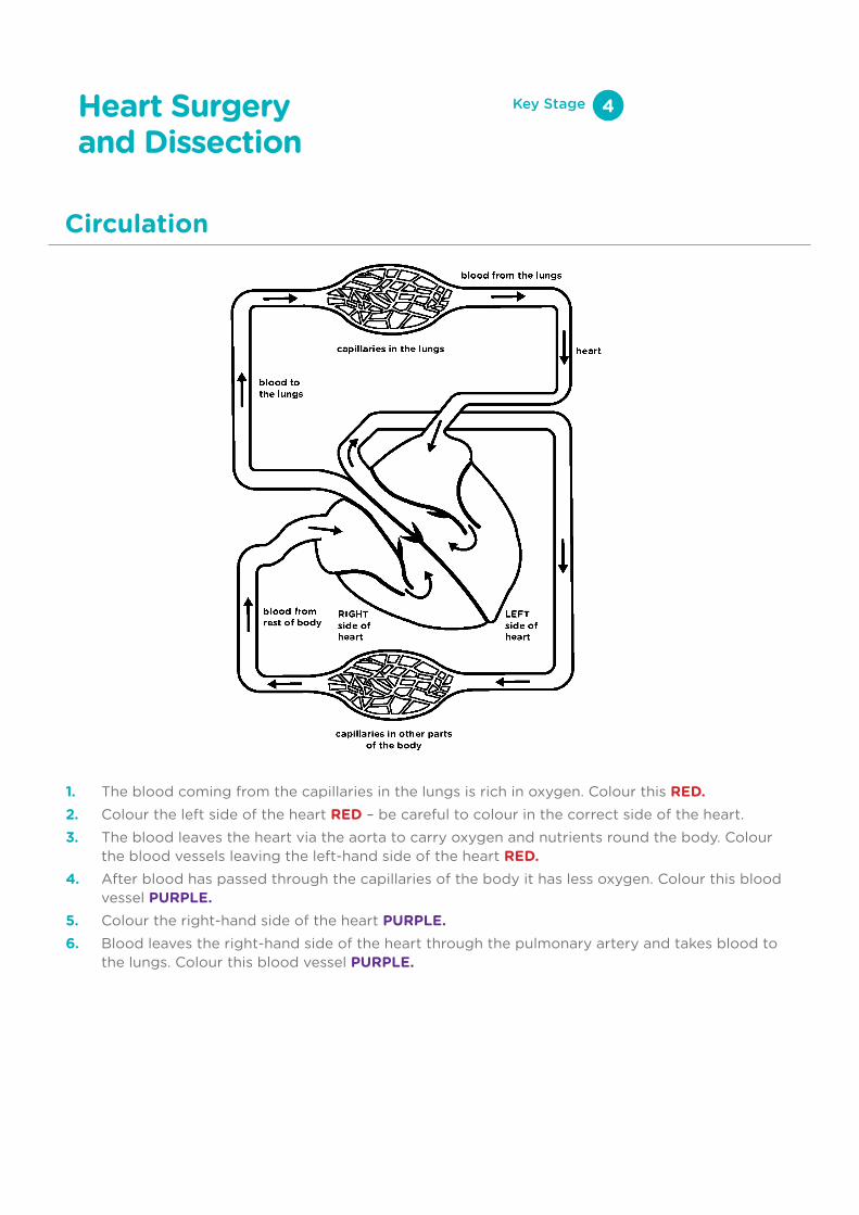
1. The blood coming from the capillaries in the lungs is rich in oxygen. Colour this RED.2. Colour the left side of the heart RED – be careful to colour in the correct side of the heart.3. The blood leaves the heart via the aorta to carry oxygen and nutrients round the body. Colour
the blood vessels leaving the left-hand side of the heart RED.4. After blood has passed through the capillaries of the body it has less oxygen. Colour this blood
vessel PURPLE.5. Colour the right-hand side of the heart PURPLE.6. Blood leaves the right-hand side of the heart through the pulmonary artery and takes blood to
the lungs. Colour this blood vessel PURPLE.
Circulation
Heart Surgery and Dissection
Key Stage 4

37 of 46
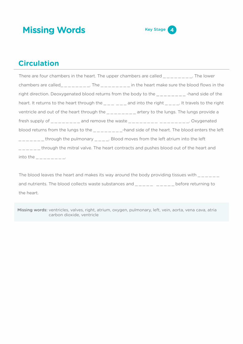
There are four chambers in the heart. The upper chambers are called _ _ _ _ _ _ _ _. The lower
chambers are called_ _ _ _ _ _ _ _. The _ _ _ _ _ _ _ _ in the heart make sure the blood flows in
the right direction. Deoxygenated blood returns from the body to the _ _ _ _ _ _ _ _ -hand side
of the heart. It returns to the heart through the _ _ _ _ _ _ and into the right _ _ _ _. It travels to
the right ventricle and out of the heart through the _ _ _ _ _ _ _ _ artery to the lungs. The lungs
provide a fresh supply of _ _ _ _ _ _ _ _ and remove the waste _ _ _ _ _ _ _ _ _ _ _ _ _ _ _ _.
Oxygenated blood returns from the lungs to the _ _ _ _ _ _ _ _-hand side of the heart. The blood
enters the left _ _ _ _ _ _ _ through the pulmonary _ _ _ _. Blood moves from the left atrium into
the left _ _ _ _ _ _ through the mitral valve. The heart contracts and pushes blood out of the
heart and into the _ _ _ _ _ _ _ _.
The blood leaves the heart and makes its way around the body providing tissues with _ _ _ _ _ _
and nutrients. The blood collects waste substances and _ _ _ _ _ _ _ _ _ _ before returning to
the heart.
Missing Words Key Stage 5
Circulation
38 of 46
There are four chambers in the heart. The upper chambers are called _ _ _ _ _ _ _ _. The lower
chambers are called_ _ _ _ _ _ _ _. The _ _ _ _ _ _ _ _ in the heart make sure the blood flows in the
right direction. Deoxygenated blood returns from the body to the _ _ _ _ _ _ _ _ -hand side of the
heart. It returns to the heart through the _ _ _ _ _ _ and into the right _ _ _ _. It travels to the right
ventricle and out of the heart through the _ _ _ _ _ _ _ _ artery to the lungs. The lungs provide a
fresh supply of _ _ _ _ _ _ _ _ and remove the waste _ _ _ _ _ _ _ _ _ _ _ _ _ _ _ _. Oxygenated
blood returns from the lungs to the _ _ _ _ _ _ _ _-hand side of the heart. The blood enters the left
_ _ _ _ _ _ _ through the pulmonary _ _ _ _. Blood moves from the left atrium into the left
_ _ _ _ _ _ through the mitral valve. The heart contracts and pushes blood out of the heart and
into the _ _ _ _ _ _ _ _.
The blood leaves the heart and makes its way around the body providing tissues with _ _ _ _ _ _
and nutrients. The blood collects waste substances and _ _ _ _ _ _ _ _ _ _ before returning to
the heart.
Missing Words Key Stage 4
Missing words: ventricles, valves, right, atrium, oxygen, pulmonary, left, vein, aorta, vena cava, atria carbon dioxide, ventricle
Circulation
39 of 46
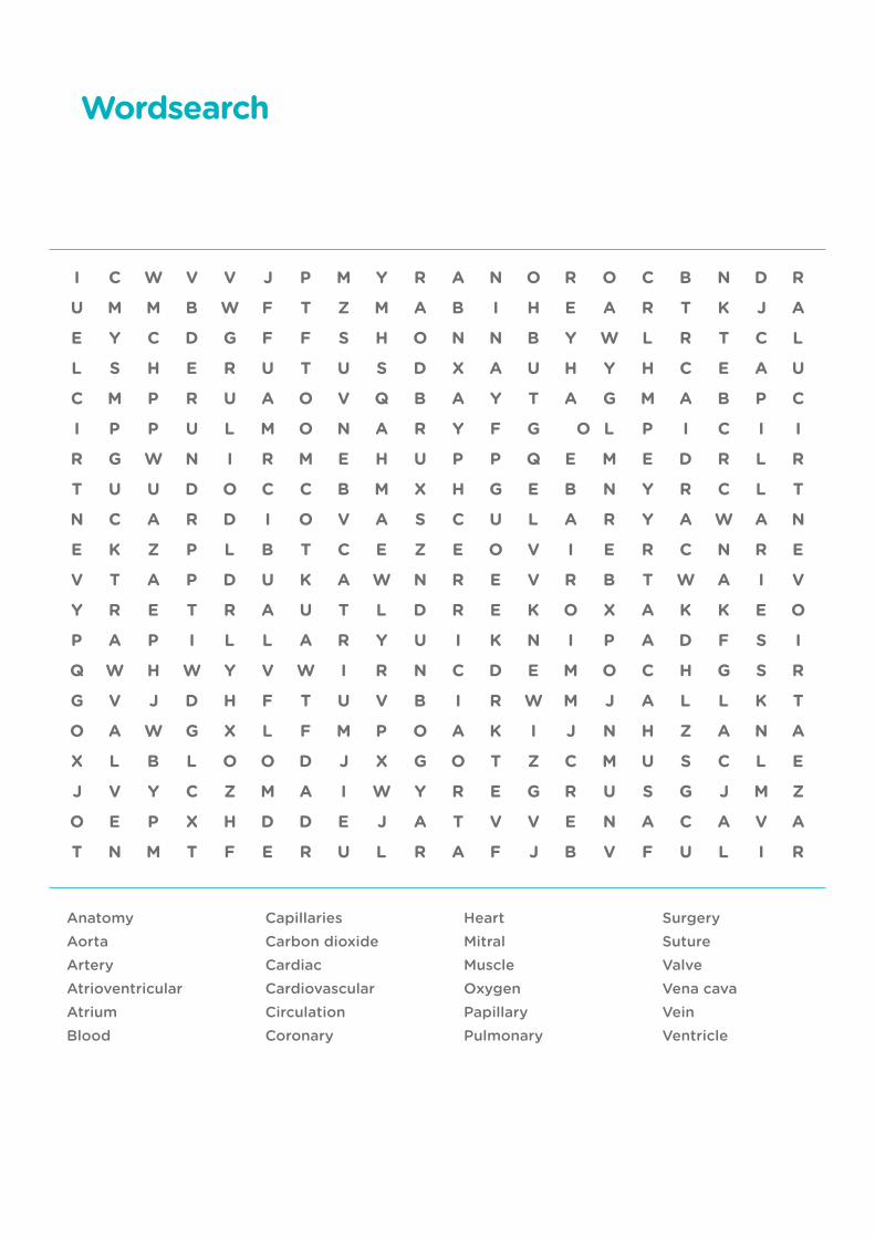
Wordsearch
AnatomyAorta ArteryAtrioventricularAtriumBlood
CapillariesCarbon dioxideCardiacCardiovascularCirculationCoronary
HeartMitralMuscleOxygenPapillaryPulmonary
SurgerySutureValveVena cavaVeinVentricle

40 of 46
Crossword
Across3. The ventricle that pumps blood to the lungs
5. The system that carries blood around the body
6. Largest artery in the body
8. The gas absorbed by the blood in the lungs
12. Blood vessels with muscular walls carrying high pressure blood
13. Relating to the heart
15. The walls of the heart are mostly made of this tissue
Down1. Main pumping chambers of the heart
2. Relating to the lungs
4. Artery supplying the heart
5. Tiny blood vessel in the tissues
7. Atrium which receives blood from the lungs
9. Large vein that returns blood to the right atrium of the heart
10. Prevents blood flowing backwards through the heart or veins
11. These carry blood back to the heart
14. Liquid that carries essential nutrients and gases around the body
9.
6.
5.
3.
1. 2.
4.
8.
10.
12.
15.
14.
11.
7.
13.
www.bbsrc.ac.uk 2 of 11
Teacher
www.bbsrc.ac.uk
Teacher
41 of 46
The walls of the ventricle push the blood out of the heart through the pulmonary artery.
Blood returns from the body to the heart along the vena cava.
Blood leaves the heart via the aorta and goes to the head and body.
Blood returns to the heart along the pulmonary vein and enters the left atrium.
The mitral valve shuts to stop blood going back into the left atrium.
The left ventricle contracts and pushes blood out of the heart and into the aorta.
The left atrium contracts and forces blood into the left ventricle through the mitral valve.
The atrium contracts to finish filling the ventricle with blood.
Blood pours into the right atrium.
The tricuspid valve shuts to stop blood going back into the atrium.
The blood travels along the pulmonary artery to the lungs where it picks up oxygen.
While the right ventricle is relaxed, blood flows into it through the open tricuspid valve.
The pulmonary valve stops blood flowing back into the right ventricle.
Answers
Card flow
www.bbsrc.ac.uk 2 of 11
Teacher
www.bbsrc.ac.uk
Teacher
42 of 46
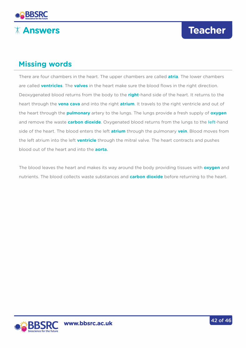
There are four chambers in the heart. The upper chambers are called atria. The lower chambers
are called ventricles. The valves in the heart make sure the blood flows in the right direction.
Deoxygenated blood returns from the body to the right-hand side of the heart. It returns to the
heart through the vena cava and into the right atrium. It travels to the right ventricle and out of
the heart through the pulmonary artery to the lungs. The lungs provide a fresh supply of oxygen
and remove the waste carbon dioxide. Oxygenated blood returns from the lungs to the left-hand
side of the heart. The blood enters the left atrium through the pulmonary vein. Blood moves from
the left atrium into the left ventricle through the mitral valve. The heart contracts and pushes
blood out of the heart and into the aorta.
The blood leaves the heart and makes its way around the body providing tissues with oxygen and
nutrients. The blood collects waste substances and carbon dioxide before returning to the heart.
Answers
Missing words
www.bbsrc.ac.uk 2 of 11
Teacher
www.bbsrc.ac.uk
Teacher
43 of 46
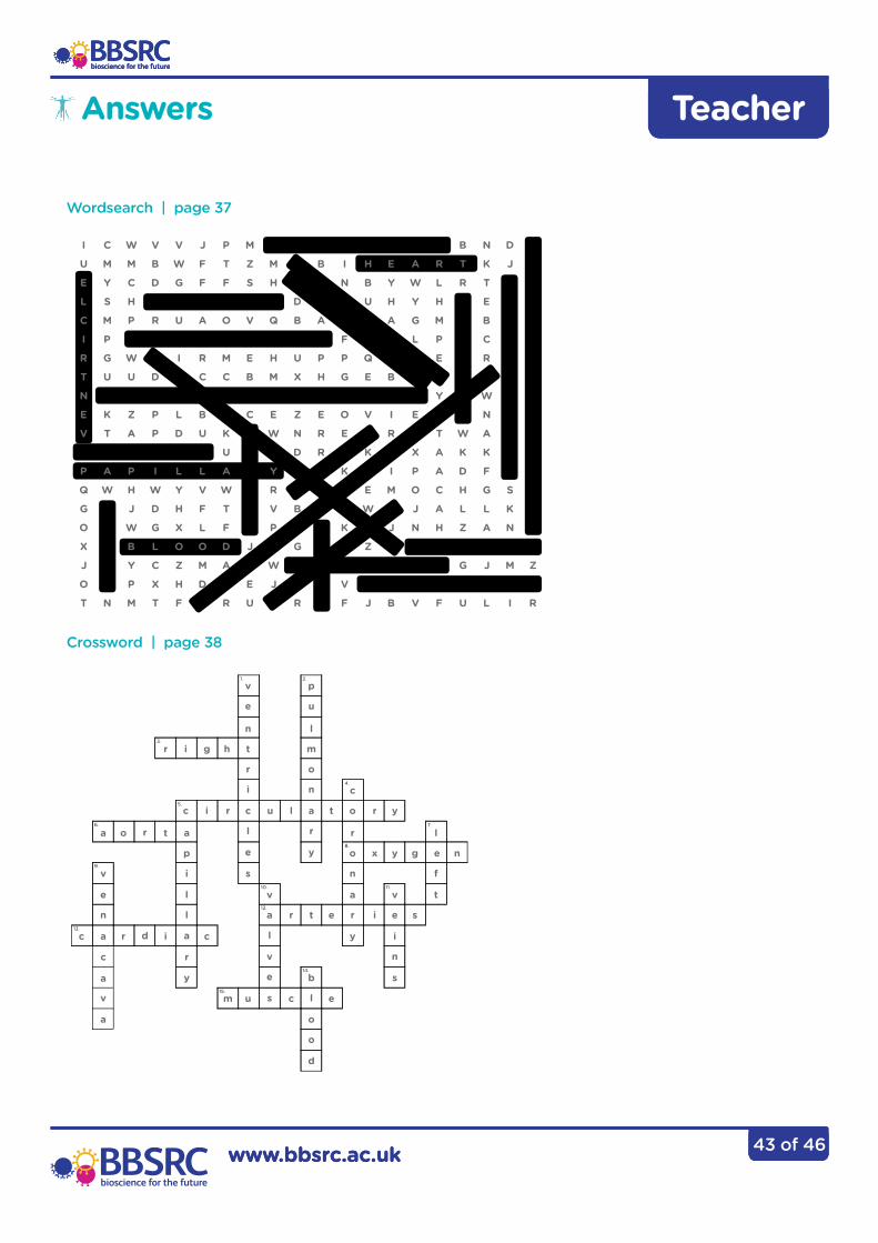
Answers
Wordsearch | page 37
Crossword | page 38
9.
6.
5.
3.
1. 2.
4.
8.
10.
12.
15.
14.
11.
7.
13.
r i g h t m
a r l
v
v p
e u
n l
c
r o
p o
a
b
v
i n
i nv f
l
l
a
y
e
y e
c
u
r
o
c u t
e
c
e
i l
c
i
t
r
r
m
a
a
a
a
c
a
t
l ae t
v
o
i
r
i
x
s
g n
y
l r
l r
r
n
c
v
e
o
n
e y
y
s
d
r
d
s
s
o
44 of 46
Glossary
Accommodation The ability to focus objects which are at different distances from the eye.
Aorta Largest blood vessel exiting the heart. Carries blood to the head and body.
Artery Blood vessel with thick muscular wall that carries blood away from the heart.
Atrioventricular Either of two heart valves through which blood flows from the atria to the ventricles; prevents return of blood to the atrium.
Atria The thin-walled upper chambers of the heart that pump blood into the ventricles. Consists of right atrium and left atrium.
Blood The fluid found in the circulatory system that carries red blood cells, white blood cells, platelets, waste and nutrients such as carbon dioxide, oxygen and glucose.
Capillaries Smallest blood vessels with very thin walls so blood can exchange dissolved food and gases with cells easily.
Cardiac Relating to the heart
Cardiovascular system The heart and the blood vessels through which blood is pumped and circulated around the body.
Coronary arteries The arteries that supply the heart with oxygenated blood.
Mitral valve A valve in the heart that is situated between the left atrium and the left ventricle, also known as the bicuspid valve.
Papillary muscles These anchor the heart valves to prevent them turning inside out.
Pulmonary Relating to the lungs.
Suture A method of joining the edges of a wound from injury or surgery, e.g. by sewing or stitching together skin, internal organs, blood vessels, and all other tissues.
45 of 46
Glossary
Valve Controls the flow of a fluid so it moves in one direction.
Vena cava The large blood vessel which receives blood from the body and head and returns it to the right atrium of the heart.
Vein Large blood vessel that carries blood back to the heart.
Ventricles The muscular lower chambers of the heart that pump blood into the pulmonary artery and aorta. Consists of left ventricle and right ventricle.
www.bbsrc.ac.uk 2 of 11
Teacher
www.bbsrc.ac.uk
Teacher
46 of 46
Author: Tristan MacLean
Copyright: © BBSRC 2014.
Design: Creative Sponge, www.creativesponge.co.uk
The materials in this resource may be reproduced for teaching purposes in universities, research institutes, schools and colleges provided that the copyright of the BBSRC and sources of the original material are acknowledged. No copies may be sold for gain.
Feedback: Please provide us with feedback so we can improve this practical guide. Email: [email protected]
Heart Surgery and Dissection