Embed Size (px)
Citation preview
Helicobacter pylori Resists the Antimicrobial Activity of Calprotectinvia Lipid A Modification and Associated Biofilm Formation
Jennifer A. Gaddy,a,b Jana N. Radin,b* Thomas W. Cullen,e* Walter J. Chazin,c,f Eric P. Skaar,a,d M. Stephen Trent,g
Holly M. S. Algooda,b,d
Veterans Affairs Tennessee Valley Healthcare Services, Nashville, Tennessee, USAa; Department of Medicine,b Department of Biochemistry,c and Department of Pathology,Microbiology and Immunology,d Vanderbilt University, Nashville, Tennessee, USA; Institute of Cellular and Molecular Biology, University of Texas at Austin, Austin, Texas,USAe; Center for Structural Biology, Vanderbilt University, Nashville, Tennessee, USAf; Department of Infectious Diseases, University of Georgia, Athens, Georgia, USAg
* Present address: Jana N. Radin, Department of Microbiology, University of Illinois, Urbana, Illinois, USA; Thomas W. Cullen, Novartis Institutes for BioMedical Research, Cambridge,Massachusetts, USA.
ABSTRACT Helicobacter pylori is one of several pathogens that persist within the host despite a robust immune response. H. py-lori elicits a proinflammatory response from host epithelia, resulting in the recruitment of immune cells which manifests as gas-tritis. Relatively little is known about how H. pylori survives antimicrobials, including calprotectin (CP), which is present duringthe inflammatory response. The data presented here suggest that one way H. pylori survives the nutrient sequestration by CP isthrough alteration of its outer membrane. CP-treated H. pylori demonstrates increased bacterial fitness in response to furthercoculture with CP. Moreover, CP-treated H. pylori cultures form biofilms and demonstrate decreased cell surface hydrophobic-ity. In response to CP, the H. pylori Lpx lipid A biosynthetic enzymes are not fully functional. The lipid A molecules observed inH. pylori cultures treated with CP indicate that the LpxF, LpxL, and LpxR enzyme functions are perturbed. Transcriptional anal-ysis of lpxF, lpxL, and lpxR indicates that metal restriction by CP does not control this pathway through transcriptional regula-tion. Analyses of H. pylori lpx mutants reveal that loss of LpxF and LpxL results in increased fitness, similar to what is observedin the presence of CP; moreover, these mutants have significantly increased biofilm formation and reduced cell surface hydro-phobicity. Taken together, these results demonstrate a novel mechanism of H. pylori resistance to the antimicrobial activity ofCP via lipid A modification strategies and resulting biofilm formation.
IMPORTANCE Helicobacter pylori evades recognition of the host’s immune system by modifying the lipid A component of lipo-polysaccharide. These results demonstrate for the first time that the lipid A modification pathway is influenced by the host’s nu-tritional immune response. H. pylori’s exposure to the host Mn- and Zn-binding protein calprotectin perturbs the function of 3enzymes involved in the lipid A modification pathway. Moreover, CP treatment of H. pylori, or mutants with an altered lipid A,exhibit increased bacterial fitness and increased biofilm formation. This suggests that H. pylori modifies its cell surface structureto survive under the stress imposed by the host immune response. These results provide new insights into the molecular mecha-nisms that influence the biofilm lifestyle and how endotoxin modification, which renders H. pylori resistant to cationic antimi-crobial peptides, can be inactivated in response to sequestration of nutrient metals.
Received 7 August 2015 Accepted 2 November 2015 Published 8 December 2015
Citation Gaddy JA, Radin JN, Cullen TW, Chazin WJ, Skaar EP, Trent MS, Algood HMS. 2015. Helicobacter pylori resists the antimicrobial activity of calprotectin via lipid Amodification and associated biofilm formation. mBio 6(6):e01349-15. doi:10.1128/mBio.01349-15.
Invited Editor Jason W. Rosch, St. Jude Children’s Research Hospital Editor Larry S. McDaniel, University of Mississippi Medical Center
Copyright © 2015 Gaddy et al. This is an open-access article distributed under the terms of the Creative Commons Attribution-Noncommercial-ShareAlike 3.0 Unportedlicense, which permits unrestricted noncommercial use, distribution, and reproduction in any medium, provided the original author and source are credited.
Address correspondence to Holly M. S. Algood, [email protected].
Helicobacter pylori is a Gram-negative microaerophilic bacte-rium that infects about half of the world’s population (1–3).
Colonization with H. pylori results in both acute and chronic gas-tritis, and in a percentage of individuals it can lead to peptic ulcerdisease or the development of stomach cancer (4, 5). As such,H. pylori has been classified by the World Health Organization asa class I carcinogen. The inflammatory response provoked byH. pylori includes infiltration of immune cells from both arms ofthe immune system into the lamina propria and gastric mucosa (6,7). The inflammatory cascade is likely initiated by early interac-tions of H. pylori with the gastric epithelial cells and production ofchemokines and cytokines, which elicit the recruitment of neutro-phils and macrophages to the site of infection. Upon activation of
an adaptive immune response, a strong T cell-mediated responseculminates in the production of proinflammatory cytokines, in-cluding gamma interferon (IFN-�) and interleukin-17 (IL-17) (6,8–18). This cytokine network results in further recruitment ofinnate immunity cells to the gastric mucosa, especially neutro-phils. Despite this robust immune response, H. pylori persistswithin the gastric niche to promote a chronic infection for theduration of the host’s life.
In order to survive the antimicrobial peptide response imposedby the vertebrate host, many Gram-negative bacteria utilize lipidA modifications (19–21). Bacteria accomplish this via severalstrategies, including adding positively charged moieties, such asphosphoethanolamine or L-4-aminoarabinose to the outer leaflet,
RESEARCH ARTICLE crossmark
November/December 2015 Volume 6 Issue 6 e01349-15 ® mbio.asm.org 1
m
bio.asm.org
on June 7, 2018 - Published by
mbio.asm
.orgD
ownloaded from
removing phosphate groups from the lipid A backbone, or chang-ing the acylation pattern (22). Altering the bacterial surface chargereduces the binding affinity of cationic antimicrobial peptides(CAMPs) with the outer membrane. CAMPs are positivelycharged peptides produced by the host innate immune system thatact by binding to bacteria through charged surface features, form-ing pores and thereby disrupting membrane integrity. For exam-ple, the enzyme responsible for dephosphorylation of the lipid A4=-phosphate group in H. pylori, LpxF, is partially responsible forthe resistance to the cationic antimicrobial peptide polymyxin(23). Thus, resistance to CAMPs via lipid A modification presentsan important mechanism by which bacteria evade the host im-mune response.
An important antimicrobial factor that is also a component ofthe host innate immune system is calprotectin (CP). CP, a het-erodimer of S100A8 and S100A9 subunits (also known as Mrp8/14, calgranulin A/B, and cystic fibrosis antigen), comprises about50% of the neutrophil’s cytoplasmic protein content and is a crit-ical component of the host nutrient-withholding process termednutritional immunity (24, 25). To prevent infection with patho-genic organisms, humans and other mammals restrict access toessential metals through nutritional immunity (26). It is clear thatnutrient limitation by the host and nutrient acquisition by bacte-ria are crucial processes in the pathogenesis of infectious diseases.CP binds Mn and Zn with high affinity, which starves bacteria ofthese essential nutrient transition metals, creating a Mn- and Zn-limited environment. CP has two transition metal-binding sites:site 1 (S1; with six His) binds Mn and Zn, and site 2 (S2; with threeHis and three Asp) binds Zn only (27, 28). Previous reports haveindicated that CP exhibits antimicrobial activity against numer-ous Gram-negative and Gram-positive pathogens (27, 29–36). Ex-pression of CP subunits S100A8 and S100A9 increases in inflamedgastric tissues of H. pylori-infected individuals. Recent work hasdemonstrated that CP inhibits H. pylori growth and has the capac-ity to alter the activity of the major inflammatory virulence factor,the cag type IV secretion system (37). However, H. pylori persistsin the gastric niche to promote chronic disease in the face of arobust immune response.
Work presented here demonstrates that H. pylori alters its lipidA molecules in response to CP, a phenotype that results in in-creased biofilm formation and increased bacterial fitness in thepresence of CP. These results also demonstrate that CP-mediatednutrient restriction is responsible for the modification of lipid A,which mirrors modifications that exist in the context of LpxF in-activation, such as increased phosphate decoration of the outermembrane, increased bacterial fitness, and increased biofilm for-mation. Taken together, the data show that CP restricts the activ-ity of LpxF (the 4=-phosphatase) and LpxL (an inner membraneacyltransferase) to promote resistance to CP by both inducingbiofilm formation and outer membrane modification. Thus, thiswork indicates that H. pylori possesses strategies to circumvent theantimicrobial activity of CP and persist in its host.
RESULTSH. pylori develops resistance to CP with exposure to subinhibi-tory doses. Transition metals are essential micronutrients re-quired by all living organisms. In response to infection, the verte-brate host produces antimicrobial proteins, such as calprotectin,lipocalin, lactoferrin, and transferrin, which sequester solublemetal ions (26, 31, 37, 38). In response to this nutritional immu-
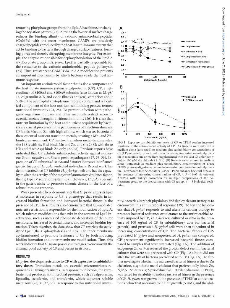
nity, bacteria alter their physiology and deploy elegant strategies tocircumvent this antimicrobial response (39). To test the hypoth-esis that H. pylori responds to and alters its cellular biology topromote bacterial resistance or tolerance to the antimicrobial ac-tivity imposed by CP, H. pylori was cultured in vitro in the pres-ence of 100 �g/ml of CP (a subinhibitory dose for bacterialgrowth), and pretreated H. pylori cells were then subcultured inincreasing concentrations of CP. The bacterial fitness of CP-pretreated H. pylori and nonpretreated H. pylori was compared.CP pretreatment significantly increased bacterial fitness com-pared to samples that were untreated (Fig. 1A). The addition ofexogenous Zn or Mn reversed the growth defect seen in bacterialcultures that were not pretreated with CP (Fig. 1A), but it did notalter the growth of bacteria pretreated with CP (Fig. 1A). To fur-ther investigate whether the increased bacterial fitness is due to Znchelation, a synthetic metal chelator that preferentially binds Zn,N,N,N=,N=-tetrakis(2-pyridylmethyl) ethylenediamine (TPEN),was tested for its ability to induce increased fitness in the presenceof CP. H. pylori was grown in the presence of TPEN at concentra-tions below that necessary to inhibit growth (5 �M), and the abil-
FIG 1 Exposure to subinhibitory levels of CP or TPEN confers increasedresistance to the antimicrobial activity of CP. (A) Bacteria were cultured inmedium alone (untreated) or medium plus subinhibitory concentrations ofCP (CP pretreated), prior to culture in increasing concentrations of calprotec-tin in medium alone or medium supplemented with 100 �M Zn chloride (�Zn) or 100 �M Mn chloride (� Mn). (B) Bacteria were cultured in mediumalone (untreated) or medium plus subinhibitory concentrations of TPEN(TPEN pretreated), prior to culture in increasing concentrations of calprotec-tin. Preexposure to zinc chelators (CP or TPEN) enhance bacterial fitness inthe presence of increasing concentrations of CP. *, P � 0.05 via one-wayANOVA with Tukey’s correction for multiple comparisons of the no-treatment group to the pretreatment with CP group. n � 3 biological repli-cates.
Gaddy et al.
2 ® mbio.asm.org November/December 2015 Volume 6 Issue 6 e01349-15
m
bio.asm.org
on June 7, 2018 - Published by
mbio.asm
.orgD
ownloaded from
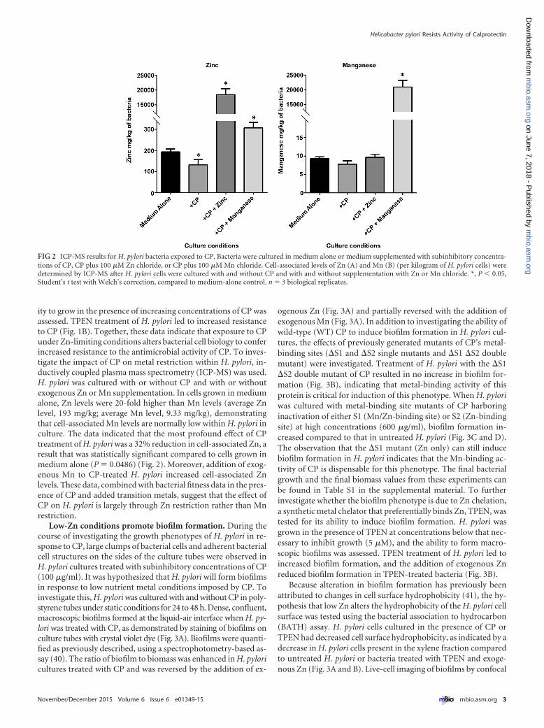
ity to grow in the presence of increasing concentrations of CP wasassessed. TPEN treatment of H. pylori led to increased resistanceto CP (Fig. 1B). Together, these data indicate that exposure to CPunder Zn-limiting conditions alters bacterial cell biology to conferincreased resistance to the antimicrobial activity of CP. To inves-tigate the impact of CP on metal restriction within H. pylori, in-ductively coupled plasma mass spectrometry (ICP-MS) was used.H. pylori was cultured with or without CP and with or withoutexogenous Zn or Mn supplementation. In cells grown in mediumalone, Zn levels were 20-fold higher than Mn levels (average Znlevel, 193 mg/kg; average Mn level, 9.33 mg/kg), demonstratingthat cell-associated Mn levels are normally low within H. pylori inculture. The data indicated that the most profound effect of CPtreatment of H. pylori was a 32% reduction in cell-associated Zn, aresult that was statistically significant compared to cells grown inmedium alone (P � 0.0486) (Fig. 2). Moreover, addition of exog-enous Mn to CP-treated H. pylori increased cell-associated Znlevels. These data, combined with bacterial fitness data in the pres-ence of CP and added transition metals, suggest that the effect ofCP on H. pylori is largely through Zn restriction rather than Mnrestriction.
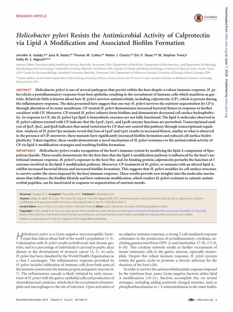
Low-Zn conditions promote biofilm formation. During thecourse of investigating the growth phenotypes of H. pylori in re-sponse to CP, large clumps of bacterial cells and adherent bacterialcell structures on the sides of the culture tubes were observed inH. pylori cultures treated with subinhibitory concentrations of CP(100 �g/ml). It was hypothesized that H. pylori will form biofilmsin response to low nutrient metal conditions imposed by CP. Toinvestigate this, H. pylori was cultured with and without CP in poly-styrene tubes under static conditions for 24 to 48 h. Dense, confluent,macroscopic biofilms formed at the liquid-air interface when H. py-lori was treated with CP, as demonstrated by staining of biofilms onculture tubes with crystal violet dye (Fig. 3A). Biofilms were quanti-fied as previously described, using a spectrophotometry-based as-say (40). The ratio of biofilm to biomass was enhanced in H. pyloricultures treated with CP and was reversed by the addition of ex-
ogenous Zn (Fig. 3A) and partially reversed with the addition ofexogenous Mn (Fig. 3A). In addition to investigating the ability ofwild-type (WT) CP to induce biofilm formation in H. pylori cul-tures, the effects of previously generated mutants of CP’s metal-binding sites (�S1 and �S2 single mutants and �S1 �S2 doublemutant) were investigated. Treatment of H. pylori with the �S1�S2 double mutant of CP resulted in no increase in biofilm for-mation (Fig. 3B), indicating that metal-binding activity of thisprotein is critical for induction of this phenotype. When H. pyloriwas cultured with metal-binding site mutants of CP harboringinactivation of either S1 (Mn/Zn-binding site) or S2 (Zn-bindingsite) at high concentrations (600 �g/ml), biofilm formation in-creased compared to that in untreated H. pylori (Fig. 3C and D).The observation that the �S1 mutant (Zn only) can still inducebiofilm formation in H. pylori indicates that the Mn-binding ac-tivity of CP is dispensable for this phenotype. The final bacterialgrowth and the final biomass values from these experiments canbe found in Table S1 in the supplemental material. To furtherinvestigate whether the biofilm phenotype is due to Zn chelation,a synthetic metal chelator that preferentially binds Zn, TPEN, wastested for its ability to induce biofilm formation. H. pylori wasgrown in the presence of TPEN at concentrations below that nec-essary to inhibit growth (5 �M), and the ability to form macro-scopic biofilms was assessed. TPEN treatment of H. pylori led toincreased biofilm formation, and the addition of exogenous Znreduced biofilm formation in TPEN-treated bacteria (Fig. 3B).
Because alteration in biofilm formation has previously beenattributed to changes in cell surface hydrophobicity (41), the hy-pothesis that low Zn alters the hydrophobicity of the H. pylori cellsurface was tested using the bacterial association to hydrocarbon(BATH) assay. H. pylori cells cultured in the presence of CP orTPEN had decreased cell surface hydrophobicity, as indicated by adecrease in H. pylori cells present in the xylene fraction comparedto untreated H. pylori or bacteria treated with TPEN and exoge-nous Zn (Fig. 3A and B). Live-cell imaging of biofilms by confocal
FIG 2 ICP-MS results for H. pylori bacteria exposed to CP. Bacteria were cultured in medium alone or medium supplemented with subinhibitory concentra-tions of CP, CP plus 100 �M Zn chloride, or CP plus 100 �M Mn chloride. Cell-associated levels of Zn (A) and Mn (B) (per kilogram of H. pylori cells) weredetermined by ICP-MS after H. pylori cells were cultured with and without CP and with and without supplementation with Zn or Mn chloride. *, P � 0.05,Student’s t test with Welch’s correction, compared to medium-alone control. n � 3 biological replicates.
Helicobacter pylori Resists Activity of Calprotectin
November/December 2015 Volume 6 Issue 6 e01349-15 ® mbio.asm.org 3
m
bio.asm.org
on June 7, 2018 - Published by
mbio.asm
.orgD
ownloaded from
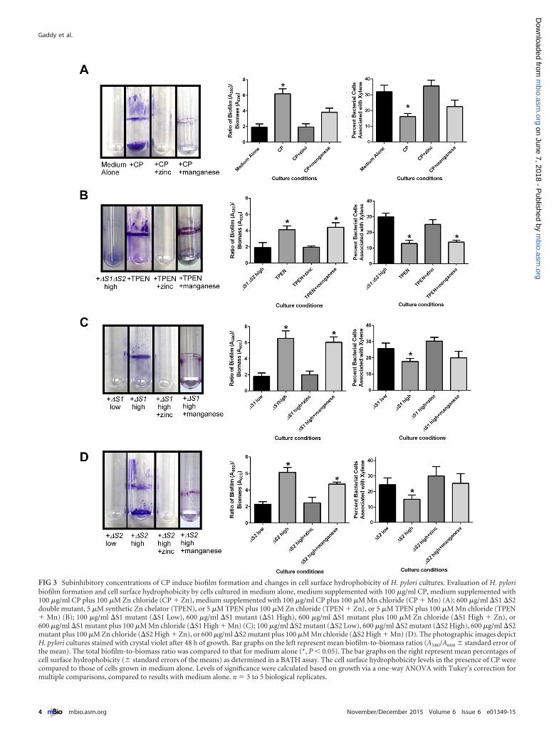
FIG 3 Subinhibitory concentrations of CP induce biofilm formation and changes in cell surface hydrophobicity of H. pylori cultures. Evaluation of H. pyloribiofilm formation and cell surface hydrophobicity by cells cultured in medium alone, medium supplemented with 100 �g/ml CP, medium supplemented with100 �g/ml CP plus 100 �M Zn chloride (CP � Zn), medium supplemented with 100 �g/ml CP plus 100 �M Mn chloride (CP � Mn) (A); 600 �g/ml �S1 �S2double mutant, 5 �M synthetic Zn chelator (TPEN), or 5 �M TPEN plus 100 �M Zn chloride (TPEN � Zn), or 5 �M TPEN plus 100 �M Mn chloride (TPEN� Mn) (B); 100 �g/ml �S1 mutant (�S1 Low), 600 �g/ml �S1 mutant (�S1 High), 600 �g/ml �S1 mutant plus 100 �M Zn chloride (�S1 High � Zn), or600 �g/ml �S1 mutant plus 100 �M Mn chloride (�S1 High � Mn) (C); 100 �g/ml �S2 mutant (�S2 Low), 600 �g/ml �S2 mutant (�S2 High), 600 �g/ml �S2mutant plus 100 �M Zn chloride (�S2 High � Zn), or 600 �g/ml �S2 mutant plus 100 �M Mn chloride (�S2 High � Mn) (D). The photographic images depictH. pylori cultures stained with crystal violet after 48 h of growth. Bar graphs on the left represent mean biofilm-to-biomass ratios (A580/A600 � standard error ofthe mean). The total biofilm-to-biomass ratio was compared to that for medium alone (*, P � 0.05). The bar graphs on the right represent mean percentages ofcell surface hydrophobicity (� standard errors of the means) as determined in a BATH assay. The cell surface hydrophobicity levels in the presence of CP werecompared to those of cells grown in medium alone. Levels of significance were calculated based on growth via a one-way ANOVA with Tukey’s correction formultiple comparisons, compared to results with medium alone. n � 3 to 5 biological replicates.
Gaddy et al.
4 ® mbio.asm.org November/December 2015 Volume 6 Issue 6 e01349-15
m
bio.asm.org
on June 7, 2018 - Published by
mbio.asm
.orgD
ownloaded from
laser scanning microscopy indicated that the tertiary structure ofthe CP-induced H. pylori biofilm was thicker than that of H. pylorigrown in the absence of CP or in the presence of CP plus anexogenous source of nutrient Zn (see Fig. S1 in the supplementalmaterial).
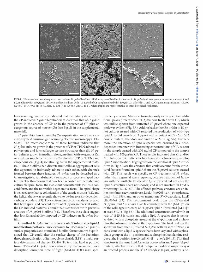
H. pylori biofilms induced by Zn sequestration were also visu-alized by field emission gun scanning electron microscopy (FEG-SEM). The microscopic view of these biofilms indicated thatH. pylori cultures grown in the presence of CP or TPEN adhered topolystyrene and formed larger tertiary structures than did H. py-lori cultures grown in medium alone, medium with exogenous Zn,or medium supplemented with a Zn chelator (CP or TPEN) andexogenous Zn (Fig. 4; see also Fig. S2 in the supplemental mate-rial). These biofilms had discrete multicellular aggregates of cellsthat appeared to intimately adhere to each other, with channelsformed between these features. H. pylori can be described as aGram-negative, spiral-shaped (S-shaped) or coccus-shaped bac-terium. The three forms that have been reported are the viable andculturable spiral form, the viable but nonculturable (VBNC) coc-coid form, and the nonviable degenerative form. The spiral shapeis believed to enhance colonization of the gastric mucosa (42), andthe helical shape was recently shown to be due to a Zn-dependentcarboxypeptidase (43). The electron microscopy analyses revealedthat both spiral and coccoid forms of H. pylori are present withinthe CP-induced biofilms, a result that is consistent with previousanalyses of H. pylori biofilms (44). Therefore, these data suggestthat low Zn availability imposed by CP induces an H. pylori bio-film.
Growth of H. pylori in the presence of CP inhibits the lipid Amodification pathway. Since exposure to CP changed H. pylori’ssurface properties and stimulated biofilm formation, we hypoth-esized that CP could alter the surface charge of the bacteriumthrough modification of its lipopolysaccharide (LPS), a major sur-face determinant of charge (45, 46). To test this, lipid A purifiedfrom CP-treated H. pylori was evaluated by matrix-assisted laserdesorption ionization–time of flight (MALDI-TOF) mass spec-
trometry analysis. Mass spectrometry analysis revealed two addi-tional peaks present when H. pylori was treated with CP, whichwas unlike spectra from untreated H. pylori where one expectedpeak was evident (Fig. 5A). Adding back either Zn or Mn to H. py-lori cultures treated with CP restored the production of wild-typelipid A, as did growth of H. pylori with a mutant of CP (�S1 �S2double mutant) that does not bind Zn or Mn (Fig. 5A). Further-more, the alteration of lipid A species was enriched in a dose-dependent manner with increasing concentrations of CP, as seenin the sample treated with 200 �g/ml CP compared to the sampletreated with 100 �g/ml CP. These results indicated that Zn and/orMn chelation by CP alters the biochemical machinery required forlipid A modification. Highlighted on the additional lipid A struc-tures in Fig. 5B are the enzymes that could account for the struc-tural features found on lipid A from the H. pylori cultures treatedwith CP. This result was specific to CP treatment of H. pylori,rather than a general stress response, because treatment of H. py-lori with the synthetic Fe chelator 2,2=-dipyridyl did not alter thelipid A structure (data not shown) and is not involved in lipid Aprocessing (23, 47–50). The affected pathway enzymes are an in-ner membrane acyltransferase, LpxL (Jhp0265), a 4=-phosphatase,LpxF (Jhp1486), and an outer membrane 3=-O-deacylase, LpxR(Jhp0634) (23). The predominant peak from the CP-treatedH. pylori lipid A is at m/z 1546.8, consistent with the [M-H]� ionof the wild-type structure of H. pylori lipid A (predicted [M-H]�
at m/z 1547.1) (Fig. 5B). The additional structure observed with anm/z of 1825.3 is consistent with a lipid A species that is penta-acylated with a phosphate group at the 4=-position and a phos-phoethanolamine residue at the 1-position. The final peak on thespectrum from the CP-treated H. pylori with an m/z of 2092.5 isconsistent with a lipid A species that is hexa-acylated with a phos-phate group at the 4=-position and a phosphoethanolamine resi-due at the 1-position (predicted [M-H]� at m/z 2091.5). This finalstructure is the same lipid A species observed in an H. pylori �lpxFmutant, which is evidence that the lipid A modification pathway isan ordered process and the 3=-O-deacylase (LpxR) activity is de-
FIG 4 CP-dependent metal sequestration induces H. pylori biofilms. SEM analyses of biofilm formation in H. pylori cultures grown in medium alone (A andD), medium with 100 �g/ml of CP (B and E), medium with 100 �g/ml of CP supplemented with 100 �M Zn chloride (D and F). Original magnification, 1,000(A to C) or 7,000 (D to F). Bars, 40 �m (A to C) or 5 �m (D to F). Micrographs are representative of three biological replicates.
Helicobacter pylori Resists Activity of Calprotectin
November/December 2015 Volume 6 Issue 6 e01349-15 ® mbio.asm.org 5
m
bio.asm.org
on June 7, 2018 - Published by
mbio.asm
.orgD
ownloaded from
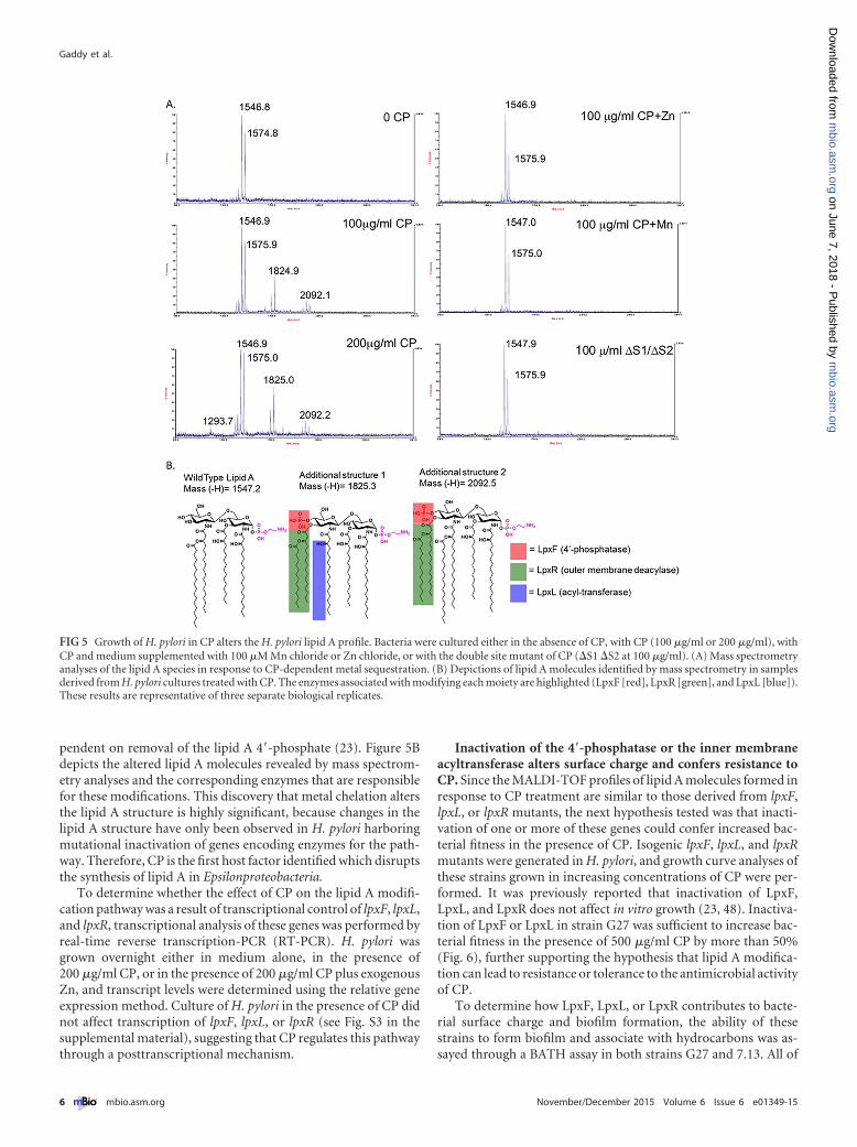
pendent on removal of the lipid A 4=-phosphate (23). Figure 5Bdepicts the altered lipid A molecules revealed by mass spectrom-etry analyses and the corresponding enzymes that are responsiblefor these modifications. This discovery that metal chelation altersthe lipid A structure is highly significant, because changes in thelipid A structure have only been observed in H. pylori harboringmutational inactivation of genes encoding enzymes for the path-way. Therefore, CP is the first host factor identified which disruptsthe synthesis of lipid A in Epsilonproteobacteria.
To determine whether the effect of CP on the lipid A modifi-cation pathway was a result of transcriptional control of lpxF, lpxL,and lpxR, transcriptional analysis of these genes was performed byreal-time reverse transcription-PCR (RT-PCR). H. pylori wasgrown overnight either in medium alone, in the presence of200 �g/ml CP, or in the presence of 200 �g/ml CP plus exogenousZn, and transcript levels were determined using the relative geneexpression method. Culture of H. pylori in the presence of CP didnot affect transcription of lpxF, lpxL, or lpxR (see Fig. S3 in thesupplemental material), suggesting that CP regulates this pathwaythrough a posttranscriptional mechanism.
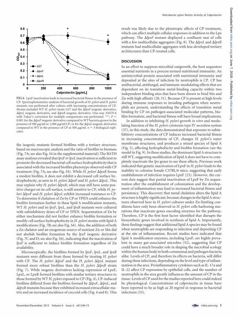
Inactivation of the 4=-phosphatase or the inner membraneacyltransferase alters surface charge and confers resistance toCP. Since the MALDI-TOF profiles of lipid A molecules formed inresponse to CP treatment are similar to those derived from lpxF,lpxL, or lpxR mutants, the next hypothesis tested was that inacti-vation of one or more of these genes could confer increased bac-terial fitness in the presence of CP. Isogenic lpxF, lpxL, and lpxRmutants were generated in H. pylori, and growth curve analyses ofthese strains grown in increasing concentrations of CP were per-formed. It was previously reported that inactivation of LpxF,LpxL, and LpxR does not affect in vitro growth (23, 48). Inactiva-tion of LpxF or LpxL in strain G27 was sufficient to increase bac-terial fitness in the presence of 500 �g/ml CP by more than 50%(Fig. 6), further supporting the hypothesis that lipid A modifica-tion can lead to resistance or tolerance to the antimicrobial activityof CP.
To determine how LpxF, LpxL, or LpxR contributes to bacte-rial surface charge and biofilm formation, the ability of thesestrains to form biofilm and associate with hydrocarbons was as-sayed through a BATH assay in both strains G27 and 7.13. All of
FIG 5 Growth of H. pylori in CP alters the H. pylori lipid A profile. Bacteria were cultured either in the absence of CP, with CP (100 �g/ml or 200 �g/ml), withCP and medium supplemented with 100 �M Mn chloride or Zn chloride, or with the double site mutant of CP (�S1 �S2 at 100 �g/ml). (A) Mass spectrometryanalyses of the lipid A species in response to CP-dependent metal sequestration. (B) Depictions of lipid A molecules identified by mass spectrometry in samplesderived from H. pylori cultures treated with CP. The enzymes associated with modifying each moiety are highlighted (LpxF [red], LpxR [green], and LpxL [blue]).These results are representative of three separate biological replicates.
Gaddy et al.
6 ® mbio.asm.org November/December 2015 Volume 6 Issue 6 e01349-15
m
bio.asm.org
on June 7, 2018 - Published by
mbio.asm
.orgD
ownloaded from
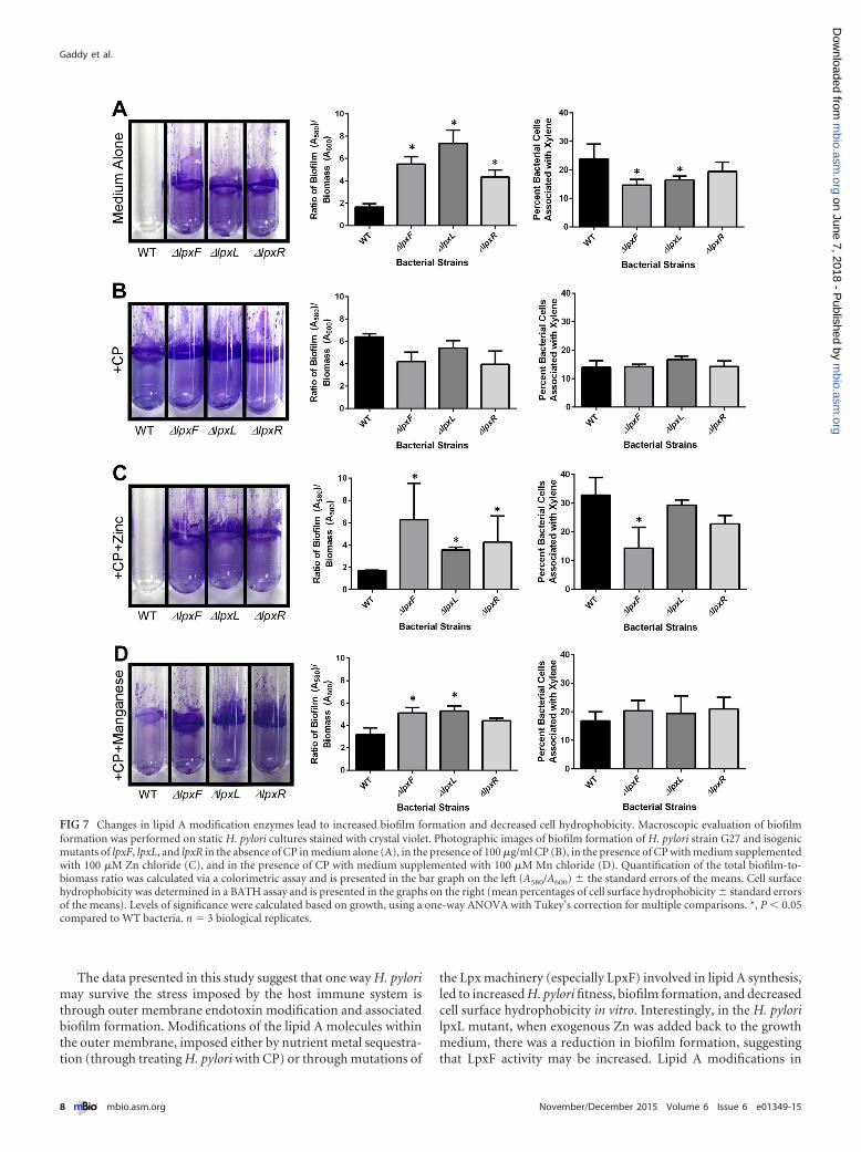
the isogenic mutants formed biofilms with a tertiary structure,based on macroscopic analysis and the ratio of biofilm to biomass(Fig. 7A; see also Fig. S4 in the supplemental material).The BATHassay analyses revealed that lpxF or lpxL inactivation is sufficient topromote the decreased bacterial cell surface hydrophobicity that isassociated with the increased biofilm phenotype observed with CPtreatment (Fig. 7A; see also Fig. S4). While H. pylori �lpxR formsa modest biofilm, it does not exhibit a decreased cell surface hy-drophobicity, as seen in H. pylori �lpxF and H. pylori �lpxL. Thismay explain why H. pylori �lpxR, which may still have some pos-itive charges on its cell surface, is still sensitive to CP, while H. py-lori �lpxF and H. pylori �lpxL exhibit increased resistance to CP.To determine if chelation of Zn by CP or TPEN could enhance thebiofilm formation further in these lipid A modification mutants,WT H. pylori and its lpxF, lpxL, and lpxR mutants were culturedwith subinhibitory doses of CP or TPEN. Sequestration of Zn byeither mechanism did not further enhance biofilm formation ormodify cell surface hydrophobicity in H. pylori strains with lipid Amodifications (Fig. 7B; see also Fig. S4). Also, the addition of botha Zn chelator and an exogenous source of nutrient Zn or Mn didnot abolish biofilm formation by the lpxF isogenic derivative(Fig. 7C and D; see also Fig. S4), indicating that the inactivation oflpxF is sufficient to induce biofilm formation regardless of Znavailability.
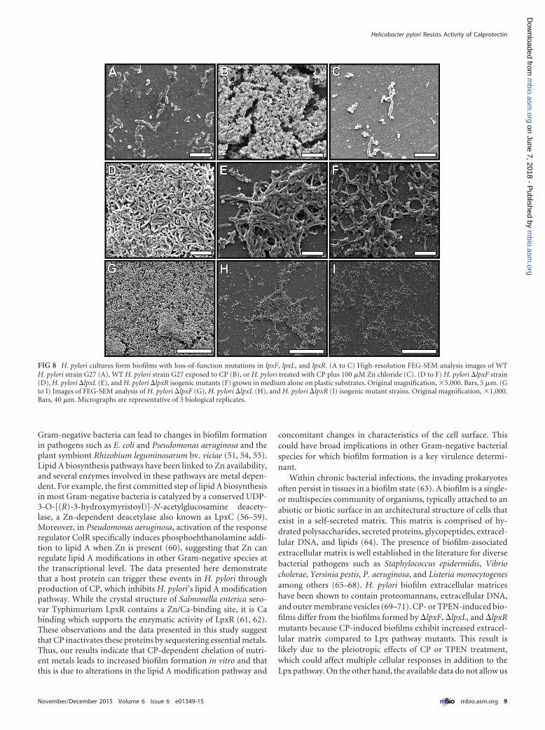
Microscopically, the biofilms formed by lpxF, lpxL, and lpxRmutants were different from those formed by treating H. pyloriwith CP. The H. pylori �lpxF and the H. pylori �lpxL mutantformed more robust biofilms than the H. pylori �lpxR strain(Fig. 7). While isogenic derivatives lacking expression of LpxF,LpxL, or LpxR formed biofilms with similar tertiary structures tothose formed by WT H. pylori exposed to CP (Fig. 4), CP-inducedbiofilms differed from the biofilms formed by �lpxF, �lpxL, and�lpxR mutants because they exhibited increased extracellular ma-trix and an increased incidence of coccoid cells (Fig. 4 and 8). This
result was likely due to the pleiotropic effects of CP treatment,which can affect multiple cellular responses in addition to the Lpxpathway. The �lpxF mutant displayed a confluent mat of cellswith a few multicellular aggregates (Fig. 8). The �lpxL and �lpxRmutants had multicellular aggregates with less-developed tertiaryarchitectures than CP-treated cells.
DISCUSSION
In an effort to suppress microbial outgrowth, the host sequestersessential nutrients in a process termed nutritional immunity. Anantimicrobial protein associated with nutritional immunity anddeposited at the sites of infection by neutrophils is CP. CP hasantibacterial, antifungal, and immune-modulating effects that aredependent on its transition metal-binding capacity within twoindependent binding sites that have been shown to bind Mn andZn with high affinity (28, 51). Because CP is present at high levelsduring immune responses to invading pathogens when neutro-phils are present, understanding the effects of transition metalbinding by CP on pathogen-associated molecular patterns, bio-film formation, and bacterial fitness will have broad implications.
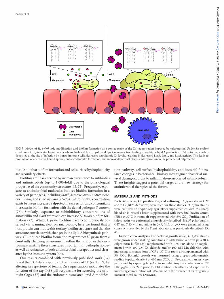
In addition to inhibiting H. pylori growth in vitro and modu-lating function of the H. pylori cytotoxin type IV secretion system(37), in this study, the data demonstrated that exposure to subin-hibitory concentrations of CP induces increased bacterial fitnessin increasing concentrations of CP, changes H. pylori’s outermembrane structures, and produces a mixed species of lipid A(Fig. 5), affecting hydrophobicity and biofilm formation (see themodel in Fig. 9). In these studies, the dominant lipid A structure isstill WT, suggesting modification of lipid A does not have to com-pletely inactivate the lpx genes to see these effects. Previous workindicated that genetic inactivation of LpxF in H. pylori leads to theinability to colonize female C57BL/6 mice, suggesting that earlyestablishment of infection requires LpxF (23). However, the cur-rent data suggest that partial inactivation of LpxF by Zn seques-tration after the establishment of colonization and the develop-ment of inflammation may lead to increased bacterial fitness andpersistence. This discovery that metal chelation alters the lipid Astructure is highly significant, because changes in the lipid A struc-tures observed here in H. pylori cultures under Zn-limiting con-ditions have only been observed in H. pylori cells harboring mu-tations that inactivate genes encoding enzymes for the pathway.Therefore, CP is the first host factor identified that disrupts thebiosynthetic genes involved in synthesis of lipid A. Importantly,these findings suggest that additional lipid A species may be foundwhen neutrophils are responding to infection and depositing CPat the site of inflammation. Recent studies have indicated thatlipid A modification enzymes, including LpxF, are highly preva-lent in many gut-associated microbes (52), suggesting that CPcould have a much broader role in shaping the microbial ecologywithin the human body in both commensal and pathogen bacteriaalike. Levels of CP, and therefore its effects on bacteria, will differduring these infections, depending on the level and type of inflam-mation in the area. Proinflammatory cytokines such as IL-17a andIL-22 affect CP expression by epithelial cells, and the number ofneutrophils in the area greatly influences the amount of CP in thetissue. Levels of CP used for the studies reported here could indeedbe physiological. Concentrations of calprotectin in tissue havebeen reported to be as high as 20 mg/ml in response to bacterialinfections (53).
FIG 6 LpxF inactivation leads to increased bacterial fitness in the presence ofCP. Spectrophotometric analysis of bacterial growth of H. pylori and H. pylorimutants was performed after culture with increasing concentrations of CP.Strains included WT H. pylori strain G27 and the �lpxF isogenic derivative,�lpxL isogenic derivative, and �lpxR isogenic derivative. One-way ANOVAwith Tukey’s correction for multiple comparisons was performed. ***, P �0.001 for the �lpxF isogenic derivative compared to WT bacteria grown in thepresence of 500 �g/ml or 1,000 �g/ml CP, or for the �lpxL isogenic derivativecompared to WT in the presence of CP at 500 �g/ml. n � 3 biological repli-cates.
Helicobacter pylori Resists Activity of Calprotectin
November/December 2015 Volume 6 Issue 6 e01349-15 ® mbio.asm.org 7
m
bio.asm.org
on June 7, 2018 - Published by
mbio.asm
.orgD
ownloaded from
The data presented in this study suggest that one way H. pylorimay survive the stress imposed by the host immune system isthrough outer membrane endotoxin modification and associatedbiofilm formation. Modifications of the lipid A molecules withinthe outer membrane, imposed either by nutrient metal sequestra-tion (through treating H. pylori with CP) or through mutations of
the Lpx machinery (especially LpxF) involved in lipid A synthesis,led to increased H. pylori fitness, biofilm formation, and decreasedcell surface hydrophobicity in vitro. Interestingly, in the H. pylorilpxL mutant, when exogenous Zn was added back to the growthmedium, there was a reduction in biofilm formation, suggestingthat LpxF activity may be increased. Lipid A modifications in
FIG 7 Changes in lipid A modification enzymes lead to increased biofilm formation and decreased cell hydrophobicity. Macroscopic evaluation of biofilmformation was performed on static H. pylori cultures stained with crystal violet. Photographic images of biofilm formation of H. pylori strain G27 and isogenicmutants of lpxF, lpxL, and lpxR in the absence of CP in medium alone (A), in the presence of 100 �g/ml CP (B), in the presence of CP with medium supplementedwith 100 �M Zn chloride (C), and in the presence of CP with medium supplemented with 100 �M Mn chloride (D). Quantification of the total biofilm-to-biomass ratio was calculated via a colorimetric assay and is presented in the bar graph on the left (A580/A600) � the standard errors of the means. Cell surfacehydrophobicity was determined in a BATH assay and is presented in the graphs on the right (mean percentages of cell surface hydrophobicity � standard errorsof the means). Levels of significance were calculated based on growth, using a one-way ANOVA with Tukey’s correction for multiple comparisons. *, P � 0.05compared to WT bacteria. n � 3 biological replicates.
Gaddy et al.
8 ® mbio.asm.org November/December 2015 Volume 6 Issue 6 e01349-15
m
bio.asm.org
on June 7, 2018 - Published by
mbio.asm
.orgD
ownloaded from
Gram-negative bacteria can lead to changes in biofilm formationin pathogens such as E. coli and Pseudomonas aeruginosa and theplant symbiont Rhizobium leguminosarum bv. viciae (51, 54, 55).Lipid A biosynthesis pathways have been linked to Zn availability,and several enzymes involved in these pathways are metal depen-dent. For example, the first committed step of lipid A biosynthesisin most Gram-negative bacteria is catalyzed by a conserved UDP-3-O-[(R)-3-hydroxymyristoyl)]-N-acetylglucosamine deacety-lase, a Zn-dependent deacetylase also known as LpxC (56–59).Moreover, in Pseudomonas aeruginosa, activation of the responseregulator ColR specifically induces phosphoehthanolamine addi-tion to lipid A when Zn is present (60), suggesting that Zn canregulate lipid A modifications in other Gram-negative species atthe transcriptional level. The data presented here demonstratethat a host protein can trigger these events in H. pylori throughproduction of CP, which inhibits H. pylori’s lipid A modificationpathway. While the crystal structure of Salmonella enterica sero-var Typhimurium LpxR contains a Zn/Ca-binding site, it is Cabinding which supports the enzymatic activity of LpxR (61, 62).These observations and the data presented in this study suggestthat CP inactivates these proteins by sequestering essential metals.Thus, our results indicate that CP-dependent chelation of nutri-ent metals leads to increased biofilm formation in vitro and thatthis is due to alterations in the lipid A modification pathway and
concomitant changes in characteristics of the cell surface. Thiscould have broad implications in other Gram-negative bacterialspecies for which biofilm formation is a key virulence determi-nant.
Within chronic bacterial infections, the invading prokaryotesoften persist in tissues in a biofilm state (63). A biofilm is a single-or multispecies community of organisms, typically attached to anabiotic or biotic surface in an architectural structure of cells thatexist in a self-secreted matrix. This matrix is comprised of hy-drated polysaccharides, secreted proteins, glycopeptides, extracel-lular DNA, and lipids (64). The presence of biofilm-associatedextracellular matrix is well established in the literature for diversebacterial pathogens such as Staphylococcus epidermidis, Vibriocholerae, Yersinia pestis, P. aeruginosa, and Listeria monocytogenesamong others (65–68). H. pylori biofilm extracellular matriceshave been shown to contain proteomannans, extracellular DNA,and outer membrane vesicles (69–71). CP- or TPEN-induced bio-films differ from the biofilms formed by �lpxF, �lpxL, and �lpxRmutants because CP-induced biofilms exhibit increased extracel-lular matrix compared to Lpx pathway mutants. This result islikely due to the pleiotropic effects of CP or TPEN treatment,which could affect multiple cellular responses in addition to theLpx pathway. On the other hand, the available data do not allow us
FIG 8 H. pylori cultures form biofilms with loss-of-function mutations in lpxF, lpxL, and lpxR. (A to C) High-resolution FEG-SEM analysis images of WTH. pylori strain G27 (A), WT H. pylori strain G27 exposed to CP (B), or H. pylori treated with CP plus 100 �M Zn chloride (C). (D to F) H. pylori �lpxF strain(D), H. pylori �lpxL (E), and H. pylori �lpxR isogenic mutants (F) grown in medium alone on plastic substrates. Original magnification, 5,000. Bars, 5 �m. (Gto I) Images of FEG-SEM analysis of H. pylori �lpxF (G), H. pylori �lpxL (H), and H. pylori �lpxR (I) isogenic mutant strains. Original magnification, 1,000.Bars, 40 �m. Micrographs are representative of 3 biological replicates.
Helicobacter pylori Resists Activity of Calprotectin
November/December 2015 Volume 6 Issue 6 e01349-15 ® mbio.asm.org 9
m
bio.asm.org
on June 7, 2018 - Published by
mbio.asm
.orgD
ownloaded from
to rule out that biofilm formation and cell surface hydrophobicityare secondary effects.
Biofilms are characterized by increased resistance to antibioticsand antimicrobials (up to 1,000-fold) due to the physiologicalproperties of the community structure (63, 72). Frequently, expo-sure to antimicrobial molecules induces biofilm formation in avariety of pathogens, including Staphylococcus aureus, Streptococ-cus mutans, and P. aeruginosa (73–75). Interestingly, a correlationexists between increased calprotectin expression and concomitantincreases in biofilm formation with the dental pathogen S. mutans(76). Similarly, exposure to subinhibitory concentrations ofamoxicillin and clarithromycin can increase H. pylori biofilm for-mation (77). While H. pylori biofilms have been previously ob-served via scanning electron microscopy, here we found that ahost protein can induce this tertiary biofilm structure and that thestructure correlates with changes in the lipid A biosynthesis path-way. CP-induced biofilm formation may protect H. pylori from aconstantly changing environment within the host or in the envi-ronment,making these structures important for pathophysiologyas well as resistance to both antimicrobial therapeutics and clear-ance by the immune system (44).
Our results combined with previously published work (37)reveal that H. pylori responds to the presence of CP (or TPEN) byaltering its repertoire of toxins. Zn sequestration modulates thefunction of the cag-T4SS pili responsible for secreting the cyto-toxin CagA (37) and the endotoxin-associated lipid A modifica-
tion pathway, cell surface hydrophobicity, and bacterial fitness.Such changes in bacterial cell biology may augment bacterial sur-vival during exposure to inflammation-associated antimicrobials.These insights suggest a potential target and a new strategy forantimicrobial therapies of the future.
MATERIALS AND METHODSBacterial strains, CP purification, and culturing. H. pylori strains G27and 7.13 (B128 derivative) were used for these studies. H. pylori strainswere cultured on tryptic soy agar plates supplemented with 5% sheepblood or in brucella broth supplemented with 10% fetal bovine serum(FBS) at 37°C in room air supplemented with 5% CO2. Purification ofcalprotectin was performed, as previously described (28). H. pylori strainsG27 and 7.13 with mutations in lpxF, lpxL, or lpxR were generated usingconstructs provided by the Trent laboratory, as previously described (23,48).
Growth curve analyses. For bacterial growth assays, H. pylori strainswere grown under shaking conditions in 60% brucella broth plus 40%calprotectin buffer (28) supplemented with 10% FBS alone or supple-mented with 100 �M Zn chloride and/or 100 �M Mn chloride, withincreasing concentrations of CP at 37°C in room air supplemented with5% CO2. Bacterial growth was measured using a spectrophotometricreading (optical density) at 600 nm (OD600). Pretreatment assays wereperformed by exposing H. pylori to subinhibitory concentrations of CP(100 �g/ml) for 24 h prior to 1:10 dilution subculture and exposure toincreasing concentrations of CP alone or in the presence of an exogenousnutrient metal source (Zn/Mn).
FIG 9 Model of H. pylori lipid modification and biofilm formation as a consequence of the Zn sequestration imposed by calprotectin. Under Zn-repleteconditions, H. pylori cytoplasmic zinc levels are high and LpxF, LpxL, and LpxR remain active, leading to wild-type lipid A production. Calprotectin, which isdeposited at the site of infection by innate immune cells, decreases cytoplasmic Zn levels, resulting in decreased LpxF, LpxL, and LpxR activity. This leads toproduction of alternative lipid A species, enhanced biofilm formation, and increased bacterial fitness and replication in the presence of calprotectin.
Gaddy et al.
10 ® mbio.asm.org November/December 2015 Volume 6 Issue 6 e01349-15
m
bio.asm.org
on June 7, 2018 - Published by
mbio.asm
.orgD
ownloaded from
Inductively coupled plasma mass spectrometry. Bacterial cells weregrown in 60% brucella broth plus 40% calprotectin buffer (28) supple-mented with 10% FBS alone or with 200 �g/ml CP, or CP plus 100 �Mexogenous metal (zinc chloride or manganese chloride) for 24 h, withshaking, at 37°C in room air supplemented with 5% CO2. Cells werepelleted at 4,000 g for 15 min before washing once with 0.5 M EDTAand three times with 5 ml of distilled water. Samples were weighed anddigested in 1 ml of 50% nitric acid overnight at 50°C. Elemental quantifi-cation was performed using a Thermo-Element 2 HR-ICPMS apparatus(Thermo, Fisher Scientific, Bremen, Germany), as previously described(30).
Biofilm assays. H. pylori biofilms were grown in 3-ml polystyrenetubes (under static conditions) in 60% brucella broth plus 40% calprotec-tin buffer (28) supplemented with 10% FBS alone or with 200 �g/ml CP,or CP plus 100 �M exogenous metal for 2 to 4 days at 37°C in room airsupplemented with 5% CO2. After biofilms were formed, they were quan-titatively analyzed, as previously described (40), by measuring the OD600,staining the biofilm with crystal violet, and resolubilizing the crystal violetin 80% ethanol–20% acetone solution before measuring the absorbance(OD) at 540 nm. The ratio of total biofilm to biomass was calculated viathe formula OD540/OD600.
BATH assay. Cell surface hydrophobicity can be determined via aBATH assay, which measures the percentage of bacterial cells that areassociated with the xylene fraction as a measure of hydrophobicity. TheH. pylori biofilm was scraped and resuspended in 1.2 ml PUM buffer(phosphate, urea, and magnesium sulfate; 0.1 M K2HPO4 · 3H2O, 50 mMKH2PO4, 30 mM urea, 0.5 M MgSO4 · 7H2O; pH 7.10), and the bacterialdensity was obtained by measuring the absorbance (OD) at 540 nm. Twohundred microliters of xylene was then added, and the sample was vor-texed vigorously and allowed to separate into aqueous and hydrocarbonfractions. The aqueous fraction was carefully pipetted out, and the bacte-rial density was measured spectrophotometrically. The percent cell sur-face hydrophobicity was determined using the following equation: {[(A540
before xylene addition) � (A540 after xylene addition)]/(A540 before xy-lene addition)} 100, as previously described (41).
Field emission gun scanning electron microscopy of biofilms. H. py-lori biofilms were imaged by FEG-SEM analysis using methods previ-ously described (40). Briefly, H. pylori biofilms were grown on poly-styrene for 48 h at 37°C in the presence of 5% CO2. Cells were fixedwith 2.0% paraformaldehyde–2.5% glutaraldehyde in 0.05 M sodiumcacodylic acid buffer for 1 h at room temperature. Samples werewashed three times with cacodylic acid buffer before sequential dehy-dration with increasing concentrations of ethanol. Samples were thendried at the critical point, mounted onto SEM stubs, painted withcolloidal silver at the sample edge, and sputter coated with 20-nm-diameter gold-palladium. Samples were visualized with an FEI Quanta250 FEG-SEM apparatus under high vacuum pressure, and micro-graphs were analyzed with ImageJ software.
Confocal laser scanning microscopy. H. pylori biofilms were grownon plastic coverslips as described above, washed three times with 1phosphate-buffered saline (PBS) and placed in 1 ml PBS before stainingwith the Live/Dead BacLight kit (Life Technologies), which is comprisedof SYTO 9 (green) and propidium iodide (red) and entails counterstain-ing with calcofluor white (blue; Sigma Aldrich). Biofilms were stainedwith 6 �l of 1.67 mM SYTO 9 and 1.67 mM propidium iodide mixture,and counterstained with 6 �l of 10-mg/ml calcofluor white solution for1 h; the biofilms were washed three times with 1 PBS and mounted ontocoverslips with ProLong gold antifade reagent (Life Technologies). Sam-ples were imaged with a Zeiss 710 confocal laser scanning microscope andanalyzed using Zen software.
Purification and analysis of lipid A. H. pylori lipid A extraction wascarried out by mild acid hydrolysis as previously described (23, 78). Formass spectrometry, lipid A was analyzed using a MALDI-TOF/TOF (ABI4700 Proteomics analyzer) mass spectrometer in the negative mode, aspreviously described (23, 78).
RNA isolation and analysis of gene expression by real-time PCR.Total RNA was isolated from H. pylori using by using Tri reagent solution(Ambion) according to the manufacturer’s instructions, with slight mod-ifications. Bacterial pellets were resuspended in Tri reagent solution, andone chloroform extraction was performed. The RNA was then mixed with70% ethanol and purified using the RNeasy minikit (Qiagen). RNA sam-ples were DNase treated using DNA-free DNase (Ambion). RNA was thenreverse transcribed using the high-capacity RNA-to-cDNA kit (AppliedBiosystems). As a control, samples were processed without reverse tran-scriptase. The cDNA and control reaction mixtures were diluted 1:10 andused in real-time PCRs. Real-time PCR was performed using a Step onePlus real-time PCR machine (Applied Biosystems), with SYBR green asthe fluorochrome. Abundance of transcript was calculated using the��CT method, with each transcript signal normalized to the abundanceof the 16S rRNA internal control. The normalized transcript signal foreach sample was then compared to similarly normalized values obtainedwith bacteria grown in vitro or under control conditions. Primer se-quences were as follows: lpxl forward, 5= TATGAGTAGGCGAGAGGCGT 3=; lpxl reverse, 5= GTAGCGGCGCGATAAAATAG 3=; lpxf forward,5= CCCTACGGCTTGCAAAATAA 3=; lpxf reverse, 5= GGGCTTGGGATCTTTCTTTC 3=; lpxr forward, 5= TTGCATGACAACCACCCTTA 3=;lpxr reverse, 5= GCGTGTTCCAGCCATAAAAT 3=.
Statistical analysis. Statistical analyses of bacterial growth, BATH as-say results, polymyxin MICs, and quantitative biofilm assays were per-formed using one-way analysis of variance (ANOVA) with Tukey’s cor-rection for multiple comparisons, or a paired two-tailed Student’s t test.All data were derived from at least three separate biological replicatesunless specified otherwise. Statistical analyses were performed usingGraphPad Prism software.
SUPPLEMENTAL MATERIALSupplemental material for this article may be found at http://mbio.asm.org/lookup/suppl/doi:10.1128/mBio.01349-15/-/DCSupplemental.
Figure S1, PDF file, 0.1 MB.Figure S2, PDF file, 0.2 MB.Figure S3, PDF file, 0.03 MB.Figure S4, PDF file, 0.3 MB.Table S1, PDF file, 0.3 MB.
ACKNOWLEDGMENTS
Core services were performed through Vanderbilt University MedicalCenter’s Digestive Disease Research Center, which is supported by NIHCore Scholarship grant P30DK058404.
Experiments were performed in part through the use of the VanderbiltUniversity Medical Center Cell Imaging Shared Resource.
REFERENCES1. Mane SP, Dominguez-Bello MG, Blaser MJ, Sobral BW, Hontecillas R,
Skoneczka J, Mohapatra SK, Crasta OR, Evans C, Modise T, Shallom S,Shukla M, Varon C, Megraud F, Maldonado-Contreras AL, WilliamsKP, Bassaganya-Riera J. 2010. Host-interactive genes in Amerindian Hel-icobacter pylori diverge from their Old World homologs and mediate in-flammatory responses. J Bacteriol 192:3078 –3092. http://dx.doi.org/10.1128/JB.00063-10.
2. Bik EM, Eckburg PB, Gill SR, Nelson KE, Purdom EA, Francois F,Perez-Perez G, Blaser MJ, Relman DA. 2006. Molecular analysis of thebacterial microbiota in the human stomach. Proc Natl Acad Sci U S A103:732–737. http://dx.doi.org/10.1073/pnas.0506655103.
3. Ghose C, Perez-Perez GI, van Doorn LJ, Dominguez-Bello MG, BlaserMJ. 2005. High frequency of gastric colonization with multiple Helicobac-ter pylori strains in Venezuelan subjects. J Clin Microbiol 43:2635–2641.http://dx.doi.org/10.1128/JCM.43.6.2635-2641.2005.
4. Noto JM, Peek RM, Jr. 2012. Helicobacter pylori: an overview. MethodsMol Biol 921:7–10. http://dx.doi.org/10.1007/978-1-62703-005-2_2.
5. Bauer B, Meyer TF. 2011. The human gastric pathogen Helicobacter pyloriand its association with gastric cancer and ulcer disease. Ulcer 2011:340157. http://dx.doi.org/10.1155/2011/340157.
6. Algood HM, Gallo-Romero J, Wilson KT, Peek RM, Jr, Cover TL. 2007.
Helicobacter pylori Resists Activity of Calprotectin
November/December 2015 Volume 6 Issue 6 e01349-15 ® mbio.asm.org 11
m
bio.asm.org
on June 7, 2018 - Published by
mbio.asm
.orgD
ownloaded from
Host response to Helicobacter pylori infection before initiation of theadaptive immune response. FEMS Immunol Med Microbiol 51:577–586.http://dx.doi.org/10.1111/j.1574-695X.2007.00338.x.
7. Wilson KT, Crabtree JE. 2007. Immunology of Helicobacter pylori: in-sights into the failure of the immune response and perspectives on vaccinestudies. Gastroenterology 133:288 –308. http://dx.doi.org/10.1053/j.gastro.2007.05.008.
8. Karttunen R, Karttunen T, Ekre HP, MacDonald TT. 1995. Interferongamma and interleukin 4 secreting cells in the gastric antrum in Helico-bacter pylori positive and negative gastritis. Gut 36:341–345. http://dx.doi.org/10.1136/gut.36.3.341.
9. Haeberle HA, Kubin M, Bamford KB, Garofalo R, Graham DY, El-Zaatari F, Karttunen R, Crowe SE, Reyes VE, Ernst PB. 1997. Differen-tial stimulation of interleukin-12 (IL-12) and IL-10 by live and killedHelicobacter pylori in vitro and association of IL-12 production withgamma interferon-producing T cells in the human gastric mucosa. InfectImmun 65:4229 – 4235.
10. Bamford KB, Fan X, Crowe SE, Leary JF, Gourley WK, Luthra GK,Brooks EG, Graham DY, Reyes VE, Ernst PB. 1998. Lymphocytes in thehuman gastric mucosa during Helicobacter pylori have a T helper cell 1phenotype. Gastroenterology 114:482– 492. http://dx.doi.org/10.1016/S0016-5085(98)70531-1.
11. Lindholm C, Quiding-Jarbrink M, Lonroth H, Hamlet A, SvennerholmAM. 1998. Local cytokine response in Helicobacter pylori-infected subjects.Infect Immun 66:5964 –5971.
12. Sommer F, Faller G, Konturek P, Kirchner T, Hahn EG, Zeus J,Rollinghoff M, Lohoff M. 1998. Antrum- and corpus mucosa-infiltratingCD4� lymphocytes in Helicobacter pylori gastritis display a Th1 pheno-type. Infect Immun 66:5543–5546.
13. Luzza F, Parrello T, Sebkova L, Pensabene L, Imeneo M, Mancuso M,La Vecchja AM, Monteleone G, Strisciuglio P, Pallone F. 2001. Expres-sion of proinflammatory and Th1 but not Th2 cytokines is enhanced ingastric mucosa of Helicobacter pylori infected children. Dig Liver Dis 33:14 –20. http://dx.doi.org/10.1016/S1590-8658(01)80130-4.
14. Itoh T, Yoshida M, Chiba T, Kita T, Wakatsuki Y. 2003. A coordinatedcytotoxic effect of IFN-gamma and cross-reactive antibodies in the patho-genesis of Helicobacter pylori gastritis. Helicobacter 8:268 –278. http://dx.doi.org/10.1046/j.1523-5378.2003.00154.x.
15. Mizuno T, Ando T, Nobata K, Tsuzuki T, Maeda O, Watanabe O,Minami M, Ina K, Kusugami K, Peek RM, Goto H. 2005. Interleukin-17levels in Helicobacter pylori-infected gastric mucosa and pathologic se-quelae of colonization. World J Gastroenterol 11:6305– 6311. http://dx.doi.org/10.3748/wjg.v11.i40.6305.
16. Caruso R, Fina D, Paoluzi O, Del Vecchio Blanco G, Stolfi C, Rizzo A,Caprioli F, Sarra M, Andrei F, Fantini M, MacDonald T, Pallone F,Monteleone G. 2008. IL-23-mediated regulation of IL-17 production inHelicobacter pylori-infected gastric mucosa. Eur J Immunol 38:470 – 478.http://dx.doi.org/10.1002/eji.200737635.
17. Sugimoto M, Ohno T, Graham DY, Yamaoka Y. 2009. Gastric mucosalinterleukin-17 and -18 mRNA expression in Helicobacter pylori-inducedMongolian gerbils. Cancer Sci 100:2152–2159. http://dx.doi.org/10.1111/j.1349-7006.2009.01291.x.
18. Horvath DJ, Jr, Washington MK, Cope VA, Algood HM. 2012. IL-23contributes to control of chronic Helicobacter pylori infection and thedevelopment of T helper responses in a mouse model. Front Immunol3:56. http://dx.doi.org/10.3389/fimmu.2012.00056.
19. Herrera CM, Crofts AA, Henderson JC, Pingali SC, Davies BW, TrentMS. 2014. The Vibrio cholerae VprA-VprB two-component system con-trols virulence through endotoxin modification. mBio 5:e02283-14.http://dx.doi.org/10.1128/mBio.02283-14.
20. Shah NR, Hancock REW, Fernandez RC. 2014. Bordetella pertussis lipidA glucosamine modification confers resistance to cationic antimicrobialpeptides and increases resistance to outer membrane perturbation. Anti-microb Agents Chemother 58:4931– 4934. http://dx.doi.org/10.1128/AAC.02590-14.
21. Rolin O, Muse SJ, Safi C, Elahi S, Gerdts V, Hittle LE, Ernst RK, HarvillET, Preston A, Pirofski L. 2014. Enzymatic modification of lipid A byArnT protects Bordetella bronchiseptica against cationic peptides and isrequired for transmission. Infect Immun 82:491– 499. http://dx.doi.org/10.1128/IAI.01260-12.
22. Olaitan AO, Morand S, Rolain J. 2014. Mechanisms of polymyxinresistance: acquired and intrinsic resistance in bacteria. Front Microbiol5:643. http://dx.doi.org/10.3389/fmicb.2014.00643.
23. Cullen TW, Giles DK, Wolf LN, Ecobichon C, Boneca IG, Trent MS.2011. Helicobacter pylori versus the host: remodeling of the bacterial outermembrane is required for survival in the gastric mucosa. PLoS Pathog7:e1002454. http://dx.doi.org/10.1371/journal.ppat.1002454.
24. Gebhardt C, Németh J, Angel P, Hess J. 2006. S100A8 and S100A9 ininflammation and cancer. Biochem Pharmacol 72:1622–1631. http://dx.doi.org/10.1016/j.bcp.2006.05.017.
25. Urban CF, Ermert D, Schmid M, Abu-Abed U, Goosmann C, NackenW, Brinkmann V, Jungblut PR, Zychlinsky A. 2009. Neutrophil extra-cellular traps contain calprotectin, a cytosolic protein complex involved inhost defense against Candida albicans. PLoS Pathog 5:e1000639. http://dx.doi.org/10.1371/journal.ppat.1000639.
26. Hood MI, Skaar EP. 2012. Nutritional immunity: transition metals at thepathogen-host interface. Nat Rev Microbiol 10:525–537. http://dx.doi.org/10.1038/nrmicro2836.
27. Damo SM, Kehl-Fie TE, Sugitani N, Holt ME, Rathi S, Murphy WJ,Zhang Y, Betz C, Hench L, Fritz G, Skaar EP, Chazin WJ. 2013.Molecular basis for manganese sequestration by calprotectin and roles inthe innate immune response to invading bacterial pathogens. Proc NatlAcad Sci U S A 110:3841–3846. http://dx.doi .org/10.1073/pnas.1220341110.
28. Kehl-Fie T, Chitayat S, Hood M, Damo S, Restrepo N, Garcia C, MunroK, Chazin W, Skaar E. 2011. Nutrient metal sequestration by calprotectininhibits bacterial superoxide defense, enhancing neutrophil killing ofStaphylococcus aureus. Cell Host Microbe 10:158 –164. http://dx.doi.org/10.1016/j.chom.2011.07.004.
29. Corbin BD, Seeley EH, Raab A, Feldmann J, Miller MR, Torres VJ,Anderson KL, Dattilo BM, Dunman PM, Gerads R, Caprioli RM,Nacken W, Chazin WJ, Skaar EP. 2008. Metal chelation and inhibition ofbacterial growth in tissue abscesses. Science 319:962–965. http://dx.doi.org/10.1126/science.1152449.
30. Hood MI, Mortensen BL, Moore JL, Zhang Y, Kehl-Fie TE, Sugitani N,Chazin WJ, Caprioli RM, Skaar EP. 2012. Identification of an Acineto-bacter baumannii zinc acquisition system that facilitates resistance tocalprotectin-mediated zinc sequestration. PLoS Pathog 8:e1003068.http://dx.doi.org/10.1371/journal.ppat.1003068.
31. Liu J, Jellbauer S, Poe AJ, Ton V, Pesciaroli M, Kehl-Fie TE, RestrepoN, Hosking MP, Edwards R, Battistoni A, Pasquali P, Lane T, ChazinW, Vogl T, Roth J, Skaar E, Raffatellu M. 2012. Zinc sequestration by theneutrophil protein calprotectin enhances Salmonella growth in the in-flamed gut. Cell Host Microbe 11:227–239. http://dx.doi.org/10.1016/j.chom.2012.01.017.
32. Loomans H, Hahn B, Li Q, Phadnis S, Sohnle P. 1998. Histidine-basedzinc-binding sequences and the antimicrobial activity of calprotectin. JInfect Dis 177:812– 814. http://dx.doi.org/10.1086/517816.
33. Lusitani D, Malawista SE, Montgomery RR. 2003. Calprotectin, anabundant cytosolic protein from human polymorphonuclear leukocytes,inhibits the growth of Borrelia burgdorferi. Infect Immun 71:4711– 4716.http://dx.doi.org/10.1128/IAI.71.8.4711-4716.2003.
34. Sohnle P, Hunter M, Hahn B, Chazin W. 2000. Zinc-reversible antimi-crobial activity of recombinant calprotectin (migration inhibitory factor-related proteins 8 and 14). J Infect Dis 182:1272–1275. http://dx.doi.org/10.1086/315810.
35. Steinbakk M, Naess-Andresen CF, Lingaas E, Dale I, Brandtzaeg P,Fagerhol MK. 1990. Antimicrobial actions of calcium binding leucocyteL1 protein, calprotectin. Lancet 336:763–765. http://dx.doi.org/10.1016/0140-6736(90)93237-J.
36. Zaia AA, Sappington KJ, Nisapakultorn K, Chazin WJ, Dietrich EA,Ross KF, Herzberg MC. 2009. Subversion of antimicrobial calprotectin(S100A8/S100A9 complex) in the cytoplasm of TR146 epithelial cells afterinvasion by Listeria monocytogenes. Mucosal Immunol 2:43–53. http://dx.doi.org/10.1038/mi.2008.63.
37. Gaddy JA, Radin JN, Loh JT, Piazuelo MB, Kehl-Fie TE, Delgado AG,Ilca FT, Peek RM, Cover TL, Chazin WJ, Skaar EP, Algood HMS. 2014.The host protein calprotectin modulates the Helicobacter pylori cag type IVsecretion system via zinc sequestration. PLoS Pathog 10:e1004450. http://dx.doi.org/10.1371/journal.ppat.1004450.
38. Raffatellu M, George MD, Akiyama Y, Hornsby MJ, Nuccio S, PaixaoTA, Butler BP, Chu H, Santos RL, Berger T, Mak TW, Tsolis RM,Bevins CL, Solnick JV, Dandekar S, Bäumler AJ. 2009. Lipocalin-2resistance confers an Advantage to Salmonella enterica serotype Typhimu-rium for growth and survival in the inflamed intestine. Cell Host Microbe5:476 – 486. http://dx.doi.org/10.1016/j.chom.2009.03.011.
Gaddy et al.
12 ® mbio.asm.org November/December 2015 Volume 6 Issue 6 e01349-15
m
bio.asm.org
on June 7, 2018 - Published by
mbio.asm
.orgD
ownloaded from
39. Diaz-Ochoa VE, Jellbauer S, Klaus S, Raffatellu M. 2014. Transitionmetal ions at the crossroads of mucosal immunity and microbial patho-genesis. Front Cell Infect Microbiol 4:2. http://dx.doi.org/10.3389/fcimb.2014.00002.
40. Gaddy JA, Tomaras AP, Actis LA. 2009. The Acinetobacter baumannii19606 OmpA protein plays a role in biofilm formation on abiotic surfacesand in the interaction of this pathogen with eukaryotic cells. Infect Immun77:3150 –3160. http://dx.doi.org/10.1128/IAI.00096-09.
41. Brossard KA, Campagnari AA. 2012. The Acinetobacter baumanniibiofilm-associated protein plays a role in adherence to human epithelialcells. Infect Immun 80:228 –233. http://dx.doi.org/10.1128/IAI.05913-11.
42. Andersen LP, Rasmussen L. 2009. Helicobacter pylori: coccoid forms andbiofilm formation. FEMS Immunol Med Microbiol 56:112–115. http://dx.doi.org/10.1111/j.1574-695X.2009.00556.x.
43. Chan ACK, Blair KM, Liu Y, Frirdich E, Gaynor EC, Tanner ME,Salama NR, Murphy MEP. 2015. Helical shape of Helicobacter pylorirequires an atypical glutamine as a zinc ligand in the carboxypeptidaseCsd4. J Biol Chem 290:3622–3638. http://dx.doi.org/10.1074/jbc.M114.624734.
44. Carron M, Tran V, Sugawa C, Coticchia J. 2006. Identification of Heli-cobacter pylori biofilms in human gastric mucosa. J Gastrointest Surg 10:712–717. http://dx.doi.org/10.1016/j.gassur.2005.10.019.
45. Kato A, Chen H, Latifi T, Groisman E. 2012. Reciprocal control betweena bacterium’s regulatory system and the modification status of its lipo-polysaccharide. Mol Cell 47:897–908. http://dx.doi.org/10.1016/j.molcel.2012.07.017.
46. Leptihn S, Har JY, Wohland T, Ding JL. 2010. Correlation of charge,hydrophobicity, and structure with antimicrobial activity of S1 and Mir-iam peptides. Biochemistry 49:9161–9170. http://dx.doi.org/10.1021/bi1011578.
47. Stead C, Tran A, Ferguson D, Jr., McGrath S, Cotter R, Trent S. 2005.A novel 3-deoxy-D-manno-octulosonic acid (Kdo) hydrolase that re-moves the outer Kdo sugar of Helicobacter pylori lipopolysaccharide. JBacteriol 187:3374 –3383. http://dx.doi.org/10.1128/JB.187.10.3374-3383.2005.
48. Stead CM, Beasley A, Cotter RJ, Trent MS. 2008. Deciphering theunusual acylation pattern of Helicobacter pylori lipid A. J Bacteriol 190:7012–7021. http://dx.doi.org/10.1128/JB.00667-08.
49. Stead CM, Zhao J, Raetz CRH, Trent MS. 2010. Removal of the outerKdo from Helicobacter pylori lipopolysaccharide and its impact on thebacterial surface. Mol Microbiol 78:837– 852. http://dx.doi.org/10.1111/j.1365-2958.2010.07304.x.
50. Tran AX, Karbarz MJ, Wang X, Raetz CRH, McGrath SC, Cotter RJ,Trent MS. 2004. Periplasmic cleavage and modification of the1-phosphate group of Helicobacter pylori lipid A. J Biol Chem 279:55780 –55791. http://dx.doi.org/10.1074/jbc.M406480200.
51. Chalabaev S, Chauhan A, Novikov A, Iyer P, Szczesny M, Beloin C,Caroff M, Ghigo JM. 2014. Biofilms formed by gram-negative bacteriaundergo increased lipid A palmitoylation, enhancing in vivo survival.mBio 5:e01116-14. http://dx.doi.org/10.1128/mBio.01116-14.
52. Cullen TW, Schofield WB, Barry NA, Putnam EE, Rundell EA, TrentMS, Degnan PH, Booth CJ, Yu H, Goodman AL. 2015. Gut microbiota.Antimicrobial peptide resistance mediates resilience of prominent gutcommensals during inflammation. Science 347:170 –175. http://dx.doi.org/10.1126/science.1260580.
53. Clohessy PA, Golden BE. 1995. Calprotectin-mediated zinc chelation as abiostatic mechanism in host defence. Scand J Immunol 42:551–556.http://dx.doi.org/10.1111/j.1365-3083.1995.tb03695.x.
54. Vigneshkumar B, Radhakrishnan S, Balamurugan K. 2014. Analysis ofPseudomonas aeruginosa PAO1 lipid A changes during the interaction withmodel organism, Caenorhabditis elegans. Lipids 49:555–575. http://dx.doi.org/10.1007/s11745-014-3898-3.
55. Vanderlinde EM, Muszynski A, Harrison JJ, Koval SF, Foreman DL,Ceri H, Kannenberg EL, Carlson RW, Yost CK. 2009. Rhizobium legu-minosarum biovar viciae 3841, deficient in 27-hydroxyoctacosanoate-modified lipopolysaccharide, is impaired in desiccation tolerance, biofilmformation and motility. Microbiology 155:3055–3069. http://dx.doi.org/10.1099/mic.0.025031-0.
56. Cole KE, Gattis SG, Angell HD, Fierke CA, Christianson DW. 2011.Structure of the metal-dependent deacetylase LpxC from Yersinia entero-colitica complexed with the potent inhibitor CHIR-090. Biochemistry 50:258 –265. http://dx.doi.org/10.1021/bi101622a.
57. Barb AW, Zhou P. 2008. Mechanism and inhibition of LpxC: an essential
zinc-dependent deacetylase of bacterial lipid A synthesis. Curr Pharm Bio-technol 9:9 –15. http://dx.doi.org/10.2174/138920108783497668.
58. Jackman JE, Raetz CRH, Fierke CA. 1999. UDP-3-O-(R-3-hydroxymyristoyl)-N-acetylglucosamine deacetylase of Escherichia coli isa zinc metalloenzyme. Biochemistry 38:1902–1911. http://dx.doi.org/10.1021/bi982339s.
59. McClerren AL, Endsley S, Bowman JL, Andersen NH, Guan Z, RudolphJ, Raetz CRH. 2005. A slow, tight-binding inhibitor of the zinc-dependentdeacetylase LpxC of lipid A biosynthesis with antibiotic activity compara-ble to ciprofloxacin. Biochemistry 44:16574 –16583. http://dx.doi.org/10.1021/bi0518186.
60. Nowicki EM, O’Brien JP, Brodbelt JS, Trent MS. 2015. Extracellular zincinduces phosphoethanolamine addition to Pseudomonas aeruginosa lipidA via the ColRS two-component system. Mol Microbiol 97:166 –178.http://dx.doi.org/10.1111/mmi.13018.
61. Rutten L, Mannie JBA, Stead CM, Raetz CRH, Reynolds CM, BonvinAMJJ, Tommassen JP, Egmond MR, Trent MS, Gros P. 2009. Active-sitearchitecture and catalytic mechanism of the lipid A deacylase LpxR ofSalmonella typhimurium. Proc Natl Acad Sci U S A 106:1960 –1964. http://dx.doi.org/10.1073/pnas.0813064106.
62. Reynolds CM, Ribeiro AA, McGrath SC, Cotter RJ, Raetz CR, TrentMS. 2006. An outer membrane enzyme encoded by Salmonella typhimu-rium lpxR that removes the 3=-acyloxyacyl moiety of lipid A. J Biol Chem281:21974 –21987. http://dx.doi.org/10.1074/jbc.M603527200.
63. Costerton JW, Stewart PS, Greenberg EP. 1999. Bacterial biofilms: acommon cause of persistent infections. Science 284:1318 –1322. http://dx.doi.org/10.1126/science.284.5418.1318.
64. Donlan RM, Costerton JW. 2002. Biofilms: survival mechanisms of clin-ically relevant microorganisms. Clin Microbiol Rev 15:167–193. http://dx.doi.org/10.1128/CMR.15.2.167-193.2002.
65. Vuong C, Voyich JM, Fischer ER, Braughton KR, Whitney AR, DeLeoFR, Otto M. 2004. Polysaccharide intercellular adhesin (PIA) protectsStaphylococcus epidermidis against major components of the human in-nate immune system. Cell Microbiol 6:269 –275. http://dx.doi.org/10.1046/j.1462-5822.2004.00367.x.
66. Fong JCN, Yildiz FH. 2008. Interplay between cyclic AMP-cyclic AMPreceptor protein and cyclic di-GMP signaling in Vibrio cholerae biofilmformation. J Bacteriol 190:6646 – 6659. http://dx.doi.org/10.1128/JB.00466-08.
67. Hinnebusch BJ, Erickson DL. 2008. Yersinia pestis biofilm in the fleavector and its role in the transmission of plague. Curr Top MicrobiolImmunol 322:229 –248. http://dx.doi.org/10.1007/978-3-540-75418-3_11.
68. Borlee BR, Goldman AD, Murakami K, Samudrala R, Wozniak DJ,Parsek MR. 2010. Pseudomonas aeruginosa uses a cyclic-di-GMP-regulated adhesion to reinforce the biofilm extracellular matrix. Mol Mi-crobiol 75:827– 842. http://dx.doi.org/10.1111/j.1365-2958.2009.06991.x.
69. Yang F, Hassanbhai AM, Chen H, Huang Z, Lin T, Wu S, Ho B. 2011.Proteomannans in biofilm of Helicobacter pylori ATCC 43504. Helicobac-ter 16:89 –98. http://dx.doi.org/10.1111/j.1523-5378.2010.00815.x.
70. Grande R, Di Giulio M, Bessa LJ, Di Campli E, Baffoni M, Guarnieri S,Cellini L. 2011. Extracellular DNA in Helicobacter pylori biofilm: a back-stairs rumour. J Appl Microbiol 110:490 – 498. http://dx.doi.org/10.1111/j.1365-2672.2010.04911.x.
71. Yonezawa H, Osaki T, Kurata S, Fukuda M, Kawakami H, Ochiai K,Hanawa T, Kamiya S. 2009. Outer membrane vesicles of Helicobacterpylori TK1402 are involved in biofilm formation. BMC Microbiol 9:197.http://dx.doi.org/10.1186/1471-2180-9-197.
72. Mah TC, O’Toole GA. 2001. Mechanisms of biofilm resistance to antimi-crobial agents. Trends Microbiol 9:34 –39. http://dx.doi.org/10.1016/S0966-842X(00)01913-2.
73. Hsu C, Lin M, Chen C, Chien S, Cheng Y, Su I, Shu J. 2011. Vancomycinpromotes the bacterial autolysis, release of extracellular DNA, and biofilmformation in vancomycin-non-susceptible Staphylococcus aureus. FEMSImmunol Med Microbiol 63:236 –247. http://dx.doi.org/10.1111/j.1574-695X.2011.00846.x.
74. Berlutti F, Ajello M, Bosso P, Morea C, Petrucca A, Antonini G, ValentiP. 2004. Both lactoferrin and iron influence aggregation and biofilm for-mation in Streptococcus mutans. Biometals 17:271–278. http://dx.doi.org/10.1023/B:BIOM.0000027704.53859.d3.
75. Morita Y, Tomida J, Kawamura Y. 2014. Responses of Pseudomonasaeruginosa to antimicrobials. Front Microbiol 4:422. http://dx.doi.org/10.3389/fmicb.2013.00422.
Helicobacter pylori Resists Activity of Calprotectin
November/December 2015 Volume 6 Issue 6 e01349-15 ® mbio.asm.org 13
m
bio.asm.org
on June 7, 2018 - Published by
mbio.asm
.orgD
ownloaded from
76. Malcolm J, Sherriff A, Lappin DF, Ramage G, Conway DI, MacphersonLMD, Culshaw S. 2014. Salivary antimicrobial proteins associate withage-related changes in streptococcal composition in dental plaque. MolOral Microbiol 29:284 –293. http://dx.doi.org/10.1111/omi.12058.
77. Bessa LJ, Grande R, Di Iorio DD, Di Giulio MD, Di Campli ED, CelliniL. 2013. Helicobacter pylori free-living and biofilm modes of growth: be-
havior in response to different culture media. APMIS 121:549 –560. http://dx.doi.org/10.1111/apm.12020.
78. Tran AX, Whittimore JD, Wyrick PB, McGrath SC, Cotter RJ, TrentMS. 2006. The lipid A 1-phosphatase of Helicobacter pylori is required forresistance to the antimicrobial peptide polymyxin. J Bacteriol 188:4531– 4541. http://dx.doi.org/10.1128/JB.00146-06.
Gaddy et al.
14 ® mbio.asm.org November/December 2015 Volume 6 Issue 6 e01349-15
m
bio.asm.org
on June 7, 2018 - Published by
mbio.asm
.orgD
ownloaded from