Embed Size (px)
Citation preview
, . 183: 453–459 (1997)
HELICOBACTER PYLORI VACUOLATINGTOXIN ACCUMULATES WITHIN THE
ENDOSOMAL-VACUOLAR COMPARTMENT OFCULTURED GASTRIC CELLS AND POTENTIATESTHE VACUOLATING ACTIVITY OF AMMONIA
. 1, . 1, . 2, . 3, . 2* . 1
1Institute of Human Physiology, University of Pavia, I-27100, Pavia, Italy2Department of Human Pathology, University of Pavia and Policlinico San Matteo Hospital, I-27100 Pavia, Italy
3Department of Medicine–Gastroenterology, II University of Naples, I-80131 Naples, Italy
SUMMARYThis study explored the relationship between vacuolating toxin and ammonia in the genesis ofHelicobacter pylori-induced vacuolation
in cultured human gastric cells and investigated the intracellular sites of toxin accumulation. Neutral red dye uptake and electronmicroscopy were used in the investigation of the respective roles of, and of the reciprocal interaction between, toxin and ammonia in cellvacuolation and ultrastructural immunocytochemistry was used for the identification of the intracellular sites of internalized toxin. Toxinwas found to cause an expansion of the endosomal compartment, where it accumulates after cellular internalization. However, toxin doesnot form large, neutral red-positive vacuoles unless combined with ammonia, whose moderate vacuolating activity is markedlypotentiated by the toxin. It is concluded that the toxin accumulated within the endosomal compartment alters the morphology andfunction of this organelle and plays a permissive role towards cell vacuolation, possibly by increasing the accumulation of protonatedammonia within endosomes. In turn, ammonia induces excessive dilatation of the endosomes with reciprocal fusion of their membranes,thus causing cytoplasmic vacuolation. ? 1997 John Wiley & Sons, Ltd.
J. Pathol. 183: 453–459, 1997.No. of Figures: 5. No. of Tables: 0. No. of References: 38.
KEY WORDS—Helicobacter pylori; vacuolating toxin; ammonia; cell vacuolation; toxin internalization; endosomes; ultrastructuralimmunocytochemistry
INTRODUCTION
Helicobacter pylori is a Gram-negative, curved orspiral bacterium which colonizes the gastric epitheliumof approximately 50 per cent of the world’s popu-lation.1,2 H. pylori is now recognized as the majorcausative agent of chronic gastritis and of peptic ulcerdisease.3–5 Mounting evidence suggests that infectionwith H. pylori increases the risk of gastric cancer,6,7 andH. pylori has recently been designated as a class Icarcinogen by the World Health Organization.8The mechanisms whereby H. pylori exerts its patho-
genetic action are not yet well understood. Biopsiesof H. pylori-colonized human gastric epithelium exhibitcellular swelling, cytoplasmic vacuolation, and expan-sion of endosomal compartments.9 H. pylori bacterialextracts induce cytoplasmic vacuolar degeneration inepithelial cells in culture.10–15 Two main H. pylori viru-lence factors are claimed to be involved in this celldamage: the so-called ‘vacuolating toxin’ and urease.Vacuolating toxin (VacA), encoded by the vacA gene,
is produced by 50–60 per cent of H. pylori strainsand migrates as an 87 kD protein under denaturingconditions.15,16 Urease, which is common to allH. pyloriclinical isolates, catalyses the hydrolysis of urea tocarbon dioxide and ammonia; ammonia is knownto cause cell vacuolation.15,16The specific roles of toxin and ammonia in the gen-
eration of cell vacuolation remain largely unclear. In aprevious study, our ultrastructural findings suggestedthat toxin- and ammonia-dependent cell vacuolationshared an endosomal origin.14 Using a panel of markersfor varying intracellular compartments, Papini et al.15confirmed our data and demonstrated that H. pylori-induced vacuoles originate from late endosomes. Themechanism by which ammonia and other weak basescause vacuolation is well known:17–19 such bases crosscell membranes in an uncharged state and are trappedby protonation within intracellular acidic compartments(endosomes, lysosomes, trans-Golgi vesicles), where theyinduce osmotic swelling. The mechanism of toxin actionis not yet clear. It has been proposed that toxin mayalter the functionality of cell compartments by acting onthe function of ion-motive ATPases13,20–22 and/or onintracellular membrane traffic.15,16 Recently, toxin hasbeen found to bind to the plasma membrane of HeLacells in vitro and to be internalized into an unknowncompartment.23
*Correspondence to: Professor E. Solcia, Department of HumanPathology, Via Forlanini 16, I-27100 Pavia, Italy.
Contract grant sponsors: Italian Ministry of Health; Italian Minis-try of University and Research; University of Pavia.
CCC 0022–3417/97/120453–07 $17.50 Received 21 March 1997? 1997 John Wiley & Sons, Ltd. Accepted 6 June 1997
The present study used a combined biochemical andelectron microscopic approach to investigate the respec-tive roles of, and the reciprocal interaction between,toxin and ammonia in the H. pylori-induced vacuolationof human gastric cells in culture and to identify theintracellular sites of internalized toxin.
MATERIALS AND METHODS
Bacterial strains and filtrate productionTwo H. pylori strains were used (1) the toxin-
producing CCUG 17874 strain (from Culture CollectionUniversity of Göteborg, Göteborg, Sweden) and (2) thetoxin-negative G21 strain (a kind gift from N. Figura,Siena, Italy).24 Bacteria were grown in Brucella broth,supplemented with 5 per cent fetal calf serum (FCS)(Gibco, Grand Island, NY, U.S.A.), for 24–36 h at37)C in a thermostatic shaker under micro-aerophilicconditions. Bacteria were then removed by centrifu-gation and the supernatants were sterilized by passagethrough a 0·22 ìm cellulose acetate filter (Nalge Co.,Rochester, NY, U.S.A.) to obtain the broth culturefiltrates (BCFs).25,26 Uninoculated broth filtrate servedas a control. To remove the ammonia content, controland BCFs were dialysed against Hanks’s balanced saltsolution (HBSS) for 36 h in dialysis tubing with a 12 kDmolecular mass cut-off (Sigma, St Louis, MO, U.S.A.).The presence and the absence of the vacuolating toxinin the respective BCFs from CCUG 17874 and G21H. pylori strains (respectively referred to as Tox+ BCFand Tox" BCF) were assessed by means of sodiumdodecyl sulphate-polyacrylamide gel electrophoresis,followed by immunoblotting with anti-toxin serum(ápKH C3 polyclonal rabbit antiserum).20,27 The ápKHC3 antiserum (kindly given by J. L. Telford, IRIS, Siena,Italy) was raised against the carboxy-terminal portion ofthe vacuolating toxin expressed as a recombinant frag-ment in Escherichia coli.27 To inactivate the toxin (whichis known to be heat-labile28), we incubated aliquots ofTox+ BCF in boiling water for 15 min.22 The toxin wasalso selectively inactivated through the incubation ofaliquots of Tox+ BCF with an anti-toxin serum thatneutralizes the toxin activity,20 at a dilution of 1:20. Theneutralizing anti-toxin serum (kindly given by T. L.Cover and M. J. Blaser, Nashville, TN, U.S.A.), wassupplied from New Zealand White rabbits which wereimmunized with the chromatographically purified toxinof H. pylori.20
Gastric epithelial cells
We used the MKN 28 cell line, which derives from ahuman gastric tubular adenocarcinoma and shows mod-erate gastric-type differentiation.29–31 The MKN 28 cellswere grown as monolayers in Dulbecco’s modifiedEagle’s medium/Ham’s nutrient mixture F-12, supple-mented with 10 per cent FCS and 1 per cent antibiotic–antimycotic solution (both from Gibco) in 35 mm plasticPetri dishes (Corning Glass Works, Corning, NY,U.S.A.) at 37)C in a humidified atmosphere of 5 per centCO2 in air.
Cell incubation
Subconfluent cell cultures were washed twice withHBSS and then incubated at 37)C in a humidifiedatmosphere of 5 per cent CO2 in air for 16 h with (1)several dilutions (in HBSS) of either control or H. pyloriBCFs in the absence of ammonia and (2) defined con-centrations of ammonium chloride (dissolved in HBSS),both alone and added to either control or H. pyloriBCFs (both diluted 1:3 in HBSS). In other experiments,after an initial 16 h incubation with one or other of thecontrol, Tox+ BCF (supplemented or not with 4 mammonium chloride), and 4 m ammonium chloridealone (step 1), cells were incubated for 5 h with HBSS(step 2) and then for 16 h with one or other of thecontrol, 4 m ammonium chloride alone, and Tox+BCF (without addition of ammonium chloride) (step 3).After each step, the incubation medium was completelyremoved and the cells were extensively washed beforethe addition of new reagents. After each step, the degreeof cell vacuolation was assayed for each condition.In contrast with the practice of previous investi-gations,10,11,13,15,20,21,32 we did not add FCS to the cellincubation medium, so as to avoid any interferenceby ammonia present and/or produced (from urea byH. pylori urease activity) in a medium supplementedwith FCS.
Neutral red dye uptake assay
At the end of incubation, the degree of cell vacuo-lation was assayed by means of neutral red dye uptake inaccordance with Cover et al.,10 and was expressed as ìgof neutral red per ìg cell protein.14 The protein contentof cell monolayers was measured in accordance withLowry et al.33 Neutral red is an acidotropic, membrane-permeant amine that accumulates in the vacuolarlumen.10,18 Neutral red uptake is a widely acceptedin vitro assay for H. pylori-induced cell vacuolation.10–15
Electron microscopy
After cell incubation, the medium was discarded andcell monolayers were washed twice with cacodylatebuffer (pH 7·3) and fixed in 2·5 per cent glutaraldehydeand 2 per cent paraformaldehyde in cacodylate bufferfor 40 min at 4)C. Fixed monolayers were scraped andcollected in cacodylate buffer, centrifuged at 10 000 g for10 min, post-fixed in 1 per cent osmium tetroxide, andthen embedded in Epon-Araldite mixture.14 Uranyl–lead-stained ultrathin sections were viewed with a ZeissEM 902 electron microscope (Oberkochen, Germany).For the ultrastructural immunolocalization of H.
pylori vacuolating toxin, we used the colloidal goldtechnique. Briefly, ultrathin sections collected on 300-mesh nickel grids were pretreated with a saturated watersolution of sodium metaperiodate for 10 min, washedwith buffer A [0·45 NaCl, 1 per cent Triton X-100,0·05 Tris–HCl (pH 7·4)], and incubated in non-immune goat serum at room temperature for 1 h, toprevent non-specific binding of immunoglobulins. Thesections were then incubated at 4)C overnight with
454 V. RICCI ET AL.
? 1997 John Wiley & Sons, Ltd. , . 183: 453–459 (1997)
ápKH C3 polyclonal rabbit antiserum diluted 1:600 inbuffer B [0·45 NaCl, 1 per cent bovine serum albumin,0·5 per cent sodium azide, 0·05 Tris–HCl (pH 7·4)].After a further wash with buffer B, binding of primaryimmunoglobulins was revealed by gold-labelled goatanti-rabbit IgG (EM GAR 20, British Bio Cell, Cardiff,U.K.) diluted 1:20 in buffer B. The sections were stainedwith uranyl and lead before electron microscopy. Theevaluation of gold labelling and of endosomal areas wasperformed by means of an IBAS 2 image analyser (Zeiss)and expressed as mean&SEM (n=20) labelling, bothper total endosomal area of the cell and per ìm2 ofendosomal area.
Statistics
The statistical significance of the differences wasevaluated by the Student’s t-test and by analysis ofvariance followed by Newman–Keuls’ Q-test.34
RESULTS
Neutral red dye uptake assayNo increase in neutral red uptake by MKN 28 cells
was found when cells were incubated in ammonia-freemedium supplemented with either Tox+ or Tox" BCF(Fig. 1). We observed that ammonium chloride aloneincreased neutral red uptake in a dose-dependent man-ner (Fig. 2). Both when ammonium chloride was addedto the uninoculated broth filtrate (control) and when it
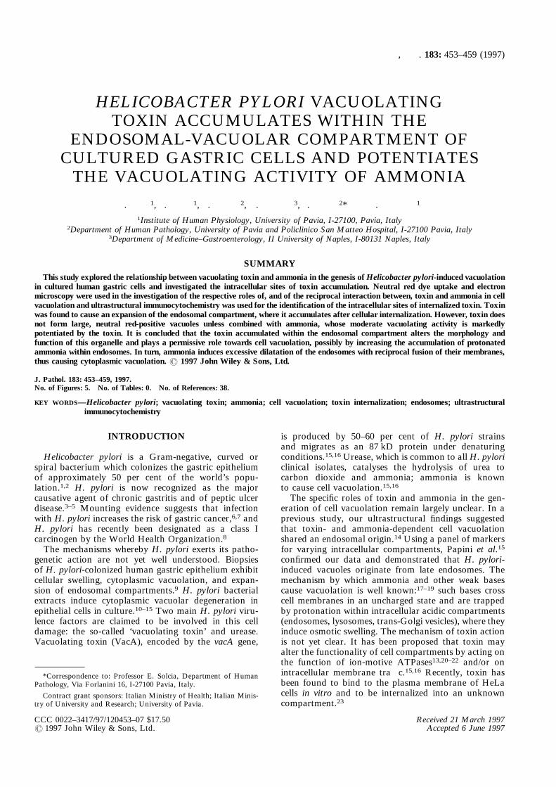
was added to the Tox" BCF, the degree of neutral reduptake was virtually identical to that of ammoniumchloride alone. When ammonium chloride was added toTox+ BCF, we observed a dose-dependent neutral reduptake which was about 2·5-fold higher than thatinduced by ammonium chloride alone. When the toxinwas inactivated by heating or by neutralization withanti-toxin serum, the amount of neutral red uptake wasvirtually identical to the amounts found for ammoniumchloride alone and for Tox" BCF (Fig. 2). To investi-gate further the respective roles of VacA toxin andammonia, we compared the neutral red uptake of MKN28 cells as variously exposed: initially, to one or other ofTox+ BCF alone, ammonium chloride alone, and Tox+BCF plus ammonium chloride; finally, after extensivewashing and 5 h incubation with HBSS, to one or otherof ammonium chloride alone and Tox+ BCF alone. Asshown in Fig. 3, treatment of cells with ammoniumchloride after exposure to the toxin induced a neutralred uptake that was identical to that of cells treated withthe two agents simultaneously. In contrast, treatmentwith Tox+ BCF alone after initial exposure to ammo-nium chloride failed to induce significant neutral reduptake (Fig. 3). Interestingly, the massive neutral reduptake induced by Tox+ BCF plus ammonium chlorideat the end of step 1 was completely reversed bysubsequent 5 h cell incubation with HBSS (Fig. 3).
Fig. 1—Neutral red dye uptake induced in MKN 28 cells by severaldilutions ofH. pylori broth culture filtrates in the absence of ammonia.Cells were incubated with (1) uninoculated broth filtrate (control), (2)broth culture filtrate from the toxin-producing CCUG 17874 strain(Tox+ BCF), and (3) broth culture filtrate from the toxin-negative G21strain (Tox" BCF). All the displayed data are the means of fiveexperiments and each SEM was less than 10 per cent of the respectivemean. No statistically significant differences were found
Fig. 2—Neutral red dye uptake induced in MKN 28 cells by definedconcentrations of ammonium chloride, alone or added to (1) uninocu-lated broth filtrate (control), (2) broth culture filtrate from thetoxin-producing CCUG 17874 strain (Tox+ BCF), (3) heat-inactivatedTox+ BCF, (4) Tox+ BCF neutralized with anti-toxin serum, and (5)broth culture filtrate from the toxin-negative G21 strain (Tox" BCF).Control and BCFs were diluted 1:3 in HBSS. All the displayed data arethe means of five experiments and each SEM was less than 10 per centof the respective mean. *P<0·05 and **P<0·001 versus all otherconditions
455CELLULAR INTERNALIZATION AND ACTIVITY OF H. PYLORI TOXIN
? 1997 John Wiley & Sons, Ltd. , . 183: 453–459 (1997)
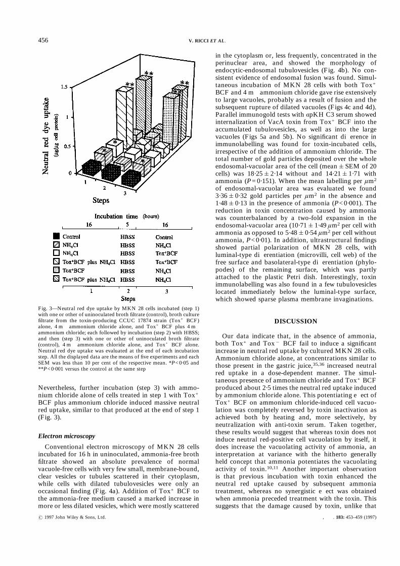
Nevertheless, further incubation (step 3) with ammo-nium chloride alone of cells treated in step 1 with Tox+BCF plus ammonium chloride induced massive neutralred uptake, similar to that produced at the end of step 1(Fig. 3).
Electron microscopy
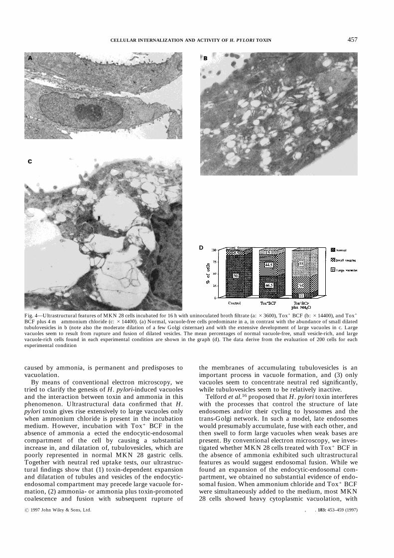
Conventional electron microscopy of MKN 28 cellsincubated for 16 h in uninoculated, ammonia-free brothfiltrate showed an absolute prevalence of normalvacuole-free cells with very few small, membrane-bound,clear vesicles or tubules scattered in their cytoplasm,while cells with dilated tubulovesicles were only anoccasional finding (Fig. 4a). Addition of Tox+ BCF tothe ammonia-free medium caused a marked increase inmore or less dilated vesicles, which were mostly scattered
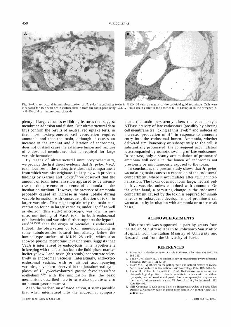
in the cytoplasm or, less frequently, concentrated in theperinuclear area, and showed the morphology ofendocytic-endosomal tubulovesicles (Fig. 4b). No con-sistent evidence of endosomal fusion was found. Simul-taneous incubation of MKN 28 cells with both Tox+BCF and 4 m ammonium chloride gave rise extensivelyto large vacuoles, probably as a result of fusion and thesubsequent rupture of dilated vacuoles (Figs 4c and 4d).Parallel immunogold tests with ápKH C3 serum showedinternalization of VacA toxin from Tox+ BCF into theaccumulated tubulovesicles, as well as into the largevacuoles (Figs 5a and 5b). No significant difference inimmunolabelling was found for toxin-incubated cells,irrespective of the addition of ammonium chloride. Thetotal number of gold particles deposited over the wholeendosomal-vacuolar area of the cell (mean&SEM of 20cells) was 18·25&2·14 without and 14·21&1·71 withammonia (P=0·151). When the mean labelling per ìm2
of endosomal-vacuolar area was evaluated we found3·36&0·32 gold particles per ìm2 in the absence and1·48&0·13 in the presence of ammonia (P<0·001). Thereduction in toxin concentration caused by ammoniawas counterbalanced by a two-fold expansion in theendosomal-vacuolar area (10·71&1·49 ìm2 per cell withammonia as opposed to 5·48&0·54 ìm2 per cell withoutammonia, P<0·01). In addition, ultrastructural findingsshowed partial polarization of MKN 28 cells, withluminal-type differentiation (microvilli, cell web) of thefree surface and basolateral-type differentiation (phylo-podes) of the remaining surface, which was partlyattached to the plastic Petri dish. Interestingly, toxinimmunolabelling was also found in a few tubulovesicleslocated immediately below the luminal-type surface,which showed sparse plasma membrane invaginations.
DISCUSSION
Our data indicate that, in the absence of ammonia,both Tox+ and Tox" BCF fail to induce a significantincrease in neutral red uptake by cultured MKN 28 cells.Ammonium chloride alone, at concentrations similar tothose present in the gastric juice,35,36 increased neutralred uptake in a dose-dependent manner. The simul-taneous presence of ammonium chloride and Tox+ BCFproduced about 2·5 times the neutral red uptake inducedby ammonium chloride alone. This potentiating effect ofTox+ BCF on ammonium chloride-induced cell vacuo-lation was completely reversed by toxin inactivation asachieved both by heating and, more selectively, byneutralization with anti-toxin serum. Taken together,these results would suggest that whereas toxin does notinduce neutral red-positive cell vacuolation by itself, itdoes increase the vacuolating activity of ammonia, aninterpretation at variance with the hitherto generallyheld concept that ammonia potentiates the vacuolatingactivity of toxin.10,11 Another important observationis that previous incubation with toxin enhanced theneutral red uptake caused by subsequent ammoniatreatment, whereas no synergistic effect was obtainedwhen ammonia preceded treatment with the toxin. Thissuggests that the damage caused by toxin, unlike that
Fig. 3—Neutral red dye uptake by MKN 28 cells incubated (step 1)with one or other of uninoculated broth filtrate (control), broth culturefiltrate from the toxin-producing CCUC 17874 strain (Tox+ BCF)alone, 4 m ammonium chloride alone, and Tox+ BCF plus 4 mammonium chloride; each followed by incubation (step 2) with HBSS;and then (step 3) with one or other of uninoculated broth filtrate(control), 4 m ammonium chloride alone, and Tox+ BCF alone.Neutral red dye uptake was evaluated at the end of each incubationstep. All the displayed data are the means of five experiments and eachSEM was less than 10 per cent of the respective mean. *P<0·05 and**P<0·001 versus the control at the same step
456 V. RICCI ET AL.
? 1997 John Wiley & Sons, Ltd. , . 183: 453–459 (1997)
caused by ammonia, is permanent and predisposes tovacuolation.By means of conventional electron microscopy, we
tried to clarify the genesis of H. pylori-induced vacuolesand the interaction between toxin and ammonia in thisphenomenon. Ultrastructural data confirmed that H.pylori toxin gives rise extensively to large vacuoles onlywhen ammonium chloride is present in the incubationmedium. However, incubation with Tox+ BCF in theabsence of ammonia affected the endocytic-endosomalcompartment of the cell by causing a substantialincrease in, and dilatation of, tubulovesicles, which arepoorly represented in normal MKN 28 gastric cells.Together with neutral red uptake tests, our ultrastruc-tural findings show that (1) toxin-dependent expansionand dilatation of tubules and vesicles of the endocytic-endosomal compartment may precede large vacuole for-mation, (2) ammonia- or ammonia plus toxin-promotedcoalescence and fusion with subsequent rupture of
the membranes of accumulating tubulovesicles is animportant process in vacuole formation, and (3) onlyvacuoles seem to concentrate neutral red significantly,while tubulovesicles seem to be relatively inactive.Telford et al.16 proposed thatH. pylori toxin interferes
with the processes that control the structure of lateendosomes and/or their cycling to lysosomes and thetrans-Golgi network. In such a model, late endosomeswould presumably accumulate, fuse with each other, andthen swell to form large vacuoles when weak bases arepresent. By conventional electron microscopy, we inves-tigated whether MKN 28 cells treated with Tox+ BCF inthe absence of ammonia exhibited such ultrastructuralfeatures as would suggest endosomal fusion. While wefound an expansion of the endocytic-endosomal com-partment, we obtained no substantial evidence of endo-somal fusion. When ammonium chloride and Tox+ BCFwere simultaneously added to the medium, most MKN28 cells showed heavy cytoplasmic vacuolation, with
Fig. 4—Ultrastructural features of MKN 28 cells incubated for 16 h with uninoculated broth filtrate (a: #3600), Tox+ BCF (b: #14400), and Tox+
BCF plus 4 m ammonium chloride (c: #14400). (a) Normal, vacuole-free cells predominate in a, in contrast with the abundance of small dilatedtubulovesicles in b (note also the moderate dilation of a few Golgi cisternae) and with the extensive development of large vacuoles in c. Largevacuoles seem to result from rupture and fusion of dilated vesicles. The mean percentages of normal vacuole-free, small vesicle-rich, and largevacuole-rich cells found in each experimental condition are shown in the graph (d). The data derive from the evaluation of 200 cells for eachexperimental condition
D
457CELLULAR INTERNALIZATION AND ACTIVITY OF H. PYLORI TOXIN
? 1997 John Wiley & Sons, Ltd. , . 183: 453–459 (1997)
plenty of large vacuoles exhibiting features that suggestmembrane adhesion and fusion. Our ultrastructural datathus confirm the results of neutral red uptake tests, inthat most toxin-promoted cell vacuolation requiresammonia and that the toxin, although it causes anincrease in the amount and dilatation of endosomes,does not of itself cause the extensive fusion and ruptureof endosomal membranes that is required for largevacuole formation.By means of ultrastructural immunocytochemistry,
we provide the first direct evidence that H. pylori VacAtoxin localizes in the endocytic-endosomal compartmentfrom which vacuoles originate. In keeping with previousfindings by Garner and Cover,23 we observed that theamount of toxin internalization appeared to be insensi-tive to the presence or absence of ammonia in theincubation medium. However, the presence of ammoniaprobably caused an increase in water uptake duringvacuole formation, with consequent dilution of toxin inlarger vacuoles. This might explain why the toxin con-centration found in larger vacuoles, under light23 as wellas electron (this study) microscopy, was low. In anycase, our finding of VacA toxin in both endosomaltubulovesicles and vacuoles further supports the hypoth-esis9,14,15,37 that the origin of vacuoles is endosomal.Indeed, the observation of toxin immunolabelling insome tubulovesicles located immediately below theluminal-type surface of MKN 28 cells, which alsoshowed plasma membrane invaginations, suggests thatVacA is internalized by endocytosis. This hypothesis isin keeping with the fact that both the fluid-phase markerlucifer yellow15 and toxin (this study) concentrate selec-tively in endosomal vacuoles. Interestingly, endocytic-endosomal vesicles, with or without accompanyingvacuoles, have been observed in the juxtalumenal cyto-plasm of H. pylori-colonized gastric foveolar-surfaceepithelium,9,38 with the implication that the basicmechanisms described here in vitro also operate in vivoon human gastric mucosa.As to the mechanism of VacA action, it seems possible
that when internalized into the endosomal compart-
ment, the toxin persistently alters the vacuolar-typeATPase activity of late endosomes (possibly by alteringcell membrane trafficking at this level)37 and induces anincreased production of H+ in response to ammoniaentry into the endosomal lumen. Ammonia, whetherdelivered simultaneously or subsequently to the cell, issubstantially protonated; the consequent accumulationis accompanied by osmotic swelling of late endosomes.In contrast, only a scanty accumulation of protonatedammonia will occur in the lumen of endosomes notpreviously or simultaneously exposed to the toxin.In conclusion, the present study shows that H. pylori
vacuolating toxin causes an expansion of the endosomalcompartment, where it accumulates after cellular inter-nalization. The toxin does not form large, neutral red-positive vacuoles unless combined with ammonia. Onthe other hand, a persisting change in the endosomalcompartment caused by the toxin is required for simul-taneous or subsequent development of prominent cellvacuolation by incubation with ammonia or other weakbases.
ACKNOWLEDGEMENTS
This research was supported in part by grants fromthe Italian Ministry of Health to Policlinico San MatteoHospital, from the Italian Ministry of University andResearch, and from the University of Pavia.
REFERENCES1. Blaser MJ. Helicobacter pylori: its role in disease. Clin Infect Dis 1992; 15:
386–393.2. Taylor DN, Blaser MJ. The epidemiology of Helicobacter pylori infections.
Epidemiol Rev 1991; 13: 42–59.3. Blaser MJ. Hypotheses on the pathogenesis and natural history of Helico-
bacter pylori-induced inflammation. Gastroenterology 1992; 102: 720–727.4. Fiocca R, Villani L, Luinetti O, et al. Helicobacter colonization and
histopathological profile of chronic gastritis in patients with or withoutdyspepsia, mucosal erosion and peptic ulcer: a morphological approach tothe study of ulcerogenesis in man. Virchows Arch A [Pathol Anat] 1992;420: 489–498.
5. NIH Consensus Development Panel on Helicobacter pylori in Peptic UlcerDisease. Helicobacter pylori in peptic ulcer disease. J Am Med Assoc 1994;272: 65–69.
Fig. 5—Ultrastructural immunolocalization of H. pylori vacuolating toxin in MKN 28 cells by means of the colloidal gold technique. Cells wereincubated for 16 h with broth culture filtrate from the toxin-producing CCUG 17874 strain either in the absence (a: #14400) or in the presence (b:#8400) of 4 m ammonium chloride
458 V. RICCI ET AL.
? 1997 John Wiley & Sons, Ltd. , . 183: 453–459 (1997)
6. Nomura A, Stemmermann GN, Chyou P-H, Kato I, Perez-Perez GI, BlaserMJ. Helicobacter pylori infection and gastric carcinoma in a population ofJapanese Americans in Hawaii, N Engl J Med 1991; 325: 1132–1136.
7. Parsonnet J, Friedman GD, Vandersteen DP, et al. Helicobacter pyloriinfection and the risk of gastric carcinoma. N Engl J Med 1991; 325:1127–1131.
8. World Health Organization. Schistosomes, liver flukes and Helicobacterpylori. IARC Monographs on the Evaluation of Carcinogenic Risks toHuman. Vol. 61. IARC, 1994: 177–240.
9. Fiocca R, Luinetti O, Villani L, Chiaravalli AM, Capella C, Solcia E.Epithelial cytotoxicity, immune responses, and inflammatory componentsof Helicobacter pylori gastritis. Scand J Gastroenterol 1994; 29(Suppl 205):11–21.
10. Cover TL, Puryear W, Perez-Perez GI, Blaser MJ. Effect of urease on HeLacell vacuolation induced by Helicobacter pylori cytotoxin. Infect Immun1991; 59: 1264–1270.
11. Cover TL, Vaughn SG, Cao P, Blaser MJ. Potentiation of Helicobacterpylori vacuolating toxin activity by nicotine and other weak bases. J InfectDis 1992; 166: 1073–1078.
12. Mégraud F, Neman-Simha V, Brügmann D. Further evidence of the toxiceffect of ammonia produced by Helicobacter pylori urease on humanepithelial cells. Infect Immun 1992; 60: 1858–1863.
13. Papini E, Bugnoli M, De Bernard M, Figura N, Rappuoli R, MontecuccoC. Bafilomycin A1 inhibits Helicobacter pylori-induced vacuolization ofHeLa cells. Mol Microbiol 1993; 7: 323–327.
14. Ricci V, Sommi P, Fiocca R, et al. Cytotoxicity of Helicobacter pylori onhuman gastric epithelial cells in vitro: role of cytotoxin(s) and ammonia. EurJ Gastroenterol Hepatol 1993; 5: 687–694.
15. Papini E, De Bernard M, Milia E, et al. Cellular vacuoles induced byHelicobacter pylori originate from late endosomal compartments. Proc NatlAcad Sci USA 1994; 91: 9720–9724.
16. Telford JL, Covacci A, Ghiara P, Montecucco C, Rappuoli R. Unravellingthe pathogenic role of Helicobacter pylori in peptic ulcer: potential newtherapies and vaccines. Trends Biotechnol 1994; 12: 420–426.
17. de Duve C, de Barsy T, Poole B, Trouet A, Tulkens P, Van Hoof F.Lysosomotropic agents. Biochem Pharmacol 1974; 23: 2495–2531.
18. Ohkuma S, Poole B. Cytoplasmic vacuolation of mouse peritoneal macro-phages and the uptake into lysosomes of weakly basic substances. J Cell Biol1981; 90: 656–664.
19. Poole B, Ohkuma S. Effect of weak bases on the intralysosomal pH inmouse peritoneal macrophages. J Cell Biol 1981; 90: 665–669.
20. Cover TL, Blaser MJ. Purification and characterization of the vacuolatingtoxin from Helicobacter pylori. J Biol Chem 1992; 267: 10570–10575.
21. Cover TL, Reddy LY, Blaser MJ. Effects of ATPase inhibitors on theresponse of HeLa cells to Helicobacter pylori vacuolating toxin. InfectImmun 1993; 61: 1427–1431.
22. Ricci V, Sommi P, Cova E, et al. Na+, K+-ATPase of gastric cells: a targetof Helicobacter pylori cytotoxic activity. FEBS Lett 1993; 334: 158–160.
23. Garner JA, Cover TL. Binding and internalization of theHelicobacter pylorivacuolating cytotoxin by epithelial cells. Infect Immun 1996; 64: 4197–4203.
24. Xiang Z, Censini S, Bayeli PF, et al. Analysis of expression of CagA andVacA virulence factors in 43 strains of Helicobacter pylori reveals thatclinical isolates can be divided into two major types and that CagA is notnecessary for expression of the vacuolating cytotoxin. Infect Immun 1995;63: 94–98.
25. Ricci V, Ciacci C, Zarrilli R, et al. Effect of Helicobacter pylori on gastricepithelial cell migration and proliferation in vitro: role of VacA and CagA.Infect Immun 1996; 64: 2829–2833.
26. Sommi P, Ricci V, Fiocca R, et al. Significance of ammonia in the genesis ofgastric epithelial lesions induced by Helicobacter pylori: an in vitro studywith different bacterial strains and urea concentrations. Digestion 1996; 57:299–304.
27. Telford JL, Ghiara P, Dell’Orco M, et al. Gene structure of theHelicobacterpylori cytotoxin and evidence of its key role in gastric disease. J Exp Med1994; 179: 1653–1658.
28. Leunk RD. Production of a cytotoxin by Helicobacter pylori. Rev Infect Dis1991; 13(Suppl 8): S686–S689.
29. Hojo H. Establishment of cultured lines of human stomach cancer. Originand their morphological characteristics. Niigata Igakukai Zasshi 1977; 91:737–752.
30. Romano M, Razandi M, Sekhon S, Krause WJ, Ivey KJ. Human cell linefor study of damage to gastric epithelial cells in vitro. J Lab Clin Med 1988;111: 430–440.
31. Ricci V, Fiocca R, Sommi P, et al. MKN 28 cell line: a useful tool to studyhuman gastric epithelial cells. Pflügers Arch 1992; 420: R182.
32. Tummuru MKR, Cover TL, Blaser MJ. Cloning and expression of ahigh-molecular-mass major antigen of Helicobacter pylori: evidence oflinkage to cytotoxin production. Infect Immun 1993; 61: 1799–1809.
33. Lowry OH, Rosebrough NJ, Farr AL, Randall RJ. Protein measurementwith the Folin phenol reagent. J Biol Chem 1951; 193: 265–275.
34. Snedecor GW, Cockran WG. Statistical Methods. Ames: The Iowa StateUniversity Press, 1967.
35. Tsujii M, Kawano S, Tsuji S, Fusamoto H, Kamada T, Sato N. Mechanismof gastric mucosal damage induced by ammonia. Gastroenterology 1992;102: 1881–1888.
36. El Nujumi AM, Rowe PA, Dahill S, Dorrian CA, Neithercut WD, McCoolKEL. Role of ammonia in the pathogenesis of the gastritis, hypergastrin-aemia, and hyperpepsinogenaemia I caused by Helicobacter pylori infection.Gut 1992; 33: 1612–1616.
37. Papini E, Gottardi E, Satin B, et al. The vacuolar ATPase proton pump ispresent on intracellular vacuoles induced by Helicobacter pylori. J MedMicrobiol 1996; 45: 84–89.
38. Solcia E, Villani L, Luinetti O, Trespi E, Fiocca R. The mucosal response toHelicobacter pylori infection and its contribution to gastric pathology.Microecol Ther 1995; 25: 121–132.
459CELLULAR INTERNALIZATION AND ACTIVITY OF H. PYLORI TOXIN
? 1997 John Wiley & Sons, Ltd. , . 183: 453–459 (1997)


![Vacuolar Transporters – Companions on a Longtime … · Vacuolar Transporters – Companions on a Longtime Journey[OPEN] Enrico Martinoia1 Department of Plant and Microbial Biology,](https://img.pdfslide.net/doc/110x75/603fbba48d3fd353b308f80e/vacuolar-transporters-a-companions-on-a-longtime-vacuolar-transporters-a-companions.jpg)