Embed Size (px)
Citation preview
www.elsevier.com/locate/addr
Advanced Drug Delivery Reviews 56 (2004) 853–868
Helminths at mucosal barriers—interaction with
the immune system
Grace Mulcahya,*, Sandra O’Neillb, Sheila Donnellyc, J.P. Daltona,d
aDepartment of Veterinary Microbiology and Parasitology, Faculty of Veterinary Medicine and Conway Institute,
University College Dublin, Belfield, Dublin 4, IrelandbSchool of Nursing, Dublin City University, Dublin 9, Ireland
cSchool of Biotechnology, Dublin City University, Dublin 9, Irelandd Institute for the Biotechnology of Infectious Disease, University of Technology, Sydney, Australia
Received 19 October 2003; accepted 3 November 2003
Abstract
Helminth parasites are the cause of very significant morbidity, mortality and economic losses in man and domestic animals.
Most parasitic helminths infect their hosts via the oral route, and live either at the mucosal surface of the gastro-intestinal tract
(GIT), or cross this mucosal barrier on their way to predilection sites. Many helminths live at mucosal surfaces, typically the gut
or respiratory tract, and some cross these barriers, either temporarily, spending a period of time in the mucosa before returning
to the mucosal surface, or to access other tissues and sites in the host. Typically, helminths induce strongly polarised Th2
responses, which are often effective in mediating protective immunity against those parasites living at mucosal surfaces, but less
so in protecting against tissue-dwelling parasites. Induction of strongly-polarised Th2 responses may impair the ability of
parasites hosts to eliminate other pathogens. Control of helminth infections relies largely on chemotherapy, together with
management and environmental measures designed to keep hosts away from infective stages. Drug resistance has become a
significant problem in some helminth populations, and this has promoted interest in the development of immunoprophylactic
strategies. However, despite intensive research efforts, helminth vaccines have not become part of regular control strategies. In
addition to the considerable technical difficulties posed in the production of vaccines against these complex organisms, further
difficulties in securing acceptance for anti-helminth vaccine by regulatory authorities and by users, will be encountered. Such
vaccines need not result in sterile immunity, as is required of anti-bacterial and anti-viral vaccines. Recent evidence indicates
that while helminths are responsible for disease, immunopathology and impairment of immunity to other pathogens, a complete
absence of helminth infection during early life may be a predisposing factor for the development of auto-immune pathology.
D 2004 Elsevier B.V. All rights reserved.
Keywords: Helminth; Mucosal immunity; Vaccine; Immunopathology
Contents
1. Introduction . . . . . . . . . . . . . . . . . . . . . . . . . . . . . . . . . . . . . . . . . . . . . . . . . . . . . 854
2. Basic helminth biology . . . . . . . . . . . . . . . . . . . . . . . . . . . . . . . . . . . . . . . . . . . . . . . . 854
0169-409X/$ - see front matter D 2004 Elsevier B.V. All rights reserved.
doi:10.1016/j.addr.2003.10.033
* Corresponding author. Tel.: +353-1-716-6180.
E-mail address: [email protected] (G. Mulcahy).

G. Mulcahy et al. / Advanced Drug Delivery Reviews 56 (2004) 853–868854
2.1. Helminths as pathogens. . . . . . . . . . . . . . . . . . . . . . . . . . . . . . . . . . . . . . . . . . . . . 856
2.2. Helminths within the gut, and across the mucosal barrier . . . . . . . . . . . . . . . . . . . . . . . . . . . . . 856
3. Characteristics of immune responses to intestinal helminths . . . . . . . . . . . . . . . . . . . . . . . . . . . . . . . . 856
3.1. Protective responses . . . . . . . . . . . . . . . . . . . . . . . . . . . . . . . . . . . . . . . . . . . . . . 857
3.2. Ineffective responses . . . . . . . . . . . . . . . . . . . . . . . . . . . . . . . . . . . . . . . . . . . . . . 858
3.3. Immunomodulation by helminths . . . . . . . . . . . . . . . . . . . . . . . . . . . . . . . . . . . . . . . . 859
4. The influence of helminth infections on resistance to other diseases . . . . . . . . . . . . . . . . . . . . . . . . . . . . 861
4.1. Impaired responses to bacterial and viral infections . . . . . . . . . . . . . . . . . . . . . . . . . . . . . . . . 861
4.2. Absence of helminth infections as a possible factor in the aetiology of immune-mediated disease . . . . . . . . . . . 862
5. Immunopathology . . . . . . . . . . . . . . . . . . . . . . . . . . . . . . . . . . . . . . . . . . . . . . . . . . . 863
6. The outlook for mucosal anti-helminth vaccines . . . . . . . . . . . . . . . . . . . . . . . . . . . . . . . . . . . . . 863
7. Exploiting helminths as therapeutic agents. . . . . . . . . . . . . . . . . . . . . . . . . . . . . . . . . . . . . . . . 864
8. Concluding remarks . . . . . . . . . . . . . . . . . . . . . . . . . . . . . . . . . . . . . . . . . . . . . . . . . . 865
References . . . . . . . . . . . . . . . . . . . . . . . . . . . . . . . . . . . . . . . . . . . . . . . . . . . . . . . . . 865
1. Introduction
Helminth parasites are responsible for significant
ill-health world-wide. The World Health Organisa-
tion estimates that soil-transmitted worms including
hookworms, ascarids and whipworms pose a health
risk to 2 billion people (one third of the world’s
population) and that water-borne helminths such a
schistosomes pose a health-risk to between 500 and
600 million people (http://whqlibdoc.who.int/hq/
2001/WHO_CDS_CPE_SMT_2001.8.pdf). Tremato-
des of the genus Fasciola are also regarded as
important causes of human disease, with an estimat-
ed 24 million people infected across 61 countries
[1]. In veterinary medicine, helminth parasites are of
constant concern to livestock producers and, indeed
to owners of companion animals, and this is
reflected in the fact that 27% of sales of veterinary
medicines are anti-parasitic drugs and this market
was worth $3.1 billion in 2002 (http://www.ifahsec.
org).
Most helminths infect their hosts via the oral
route, and therefore, either live at the mucosal
surface of the gastro-intestinal tract (GIT), or traverse
this mucosal barrier on their way to their predilection
site. Major helminth parasites of man and animals
which inhabit or traverse the mucosa of the GIT are
shown in Table 1.
While a wide range of effective anthelmintic drugs
are available, their use is associated with diverse
problems such as pathology arising before treatment,
acquisition of drug resistance, and, in veterinary med-
icine, drug residue issues. The development of effec-
tive anti-helminth vaccines would provide a solution to
these problems. However, the development of such
vaccines has been slow due to the complexities of
helminth life-cycles, and the ability of these parasites
to evade and modulate host immune responses. The
increased availability of helminth parasite genomic
information, together with new and improved methods
of methods of gene expression (reviewed in [2]),
promise to provide enhanced capabilities for helminth
vaccine development.
It has recently become clear that helminth patho-
gens are important not only because of the patho-
genic effects directly attributable to them, but
because they interact with the host immune system
in ways which may impair the response to other
pathogens and/or cause immunopathology. In this
review, we present an overview of the biology of
parasitic helminths, and of the mechanisms whereby
they cause intestinal disease. We describe the spe-
cific details of some interactions of intestinal hel-
minths with their hosts, the characteristics of immune
responses to such parasites, and the outlook for new
prophylactic and therapeutic interventions.
2. Basic helminth biology
Helminths (worms) are classified zoologically as
roundworms (nematodes) or flatworms (tapeworms or
cestodes, and flukes or trematodes). Amongst the
nematodes, only a minority are parasitic. The free-

Table 1
Helminth parasites of the GIT which are of major medical or veterinary importance
Parasite group Host location Significance
Ascarids Small intestine, some with migratory larval stages Ascaris lumbricoides infecting large
numbers of people in underdeveloped
countries, ascarids of domestic animals
causing production losses, and some,
such as T. canis, causing zoonotic
disease
Fasciola spp. Liver flukes, which migrate across intestinal mucosa
and peritoneal cavity to gain access to hepatic tissue
Large numbers of people in some
developing countries such as Bolivia,
Egypt, are infected. Major pathogen of
ruminants in both temperate
(F. hepatica) and tropical (F. gigantica)
areas
Hookworms Small intestine Mouthparts adapted for bloodsucking,
with the more pathogenic species
causing severe anaemia in man and
animals
Lungworms Airways or lung parenchyma Those species living at the airway
mucosa are generally more pathogenic
than those living in lung parenchyma.
Immune responses are often protective
against challenge but may also cause
immunopathological lung damage
Schistosomes Blood flukes whose main predilection site is
mesenteric veins,
thus allowing eggs to be layed into the intestinal
lumen and pass
out with host faeces
Major cause of morbidity and mortality
in man and animals in tropical areas.
Definitive hosts become infected by
skin penetration by infective stages
(cercariae) emerging from water snails
Strongyloides spp. Small intestine, but sometimes also cause skin
irritation
Large numbers of humans infected in
developing countries. Also affects
neonatal livestock
Tapeworms Adults in small intestine, intermediate stages
in a variety of tissues
Intestinal stages generally tolerated
without significant pathology.
Intermediate stages may cause serious
disease, for example neurocysticercosis
by T. solium, and echinococcosis
Trichinella spp. Adults with brief life-span in small intestine,
but larvae in tissues remain
viable for years and are a source of infection for
carnivorous hosts
Human infection generally controlled
satisfactorily by meat inspection, but
still a cause of muscle pathology
following consumption of game
Trichostrongylid nematodes Various parts of the GIT Of greatest importance in ruminants.
Several species have developed drug
resistance, thus stimulating vaccine studies
G. Mulcahy et al. / Advanced Drug Delivery Reviews 56 (2004) 853–868 855
living nematode Caenorhabditis elegans has been
used extensively in the fields of basic nematode
biology and genomics. All flatworms adopt a parasitic
life-cycle. Where there has been a co-evolution of
helminths with their hosts, and where parasite burdens
are not extreme, it is common for helminth infections
to result in only very minor, if any, clinical disease.
This is the case, for example, for infection of man
with the ‘‘beef tapeworm’’, Taenia saginata. Typical-
ly, helminth infections become clinically important in
individuals or populations when environmental con-
ditions promote a build-up of infectious stages, and
therefore, very large parasite burdens. Helminths may
also cause disease when host populations previously
unexposed to a particular species become infected,
due to a change in the environment, or when individ-

G. Mulcahy et al. / Advanced Drug Delivery Reviews 56 (2004) 853–868856
uals are immunosuppressed or nutritionally compro-
mised. In many cases, neonates are more susceptible
to helminth infection than adults. In some cases,
resistance develops as a result of active immunity,
but there are also many examples where innate age
resistance plays a role.
Most parasitic helminths have at least one stage of
their life-cycle where they are free-living. With many
nematodes, for example, eggs passed in the faeces of
infected hosts develop through several larval stages
in the environment, with infection of a new host
taking place through ingestion of an infective larva
(often the third larval stage). In the case of some
roundworms and all flatworms, life-cycles are indi-
rect, with at least two and possibly more hosts
involved.
2.1. Helminths as pathogens
Helminths which live in the GIT of their hosts
include a wide variety of taxonomic groups, across
both flatworms and roundworms. In many cases, such
as in most tapeworm infections, they are tolerated well
and are responsible for few, if any, pathogenic effects.
Where disease does occur as a result of intestinal
helminthosis, it is often the case that significant
pathology is only seen when parasite burdens exceed
a certain threshold. Various pathogenic mechanisms
can be involved including loss of blood due to parasite
feeding [3], competition for nutrients or nutritional
dysfunction [4], immunopathology [5,6], interference
with gut motility and/or neuromuscular control [7]
and blockage of the intestine [8].
Helminths often cross the mucosal barrier of the
GIT to gain access to other sites within the host.
Another mucosal site favoured by helminths is the
respiratory tract. Typically, helminths arrive here
after travelling through the blood vascular system
and then crossing over to the airways at the lung
capillaries.
2.2. Helminths within the gut, and across the mucosal
barrier
It seems probable that parasitic nematodes
evolved from free-living populations which were
transported from their environmental niche (soil
and water) into the GIT of higher organisms. Evi-
dence of such a pattern can be seen in some
nematode groups (such as the genus Strongyloides)
which are still at the borderline between a parasitic
and free-living life-cycle. These nematodes can be
parasitic in a variety of hosts, including man. In this
genus both parasitic and free-living life-cycles can
occur, depending on environmental conditions. When
the latter are unfavourable, the existence of parasitic
forms ensures the survival of populations, which can
revert back to free-living forms when conditions
improve. Infection of hosts can take place either
orally, or through skin.
Helminths which live at mucosal sites such as the
GIT are, strictly speaking, not truly living within the
host organism, and are therefore, in some ways less
exposed to host immune defence mechanisms than
helminths living within host tissues. However, the
typical Th2 immune response evoked by helminths,
which among its effector mechanisms includes in-
creased gut motility, may be particularly suited to the
elimination of intestinal parasites.
Another evolutionary step may have occurred
when helminths living within the GIT of their hosts
crossed this mucosal barrier to continue their life-
cycle elsewhere, within host tissues, in some cases
returning to the intestine to provide an easy access
for reproductive stages to the outside environment.
There are many such examples among parasitic
helminths—including the liver and blood flukes,
Schistosoma and Fasciola), some ascarids such as
Toxocara canis. The evolution of such migratory
patterns among parasitic helminths may have been
driven by requirements to evade immune defence
mechanisms, targeted specifically at helminths inhab-
iting mucosal sites. In order to penetrate mucosal
surfaces and host tissues, helminths use a variety of
mechanical and chemical tools including tough mod-
ifications of the surface (hooks, lancets, spines) and
proteolytic enzymes (reviewed in [9]).
3. Characteristics of immune responses to
intestinal helminths
Host immune responses to helminths living in the
GIT are well-defined. Some general principles in
relation to such responses can be stated. Innate
immunity, often age-related, is important in many
G. Mulcahy et al. / Advanced Drug Delivery Reviews 56 (2004) 853–868 857
cases. This may be due to physico-chemical differ-
ences in the gut environment in adult as compared
with young hosts. In dogs infected with the ascarid
parasite T. canis, for example, adult worms are
expelled from the intestine when the host is between
3 and 6 months of age, and new infections do not
establish as adults, following egg infection, in dogs
older than 6 months [10]. This phenomenon does not
depend on prior exposure to the parasite.
Distinct immune responses often occur to antigens
expressed at different stages of the parasitic life-
cycle. This is especially true where some stages are
migratory. Responses to gut-dwelling stages are often
polarised Th2 responses, characterised by eosinophilia,
mastocytosis and IgE production. One effect of such a
response in the GIT is stimulation of smooth muscle,
increased gut motility, and diarrhoea [11]. Clearly, this
effect is likely to promote parasite expulsion, but
diarrhoea is also one of the pathogenic effects associ-
ated with GIT helminth infection. This illustrates
another common feature of mucosal helminth infec-
tions, namely, that pathology often results from the
host’s own immune response. Allied to this, polar-
isation of the immune response to Th2 also down-
regulates the Th1 cell subset [12], which may have
implications for the ability of hosts with helminth
burdens to mount protective immune responses to
intracellular pathogens.
3.1. Protective responses
There are many examples of intestinal helminth
infections which are contained, and ultimately elim-
inated, by the classic ‘‘anti-helminth’’ Th2 response
described above. In man, for example, such
responses are involved in protection against hook-
worm infection [13]. Many studies on protective
immunity to gastro-intestinal nematodes come from
examples important in veterinary medicine, or from
experimental animal models. One of the most exten-
sively studied parasites in this respect is Ostertagia
ostertagi, a nematode parasite which lives in the
abomasum of cattle and causes serious economic
losses to beef and dairy producers. Within a number
of days following infection, and persisting for the
duration of infection, there is a considerable increase
in the size of regional lymph nodes draining the site
of infection [14], and also an infiltration of lympho-
cytes at the site of infection [15]. This is accompa-
nied by increased numbers of mast cells and
eosinophils [16] and by increases in smooth muscle
activity [17]. Despite these changes, immunity to
Ostertagia infection develops slowly, so that animals
during their first season of parasite exposure remain
comparatively susceptible, and eliminate existing
infections relatively slowly in spite of immunopath-
ological consequences such as diarrhoea and loss of
specialisation in the epithelial lining of the aboma-
sum. Markers of developing protective immunity
include lowered parasite egg production, stunting
of worms, and increased parasite development times.
The outcome for the animal is solid immunity, but
only after several months of parasite exposure and,
in cases where parasite burdens exceed a certain
threshold, clinical disease. The outcome for the
parasite is the establishment of relatively long-stand-
ing infections in young animals ensuring sufficient
reproductive capacity for survival until the next
generation of susceptible hosts becomes available,
i.e. from one grazing season to another. Although
eliciting similar immune-effector mechanisms, immu-
nity to another gastro-intestinal nematode of veteri-
nary importance, Nematodirus battus, is extremely
rapid [18]. This parasite is a serious intestinal path-
ogen of lambs, but sheep over the age of about 6
months have a sold age-immunity. Infected lambs
develop severe diarrhoea and a Th2-skewed immune
response, with worm expulsion, associated with
shedding of duodenal villi, occurring about 3 weeks
post-infection [18].
The role of goblet cells and intra-epithelial gy-lymphocytes in immune-mediated expulsion of hel-
minth parasites is receiving increasing attention.
The higher numbers of goblet cells recruited to
the GIT in helminth infection, associated with
increased mucus production and in some cases
parasite expulsion [19], may be under the control
of gy T-cells [20]. The role of the latter as a bridge
between the innate and adaptive immune systems is
gaining currency, and new information on the
existence of various subsets within the population
promises to throw further light on this role
(reviewed in [21]). A schematic showing some of
the mechanisms that may contribute to Th2 medi-
ated protective immunity against intestinal hel-
minths is shown in Fig. 1.
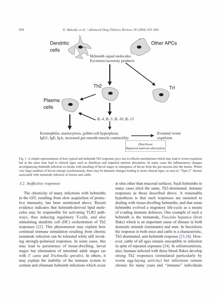
Fig. 1. A simple representation of how typical anti-helminth Th2 responses give rise to effector mechanisms which may lead to worm expulsion
but at the same time lead to clinical signs such as diarrhoea and impaired nutrient absorption. In many cases the inflammatory changes
accompanying helminth infection co-incide with moulting of larval stages or emergence of larvae from the gut mucosa into the lumen. Where
very large numbers of larvae emerge synchronously, there may be dramatic changes leading to acute clinical signs, as seen in ‘‘Type-2’’ disease
G. Mulcahy et al. / Advanced Drug Delivery Reviews 56 (2004) 853–868858
3.2. Ineffective responses
The chronicity of many infections with helminths
in the GIT, resulting from slow acquisition of protec-
tive immunity, has been mentioned above. Recent
evidence indicates that helminth-derived lipid mole-
cules may be responsible for activating TLR2 path-
ways, thus inducing regulatory T-cells, and also
stimulating dendritic cell (DC) orchestration of Th2
responses [22]. This phenomenon may explain how
continual immune stimulation resulting from chronic
nematode infection can be tolerated while still invok-
ing strongly-polarised responses. In some cases, this
may lead to persistence of tissue-dwelling, larval
stages but elimination of intestinal adult stages (as
with T. canis and Trichinella spiralis). In others, it
may explain the inability of the immune system to
contain and eliminate helminth infections which occur
associated with nematode infection in horses and cattle.
at sites other than mucosal surfaces. Such helminths in
many cases elicit the same, Th2-dominated, immune
responses as those described above. A reasonable
hypothesis is that such responses are unsuited to
dealing with tissue-dwelling helminths, and that some
helminths evolved a migratory life-cycle as a means
of evading immune defences. One example of such a
helminth is the trematode, Fasciola hepatica (liver
fluke) which is an important cause of disease in both
domestic animals (ruminants) and man. In fasciolosis
the response in both mice and cattle is a characteristic,
Th2-dominated, anti-helminth response [23,24]. How-
ever, cattle of all ages remain susceptible to infection
in spite of repeated exposure [24]. In schistosomiosis,
also, humans infected with these blood flukes develop
strong Th2 responses (stimulated particularly by
worm egg-laying activity) but infections remain
chronic for many years and ‘‘immune’’ individuals

G. Mulcahy et al. / Advanced Drug Delivery Reviews 56 (2004) 853–868 859
remain susceptible to new infection [25,26]. Further
examples of typical anti-helminth responses being
ineffective in the face of tissue-dwelling helminths
comes from the filarial nematodes, where Onchocerca
volvulus infection of man (the cause of river blind-
ness), and Dirofilaria immitis infection of dogs (heart-
worm) result in typical Th2 responses, yet chronic
infections are the norm and exposed individuals
remain susceptible to new infection [27].
3.3. Immunomodulation by helminths
As part of their adaptation to a parasitic life-cycle,
helminths have of necessity acquired the means of
dealing with host immune responses. One such mech-
anism of immune evasion may have been the devel-
opment of migratory strategies, away from mucosal
immune defence mechanisms, as described above,
coupled with induction of immunoregulatory cell
subsets. There is also much evidence in the literature
for specific modulatory mechanisms evoked by hel-
minths to interfere with particular effector mecha-
nisms evoked following induction of immune
responses. Such mechanisms contribute to the chro-
nicity of infections, and their persistence in adult
populations, but they can also contribute to immuno-
pathology, either by inducing potent inflammatory
reactions which damage host tissues, or by impinging
on host responses to other pathogens.
There are numerous mechanisms documented for
helminth immunomodulation including down-regula-
tion of Th1 responses [26,28] associated with the
intense stimulation of Th2 responses, secretion of
anti-effector molecules, inhibition of effector cells,
inhibition of complement components, induction of
non-specific proliferative responses, and inhibition of
proliferative responses.
The complement system is an important effector of
the innate and adaptive immune response. The system
consists of a group of serum proteins that activate
each other in a specifically ordered manner to gener-
ate biologically active molecules. These mediate the
elimination of pathogens directly through cell lysis or
indirectly through opsonisation and induction of in-
flammation. Following infection, helminth parasites
typically activate immune recognition, represented by
the complement pathway, which contributes to the
host inflammatory response, specifically in the recruit-
ment of eosinophils and Non-T-Non-B cells. The
juvenile stages of many parasites are susceptible to
lysis by this cytolytic response [29–31]. However,
following development to the adult phase of the life
cycle, many parasites develop resistance to comple-
ment-mediated killing. It has been suggested that this
may occur due to a number of immunoevasive strat-
egies adopted by helminth worms. For example,
schistosomes secrete proteases which cleave peptides
anchoring the glycocalyx coat to the tegument of the
worm. This results in the shedding, not only of the
parasite coat but also of surface-bound components of
the complement system [32].
Helminth parasites have also developed a number
of immunomodulatory mechanisms, which inhibit
complement killing via either the alternative or clas-
sical pathway. Complement activation of either path-
way converges at the level of C3 with C3 convertase
formation being critical to its function. Regulation of
convertase enzyme activity is tightly controlled by a
number of membrane-associated or secreted regulato-
ry proteins, which prevent complement-mediated he-
molysis by blocking the assembly or accelerating the
decay of C3 convertase. Parasite worms, which spend
a large proportion of their life cycle in the host
vascular system, have developed the ability to acquire
these regulatory proteins as part of their surface coat.
In this way, the parasite inhibits the synthesis of
enzyme, therefore, preventing induction of the classi-
cal complement pathway. For example, both Echino-
coccus granulosus and Taenia taeniaeformis sequester
host-derived negative regulator, factor H, while Schi-
tosoma mansoni adsorbs host decay accelerating fac-
tor (DAF) from erythrocytes [33,34]. Such acquisition
of regulatory factors from the complement system
ensures survival of the worm. However, this disrup-
tion of the C3 convertase enzyme, a pivotal compo-
nent of both the classical and alternative pathway,
undoubtedly affects the susceptibility of the host to
concomitant bacterial or viral infections.
The alternative complement pathway requires the
convertase activation of C3b, which binds to the
surface of the invading pathogen preparing it for
opsonisation. Two serine proteases isolated from S.
mansoni have demonstrated the ability to cleave the
C3b component of the human complement system
[32], thereby inactivating the cascade of events lead-
ing to induction of the alternative pathway. While this

G. Mulcahy et al. / Advanced Drug Delivery Reviews 56 (2004) 853–868860
determines that the parasite is resistant to comple-
ment, it may also result in increased host susceptibility
to concurrent infection by other pathogens or failure
of vaccines to confer protection. It is widely accepted
that C3d, a downstream product of C3b, is an effec-
tive adjuvant for inducing immune responses to a
variety of antigens [35–37]. Therefore, immunomo-
dulation by helminths resulting in the inactivation of
C3b and consequential removal of its cascade prod-
ucts, may mediate susceptibility to the host towards
infectious agents and reduce efficacy of a wide range
of vaccines.
The activation of complement through the classical
pathway requires C1q for initiation. Helminths have
adapted a wide range of mechanisms to inhibit the
function of this component of the classical pathway.
The gut-associated antigen, CAA of S. mansoni
behaves like a receptor for C1q preventing activation
of the precursor C1 complement component thus
protecting the schistosome gut from complement
mediated attack [38]. Paramyosins from S. mansoni
and T. solium inhibit C1 function by binding to C1q,
resulting in the inactivation of C4, an essential ele-
ment in the induction of inflammation [39]. Trans-
genic mice deficient for the C1q component are
known to lack the classical pathway of complement
activation. We hypothesise that a similar immune
deficiency may, therefore, exist during infection with
helminth parasites, leading to increased susceptibility
to bacterial or viral infections. It has been demonstrat-
ed that C1q is a vital element in protection against
bacteria. Mortality of C1q deficient mice to polymi-
crobial peritonitis was 83% compared to 42% for wild
type. The same strain of mice were found to be
significantly more susceptible to Salmonella infection,
reflected in increased hepatic and splenic bacterial
counts [40,41]. These observations further support our
suggestion of helminth-induced predisposition to con-
current bacterial infection.
Parasitic helminths possess mechanisms to disable
the short-range offences of effector cells, such as toxic
reactive oxygen products of leucocytes (eosinophils
and neutrophils) and macrophages or reactive nitrogen
intermediates generated by macrophages. S. mansoni
and F. hepatica are highly resistant to killing by
reactive nitrogen intermediates generated by lipopoly-
saccharide (LPS)-stimulated macrophages and by
chemically-generated reactive oxygen intermediates
(ROI). Inactivation of ROI involves oxidant-scaveng-
ing enzymes such as superoxide dismutase (SOD),
glutathione peroxidase and glutathione-S-transferase
(GST) [42]. SOD is also found at the surface of adult
Brugia worm where resistance to killing by superox-
ide is observed [43].
While phagocytosis by macrophages is primarily
mediated by reactive oxygen and nitrogen intermedi-
ates, neutrophils contain an abundant array of mole-
cules, which are involved in oxygen-independent
killing of pathogens. Among these, a-defensins, elas-
tase and neutral proteinases (cathepsin G) cause lethal
damage to membranes by forming destabilising ion-
channels and degrading the extracellular matrices of
pathogen membranes. Serine proteinase inhibitors
(serpins) secreted by pathogens play a vital role in
inhibiting the activity of these enzymes. Serpins from
viruses have been implicated in pathogen evasion of
the host defence system. Recombinant Bm-SPN-2, a
protein found in the microfilarial stages of B. malayi
was found to specifically inhibit enzymatic activity of
human neutrophil cathepsin G and human neutrophil
elastase [44]. It is possible that Bm-SPN-2 could
function as a stage-specific serpin in the blood stream
protecting the parasite from attack from neutrophils
[44]. F. hepatica has also been shown to secrete a
Kunitz-type serine proteinase inhibitor, which is
expressed in the gut, parenchyma and tegument of
the adult worm. This protein is thought to inhibit the
activity of neutrophil elastate [45].
In order for T-cells to respond to foreign particles,
the antigen must be broken into peptide fragments and
presented via MHC molecules. The processing of
peptides depends upon whether the antigen is exoge-
nous (generally bacterial or viral origin) or endoge-
nous (generally viral in origin). Antigen presenting
cells that present exogenous antigens include macro-
phages, B-cells, DCs and epithelial cells whereas
endogenous particles are generally synthesised by
viruses within the cell and can be presented via all
other nucleated cell types. Processing of exogenous
antigens begins when antigens are internalised via
endocytosis. Following endocytosis, endosome–lyso-
some fusion occurs and degradation of antigen com-
mences within these acid vesicles. Initially the tertiary
structure of the protein is destroyed by the reduction of
disulphide bonds, thus making it more accessible to
other degradative enzymes. Enzymes involved in the

G. Mulcahy et al. / Advanced Drug Delivery Reviews 56 (2004) 853–868 861
degradation of proteins include cathepsin B, D and E.
The antigen is further processed by endosomal pro-
teases and the resulting peptides fragments form com-
plexes with Class II MHCmolecules that are expressed
on the cell surface. Helminths are known to produce a
number of protease inhibitors that interfere with pro-
cessing of exogenous antigens. PI-3 is an aspartyl
proteinase inhibitor, which is thought to interfere with
cathepsin-E activity [46]. Cathepsin E is known to be
involved in the processing of antigens and of invariant
chains. It is a major aspartic proteinase in a murine
antigen-presenting cell line, A20 [47] and is thought to
play a major role in antigen processing. This enzyme is
localised to a non-lysosomal compartment of the
endosomal system in these cells. Functional studies
using a highly specific inhibitor of cathepsin E have
shown that this enzyme is essential for the processing
of ovalbumin by this cell line [47].
During cell-mediated immunity, T-cells secrete
cytokines which activate macrophages to become
efficient phagocytes, with cells activated by Th1
pro-inflammatory cytokines, such as IFNg, being
critical in combating infection with intracellular
micro-organisms. In contrast, Th2 cytokines, such
as those observed following a helminth infection,
activate macrophages toward a down-regulatory phe-
notype. In support of this, recent studies have
described an innate response that leads to expansion
of suppressor macrophage populations. These im-
munoregulatory cells, termed natural suppressor
(NS) cells, originate from granulocyte–monocyte
progenitors and are capable of inhibiting prolifera-
tive responses of naıve or activated T and B cells.
Two different subclasses of NS have been described:
(1) alternatively activated macrophages (AA), which
are IL-4 dependent and (2) classically activated
macrophages (CA), which are IFNg-dependent. IL-
4 dependent macrophages recruited to the peritoneal
cavity in mice infected with B. malayi actively
suppress the proliferation of lymphocytes co-cul-
tured in vitro [49]. These AA macrophages block
proliferation by cell-to-cell contact, implicating a
receptor-mediated mechanism.
Following successful migration to the lymphoid
tissue, immune responses to pathogens are initiated
when DCs present MHC class II antigen peptide
complexes to CD4+ T cells. In combination with co-
stimulation, this signal drives T cells to produce IL-2
and enter the cell cycle. There is increasing evidence
that microbes drive the development of protective
Th1 of Th2 cells via their effects on APC with
suggestions that DC’s carry a signal determining
the polarisation of naıve T cells into either Th1 or
Th2. Exposure of DC to Th2-inducing soluble egg
antigen of schistosomes was found to be sufficient to
induce the ability to promote Th2 responses when
subsequently injected into mice or co-cultured with
naıve CD4+ cells in vitro [48]. This indicates that
pathogens, or their signature molecules, can induce
biased immune responses by direct priming of DC’s,
and fully matured DC’s are resistant to repolarisation
by microbial stimuli. Carbohydrate ligands are in-
creasingly recognised as important in helminth
immuno-regulatory activity [50]. These observations
have implications in the field, where populations
endemic for Th2-inducing parasitic worms may be
unable subsequently to prime effective subsequent
Th1 response essential for defence against bacterial/
viral infection and protection by vaccination.
Helminths are also known to suppress lymphocyte
responsiveness directly. This has been shown for
example in F. hepatica in a Bordetella pertussis
mouse infection model [51], in onchocerciasis [52]
and in schistosomiasis [53].
4. The influence of helminth infections on
resistance to other diseases
4.1. Impaired responses to bacterial and viral
infections
There is growing epidemiological evidence to sup-
port the hypothesis that helminth parasites cause
impaired immune responses to bystander bacterial
and viral infections. Studies in South Africa, for
example, demonstrated a significant correlation be-
tween total serum IgE levels, anti helminth-specific
IgE and the incidence of tuberculosis [54]. In countries
such as Asia and South America, co-infection with
schistosomes and hepatitis B virus is a frequent event
with viral clearance dependent upon the intra-hepatic
production of Th1 cytokines [55–57]. In addition,
individuals co-infected with HIV and S. mansoni have
higher viral loads than individuals from non-endemic
regions [58]. Moreover, when anti-helminth chemo-

G. Mulcahy et al. / Advanced Drug Delivery Reviews 56 (2004) 853–868862
therapy was administered to S. mansoni infected
individuals a reduced HIV viral plasma load, similar
to that of individuals from non-endemic regions was
observed [58,59]. This evidence suggests that treat-
ment of helminth parasites will restore the immune
response and result in clearance of infection or retard-
ed progression of diseases such as HIV. Similar obser-
vations have been made in animals co-infected with F.
hepatica and Salmonella dublin where spontaneous
clearance of the latter was observed following anti-
helminth therapy.
In addition, infection with these parasites also
impairs the protective immune response to vaccina-
tion. S. mansoni infected patients, particularly those
with hepato-intestinal disease, showed a diminished
ability to mount an immune response to Salmonella
typhi after immunisation with a typhoid vaccine
[60]. Similarly, populations harbouring pre-existing
trematode or nematode infections have diminished
protective immunity to tuberculosis induced by vac-
cination. Significant improvement in the bacterial-
specific immune responses occurs following anti-
helminth therapy [61,62]. Concurrent infection with
helminths such as Ascaris, Onchercerca and Schis-
tosoma also diminished the magnitude of the Th1
immune response to tetanus, diptheria and cholera
toxins which was restored following helminth spe-
cific chemotherapy [63,64]. It is suggested that large-
scale programs to eradicate helminths would have a
significant impact on the incidence of bacterial and
viral infections throughout the third world.
Studies in experimental animals have supported
these observations, with many authors describing a
generalised imbalance of the Th1/Th2 immune re-
sponse following helminth infection affecting
responses to unrelated antigens. For example, mice
infected with S. mansoni displayed reduced Th1 cyto-
kine response to sperm myoglobin [65]. Similarly, co-
infection of mice with S. mansoni and recombinant
vaccinia virus expressing HIV gp160 envelope glyco-
protein, exhibited delayed clearance of the vaccinia
virus, which was associated with the suppression of
IFN-g secretion from CD8+T cells [66]. Moreover,
mice infected with F. hepatica and B. pertussis exhibit
delayed bacterial clearance from the lungs, directly
associated with inhibition of B. pertussis-specific
IFNg production [51]. This immune modulation has
been further characterised by a variety of studies
demonstrating impairment of immune responses in
specific circumstances. These include, for example,
delayed rejection of skin grafts, modulation of Heli-
cobacter-induced gastritis and absence of delayed type
hypersensitivity responses which were restored by
specific anti-helminth therapy [67,68]. Recently
O’Neill et al. [69] demonstrated that a single purified
enzyme Cathepsin L, secreted by F. hepatica, down-
regulates Th1 immune responses to a non-parasitic
bacterial pathogen. Furthermore, it was demonstrated
that suppression of the bacterial-specific Th1 immune
response was partially dependent upon IL-4. Cathepsin
L is a proteolytic enzyme that plays a vital role in the
acquisition of nutrient and migration within the host
and was also shown to prevent antibody mediated
eosinophil attachment.
4.2. Absence of helminth infections as a possible
factor in the aetiology of immune-mediated disease
In addition to inducing increased susceptibility to
some intracellular pathogens and impairing vaccine
responses, there is increasing evidence that anti-hel-
minth responses, or a lack of them, may be responsi-
ble for the increased prevalence of certain diseases in
affluent, western societies where intestinal helminth
infection is not widespread. Included under this head-
ing are diseases such as asthma, atopy, type-1 diabetes
and inflammatory bowel disease (IBD). The so-called
‘‘hygiene hypothesis’’ states that since gastro-intesti-
nal helminths are among those pathogens likely to
have co-evolved over the longest period of time with
their hosts, lack of exposure to these organisms early
in life in developed societies may predispose certain
individuals to immunopathological conditions such as
those listed. There are strong epidemiological corre-
lates between low exposure to helminths and high
levels of allergic/immunopathological conditions
within individual societies [70–72]. The mechanisms
involved are not yet clear, although there are pieces of
experimental evidence beginning to emerge which
offer at least partial explanation for this phenomenon.
For example, the non-obese diabetic (NOD) mouse
develops type-1 diabetes when raised under SPF
conditions [73]. However, development of the disease
can be prevented by infection with S. mansoni [74].
One possible explanation for autoimmune diseases
in this scenario which is gaining currency is the
G. Mulcahy et al. / Advanced Drug Delivery Reviews 56 (2004) 853–868 863
‘‘gatekeeper’’ hypothesis [71]; lack of exposure to
certain pathogens which the host has evolved to expect
early in life may result in failure of regulatory T-cells
(Tr1) to delete those clones which are specific to self-
antigens but also cross-react with pathogen epitopes.
This hypothesis could also explain the pathogenesis of
auto-immune bowel disease even where there is a
well-recognised association with sensitivity to one
particular antigen, such as Crohn’s disease. Epidemi-
ological links between type-1 diabetes and Crohn’s
disease have been observed [75].
5. Immunopathology
The immune response is a two-edged sword when
it comes to helminth infection. Although helminths
can impair protective immune responses both to
themselves and to other pathogens, they may also be
associated with severe inflammatory reactions and
immunopathology. The example of bovine ostertagio-
sis is as apt here as it is in discussing the genesis of
protective immune responses to intestinal helminths.
The clinical signs of this condition include diarrhoea,
inappetance and weight loss, all of which can be
traced to changes in the architecture and chemistry
of the abomasal epithelium which result from the
inflammatory and immune response to parasite chal-
lenge. Diarrhoea results, in part, from increased stim-
ulation of smooth muscle contractility along the
length of the GIT. It is also due to hyperplasia and
loss of specialised cells in the abomasal glands, which
in turn leads to an elevated pH because of impaired
acid secretion, elevated levels of gastrin and continued
overgrowth of non-specialised cells. These changes
seem to be due to a response to parasite antigen is
shown by the fact that the most severe changes
coincide with a moult and emergence from the gastric
glands of larval worms, and that some changes
(elevated serum gastrin) occur in immune animals
following secondary challenge [76]. An understand-
ing of immunopathology as a consequence of immune
responses to helminth infection has implications for
the design of anti-helminth vaccines. Clearly, there is
a risk that mimicking the host response to infection
may carry the risk of inducing undesirable pathology.
However, there are some indications that in some
experimental infections at least, a distinction can be
drawn between the mechanisms underlying protection
and immunopathology. Garside et al. [77] have shown
that abrogation of the mediators of enteropathy such
as iNOS and TNF does not prevent expulsion of T.
spiralis in mice. In addition experimental vaccines
against schistosomiasis, using IL-12 as adjuvant, are
able to diminish the immunopathology associated
with egg granulomata [78].
6. The outlook for mucosal anti-helminth vaccines
The first vaccine effective at protecting a target
species against a helminth parasite has been available
since the 1950s. Irradiated Dictyocaulus viviparus
larvae, administered orally to calves, are effective in
inducing protective immunity without contributing
significantly to pasture contamination or inducing
disease signs. On the advent of this vaccine, it was
widely predicted that many others would follow, and
that helminth vaccines would become a useful tool in
the prevention of disease in man and animals. Unfor-
tunately, however, it proved a much more difficult task
to produce vaccines effective against other helminth
parasites, and, to date, they have not made a major
impact on medical or veterinary practice. One of the
first successful attempts to produce a recombinant anti-
helminth vaccine was described by Johnson et al. [79].
This vaccine was developed using a recombinant
antigen (To45W) to protect sheep against infection
with the larval stage (cysticercus) of a tapeworm,
Taenia ovis. Although the vaccine was very effective
it was not ultimately commercialised, due to lack of a
sufficient market. Work is continuing, however, in
using the same approach to produce effective vaccines
against other tapeworms of greater pathogenic signif-
icance in animals and man, including Taenia solium, a
tapeworm which cycles between pigs and man and
causes the serious disease of neurocysticercosis
(reviewed in [80]). Promising experimental vaccines
to hookworms [81], schistosomes [25] and liver flukes
[82] currently being developed are likely to yield at
least some commercial products within the next de-
cade. Vaccines against F. hepatica developed in our
laboratory can reliably deliver substantial protection
(55–72%) to cattle in terms of reduction in worm
burden, coupled with an almost complete anti-embry-
onation effect [83]. These effects combine to render
G. Mulcahy et al. / Advanced Drug Delivery Reviews 56 (2004) 853–868864
the transmission-blocking efficacy of these vaccines
equal to almost 100%. To date, however, these vac-
cines have not incorporated antigens specific to the
intestinal stages of the flukes (newly excysted juve-
niles, NEJs). These traverse the intestinal mucosa and
cross into the peritoneal cavity within 24 h of infection,
so vaccines inducing responses targeted at these stages
would need to evoke rapidly-active effector mecha-
nisms at the mucosal surface.
Notably, vaccines effective against helminths liv-
ing in the gastrointestinal or respiratory tract mimic
the Th2 response induced by infection [81,84]. In
contrast, protection can often be correlated with Th1
responses for vaccines which are protective against
tissue-dwelling parasites including F. hepatica [83]
and juvenile tapeworms [85].
A new impetus to the quest for anti-helminth
vaccines has been provided by the increasing prob-
lems posed by acquisition of drug resistance by
helminth populations, and by logistical difficulties
and cost associated with the requirement for repeated
drug administration as a control method. Recent
increases in the amount of information available from
parasite genome projects will also have a major
impact on the rate of progress towards effective
vaccines [2]. Gene discovery and identification of
molecules for vaccine applications is already happen-
ing as a result of genome projects focused on schis-
tosomes and Onchocerca spp. [86], while projects to
sequence the genomes of other major pathogenic
helminths including F. hepatica, are underway. Recent
advances will allow us to use biotechnology to un-
derstand the parasite proteome and metabolome, and
to use improved recombinant expression systems
(yeast, baculovirus and even transfected helminth cell
lines), to produce helminth proteins in as near as
possible to their natural state. These new tools hold
much promise for overcoming serious technical diffi-
culties associated with helminth vaccines.
However, in order to reach the goal of commercial
application, the hurdles to be overcome by helminth
vaccines include not only technical and scientific
problems, but also issues with regulatory agencies
and public acceptance. Certainly for helminth vaccines
for veterinary applications, appropriate criteria for
measuring vaccine efficacy differ from those accepted
for bacterial and viral vaccines, where sterile immunity
is achievable. Where helminths are concerned, sterile
immunity may be neither desirable nor achievable.
Vaccines capable of maintaining parasite burdens
below pathogenic levels, and/or having a transmission
blocking effect, while still maintaining a level of
infection sufficient for immune challenge, represent
the ideal. However, the regulation and acceptance of
such vaccines will clearly require different standards
from those applied to date. The prospects for vaccines
for the control of helminth infections are further
discussed in Ref. [87].
7. Exploiting helminths as therapeutic agents
Mirroring current interest in the role of helminth
infection in immunopathological conditions, there
have been some attempts to use helminths as tools
to alleviate such diseases [88]. Given public percep-
tions of ‘‘worms’’ as associated with lack of hygiene,
poverty and poor living conditions, it is probably
unlikely that ‘‘worms on prescription’’ would ever
become widely accepted, even as a therapy for
diseases which have a devastating impact on suffer-
ers’ quality of life. However, knowledge of the
mechanisms whereby strong induction of Th2
responses can prevent imnmunopathology opens up
possibilities of more acceptable novel therapies for
IBD and other autoimmune conditions. Those which
have been used in clinical situations to date include
cytokines [89], immunomodulatory agents directed at
pathogenic effector mechanisms [90], and probiotics
which are capable of blocking or reducing such
effector mechanisms [91].
The dramatic increase in such immune-mediated
conditions in developed society as asthma and atopic
allergy may mean, however, that a preventive ap-
proach on a population basis, rather than treatment of
individual clinical cases, might be advisable. Such an
approach might take the form of immunomodulatory
vaccination in childhood, capable of providing the
same type of stimulus as helminth infection. Clearly,
given recent history of public mistrust and suspicion of
childhood vaccinations, this approach would not be
readily accepted. In veterinary medicine, there is
widespread recognition that at least in production
animals, ‘‘a few parasites is better than none’’, from
the point of view of acquisition of protective immuni-
ty, decreasing required dosing frequency and thereby

Fig. 2. The co-evolution of intestinal helminth parasites with their hosts may be responsible for immunopatholgical events where circumstances
intervene to ensure that either very heavy infections, or none at all, take place. Generally, infections with a few parasites are compatibile with the
survival of both host and parasite. Large parasite burdens may build up where animals or people are crowded together in environments that
facilitate faecal-oral transmission of infection, and as a result the hosts may respond sub-optimally to vaccination or to infection with other
pathogens. In developed society, on the other hand, lack of exposure to helminth infection may predispose to auto-immunity, ad proposed by the
‘‘hygiene hypothesis’’. The immunopatholgical effects of too many or too few helminths, considered together, can be considered as the basis for
the ‘‘Goldilocks hypothesis’’, with an optimum level of exposure (not too much, not too little, but just right), conducive to immunological
balance. The induction of regulatory T-cells by helminths and other pathogens may be crucial in achieving this balance.
G. Mulcahy et al. / Advanced Drug Delivery Reviews 56 (2004) 853–868 865
slowing the rate of drug resistance in parasite popula-
tions. As the ‘‘hygiene hypothesis’’ and the ‘‘gateway
hypothesis’’ have gained currency as possible explan-
ations for immunopathological consequences of an
absence of helminth infection, it is possible to provide
a broader view of the relationship between helminth
parasites, the immune system and disease by means of
the ‘‘Goldilocks hypothesis’’ (Fig. 2).
8. Concluding remarks
Pathogenic helminths of man and animals, most of
which have contact with the mucosa of the GIT for all
or part of their life-cycle, have been among the most
difficult category of pathogens to target with appro-
priate immunoprophylactic strategies. Rapid develop-
ments in parasite genomics, bioinformatics and
recombinant expression technology are now begin-
ning to have a positive impact on the development of
helminth vaccines. Throughout the history of immu-
nology, and particularly since the advent of the Th1/
Th2 paradigm, the study of immune responses to
helminths has been rewarding in what it has revealed
about immunoevasion and immunomodulatory mech-
anisms. It now seems that parasites have also some-
thing to teach us about immunoregulation, avoidance
of immunopathology, and the benefits of an immune
system which, like little bear’s porridge, is neither too
hot not too cold, but just right.
References
[1] M.G. Chen, K.E. Mott, Progress in assessment of morbidity
due to Fasciola hepatica infection. A review of recent lite-
rature, Trop. Dis. Bull. 87 (1990) 1–38.

G. Mulcahy et al. / Advanced Drug Delivery Reviews 56 (2004) 853–868866
[2] J.P. Dalton, P.J. Brindley, D.P. Knox, C.P. Brady, P.J. Hotez, S.
O’Neill, S.M. O’Neill, G. Mulcahy, A. Loukas, Helminth
vaccines: from mining genomic information for vaccine tar-
gets to systems used for protein expression, Int. J. Parasitol. 33
(2003) 621–640.
[3] L.M. Harrison, A. Nerlinger, R.D. Bungiro, J.L. Cordova, P.
Kuzmic, M. Cappello, Molecular characterization of Ancylos-
toma inhibitors of coagulation factor Xa. Hookworm antico-
agulant activity in vitro predicts parasite bloodfeeding in vivo,
J. Biol. Chem. 277 (2002) 6223–6229.
[4] D.W. Crompton, M.C. Nesheim, Nutritional impact of intes-
tinal helminthiasis during the human life cycle, Annu. Rev.
Nutr. 22 (2002) 35–59.
[5] C.E. Lawrence, J.C. Paterson, X.Q. Wei, F.Y. Liew, P. Gar-
side, M.W. Kennedy, Nitric oxide mediates intestinal patho-
logy but not immune expulsion during Trichinella spiralis
infection in mice, J. Immunol. 164 (2000) 4229–4234.
[6] O.B. Balemba, G.K. Mbassa, R.J. Assey, C.K. Kahwa, A.E.
Makundi, A. Hay-Schmidt, V. Dantzer, W.D. Semuguruka,
Lesions of the enteric nervous system and the possible role
of mast cells in the pathogenic mechanisms of migration of
schistosome eggs in the small intestine of cattle during Schis-
tosoma bovis infection, Vet. Parasitol., (2000) 57–71.
[7] G. Barbara, B.A. Vallance, S.M. Collins, Persistent intestinal
neuromuscular dysfunction after acute nematode infection in
mice, Gastroenterology 113 (1997) 1224–1232.
[8] B. Mukhopadhyay, S. Saha, S. Maiti, D. Mitra, T.J. Bane-
rjee, M. Jha, M. Mukhopadhyay, N. Samanta, S. Das, Clin-
ical appraisal of Ascaris lumbricoides, with special
reference to surgical complications, Pediatr. Surg. Int. 17
(2001) 403–405.
[9] J. Tort, P.J. Brindley, D. Knox, K.H. Wolfe, J.P. Dalton, Pro-
teinases and associated genes of parasitic helminths, Adv.
Parasitol. 43 (1999) 161–266.
[10] S. Lloyd, S. Kristensen, E.J. Soulsby, The effect of cortico-
steroid therapy on infection with Toxocara canis in dogs,
Z. Parasitenkd. 66 (1981) 57–61.
[11] B.A. Vallance, S.M. Collins, The effect of nematode infection
upon intestinal smooth muscle function, Parasite Immunol. 5
(1998) 249–253.
[12] A. Boitelle, H.E. Scales, C. Di Lorenzo, E. Devaney, M.W.
Kennedy, P. Garside, C.E. Lawrence, Investigating the impact
of helminth products on immune responsiveness using a TCR
transgenic adoptive transfer system, J. Immunol. 171 (2003)
447–454.
[13] R.J. Quinnell, M.E. Woolhouse, E.A. Walsh, D.I. Pritchard,
Immunoepidemiology of human necatoriasis: correlations be-
tween antibody responses and parasite burdens, Parasite Im-
munol. 17 (1995) 313–318.
[14] R. Gasbarre, L.C. Gasbarre, Limiting dilution analyses for the
quantification of cellular immune responses in bovine osterta-
giasis, Vet. Parasitol. 20 (1986) 133–147.
[15] S. Almeria, A. Canals, D.S. Zarlenga, L.C. Gasbarre, Quan-
tification of cytokine gene expression in lamina propria
lymphocytes of cattle following infection with Ostertagia
ostertagi, J. Parasitol. 83 (1997) 1051–1055.
[16] D.G. Baker, J.L. Stott, L.J. Gershwin, Abomasal lymphatic
lymphocyte subpopulations in cattle infected with Ostertagia
ostertagi and Cooperia sp, Vet. Immunol. Immunopathol. 39
(1999) 467–473.
[17] A. Balic, V.M. Bowles, E.N. Meeusen, The immunobiology of
gastrointestinal nematode infections in ruminants, Adv. Para-
sitol. 45 (2000) 181–241.
[18] M.D. Winter, Nematodirus battus 50 years on—a realistic
vaccine candidate? Trends Parasitol. 18 (2002) 298–301.
[19] Y. Nawa, N. Ishikawa, K. Tsuchiya, Y. Horii, T. Abe, A.I.
Khan, Bing-Shi, H. Itoh, H. Ide, F. Uchiyama, Selective ef-
fector mechanisms for the expulsion of intestinal helminths,
Parasite Immunol. 16 (1994) 333–338.
[20] F. Bozic, A. Marinculic, E. Durakovic, Analysis of intestinal
intraepithelial lymphocyte populations in experimental Trichi-
nella spiralis infection of mice, Folia Parasitol. (Praha) 47
(2000) 55–59.
[21] A. Hayday, R. Tigelaar, Immunoregulation in the tissues by
gammadelta T cells, Nat. Rev. Immunol., (2003) 233–242.
[22] D. van der Kleij, E. Latz, J.F. Brouwers, Y.C. Kruize, M.
Schmitz, E.A. Kurt-Jones, T. Espevik, E.C. de Jong, M.L.
Kapsenberg, D.T. Golenbock, A.G. Tielens, M. Yazdan-
bakhsh, A novel host–parasite lipid cross-talk. Schistosomal
lyso-phosphatidylserine activates toll-like receptor 2 and
affects immune polarization, J. Biol. Chem. 277 (2002)
48122–48129.
[23] S.M. O’Neill, M.T. Brady, J.J. Callanan, G. Mulcahy, P. Joyce,
K.H. Mills, J.P. Dalton, Fasciola hepatica infection downre-
gulates Th1 responses in mice, Parasite Immunol. 22 (2000)
147–155.
[24] D. Clery, P. Torgerson, G. Mulcahy, Immune responses of
chronically infected adult cattle to Fasciola hepatica, Vet.
Parasitol. 62 (1996) 71–82.
[25] A. Capron, G.J. Riveau, P.B. Bartley, D.P. McManus, Pros-
pects for a schistosome vaccine, Curr. Drug Targets Immune
Endocr. Metabol. Disord. 2 (2002) 281–290.
[26] E.J. Pearce, A. La Flamme, E. Sabin, L.R. Brunet, The
initiation and function of Th2 responses during infection
with Schistosoma mansoni, Adv. Exp. Med. Biol. 452
(1998) 67–73.
[27] D. Abraham, R. Lucius, A.J. Trees, Immunity to Onchocerca
spp. in animal hosts, Trends Parasitol. 18 (2002) 164–171.
[28] J.M. Grzych, E. Pearce, A. Cheever, Z.A. Caulada, P. Caspar,
S. Heiny, F. Lewis, A. Sher, Egg deposition is the major
stimulus for the production of Th2 cytokines in murine Schis-
tosomiasis mansoni, J. Immunol. 146 (1991) 1322–1327.
[29] N. Aime, A. Haque, B. Bonnel, G. Torpier, A. Capron, Neu-
trophil-mediated killing of Dipetalonema viteae microfilariae:
simultaneous presence of IgE, IgG antibodies and complement
is required, Immunology 51 (1984) 585–594.
[30] D.D. Heath, B. Holcman, R.J. Shaw, Echinococcus granulo-
sus: the mechanism of oncosphere lysis by sheep complement
and antibody, Int. J. Parasitol. 24 (1994) 929–935.
[31] M.D. Rickard, L.M. Mackinlay, G.J. Kane, R.M. Matossian,
J.D. Smyth, Studies on the mechanism of lysis of Echinococ-
cus granulosus protoscoleces incubated in normal serum, J.
Helminthol. 51 (1977) 221–228.
[32] M. Marikovsky, R. Arnon, Z. Fishelson, Proteases secreted by

G. Mulcahy et al. / Advanced Drug Delivery Reviews 56 (2004) 853–868 867
transforming schistosomula of Schistosoma mansoni promote
resistance to killing by complement, J. Immunol. 1141 (1998)
273–278.
[33] A. Diaz, A. Ferreira, R.B. Sim, Complement evasion by Echi-
nococcus granulosus: sequestration of hostfactor H in the
hydatid cyst, J. Immunol. 158 (1997) 3779–3786.
[34] R.D. Horstmann, Target recognition failure by the nonspecific
defence system: surface constituents of pathogens interfere
with the alternative pathway of complement activation, Infect.
Immun. 60 (1992) 721–727.
[35] P.W. Dempsey, M.E. Allison, S. Akkaraju, C.C. Goodnow,
C3d of complement as a molecular adjuvant: bridging innate
and acquired immunity, Science 271 (1996) 348–350.
[36] T.D. Green, B.R. Newton, P.A. Rota, Y. Xu, H.L. Robinson,
Ross C3d enhancement of neutralizing antibodies to measles
hemagglutinin, Vaccine 20 (2001) 242–248.
[37] T.M. Ross, Y. Xu, R.A. Bright, H.L. Robinson, C3d enhance-
ment of antibodies to hemagglutinin accelerates protection
against influenza virus challenge, Nat. Immunol. 1 (2000)
127–131.
[38] G.J. van Dam, J. Seino, J.P. Rotmans, M.R. Daha, A.M.
Deelder, Schistosoma mansoni circulating anodic antigen
but not circulating cathodic antigen interacts with complement
component C1q, Eur. J. Immunol. 23 (1993) 2807–2812.
[39] A. Weller, Paramyosin inhibits complement C1, J. Immunol.
148 (1992) 124–128.
[40] I. Celik, C. Stover, M. Botto, S. Thiel, S. Tzima, D. Kunkel, M.
Walport, W. Lorenz, W. Schwaeble, Role of the classical path-
way of complement activation in experimentally induced poly-
microbial peritonitis, Infect. Immun. 69 (2001) 7304–7309.
[41] J. Warren, P. Mastroeni, G. Dougan, M. Noursadeghi, J. Co-
hen, M.J. Walport, M. Botto, Increased susceptibility of C1q-
deficient mice to Salmonella enterica serovar Typhimurium
infection, Infect. Immun. 70 (2002) 551–557.
[42] D. Piedrafita, T.W. Spithill, J.P. Dalton, P.J. Brindley, M.R.
Sandeman, P.R. Wood, J.C. Parsons, Juvenile Fasciola hepa-
tica are resistant to killing in vitro by free radicals compared
with larvae of Schistosoma mansoni, Parasite Immunol. 22
(2000) 287–295.
[43] X. Ou, R. Thomas, M.R. Chacon, L. Tang, M.E. Selkirk,
Brugia malayi: differential susceptibility to and metabolism
of hydrogen peroide in adults and microfilariae, Exp. Para-
sitol. 80 (1995) 530–540.
[44] X.X. Zang, M. Yaxdanbakhsh, H. Kiang, M.R. Kanost,
R.M. Maizels, A novel serpin expressed by the blood-borne
microfilariae of the parasitic nematode Brugia malayi inhi-
bits human neutrophil serine proteinases, Blood 94 (1999)
1418–1428.
[45] S.E. Bozas, M. Panaccio, J. Creaney, M. Dosen, J.C. Parsons,
G.V. Vlasuk, I. Walker, T.W. Spithill, Characterisation of a
novel Kuntiz-type molecule from trematode Fasciola hepati-
ca, Mol. Biochem. Parasitol. 74 (1995) 19–24.
[46] T. Kageyama, Molecular cloning, expression and character-
ization of an Ascaris inhibitor for pepsin and cathepsin E,
Eur. J. Biochem. 253 (1998) 804–809.
[47] K. Bennett, T. Levine, J.S. Ellis, R.J. Peanasky, I.M. Samloff,
J. Kay, B.M. Chain, Antigen processing for presentation by
class II major histocompatibility complex requires cleavage by
cathepsin E, Eur. J. Immunol. 22 (1992) 1519–1524.
[48] A.S. MacDonald, P. Loke, R. Martynoga, I. Dransfield, J.E.
Allen, Cytokine-dependent inflammatory cell recruitment pat-
terns in the peritoneal cavity of mice exposed to the parasitic
nematode Brugia malayi, Med. Microbiol. Immunol. (Berlin)
192 (2003) 33–40.
[49] A.S. MacDonald, A.D. Straw, N.M. Dalton, E.J. Pearce, Cut-
ting edge: Th2 response induction by dendritic cells: a role for
CD40, J. Immunol. 168 (2002) 537–540.
[50] C. Faveeuw, T. Mallevaey, K. Paschinger, I.B. Wilson, J.
Fontaine, R. Mollicone, R. Oriol, F. Altmann, P. Lerouge,
M. Capron, F. Trottein, Schistosome N-glycans containing
core alpha 3-fucose and core beta 2-xylose epitopes are strong
inducers of Th2 responses in mice, Eur. J. Immunol. 33 (2003)
1271–1281.
[51] M.T. Brady, S.M. O’Neill, J.P. Dalton, K.H.G. Mills, Fasciola
hepatica suppresses a protective Th1 response against Borde-
tella pertussis, Infect. Immun. 67 (1999) 5372–5378.
[52] J. Satoguina, M. Mempel, J. Larbi, M. Badusche, C. Loliger,
O. Adjei, G. Gachelin, B. Fleischer, A. Hoerauf, Antigen-
specific T regulatory-1 cells are associated with immunosup-
pression in a chronic helminth infection (onchocerciasis), Mi-
crob. Infect. 13 (2002) 1291–1300.
[53] M.A. Marshall, D. Jankovic, V.E. Maher, A. Sher, J.A. Ber-
zofsky, Mice infected with Schistosoma mansoni develop a
novel non-T-lymphocyte suppressor population which inhibits
virus-specific CTL induction via a soluble factor, Microb.
Infect. 13 (2001) 1051–1061.
[54] A.D. Beyers, A. van Rie, J. Adams, G. Fenhalls, R. Gie, N.
Beyers, Signals that regulate the host response to Mycobac-
terium tuberculosis, Novartis Found Symp. 217 (1998)
145–157.
[55] S. Bassily, M.A. Dunn, Z. Farid, M.E. Kilpatrick, N.A. El-
Masry, I.A. Kamel, M. El Alamy, B.L. Murphy, Chronic hep-
atitis B in patients with schistosomiasis mansoni, J. Trop.
Med. Hyg. 86 (1983) 67–71.
[56] M.A. Madwar, M. el Tahawy, G.T. Strickland, The relationship
between uncomplicated schistosomiasis and hepatitis B in-
fection, Trans. R. Soc. Trop. Med. Hyg. 83 (1989) 233–236.
[57] H. McClary, R. Koch, F.V. Chisari, L.G. Guidotti, Inhibition of
hepatitis B virus replication during Schistosoma mansoni in-
fection in transgenic mice, J. Exp. Med. 192 (2000) 289–294.
[58] Z. Bentwich, A. Kalinkovich, Z. Weisman, G. Borkow, N.
Beyers, A.D. Beyers, Can eradication of helminthic infections
change the face of AIDS and tuberculosis? Immunol. Today
20 (1999) 485–487.
[59] N. Galai, A. Kalinkovich, R. Burstein, D. Vlahov, Z. Bent-
wich, African HIV-1 subtype C and rate of progression among
Ethiopian immigrants in Israel, Lancet 349 (1997) 180–181.
[60] M.I. Muniz-Junqueira, J. Tavares-Neto, A. Prata, C.E. Tosta,
Antibody response to Salmonella typhi in human schistoso-
miasis mansoni, Rev. Soc. Bras. Med. Trop. 29 (1996)
441–445.
[61] A. Rougemont, M.E. Boisson-Pontal, P.G. Pontal, F. Gridel, S.
Sangare, Tuberculin skin tests and B.C.G. vaccination in hyper-
endemic area of onchocerciasis, Lancet 1 (8006) (1977) 309.

G. Mulcahy et al. / Advanced Drug Delivery Reviews 56 (2004) 853–868868
[62] I. Malhotra, P. Mungai, A.Wamachi, J. Kioko, J.H. Ouma, J.W.
Kazura, C.L. King, Helminth- and Bacillus Calmette-Guerin-
induced immunity in children sensitized in utero to filariasis
and schistosomiasis, J. Immunol. 162 (1999) 6843–6848.
[63] E.A. Sabin, M.I. Araujo, E.M. Carvalho, E.J. Pearce, Impair-
ment of tetanus toxoid-specific Th1-like immune responses in
humans infected with Schistosoma mansoni, J. Infect. Dis. 173
(1996) 269–272.
[64] M.A. Haseeb, J.P. Craig, Suppression of the immune response
to diphtheria toxoid in murine schistosomiasis, Vaccine 15
(1997) 45–50.
[65] M.C. Kullberg, E.J. Pearce, S.E. Hieny, A. Sher, J.A. Berzof-
sky, Infection with Schistosoma mansoni alters Th1/Th2 cy-
tokine responses to a non-parasite antigen, J. Immunol. 148
(1992) 3264–3270.
[66] J.K. Actor, M. Shirai, M.C. Kullberg, R.M. Buller, A. Sher,
J.A. Berzofsky, Helminth infection results in decreased virus-
specific CD8+ cytotoxic T-cell and Th1 cytokine responses as
well as delayed virus clearance, Proc. Natl. Acad. Sci. 90
(1993) 948–952.
[67] F.G. Araujo, P.M. Coelho, L.H. Pereira, J. Pellegrino, Schis-
tosoma mansoni: impairment of the cell-mediated immune
response in mice, Clin. Exp. Immunol. 28 (1977) 289–291.
[68] M.I. Muniz-Junqueira, C.E. Tosta, A. Prata, T cell-dependent
immunodepression in vivo in Schistosoma mansoni infected
patients, Rev. Soc. Bras. Med. Trop. 23 (1990) 27–31.
[69] S.M. O’Neill, K.H. Mills, J.P. Dalton, Fasciola hepatica cath-
epsin L cysteine proteinase suppresses Bordetella pertussis-
specific interferon-gamma production in vivo, Parasite Immu-
nol. 23 (2001) 541–547.
[70] D.E. Elliott, J.F.J.R. Urban, C.K. Argo, J.V. Weinstock, Does
the failure to acquire helminthic parasites predispose to
Crohn’s disease? FASEB J. 14 (2000) 1848–1855.
[71] E.A. Gale, A missing link in the hygiene hypothesis? Diabe-
tologia 45 (2000) 588–594.
[72] C. Palmas, F. Gabriele, M. Conchedda, G. Bortoletti, A.R.
Ecca, Causality or coincidence: may the slow disappearance
of helminths be responsible for the imbalances in immune
control mechanisms? J. Helminthol. 77 (2002) 147–153.
[73] J.A. Todd, A protective role of the environment in the
development of type 1 diabetes? Diabetes Med. 8 (1991)
906–910.
[74] P. Zaccone, Z. Fehervari, F.M. Jones, S. Sidobre, M. Kro-
nenberg, D.W. Dunne, A. Cooke, Schistosoma mansoni anti-
gens modulate the activity of the innate immune response
and prevent onset of type-1 diabetes, Eur. J. Immunol. 33
(2003) 1439–1449.
[75] R. Troncone, A. Franzese, G. Mazzarella, F. Paparo, R. Auric-
chio, I. Coto, M. Mayer, L. Greco, Gluten sensitivity in a
subset of children with insulin dependent diabetes mellitus,
Am. J. Gastroenterol. 98 (2003) 590–595.
[76] H.V. Simpson, Pathophysiology of abomasal parasitism: is the
host or parasite responsible? Vet. J. 160 (2000) 177–191.
[77] P. Garside, M.W. Kennedy, D. Wakelin, C.E. Lawrence, Im-
munopathology of intestinal helminth infection, Parasite Im-
munol. 22 (2000) 605–612.
[78] A. Sher, D. Jankovic, A. Cheever, T. Wynn, An IL-12-based
vaccine approach for preventing immunopathology in schis-
tosomiasis, Ann. New York Acad. Sci. 795 (1996) 202–207.
[79] K.S. Johnson, G.B. Harrison, M.W. Lightowlers, K.L. O’Hoy,
W.G. Cougle, R.P. Dempster, S.B. Lawrence, J.G. Vinton,
D.D. Heath, M.D. Rickard, Vaccination against ovine cysti-
cercosis using a defined recombinant antigen, Nature 338
(1989) 585–587.
[80] M.W. Lightowlers, Vaccines for prevention of cysticercosis,
Acta Trop. 87 (2003) 129–135.
[81] N. Girod, A. Brown, D.I. Pritchard, E.E. Billett, Successful
vaccination of BALB/c mice against human hookworm (Ne-
ecator americanus): the immunological phenotype of the pro-
tective response, Int. J. Parasitol. 33 (2003) 1–80.
[82] G. Mulcahy, J.P. Dalton, Cathepsin L proteinases as vaccines
against infection with Fasciola hepatica (liver fluke) in rumi-
nants, Res. Vet. Sci. 70 (2001) 83–86.
[83] G. Mulcahy, F. O’Connor, S. McGonigle, A. Dowd, D.G.
Clery, S.J. Andrews, J.P. Dalton, Correlation of specific anti-
body titre and avidity with protection in cattle immunized
against Fasciola hepatica, Vaccine 16 (1998) 932–939.
[84] C. McGuire, W.C. Chan, D. Wakelin, Nasal immunisation
with homogenate and peptide antigens induces protective im-
munity against Trichinella spiralis, Infect. Immun. 70 (2002)
7149–7152.
[85] A. Toledo, G. Fragoso, C. Larralde, G. Rosas, M. Hernandez,
G. Gevorkian, F. Lopez-Casillas, B. Hernandez, G. Acero, M.
Huerta, E. Sciutto, Two epitopes shared by Taenia crassiceps
and Taenia solium confer protection against murine T. crassi-
ceps cysticercosis along with a prominent T1 response, Infect.
Immun. 69 (2001) 1766–1773.
[86] S.A. Williams, D.A. Johnston, Helminth genome analysis: the
current status of the filarial and schistosome genome projects.
Filarial Genome Project. Schistosome Genome Project, Para-
sitology 118 (1999) S19–S38 (Suppl.).
[87] J.P. Dalton, G. Mulcahy, Parasite vaccines—a reality? Vet.
Parasitol. 98 (2001) 149–167.
[88] T. von Schrenck, New approach to chronic inflammatory bow-
el diseases. Worm ova by prescription? MMW Fortschr.
Med. 142 (2000) 16.
[89] K. Mitsuyama, A. Toyonaga, M. Sata, Cytokine-targeted ther-
apeutic approaches for inflammatory bowel disease, Drugs
Today (Barc.) 36 (2000) 281–293.
[90] S. Travis, Recent advances in immunomodulation in the treat-
ment of inflammatory bowel disease, Eur. J. Gastroenterol.
Hepatol. 15 (2003) 215–218.
[91] D. Jonkers, R. Stockbrugger, Probiotics and inflammatory
bowel disease, J. R. Soc. Med. 96 (2002) 167–171.
![2016 Inflammatory Bowel Disease: Global view Influence of … · 2017-04-28 · enhancement of the mucosal barrier[22,23]. Reduced exposure to helminths in developed societies has](https://img.pdfslide.net/doc/110x75/5f223302b4c17525697f5d72/2016-inflammatory-bowel-disease-global-view-influence-of-2017-04-28-enhancement.jpg)