Embed Size (px)
Citation preview

HEMANGIOBLASTS: FROM HEMATOPOIETIC STEM CELLS TO
ENDOTHELIAL PROGENITOR CELLS AND THEIR EFFECTOR MOLECULES
By
STEVEN MITCHELL GUTHRIE
A DISSERTATION PRESENTED TO THE GRADUATE SCHOOL OF THE UNIVERSITY OF FLORIDA IN PARTIAL FULFILLMENT
OF THE REQUIREMENTS FOR THE DEGREE OF DOCTOR OF PHILOSOPHY
UNIVERSITY OF FLORIDA
2005

Copyright 2005
by
Steven Mitchell Guthrie

This work is dedicated to my mother, Bernadette Guthrie, and my father, Edwin Guthrie.

iv
ACKNOWLEDGMENTS
I would first like to thank my mentor, Dr. Edward Scott, for the excellent training
and opportunities I received during my stay in his lab. I would also like to thank all of
my committee members-- Dr. Maria Grant, Dr. Jorg Bungert, Dr. Bryon Petersen, and Dr.
Naohiro Terada-- for their time, energy, and guidance. I thank Dr. Chris Cogle, also my
good friend, for his constant sharing of ideas and discussions including but not limited to
science. My deepest thanks also are extended to current and past members of the “Scott
lab,” especially Gary Brown for has vast animal knowledge and expertise; Doug Smith
for his very capable FACS analysis and confocal imaging; Chris Culler and Dustin Hart
for maintaining an organized and efficient lab; Jen Targac for always cleaning up after
me; and Jason Butler with whom I worked side by side on many experiments. In
addition, I would like to thank Jeff Harris, Dr. Robert Fisher, Dr. Ron Sanders and
especially Dr. Bill Slayton, Chris Bray, and Greg Marshall for constant and engaging
discussion.
Outside the lab, and most importantly, I would like to thank my parents and my
sister, Alisa, in Pennsylvania. Although we were separated by a thousand miles, I could
always hear their encouragement and feel their caring. Finally I would like to thank my
fiancé, Dr. Christina Covelli, who has been through thick and thin during my science
career and has provided strength, wisdom, motivation, and love.
v
TABLE OF CONTENTS page
ACKNOWLEDGMENTS.............................................................................................. iv
LIST OF FIGURES.......................................................................................................vii
ABSTRACT ................................................................................................................viii
CHAPTER
1 INTRODUCTION AND BACKGROUND INFORMATION .................................. 1
Hematopoiesis and Vasculogenesis during Embryonic Development........................ 2 Formation of Blood Vessels in Adults ...................................................................... 3 Regulation of Neovascularization............................................................................. 5 Stem Cell Transplantation ........................................................................................ 8 Endothelail Progenitor Cells for Neovascularization................................................. 9 Nitric Oxide as Potential Regulator of Vascular Formation..................................... 11 Role of Nitric Oxide Synthase in Vessel Formation................................................ 13
2 GENERAL METHODS AND MATERIALS......................................................... 16
Generating the GFP/BL6 Chimera.......................................................................... 16 Harvesting Bone Marrow ................................................................................ 16 Initial Purification of Hematopoietic Stem Cells by Magnetic Activated Cell
Sorting......................................................................................................... 17 Final Purification of Hematopoietic Stem Cells by Flourescence Activated
Cell Sorting.................................................................................................. 18 Harvesting of BL6 Rescue Marrow with Hematopoietic Stem Cells
Depletion, and Irradiation of Recipient Animals........................................... 19 Purified Green Fluorescence Protein Hematopoietic Stem Cells and Depleted
Rescue Marrow Transplantation and Ensuing Animal Husbandry Concerns. 20 Verification of Multilineage Reconstitution..................................................... 21 Induction of Retinal Ischemia.......................................................................... 21 Eye Removal................................................................................................... 23
3 THE HEMATOPOIETIC STEM CELL HAS HEMANGIOBLAST ACTIVITY ... 25
Adult Hematopoietic Stem Cells............................................................................. 26 Diabetic Retinopathy.............................................................................................. 29

vi
Angiogenesis vs. Neovascularization...................................................................... 30 Results ................................................................................................................... 31
The C57BL6.GFP Chimera............................................................................. 31 Assessment of Green Fluorescence Protein Retinal Blood Vessel Endothelial
Cells ............................................................................................................ 34 The Hematopoietic Stem Cells has Hemangioblast Function ........................... 38
Discussion.............................................................................................................. 39
4 MODULATORS OF HSC/HEMANGIOBLAST ACTIVITY ................................ 42
Results ................................................................................................................... 48 Inducible Nitric Oxide Synthase and Endothelial Nitric Oxide Synthase
Green Fluorescence Protein Chimeras Demonstrated Robust Hematopoietic Stem Cells Engraftment. .............................................................................. 48
The Nitric Oxide Pathway Affects Blood Vessel Formation ............................ 51 The Nitric Oxide Synthase Pathway Affects Blood Vessel Branching
Characteristics. ............................................................................................ 55 Nitric Oxide Production Effect on Vasculature in Non-ocular Tissue............... 57 Quantitation and Location of Nitric Oxide Synthase Produced in Knockout
Animals. ...................................................................................................... 60 Discussion.............................................................................................................. 61
5 LIMITATIONS OF STEM CELL RESEARCH AND ETHICAL CONSIDERATIONS ............................................................................................. 64
Biological Limitations............................................................................................ 64 Ethics ..................................................................................................................... 66 Concerns Over Stem Cell Use ................................................................................ 67
6 GENERAL CONCLUSIONS................................................................................. 72
LIST OF REFERENCES .............................................................................................. 74
BIOGRAPHICAL SKETCH......................................................................................... 86

vii
LIST OF FIGURES
Figure page 2-1. Fluorescence activated cell sorting gates for isolating HSC ...................... 19
3-1. Reanalysis of HSC post-enrichment used for transplantation .................... 32
3-2. HSC can engraft multiple lineages long-term and self-renew.................... 33
3-3. HSC can produce all hematopoietic lineages clonally ............................... 34
3-4. Donor-derived HSC contribute to endothelial cells of blood vessels in the eye...................................................................................................... 36
3-5. Donor-derived HSC produce functional endothelial cells surrounding blood vessel lumens.................................................................................. 37
3-6. The HSC is self-renewing and can clonally form endothelial cells ............ 40
4-1. NOS knockout animals exhibit long-term, multi-lineage, donor GFP peripheral blood engraftment.................................................................... 50
4-2. The iNOS pathway modulates hemangioblast neovascularization ............. 52
4-3. The eNOS pathway modulates hemangioblast neovascularization ............ 54
4-4. The nitric oxide pathway alters hemangioblast blood vessel formed branching characteristics .......................................................................... 56
4-5. Chronic vascular injury in eNOS.GFP chimeras induces widespread hemangioblast activity from adult HSC .................................................... 58
4-6. Donor-derived cells lining vascular lumens in eNOS.GFP animals are MECA-32 positive.................................................................................... 60
4-7. Nitric oxide production is dysregulated in eNOS knockout animals.......... 62
5-1. Propidium iodide staining of circulating EPC does not indicate abnormal ploidy....................................................................................................... 66

viii
Abstract of Dissertation Presented to the Graduate School of the University of Florida in Partial Fulfillment of the Requirements for the Degree of Doctor of Philosophy
HEMANGIOBLASTS: FROM HEMATOPOIETIC STEM CELLS TO ENDOTHELIAL PROGENITOR CELLS AND THEIR EFFECTOR MOLECULES
By
Steven Mitchell Guthrie
May 2005
Chair: Edward Scott Major Department: Molecular Genetics and Microbiology
Research in the field of stem cell has received much attention in the past few years.
Stem cells hold tremendous potential for treating many debilitating conditions and
diseases. My study describes how the hematopoietic stem cell is plastic, or capable of
producing non-hematopoietic tissue in addition to all of the expected blood lineages.
Specifically, the hematopoietic stem cell is capable of producing endothelial cells of
blood vessels. I describe this through a series of experiments where I transplanted a
single hematopoietic stem cell into a lethally irradiated recipient and reconstituted all of
the blood lineages. This single cell was then able to produce endothelial cells under
conditions of injury and ischemia in an attempt to relieve the ischemic pressure. I found
that the hematopoietic stem cell can function as a hemangioblast, capable of producing
not all of the blood lineages and also blood vessels. This activity suggests the possibility
of modulating this hemangioblast activity.

ix
I determined that two genes play a role in blood-pressure maintenance and immune
responses in the Nitric Oxide Synthase pathway. These genes are also able to modulate
hemangioblast function in mice. This ability to alter blood vessel formation would be
extremely useful in conditions of pathologic blood vessel growth such as diabetic
retinopathy, the leading cause of blindness worldwide, or tumor blood vessel growth
where decreasing the blood supply could starve the cancer cells. Conversely, wound
healing, and therapy for conditions such as stroke or cardiac ischemia, would benefit
from increased blood vessel growth. This knowledge can be directly applied by using
pharmacological agents that either inhibit or upregulate the Nitric Oxide Synthase genes
to modulate blood vessel formation for therapies useful in human patients.

1
CHAPTER 1 INTRODUCTION AND BACKGROUND INFORMATION
The discovery of the ability of stem cells to differentiate along alternative
developmental fates heralded a new tool for the treatment of many debilitating diseases.
The ability of exogenous cells to home to areas of injury, take up residence, and
reprogram themselves to new tissue types allows for functional repair of dysfunctional
tissues. While in some tissue types this has been known to occur, such as vasculature
reperfusion in wound healing, the exact cells contributing to the endothelial tissue were
identified only recently. Elucidation of the contributing cell to certain types of vascular
repair, viz. hematopoietic stem cells, now allows exploration of the molecules that play
parallel roles in both hematopoiesis and blood vessel formation. Indeed, tailoring of the
hematopoietic stem cell’s hemangioblast activity could improve currently limited
palliative care for conditions such as diabetic retinopathy or could provide an improved
targeted approach for tumor growth suppression and elimination. The potential for
clinical therapies is profound.
The unifying goal of my study was to further describe the characteristics of the
hematopoietic stem cell in relation to its plastic ability to produce the endothelial tissue
lining the blood vessel walls. I do this immersed in the current environment of sanguine
skepticism towards stem cell plasticity highlighting how this work addresses the
controversy. I begin by outlining the backdrop for current research and provide a
barometer to measure the current stem cell climate. I outline the limitations to stem cell
research in relation to the hematopoietic exploration along with the methods by which
2
they were addressed. Chapter 2 describes the development of a novel, robust, and
reproducible model for inducing hematopoietic stem cell hemangioblast activity thereby
promoting an alternative developmental fate along the endothelial lineage. Chapter 3
underscores how this model was applied to the critiques of stem cell plasticity and how
the hematopoietic stem cell functions in conditions of injury. There are many biological
molecules that can modulate hematopoiesis and neovascularization. In chapter 4 I
describe how nitric oxide has the ability to play a significant role in hematopoietic stem
cell derived hemangioblast activity. Finally, in chapter 5 I will identify some of the
limitations of stem cell based research and therapy including both biological and ethical
implications.
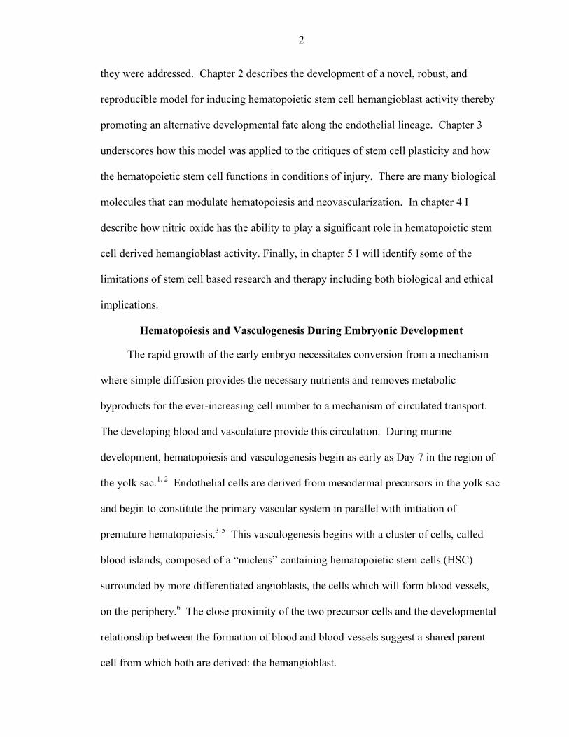
Hematopoiesis and Vasculogenesis During Embryonic Development
The rapid growth of the early embryo necessitates conversion from a mechanism
where simple diffusion provides the necessary nutrients and removes metabolic
byproducts for the ever-increasing cell number to a mechanism of circulated transport.
The developing blood and vasculature provide this circulation. During murine
development, hematopoiesis and vasculogenesis begin as early as Day 7 in the region of
the yolk sac.1, 2 Endothelial cells are derived from mesodermal precursors in the yolk sac
and begin to constitute the primary vascular system in parallel with initiation of
premature hematopoiesis.3-5 This vasculogenesis begins with a cluster of cells, called
blood islands, composed of a “nucleus” containing hematopoietic stem cells (HSC)
surrounded by more differentiated angioblasts, the cells which will form blood vessels,
on the periphery.6 The close proximity of the two precursor cells and the developmental
relationship between the formation of blood and blood vessels suggest a shared parent
cell from which both are derived: the hemangioblast.

3
Until the Day 10 of development, the yolk sac remains the primary site of
hematopoiesis. Around Day 12 the liver which then becomes the primary site of
hematopoiesis.7 However, there are other regions of potential hematopoiesis in the para-
aortic splanchnopoeura (PAS) from Day 8.5 to 10, and the aorta-gonad-mesonephrous
(AGM) region from Day 10.5 through Day 12.7-11 The potential of these areas was
determined through a series of transplantation studies where cells isolated from these
regions are able to rescue lethally irradiated recipients.12-14 This hematopoietic rescue
capability defines the first location from where functionally defined HSC arise.
Endothelial cells on the ventral surface of the aorta are derived from the PAS/AGM
regions, and HSC are also found nestled in the endothelial floor of the aorta, again
suggesting the that this area contains cells which are have the capabilities of the
hemangioblast.12
Formation of Blood Vessels in Adults
Vasculogenesis and angiogenesis are two distinct roles of the hemangioblast.
Vasculogenesis is defined as the de novo generation of blood vessels via the recruitment
of undifferentiated progenitor cells to the site of vessel formation where they differentiate
into vascular endothelium.11 During embryonic development, the vascular system is
formed through vasculogenesis. After development is complete, new blood vessel
formation is attributed to the process of angiogenesis where vessels are formed by
sprouting from the pre-existing vasculature.15 Until 1991, angiogenesis was thought to
occur by the proliferation of resident endothelial cells at the site where new vessels are
forming, but George et al.16 showed that endothelial cells circulate in the blood. They
found that peripheral blood contained endothelial cells by staining blood samples with the

4
endothelial cell specific antibody S-Endo 1 and analyzing these cells by Fluorescence
Activated Cell Sorting (FACS). The discovery of circulating endothelial cells
unvaryingly leads us to question where these cells are derived.
There are two possibilities of circulating endothelial cell parentage: the existing
vasculature where cells extrude themselves from blood vessel walls and enter the
circulation; the bone marrow itself, via an endothelial cell progenitor (EPC) intermediate.
Several studies describe endothelial cells which derived from the bone marrow.17-22 If
this is the case, the HSC and EPC populations could possible be distinguished through
their cell surface marker expression, or through “tagging” of the parent cell. No studies
have yet directly addressed this question; however there is significant indirect evidence
linking endothelial cells to the EPC and its involvement in adult neovascularization. One
such study described several cell surface antigens present on the EPC, such as CD133
and CD34, that are also present on the HSC.23 However, there are differences in the two
populations, namely that fetal liver kinase-2 (VEGFR-2) expression is only found on
committed progenitors.24 This is one of the first hints that EPC may be a more
differentiated or committed HSC daughter cell. CD 34 positive cells can phenotypically
function as endothelial cells after several days of culture on fibronectin. They are
capable of incorporating acetylated LDL, producing nitric oxide when stimulated with
VEGF, and express of PECAM-1 and Tie-2, both of which are specific to endothelial
cells.25 CD133 positive cells appear to be a more immature subgroup of the CD34
population. The CD133 positive cells are able to repopulate the bone marrow
compartment of radioablated sheep, and evidence shows that a subset of cells which are
CD34, CD133 and VEGFR-2 positive may be EPC.26-28 CD133 and CD34 positive cells

5
are believed to be more primitive EPC because they lack VE-cadherin or Von Willebrand
expression. Only 3% of these cells express VEGFR-2.27 CD34 negative, CD133
positive, and VEGFR-2 positive cells may represent a more mature or further
differentiated population of endothelial cells.
The exact markers and phenotype of EPC are not known, and the conditions under
which these cells are stimulated to proliferate, circulate, and home to sites of injury are
poorly understood. There is disparity in the amount of neovascularization occuring in
certain vascular beds with some tissue-types experiencing significantly more vessel
formation in relation to others. In addition, the wide range of ischemia models utilized
for study have been found to induce different levels of neovascularization. Crosby et
al.29 have shown that up to 11% of endothelial cells contributing to neovascularization
are EPC derived. This contribution occurred during injury and was not observed under
normal physiologic conditions. Grant et al.30 demonstrated that circulating endothelial
cells, specifically endothelial cells which contribute to the formation of blood vessels
during injury repair, arise from the HSC through an EPC intermediate. This finding lends
to the possibility of regulating vessel formation at a precursor level through a molecular
mediator. The ability to orchestrate formation of blood vessels is highly desired for
conditions in which pathological vascular growth, or lack of growth, and leads to
damaging conditions ultimately decreasing the quality of life.
Regulation of Neovascularization
Vascular endothelial cells maintain a tight border between the circulating blood and
the outside tissue. This monolayer of cells acts as a non-adherent surface where
circulating cells cannot interact and adhere without the presence of certain surface
markers, such as the integrins or selectins of the cellular adhesion molecule family.
6
While this boundary must necessarily remain intact, mechanisms exist in which cells
within the blood can extravasate into the surrounding tissue in order to fight infection or
provide repair. Conversely, mechanisms exist by which cells in tissue can enter the
bloodstream illustrated by bone marrow cells ability to proliferate in the bone marrow
compartment, migrate to the inner marrow vessels, and enter the circulation. Endothelial
cells generally have a very low level of apoptosis and thus a low turnover rate. Cells in
certain organs, such as the eye, can live for years without being replaced.31 As a result,
there are infrequent endothelial cells circulating in healthy adults usually numbering 1-3
per milliliter of blood.32 This emphasizes how the steady state of endothelial cells is non-
dividing unless stimulated by injury when mechanisms to upregulate endothelial mitosis
stimulate proliferation.
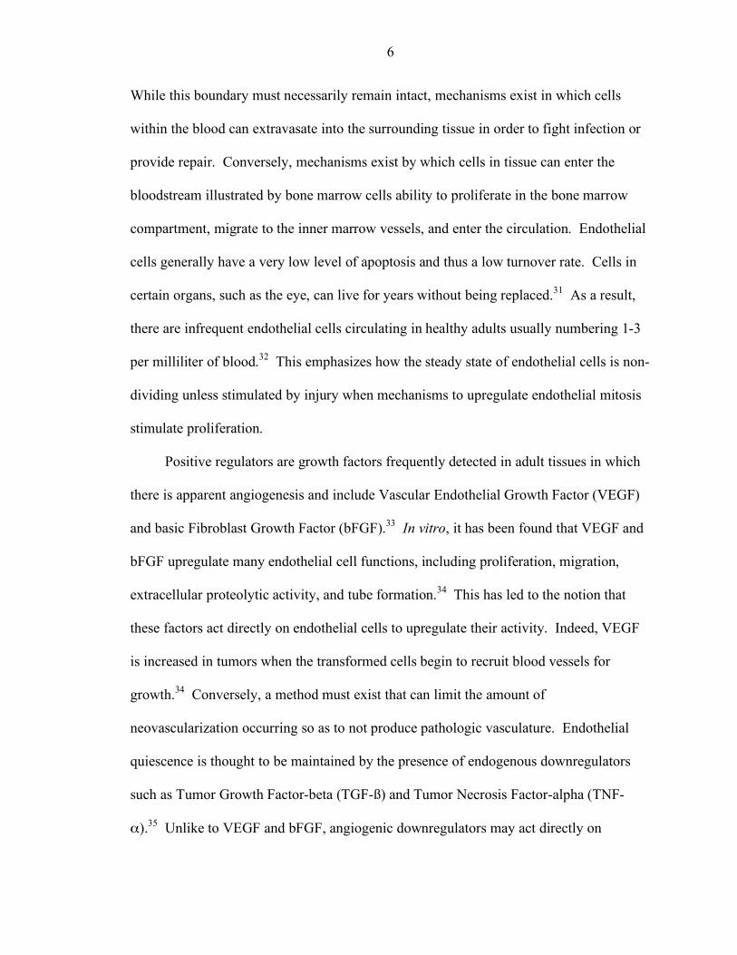
Positive regulators are growth factors frequently detected in adult tissues in which
there is apparent angiogenesis and include Vascular Endothelial Growth Factor (VEGF)
and basic Fibroblast Growth Factor (bFGF).33 In vitro, it has been found that VEGF and
bFGF upregulate many endothelial cell functions, including proliferation, migration,
extracellular proteolytic activity, and tube formation.34 This has led to the notion that
these factors act directly on endothelial cells to upregulate their activity. Indeed, VEGF
is increased in tumors when the transformed cells begin to recruit blood vessels for
growth.34 Conversely, a method must exist that can limit the amount of
neovascularization occurring so as to not produce pathologic vasculature. Endothelial
quiescence is thought to be maintained by the presence of endogenous downregulators
such as Tumor Growth Factor-beta (TGF-ß) and Tumor Necrosis Factor-alpha (TNF-
α).35 Unlike to VEGF and bFGF, angiogenic downregulators may act directly on
7
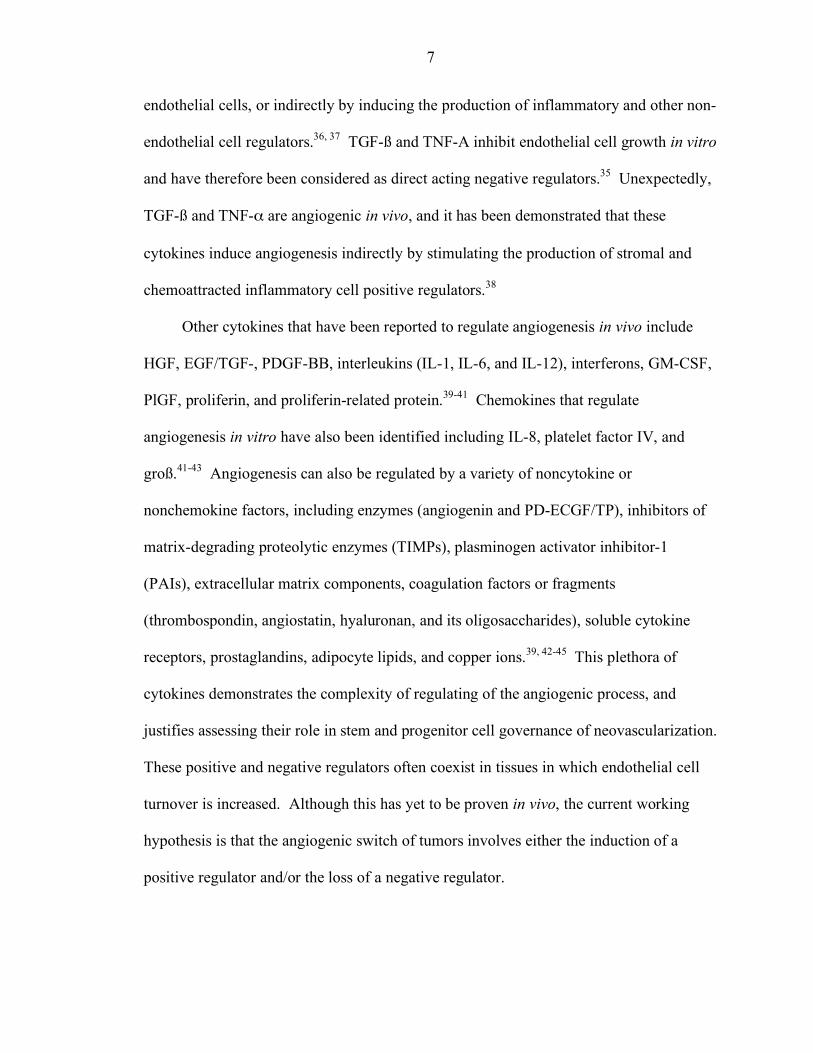
endothelial cells, or indirectly by inducing the production of inflammatory and other non-
endothelial cell regulators.36, 37 TGF-ß and TNF-A inhibit endothelial cell growth in vitro
and have therefore been considered as direct acting negative regulators.35 Unexpectedly,
TGF-ß and TNF-α are angiogenic in vivo, and it has been demonstrated that these
cytokines induce angiogenesis indirectly by stimulating the production of stromal and
chemoattracted inflammatory cell positive regulators.38
Other cytokines that have been reported to regulate angiogenesis in vivo include
HGF, EGF/TGF-, PDGF-BB, interleukins (IL-1, IL-6, and IL-12), interferons, GM-CSF,
PlGF, proliferin, and proliferin-related protein.39-41 Chemokines that regulate
angiogenesis in vitro have also been identified including IL-8, platelet factor IV, and
groß.41-43 Angiogenesis can also be regulated by a variety of noncytokine or
nonchemokine factors, including enzymes (angiogenin and PD-ECGF/TP), inhibitors of
matrix-degrading proteolytic enzymes (TIMPs), plasminogen activator inhibitor-1
(PAIs), extracellular matrix components, coagulation factors or fragments
(thrombospondin, angiostatin, hyaluronan, and its oligosaccharides), soluble cytokine
receptors, prostaglandins, adipocyte lipids, and copper ions.39, 42-45 This plethora of
cytokines demonstrates the complexity of regulating of the angiogenic process, and
justifies assessing their role in stem and progenitor cell governance of neovascularization.
These positive and negative regulators often coexist in tissues in which endothelial cell
turnover is increased. Although this has yet to be proven in vivo, the current working
hypothesis is that the angiogenic switch of tumors involves either the induction of a
positive regulator and/or the loss of a negative regulator.

8
Stem Cell Transplantation
The adult bone marrow (BM) is a rich reservoir of tissue specific stem and
progenitor cells. BM cells may be a source of EPC. Therefore tapping into BM in
combination with neovascularization regulators may provide significant and manageable
therapy. Stimulation of angiogenesis may be of benefit in wound healing and fracture
repair. Therapeutic growth will also be beneficial in the treatment of ischemia, and
substantiated by extensive experimental data.46-49 Pesce et al.49 demonstrated that under
ischemic conditions, transplanted umbilical cord cells gave rise to enhanced arteriole
length and density along with skeletal muscle fibers. Another group transplanted early
bone marrow cells into nonirradiated, aged mice and found a contribution to vasculature
from subsequently transplanted neonatal myocardium.48 In addition, Orlic et al.50
demonstrated that bone marrow cells can differentiate into myocytes and vascular
structures. They also mobilized bone marrow cells with stem cell factor and granulocyte-
colony stimulating factor and found that marrow cells could home to infarcted regions of
the heart, replicate, differentiate, and ultimately promote myocardial repair.51 This could
lead to significant alterations and improvements in treatment for cardiac ischemia.
Current therapy for myocardial ischemia relies on drugs that reduce myocardial
oxygen demand, mechanical endovascular revascularization procedures (angioplasty), or
bypass surgery.52 However, compensatory neovascularization is an important
physiological process that occurs in chronic myocardial ischemia.53 It has recently been
demonstrated in experimental models of myocardial ischemia and infarction in the pig
and rat that VEGF and VEGF receptors 1 and 2 are increased in chronically ischemic
myocardium and also in regions of ischemia surrounding an area of infarction.54-56 Those
studies demonstrated that the VEGF ligand is upregulated in cardiomyocytes and its
9
cognate receptors exhibited increased expression in endothelial cells. Further studies
have revealed that hypoxia is a potent inducer of VEGF in cultured cardiac myocytes.57
Correspondingly, escalated bFGF activity has been shown in myocardium after coronary
artery ligation.58 This occurs in parallel with an increase in collateral blood flow in dogs,
and elevated levels of bFGF (but not VEGF) have been detected in the pericardial fluid of
patients with unstable angina.52 These observations on the molecular mechanisms of
physiological angiogenesis in ischemic myocardium led to the notion that cell based
therapy or pharmacological stimulation of angiogenesis may augment or even replace
more conventional forms of therapy. As will be described next, this notion has recently
received considerable experimental support in animal models.
Vascular healing may be mediated in part by the recruitment of EPC. In several
studies, genetically marked bone marrow-derived EPC were recruited to the ischemic
limbs of mice.11,17 In addition, transplantation of mature endothelial cells (EC) derived
from in vitro generated, human bone marrow-derived, multipotent adult progenitor cells
has facilitated revascularization of various tissues.59 The physiologic significance of
EPCs and EC in neovascularization was further underscored when thoracic aorta from
adult dogs previously transplanted with haploidentical bone marrow,were replaced with
Dacron grafts impervious to the ingrowth of established EC. In 3 month old grafts, the
newly established EC layer were determined to arise from donor derived cells from the
bone marrow.58 These findings indicate that EC derived from the EPC of bone marrow
origin can contribute to new blood vessel formation.
EPC for Neovascularization
This low number of EPC in the circulation increases dramatically under conditions
such as acute stress or injury to vasculature walls where there is a large apoptotic event of

10
EC. Normal replacement of the EC is usually accomplished by the surrounding local
endothelial cells which increase their proliferation and migrate to the areas of ischemia.
The terminally differentiated EC, however, are not able to proliferate considerably and
may not have the capacity to provide for the demand for new vessels. As described in
numerous stuidies, researchers have isolated circulating cells that are bone marrow
derived yet have endothelial potential—the EPC. These EPC are capable of lessening the
ischemic pressure of injured organs by revascularizing injured areas and restoring organ
function.
Our current understanding of the neovascularization process is founded on the
classical light-microscopy observations made by Clark and Clark in 1953.60 They were
among the first to reveal the sequence of events leading to the formation of new capillary
blood vessels in the translucent tails of amphibian larvae. These and later observations in
nondevelopmental settings provided a detailed histological account of new blood vessel
formation.61, 62 On these pioneering results our current knowledge was founded. Clark
and Clark described a local angiogenic stimulus that causes endothelial cells of
preexisting capillaries or postcapillary venules to become activated. Although the precise
molecular consequences of this activation process remain to be clearly defined, activated
blood vessels are vasodilated, have increased vascular permeability, and experience
accumulation of extravascular fibrin as well as proteolytic degradation of the basement
membrane of the parent vessel.46-48 The endothelial cells then extend thin cytoplasmic
arms which direct migration into the surrounding matrix towards the angiogenic stimulus.
Migrating endothelial cells elongate and align with one another to form a capillary sprout,
and endothelial cell division, which occurs proximal to the migrating tip, further

11
increases the length of the sprout. The solid sprout gradually develops a lumen proximal
to the region of proliferation. Contiguous tubular sprouts fuse at their tips to form a
functional capillary loop in which blood flow is soon established. Vessel maturation is
accomplished by reconstitution of the basement membrane and recruitment of mural
cells.49 These cellular functions contribute to the formation of patent, endothelium-lined,
blood vessel structures.
Nitric Oxide as Potential Regulator of Vascular Formation
The process of angiogenesis in the adult is a complex sequence of growth factor
release, vasodilation, and recruitment or proliferation of endothelial cells to build the
vessels. These events are heralded by EC activation, most notably vasodilation, which
facilitates growth by granting access for cells to enter the area and remove any damaged
and dead cells/debris, increases nutrient depositing and breakdown of existing
extracellular matrix, and allows cells to establish permanent residence. One of the
molecules which has been shown to play an extensive role in vasodilation is Nitric Oxide
(NO).
NO has been used in nature for over 250 million years, longer than mammals have
existed. The horseshoe crab uses NO to prevent blood cell aggregation, and this function
is still retained in mammals. Other kingdom and phyla also utilize NO including fireflies
for their flashes, and plants that use NO’s cytotoxic effects to fight infection. Victorian
physicians recognized its vasodilatory effect, even if they did not understand its
mechanism, and its medicinal value was written in a Sherlock Holmes story.130 The
medical uses for NO continued into World War I where doctors noticed that factory
workers in ammunition plants had lower blood pressures. This led directly to the
nitroglycerine tablet still used today to treat angina. The gas molecule itself, however,

12
was considered only a pollutant until recently. In the early 1990s the journal Science
named it molecule of the year. During this time over 250 articles per month were written
further characterizing NO and its effects. Robert F. Furchgott, Louis J. Ignarro, and
Ferid Murad received the Nobel Prize in Medicine in 1998 for their work on "nitric oxide
as a signaling molecule in the cardiovascular system." One historic irony is that Alfred
Nobel made his fortune by making dynamite from nitroglycerine, a known NO donor.
NO is unique among physiologic substances in the body as it is the only gas
produced in mammals that has a biological effect. This singular messenger molecule is
involved in the regulation of diverse physiologic functions including central and
peripheral nerve cell neurotransmission, promotion of the cytotoxic actions of immune
cells, and preventing/increasing leukocyte adhesion.63-67 It also has profound vasomotor
regulatory affect on vascular beds, specifically the regulation of smooth muscle
contractility and thus vasodilation.63, 64
Three distinct isoforms of the enzyme that synthesizes NO (NOS) have been
identified, all of which share a 50-60% homology.67 Two isoforms are constitutively
active: the form expressed primarily in neuronal tissue (nNOS) and the form first found
in vascular endothelial tissue (eNOS). The third form’s activity can be induced in a
variety of cell types usually in response to inflammatory signals and bacterial products,
and has been named inducible NOS (iNOS). Each of the three isoforms require
homodimerization for activity. The C-terminal portion of the NOS protein closely
resembles the cytochrome P-450 reductase possessing many of the same cofactor binding
sites.68 The extreme C-terminus contains an NAPDH binding region, conserved in all
three isoforms, that exactly aligns with the binding region of the cytochrome P-450.68
13
Following this is a flavin adenine dinucleotide and flavin mononucleotide consensus
sequence that is self-sufficient, unlike the P-450 enzyme, in that the oxygenation of its
substrate L-arginine occurs at the heme site in the N-terminal region.69 NO is generated
via a 5-electron oxidation of a terminal guanidinium nitrogen on L-arginine.68
Most of the physiologic actions of NO are brought about by the activation of
soluble guanylate cyclase. Binding of NO to the heme moiety of the enzyme causes a
conformational change that upregulates the activity over 400-fold resulting in the
formation of the intracellular second messenger cyclic GMP.70 NO has numerous
angiogenic effects, including (but not limited to) increasing matrix metalloprotinase
(MMP) expression and tyrosine phosphorylation of proteins in sprouting tips of
capillaries.65 Inhibiting NO production has been shown to decrease capillary formation
in rats with portal hypertension.66 In addition, DNA synthesis can be impaired by the
inhibitory effect of NO on ribonucleotide reductase which addresses the cytotoxic and
cytostatic effect of NO during an immune response. In the aqueous environment of the
cytosol, NO interacts with water to form the free radical peroxynitrate.67 Peroxynitrate
interacts with DNA leading to oxidation and initiation of a complex series of
transformations involving base damage or strand breaks as well as reactions with the
deoxyribose portion of the DNA.71 The DNA damage itself, along with the cell cycle
arrest as repeated and costly DNA repair occurs, ultimately leads to apoptosis.
Role of NOS in vessel formation
The process of angiogenesis can be divided into two components: endothelial cell
proliferation and blood vessel tube formation. The potent angiogenic agent VEGF
stimulates NO release from endothelial cells.72 VEGF-induced NO release has been
shown to modulate angiogenesis both in vitro and in vivo.73, 74 The adult mouse model we

14
have developed utilizes the angiogenic influence of VEGF as we artificially increase
local expression of this growth factor in the retina mimicking the pathophysiology that
occurs in diseases associated with retinal neovascularization such as Diabetic
Retinopathy and Retinopathy of Prematurity. The established resident vascular
endothelial cells, the endothelial cells found in the circulation, and those derived from
HSC all respond to VEGF and influence local NO concentration. NO is crucial for the
myriad of physiological vascular functions, and its inappropriate production and release
has been linked to several pathologies.75 Consequently, agents which modulate NO
activity could find beneficial use in a therapeutic setting. As has been shown, NO plays
an integral role in blood vessel formation, and consequently makes a good starting
candidate for manipulating hemangioblast function.
The two isoforms which have a direct influence over endothelial cells are the iNOS
and eNOS isoforms as nNOS is found only in neuronal tissue.67 The role of eNOS in
angiogenesis is complex. Brooks et al. have demonstrated that eNOS deficiency, either
through gene disruption or through pharmacological inhibition, significantly protects the
developing retina from oxygen-induced retinopathy.76 The fact that nonspecific
inhibitors of NOS activity produced quantitatively similar levels of vaso-obliteration
compared to eNOS gene disruption also suggests that eNOS may be an isoform involved
in blood vessel regulation. Evidence suggests that NO and VEGF are reciprocally
regulated such that stimulation of VEGFR-2 activates eNOS leading to NO formation.76
NO inhibits VEGF production in adjacent cells by a paracrine feedback mechanism
involving inhibition of AP-1 binding to the VEGF promoter.77
15
iNOS has consensus sequences in its promoter for the transcription factors
hypoxia inducible factor (HIF) and NF-kappa B, both of which are activated under
conditions of ischemia.78 Consequently, iNOS is thought to be induced under conditions
of ischemia. Sennlaub et al. perfused retinas of wild type and iNOS knockout (iNOS -/-)
mice exposed to hypoxic conditions. They found that iNOS -/- animals had normal
intraretinal vasculature patterning whereas wild type animals had persistent avascular
areas.79 Interestingly, there was a reduction in preretinal neovascularization in iNOS -/-
mice indicating a dual role of iNOS in distinct retina layers. They corroborated these
observations with pharmacological inhibition of iNOS which increased retinal
neovascularization and decreased preretinal neovascularization. They found that
pathological intraretinal neovascularization was more severe in iNOS expressing
animals.80 These studies suggest that NO can be an important modulator of angiogenesis
in the retina, and that local levels of NO can influence the location and degree of
neovascularization. To our knowledge our model is the only one which allows for the
simultaneous examination of preretinal and intraretinal neovascularization at the same
time in an adult animal. We will use this model to understand the requirement of
beneficial intraretinal neovascularization compared to pathological preretinal
neovascularization allowing for the dissection of NO and other molecules which affect
vascular growth.

16
CHAPTER 2 GENERAL METHODS AND MATERIALS
The methods detailed below are used extensively in each chapter. Any
modifications made to this framework during an experiment are noted in the specific
chapter. Methods will be described in this basic outline: (1) the generation of the
GFP/BL6 chimera, (2) the induction of the retinal neovascularization, (3) the enucleation
of the eye for mounting, (4) examination of neovascularization via confocal microscopy
and (5) immunohistochemistry staining of serial sections.
Generating The GFP/BL6 Chimera
The generation of the chimeric GFP/BL6 animal will be described below. This
includes the harvesting of bone marrow from the GFP donor animal, the purification and
preparation of the marrow for FACS sorting of HSC, the preparation of the C57BL6
rescue marrow and recipient animals, and the HSC transplant and commensurate animal
husbandry concerns.
Harvesting Bone Marrow
The generation of the GFP/BL6 chimera animals requires extensive animal use and
cell manipulation. The transgenic mouse used as the donor strain was obtained from
Andras Nagy at mount Sanai in Toronto Canada.81 The strain carries green fluorescent
protein (GFP) driven by chicken beta-actin promoter and CMV intermediate early
enhancer and is ubiquitously expressed. The BL6 females were obtained from Jackson
Laboratories (Bar Harbor, Maine) and were at least 5 weeks old at the time of bone
marrow transplantation. Recent controversy concerning the events during stem cell

17
transdifferentiation for repair has led to the possibility that this may not be an inherent
ability stem cells, but rather a fusion event occurring between the stem cell and target
tissue. The transplantation of male HSC into female recipients directly addresses this
issue by allowing for fluorescent in situ hybridization of tissue samples looking for the Y
chromosome and determination if a fusion event has occurred. After fully-grown GFP
males are euthanized and sacrificed, the long bones in the legs were immediately
removed. All muscle, tendon, and ligature was dissected from the bone which was
immediately placed in ice-cold PBS. Each bone end was then pruned back about 1-2
millimeters to expose the hollow core of the marrow space. The bone marrow was
flushed out into a tissue culture treated plate by inserting a 26-gauge needle into one end
of the bone and washing 1-2 milliliters of Dulbecco’s Modified Eagle’s Medium (Gibco)
through the hollow bone core. The cells were kept on ice at all times. The liberated
marrow was then triturated with a 26-gauge needle to break up the cell clumps and
allowed to adhere to a tissue culture treated plate (Gibco) for 120 minutes. This step
allows for an initial enrichment of HSC from other adherent progenitor cells such as
mesenchymal stem cells (MSC) since hematopoietic progenitor and stromal cells adhere
to the tissue culture treated plastic, while HSC will remain suspended in the media. The
complete volume of media containing the nonadherent HSC was then gently drawn up,
washed in >10mL volume of cold media, and pelleted by centrifugation at 1000 x g
performed at 4 degrees Celsius. The cells were resuspended and stained as outlined by
the protocol of the Milteny MACS system in the following section.
Initial Purification of HSC by MACS
Initial HSC purification was done through sorting of the cells by magnetic beads
using the Milteny Magnetic Activated Cell Sorting (MACS) system. Briefly, cells were

18
stained with an antibody conjugated to a magnetic bead. The antibody, and subsequently
the bead, is bound to the cell. When these cells are then run over a column in the
presence of a magnetic field, those cells which have the specific surface antigens, and
thus the antibody-bead bound to them, will adhere to the column (termed positive
fraction). Cells which do not present that surface marker (negative fraction) will pass
directly through the magnetic field and be removed from the positive fraction of cells.
The magnetic field can then be removed and the positive fraction collected from the
column.
To begin the MACS enrichment, cell number and viability were determined from
the total marrow flushed from the long bones to ensure that the correct amount of
antibody, beads, and staining volume will be used. To determine the cell number, I
resuspended the washed cells in trypan blue and counted bright cells using a
hemacytometer under a phase-contrast microscope. The enumerated cells were then
washed in >10mL cold PBS and stained with Sca-1 microbeads (Milteny) in appropriate
volume. The cells were run over 2 separate columns to insure enrichment, and the flow-
through was discarded and the positive fraction retained. At this time a >90% Sca-1
positive purity typically has been achieved. After enrichment, cells were immediately
pelleted and placed back on ice for fluorescent antibody staining for FACS sorting.
Final Purification of HSC by FACS
Again all antibody concentrations and incubation times were followed according to
the parameters described by the manufacturer guidelines. For HSC purification I used
three different fluorochromes: C-KIT conjugated to APC, biotynylated Sca-1 (with
Streptavidin-PharRed secondary antibody), and the lineage markers B220, CD3, CD4,
CD8, CD11B, GR-1, and TER-119 all directly conjugated to PE (Pharmingen). The

19
FACSvantage SE is able to isolate single cells based on the surface antigen bound by
antibodies and hence the spectrum of absorbance and fluorescence emitted by that cell.
Two rounds of purification are needed to ensure complete removal of all non-HSC cells.
See Figure 2-1 for of an example of the gates used to enrich and isolate single HSC.
Figure 2-1. Fluorescence activated cell sorting gates for isolating HSC. HSC were removed from bone marrow, enriched by MACS, and stained for SKL surface expression. First panel: Forward and Side Scatter of MACS enriched cells with gate R1 drawn. Second panel: Cells are enriched for GFP and Lineage positive cells (B220, CD3, CD4, CD11b, Gr-1, Ter-119) are depleted excluding gate R2. Third panel: Sca-1 and c-kit positive cells from gate R1 and R2 are enriched in gate R3. Cells are then further enriched by gate R4 based on the same parameters. Panel 4: Reanalysis of cells based on Sca-1 and c-kit expression. These doubly sorted enriched cells were used for transplantation.
The flow rate is set at 10,000 events per second with no greater than a 10% abort
proportion. The cells were then collected in media immediately after completion of the
sort, isolated, and injected into the recipient animals following "rescue" marrow isolation
and recipient preparation kept on ice at all times.
Harvesting of BL6 Rescue Marrow with HSC Depletion, and Irradiation of Recipient Animals.
The harvesting of non-GFP female BL6 marrow was performed in the same
manner as the HSC, except these cells were not given time to adhere to the tissue culture
treated plate. Once the marrow was flushed, washed, and counted, a Sca-1 depletion was
done to remove any HSC from the rescue marrow which would compete with the donor

20
GFP HSC. This rescue dose is administered for twofold reasons. The immune system of
the irradiated animal will experience an interruption and often the animal will become
anemic. Until the HSC can engraft and repopulate hematopoiesis, these short term rescue
progenitors will help the animal mount an immune response and provide the necessary
blood products as needed. Again cells were stained as described in the MACS magnetic
bead section, but this time the cells were Sca-1 depleted three times to ensure that the
rescue marrow was devoid of HSC. Recipient BL6 mice were finally irradiated with 950
RADS of gamma radiation to prepare the bone marrow for transplantation.
Purified GFP HSC and Depleted Rescue Marrow Transplantation and Ensuing Animal Husbandry Concerns
The HSC depleted rescue marrow was count as above and 1 x 10⁶ cells in a 100
microliter volume were aliquoted into a fresh Eppendorf tube. The highly enriched HSC
were then singly isolated in the following manner. A volume of the sorted sample was
placed on a glass drop slide and examined under a phase-contrast microscope. The cells
were diluted to a concentration where single cells can be visualized, isolated, and
captured one at a time with a micropipette. Under the scope a single, round, bright,
viable cell was isolated and drawn up into a pulled glass micropipette by mouth pipetting
with a suction tube. The needle was examined to visualize the cell to ensure that only
one cell was drawn. The cell was then place into the 100 microliter aliquot containing
the HSC depleted rescue dose. The rescue/single HSC mixture was drawn into a fresh
insulin needle and syringe to ensure no contamination of other samples. Finally, an
anaesthetized, irradiated BL6 animal was injected in the retro-orbital sinus cavity. The
animals were monitored until they overcome the effects of the anesthetic and then be

21
placed on a regime of antibiotics for the next month until multilineage engraftment had
been verified.
Verification of Multilineage Reconstitution
The recipient animals were given one month for the HSC to home to the bone
marrow niche and begin to divide to produce progenitor cells which will contribute to the
various hematopoietic cell lineages. Determination of engraftment was resolved by
peripheral blood sampling and FACS analysis to determine whether the marrow was
repopulated or if the animal’s native marrow recovered. Each animal had a peripheral
blood sample drawn through a tail vein bleed and the blood was collect in a tube
containing PBS and 5mM EDTA to act as an anticoagulant. The erythrocytes were
removed with a FICOLL PLAQUE (Amersham Biosciences) purification. Briefly, the
blood/PBS sample was layered on top of two times greater volume of FICOLL. The
emulsion was centrifuged and the “buffy” layer containing the nucleated cells at the
interface was removed. The lymphocyte layer containing the nucleated cells was washed
in 5X volumes of PBS and stained with the various lineage marker antibodies conjugated
to PE. Samples were analyzed by FACS caliber, and animals exhibiting GFP positive
cells of the various lineages were scored positive for engraftment. The positive animals
were then monitored an additional three months where multi-lineage reconstitution is
reconfirmed to demonstrate long-term engraftment by HSC. Exogenous growth factor
was then administered as described below.
Induction of Retinal Ischemia
The next step involves administration of an endogenous growth factor and vessel
damage in order to promote blood vessel growth in the retina. Fully and robustly
engrafted animals were selected and anaesthetized. VEGF was administered directly into
22
the vitreous using a 36-gauge needle and Hamilton syringe. Either purified (40ug/kg)
VEGF protein (Sigma) or (2 x 10⁸ particles) AAV-VEGF (VectorCore, UF), where CMV
promoter drives expression of VEGF in an Adeno Associated Vector, was used. VEGF is
an endothelial cell-specific mitogen which is transcriptionally regulated by the
cytomegalovirus promoter/enhancer when packaged in AAV. AAV mediates long-term
expression in nondividing cells, which allows for stable expression and constant amounts
of VEGF to reach the area of ischemia to promote neovascularization.30
The study of clinical diseases such as Diabetic Retinopathy and Retinopathy of
Prematurity has led to an understanding of the pathology which occurs in these diseases.
In these conditions the eye "detects" a lack of oxygen, either due to the diabetic condition
leading to leaky vessels, or the removal of a prematurely born baby from an incubator’s
oxygen-rich environment. The model takes advantage of this neovascularization by
creating a local region of ischemia in the eye through cauterizing of large blood vessels
with a laser. As a result, the cells signal new blood vessel growth in the region in an
attempt to relieve the ischemic pressure.
Peak expression of VEGF by AAV has been determined to be at 3-6 weeks,
therefore the physical disruption of the blood vessels is done during this time
(unpublished data). First, mice were anaesthetized normally with a general anesthetic,
and concurrently a 10% sodium fluorescein (Akorn) solution was administered
intraperitineally. This dye labels blood vessels facilitating visualization during
photocoagulation. The eyes were dilated with 1% atropine (Akorn) for 5 minutes,
washed with PBS (Gibco), and subsequently dilated with 2.5% phenylephrin (Akorn) for
5 minutes. Immediately after the two 5 minute treatments the mice underwent laser

23
treatment. An Argon Green laser system (HGM Corporation) was used for retinal vessel
photocoagulation with the aid of a 78-diopter lens. The blue-green argon laser
(wavelength 488-514 nm) was applied to various venous sites juxtaposed the optic nerve.
The venous occlusion were accomplished with >60 burns of 1-sec duration, 50 millimeter
spot size, and 50-100 milliwatt intensity. Again the animals were allowed to recover for
30 days while the transplanted HSC, directed by the ischemia and induced by the VEGF,
contributed to the neovascularization in order to relieve the hypoxia produced by the
cauterizing of the existing vessels.
Eye Removal
One month after ischemic injury the eyes were ready to be enucleated and
neovascularization imaged by confocal microscopy. Mice were first anesthetized and
then perfused while sedated. Peripheral blood and bone marrow was collected to confirm
donor contribution analysis by FACS with lineage specific antibodies conjugated to PE
(BD BioSciences) similarly to the procedure outlined above. First, the chest cavity was
opened and the ribs cut away to expose the heart completely. The left atria was
punctured with a 26-gauge needle and injected with >3 mL of 50 mg/mL tetramethyl
rhodamine isothiocyanate (TRITC)-conjugated dextran (160,000 avg. MW, Sigma
Chemical) in phosphate-buffered formaldehyde, pH 7.4. The perfusion was performed
slowly into the left ventricle and is integral for the functional assay. Immediately
afterwards the eyes were removed by sliding a curved forceps underneath the eyeball and
pulling the globe out. The eye was punctured with a 26-gauge needle to allow complete
perfusion. The eye was placed in fresh 4% PFA and shaken at room temperature for 30
minutes. The globe was then transferred to 1X PBS and washed by shaking at room
temperature for 30 minutes to overnight. After washing with PBS the eyes were

24
dissected. To do this I placed the eye under a surgical microscope and made an initial
incision in the cornea. The opening was enlarged until it could accommodate the lens of
the eye. The lens was gently pushed forward until it exited through the hole cut in the
cornea. The remaining cornea was then trimmed to where the sclera and cornea meet.
The retina was dissected away from the retina pigment epithelial (RPE). To do this I
gently pushed down on the posterior portion of the RPE and rolled the forceps forward.
The retina then detached and was readily mounted. The thickness of the retina (>200um)
prevents adequate perfusion of antibody, therefore the retina was placed on a glass slide
and 5-6 cuts were made around the periphery so that the retina lies flat when mounted.
The tissue was placed in Vectashield mounting medium (Vector Laboratories) to inhibit
photo-bleaching. The retinas were immediately imaged. I used an Olympus IX-70, with
inverted stage, attached to the Bio-Rad Confocal 1024 ES system for fluorescence
microscopy. A Krypton-Argon laser with emission detector wavelengths of 598nm and
522nm differentiated the red and green fluorescence. The lenses used in our system were
the (Olympus) 10X/0.4 Uplan Apo, 20X/0.4 LC Plan Apo, 40X/0.85 Uplan Apo,
60X/1.40 oil Plan Apo and 100X/1.35 oil Uplan Apo. The software was OS/2 Laser
Sharp.

25
CHAPTER 3 THE HEMATOPOIETIC STEM CELL HAS HEMANGIOBLAST ACTIVITY
During development there are several types of stem cells broadly classified based
on their ability for form specific tissue types. After fertilization during the first few days
of division, the embryonic cells are described as totipotent. They have the capacity to
produce all the cells, tissues and organs that make up the body along with all of the
extraembryonic tissue of the trophectoderm. After the first four to five cell divisions, the
embryo forms a hollow sphere called the blostocyst. The blastocyst contains a population
of cells located in the inner wall which are capable of producing each of the over two
hundred different cell types of an organism. These differ from the totipotent cells in that
no one of them can produce an entire organism, nor can they produce the cells of the
trophectoderm. Finally, after birth and into adulthood, several types of tissues have cells
residing within them which are able to produce the tissue type where they reside. This
can occur constantly, such as the hematopoietic stem cell producing all of the blood cells,
or only in times of stress or injury such as the oval cells producing hepatocytes. These
stem cells are called multipotent, and in most cases under “normal” conditions these cells
are thought to produce only one cell type.
In the adult, stem cells are believed to define unspecialized cells that can self-renew
(or proliferate) for extended periods of time without differentiating. This process is not
well understood, but is believed to involve asymmetric cell division where a copy of
itself is produced along with a further differentiated daughter cell. These stem cells
exhibit a stable, normal chromosome complement and cannot perform any specialized
26
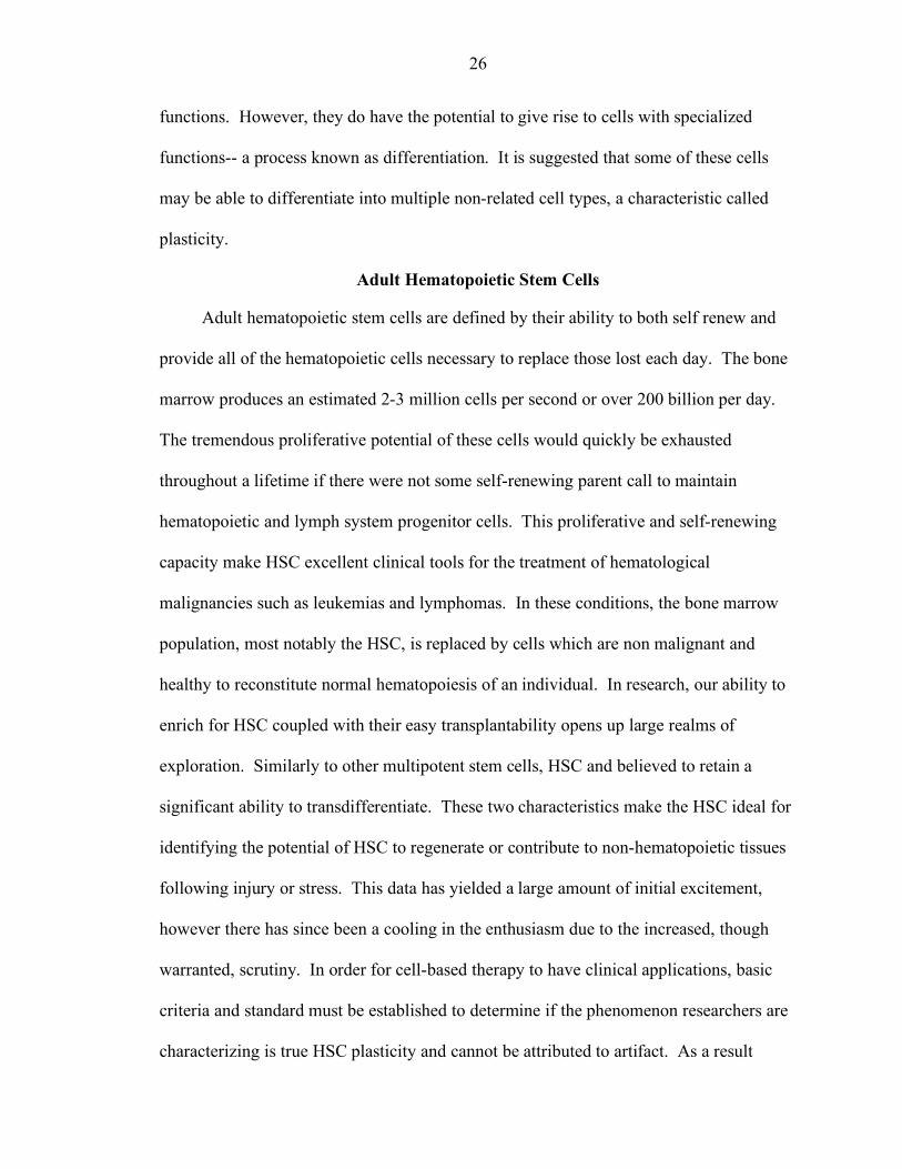
functions. However, they do have the potential to give rise to cells with specialized
functions-- a process known as differentiation. It is suggested that some of these cells
may be able to differentiate into multiple non-related cell types, a characteristic called
plasticity.
Adult Hematopoietic Stem Cells
Adult hematopoietic stem cells are defined by their ability to both self renew and
provide all of the hematopoietic cells necessary to replace those lost each day. The bone
marrow produces an estimated 2-3 million cells per second or over 200 billion per day.
The tremendous proliferative potential of these cells would quickly be exhausted
throughout a lifetime if there were not some self-renewing parent call to maintain
hematopoietic and lymph system progenitor cells. This proliferative and self-renewing
capacity make HSC excellent clinical tools for the treatment of hematological
malignancies such as leukemias and lymphomas. In these conditions, the bone marrow
population, most notably the HSC, is replaced by cells which are non malignant and
healthy to reconstitute normal hematopoiesis of an individual. In research, our ability to
enrich for HSC coupled with their easy transplantability opens up large realms of
exploration. Similarly to other multipotent stem cells, HSC and believed to retain a
significant ability to transdifferentiate. These two characteristics make the HSC ideal for
identifying the potential of HSC to regenerate or contribute to non-hematopoietic tissues
following injury or stress. This data has yielded a large amount of initial excitement,
however there has since been a cooling in the enthusiasm due to the increased, though
warranted, scrutiny. In order for cell-based therapy to have clinical applications, basic
criteria and standard must be established to determine if the phenomenon researchers are
characterizing is true HSC plasticity and cannot be attributed to artifact. As a result

27
several stringent criteria have been outlined which must be fulfilled in order to
demonstrate true plasticity.
The criteria demonstrating HSC plasticity is three-fold. First, the cell must be
capable of self-renewing and homing to the bone marrow thereby reconstituting
hematopoiesis for the lifetime of the organism. This is necessary so that short term
progenitors are not used as therapy which may slowly die off as progenitors differentiate
and are not replaced. Long-term repopulating self-renewing cells must be transplanted so
that the therapy would not fail and the disease or pathologic condition reemerge.
Secondly, the bone marrow contains a myriad of cell types ranging from those along any
point of hematopoietic development to the supporting cells of the stroma. During a bone
marrow transplant, a number of these cells could be transplanted with the bolus
containing the enriched HSC no matter stringent the purification parameters. These
“contaminating” cells could contribute to the tissue type where the donor-derived tagged
cells are found confounding results. In order to conclusively demonstrate the plasticity of
the HSC, clonal studies must be done. Through clonal transplants, a single cell must be
shown to be able to produce the blood along with the non-hematopoietic tissue. These
experiments exclude the possibility of several different cells accomplishing different
roles, and tissue which arises from the donor must necessarily be from the single cell.
Finally, for these cell based therapies to be practical it must be demonstrated that the
plasticity measured is robust and functional transdifferentiation into the non-
hematopoietic tissue. Many cells, especially those of the immune system, are capable of
assuming the general morphology or even surface marker expression of cells they are
nearby either due to stimulation or macrophage engulfment. It must be demonstrated that

28
the cells are physiologically performing the role of the tissue they are replacing, i.e. cells
that are residing in the pancreas having the morphology and characteristics of beta cells
must actually produce insulin to be therapeutic. In addition, a few isolated cells capable
of producing insulin will not rescue a person from diabetes, therefore the
transdifferentiation or plasticity must be robust producing a physiologically relevant
amount of tissue. Only when these three stringent criteria have been met can the cell be
classified as plastic. To date there has been relatively few examples fulfilling all three,
although those that have present some exciting potential.
One of the initial studies have shown that after long term stable hematopoietic
reconstitution by a single bone marrow HSC, donor-derived cells could be found in
multiple tissues including the brain, skeletal and cardiac muscle, liver, and endothelial
cells.82 This elegant work used a homing assay to isolate HSCs which presented stem
cell specific surface markers and then were able to successfully home to the bone marrow
niche. These homed cells were then isolated and single cells were transplanted into
lethally irradiated recipients. While this work was of note, there was as significantly low
level of contribution to the various tissues and there was no functional assay of the donor-
derived cells. It does, however, suggest the exciting possibility of regeneration of various
damaged tissues by HSC-derived progenitors. Two notable studies also demonstrated the
plasticity of the HSC in liver to replace hepatocytes injured chemically.83, 84 Excitingly,
these cells were able to restore liver function, however, clonal assays were not done in
these transplant studies. In addition, Orlic et al. demonstrated the functional recovery of
cardiac muscle through HSC transplantation.50 After these initial pioneering papers a
flood of work was embarked upon, however since then the tide was stemmed due to the

29
difficulty of meeting all three criteria.85 Grant et al. has developed a model mimicking
diabetic retinopathy, and using this model we have been able to expand the understanding
of HSC while fulfilling the three plasticity criteria.30
Diabetic Retinopathy
Diabetic retinopathy is the leading source of legal blindness among working-age
Americans. It is caused by damage to the small blood vessels in the retina as a result of
diabetes mellitus. It is estimated that over fourteen million people in the United States
have diabetes with approximately half of these individuals not yet diagnosed and unaware
of the condition. Ninety percent of patients with diabetes have noninsulin-dependent
diabetes mellitus (NIDDM) and control their blood sugar with oral medications or diet
alone. The other ten percent have insulin-dependent diabetes mellitus (IDDM), and must
use insulin injections daily to regulate their blood sugar levels. Although diabetic
retinopathy is frequently seen in both types of diabetes, patients with IDDM are at greater
risk for Diabetic Retinopathy complications. The risk increases over time for all patients
with diabetes. After five years, approximately one-quarter of patients with IDDM have
retinopathy and by fifteen years, nearly everyone with IDDM experiences retinal damage.
Diabetics as a group have twenty-five times the usual risk of blindness.
The entire vasculature of a diabetic individual experiences the pathologic changes
including plaque formation and swelling of the endothelial cells. These vessels have a
diminished capacity to carry blood, and consequently all downstream tissue becomes
ischemic. This ischemia causes changes in existing vasculature by stimulating
compensatory growth. This pathologic growth is unstable and the vessels are fragile. As
a result their rupture can cause leakage of blood into the vitreous and consequently vision
loss.

30
Once pathologic retinopathy has developed, laser photocoagulation is currently the
mainstay of treatment. Laser surgery has been used in the treatment of diabetic
retinopathy for more than twenty years and its benefit has been clearly established. The
abnormal neovascular vessels of proliferative diabetic retinopathy are treated with
panretinal laser photocoagulation (PRP). This type of laser involves treatment to the
peripheral retina which is not receiving adequate blood flow due to the vessel pathology.
By photocoagulating the ischemic regions the stimulus that drives the neovascular
process may be halted. This type of laser treatment is frequently successful in stopping
the growth of the abnormal vessels, but in some cases they may regress. It is not without
side effects as some loss of peripheral and color vision is normal following this type of
treatment. Ironically it is the existing PRP laser treatment in humans from which we
developed our mouse neovascularization model described in chapter 2 and used
throughout this body of work.
Angiogenesis vs. Neovascularization
Our diabetic model is an example of neovascularization. During
neovascularization, de novo blood vessels are formed which are not derived from
preexisting vasculature. The cells which contribute to neovascularization are derived
from a distant source, namely the HSC residing in the bone marrow. Contrastingly,
angiogenesis is the process of endothelial cell sprouting from pre-existing vasculature.15
Local endothelial cells, even with their diminished capacity to divide, are able to produce
enough daughter cells to supply blood vessel lining, i.e. normal endothelial cells turnover
is replaced by neighboring cells. Under conditions of severe injury or in some pathologic
condition such as diabetic retinopathy, these vessels are derived from the EPC. In vitro
studies have shown that EPC are capable of producing tube-like structures under culture

31
conditions and can be derived from bone marrow cells.18, 86, 87 Pro-angiogenic factors
such as VEGF and GM-CSF increase the number of circulating EPC in the adult and
have been shown to promote blood vessel growth.88, 89 In addition,
hydroxymethlyglutaryl-CoA reductase inhibitors are efficient stimulators of EPC
transdifferentiation and formation of endothelial cells involving the Akt protein kinase
pathway.90 In vivo, several groups have shown that EPC contribute to blood vessels in
adult organisms to relieve cardiac ischemia, however these models used short-term
progenitor cells in an acute injury model.29, 91, 92 While clearly the EPC can functionally
provide therapy for ischemic injury, these studies did not demonstrate whether these EPC
were derived from the HSC or from some other cell such as the mesenchymal stem cell.
During development, the pluripotent progenitors which contribute to the formation
of both blood and blood vessels are the hemangioblasts.93-96 The hemangioblast
phenotype can also be derived in vitro from embryonic stem cells when cultured with
VEGF.93 The presence of an adult hemangioblast in vivo and the role bone marrow
derived cells play in neovascularization, however, is incomplete. The work described
here will elucidate the role HSC derived cells have in promoting or contributing to
neovascularization and describe the plastic nature of these cells in ischemic tissue.
Results
The methods used to obtain the following results are described in detain in chapter
two. Any alterations or additions of the model described will be noted.
The C57BL6.GFP Chimera
As described above, there are three stringent criteria for the demonstration of HSC
plasticity. Briefly, the criteria are 1) the cell must be self renewing and able to provide
all of the blood and blood products for the entire life or the organism, 2) the cell must be

32
able to do so clonally, and 3) the cell must product functional non-hematopoietic tissue in
a robust manner. The C57BL6.GFP chimera studies will directly address these three
criteria. To address question one, HSC were isolated from a donor GFP animal as
described. Figure 3-1 is an example of the enriched HSC. The row of panels was
obtained from a whole bone marrow preparation purified with a FICOLL gradient. A
vast majority of cells are lineage positive (>80%) and Sca-1 negative (>93%) indicating
that the bulk of the cellular mass in the marrow is progenitor cells. Once the cells have
been enriched for HSC with MACS and FACS, a high proportion of cells have the
expected surface marker phenotype of the HSC (>98% Sca-1 positive and >99% lineage
negative).
Figure 3-1. Reanalysis of HSC post-enrichment used for transplantation. HSC were
flushed from the bone marrow, enriched by MACS, stained for the SKL surface markers, and enriched by FACS. Panel 1: Sca-1 expression of enriched HSC achieving 98% purity. Panel 2: Cells expressing any of the lineage markers were depleted to a 99% purity. Panel 3: 99% of the enriched cells express the pan-hematopoietic marker CD45.
These cells were then transplanted into a lethally irradiated recipient and allowed to
long term engraft for three months. Once long term multilineage engraftment was
demonstrated in the peripheral blood of the primary recipient, the animal was sacrificed
and the GFP HSC isolated from the marrow. These cells were once again transplanted
into secondary lethally irradiated recipients and allowed to engraft for four months. This

33
combined total represents much longer than any short-term progenitor would be able to
provide hematopoiesis. Figure 3-2 depicts a representative FACS analysis of the
peripheral blood of a serially transplanted mouse with donor GFP HSC. Significant
proportions of the T-cell (CD4), B-cell (B220) and mylomonocytic (CD11b) lineages are
donor derived (see methods chapter for description of GFP standardization). This
contribution could only be from a long term repopulating, and thus self-renewing HSC.
Figure 3-2. HSC can engraft multiple lineages long-term and self-renew. Enriched
HSC were transplanted into a primary recipient and hematopoietic reconstitution was demonstrated long-term. HSC were then isolated from the primary recipients and transplanted into lethally irradiated secondary recipients. Peripheral blood was isolated from secondary recipients and stained for various hematopoietic lineages. Panel 1: CD4 (T-cell) lineages were donor-derived. Panel 2: B220 (B-cell) lineages were donor-derived. Panel 3: CD11b (Mylomonocytic) lineages were donor-derived.
The second criteria addresses the clonality of the HSC in its ability to produce all
the blood lineages from once single cell. These experiments will also be crucial to
demonstrate the ability of the HSC to produce an alternative non-hematopoietic tissue
type. In these experiments, HSC were purified as above, except that during the final
transplanting into the lethally irradiated recipients, one single cell was isolated and
transplanted along with non-GFP rescue progenitor cells. Figure 3-3 is the peripheral
blood mononuclear cells stained with the same lineage markers, T-cell (CD4), B-cell
(B220) and mylomonocyte (CD11b). This figure demonstrates the clonal ability of the

34
HSC in hematopoiesis, or the capability of a single cell to provide all of the blood
lineages. Each of these cohorts was then placed into the neovascularization model.
Figure 3-3. HSC can produce all hematopoietic lineages clonally. Single enriched
HSC were transplanted into lethally irradiated recipients. Peripheral blood was isolated and stained for various hematopoietic lineages. Panel 1: CD4 (T-cell) lineages were donor-derived. Panel 2: B220 (B-cell) lineages were donor-derived. Panel 3: CD11b (Mylomonocytic) lineages were donor-derived.
Assessment of GFP Retinal Blood Vessel Endothelial Cells
Once long term multilineage engraftment has been demonstrated in these animals,
exogenous growth factor (VEGF) was administered to prime the system for blood vessel
growth. As noted, VEGF is a potent stimulator of endothelial recruitment and blood
vessel formation. The VEGF is packaged into AAV which infects the cells of the retina
and causes overexpression and accumulation of the protein. Indeed, the vitreous of the
eye is almost completely lacking proteases, so there is ample signal for the endothelial
cell formation of blood vessels. After one month to allow for peak VEGF expression, the
major blood vessels of the eye are photocoagulated with a laser. This ischemic injury,
combined with the VEGF, elicits a dramatic neovascular response in the retina. One
month after photocoagulation the animals were sacrificed to measure the amount of HSC
contribution to the new vasculature. The mice were perfused with Hoechst stain to mark
the nuclei of cells and delineate vessel lumens. Eyes were removed for sectioning and
immunohistochemical analysis of the donor cells for both blood and endothelial cell

35
surface phenotypes. This was done to determine whether the cells were truly
transdifferentiated into endothelial cells, or if they were invading leukocytes or
macrophages. Eyes were sectioned along both sides of the optic nerve, and more than 30
sections were obtained from each eye. The sections were stained with hematoxylin,
Factor VIII, platelet endothelial cell adhesion molecule, or mouse endothelial cell
antigen-32.
Figure 3-4 shows the GFP cells which surround the lumen of the newly
formed vessels. These same sections when counterstained with the endothelial specific
markers demonstrate that the cells lining the lumen of the vessels are endothelial in
nature. Each row is of a different capillary tuft, and each is stained with a different
endothelial marker. The top row is stained with Factor VIII conjugated to PE. Panel C
shows that the vessel lumen is endothelial, as expected, and the GFP cells seen in panel B
colocalized with Factor VIII which merge yellow in D. Panel F shows another vessel
with donor derived cells (GFP in Panel F) which costain with PECAM in panel G.
Another vessel has the same donor derived endothelial phenotype expressing MECA-32
(Panel J and K). Finally, in these vessels when stained with CD45, a hematopoietic
specific marker, the GFP cells did not express CD45 and had entirely adopted the
endothelial phenotype.
This result was also readily observed on a whole mounted retina. Figure 3-5
shows an entire retina from an animal perfused with the red fluorescent dye as described
in the methods chapter. Under low power (Panel A), areas of donor derived GFP cells
are visible contributing to the vasculature in the treated eyes. The contralateral, untreated
eye has no such endothelial contribution, although areas where the blood was not

36
removed by the perfusion can be seen containing the GFP hematopoietic cells (Panel B).
Under higher power magnification, GFP cells can be seen wrapping around the vessels
containing the red dye in various stages of blood vessel formation (Panels C-F). Of note,
this HSC contribution to endothelial cells did not occur where no ischemic injury was
induced.
Figure 3-4. Donor-derived HSC contribute to endothelial cells of blood vessels in the
eye. Neovascularization was induced in HSC engrafted animals. Retinas were sectioned and stained with endothelial specific markers. Panel A; A treated animal was perfused with a buffer containing Hoescst dye to delineate vessel lumen and a treated control retina was cross-sectioned. Panel B: The same cross-section had GFP donor-derived cells lining the blood vessel lumen. Panel C: The same cross-section was stained with an antibody to Factor VIII conjugated to PE to stain endothelial cells. Panel D: Merged images of B and C demonstrating endothelial cells which were donor-derived. Panels E-H: Another cross-section was stained with Platelet Endothelial Cell Adhesion Molecule-1 and illustrated in the same manner as A-D. Panels I-L: Another cross-section was stained with Mouse Endothelial Cell Adhesion-32 and illustrated in the same manner as A-D. Magnification is x60.
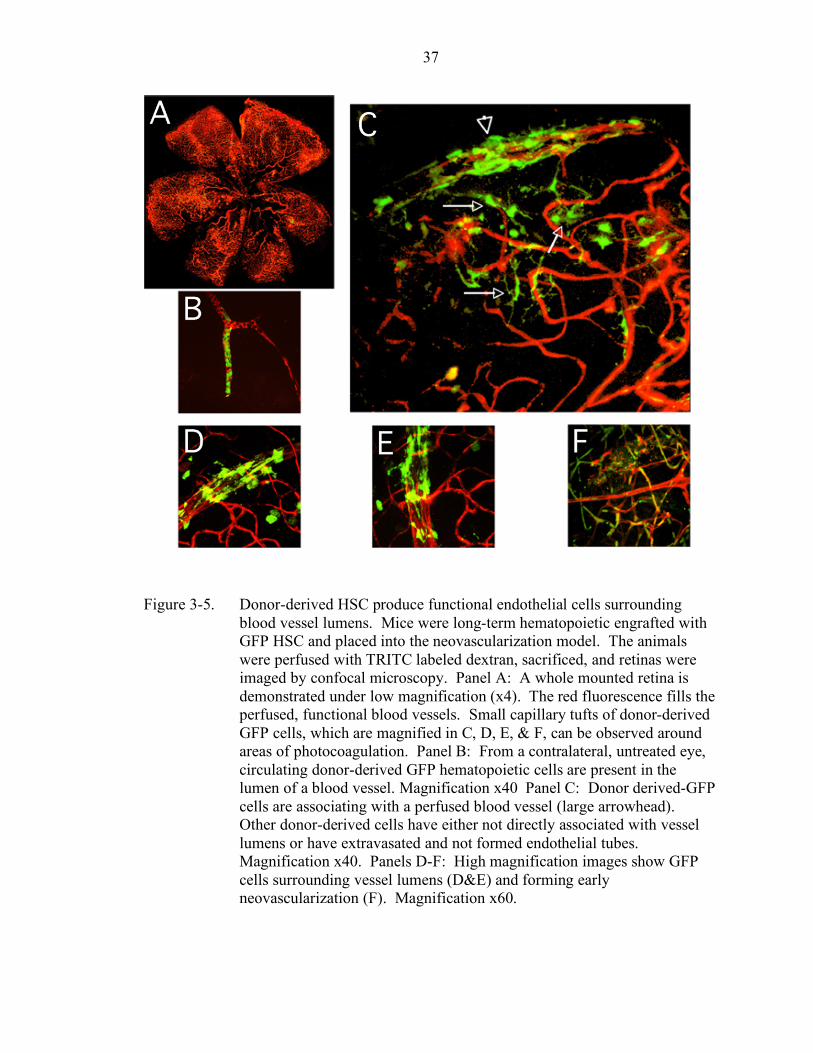
37
Figure 3-5. Donor-derived HSC produce functional endothelial cells surrounding blood vessel lumens. Mice were long-term hematopoietic engrafted with GFP HSC and placed into the neovascularization model. The animals were perfused with TRITC labeled dextran, sacrificed, and retinas were imaged by confocal microscopy. Panel A: A whole mounted retina is demonstrated under low magnification (x4). The red fluorescence fills the perfused, functional blood vessels. Small capillary tufts of donor-derived GFP cells, which are magnified in C, D, E, & F, can be observed around areas of photocoagulation. Panel B: From a contralateral, untreated eye, circulating donor-derived GFP hematopoietic cells are present in the lumen of a blood vessel. Magnification x40 Panel C: Donor derived-GFP cells are associating with a perfused blood vessel (large arrowhead). Other donor-derived cells have either not directly associated with vessel lumens or have extravasated and not formed endothelial tubes. Magnification x40. Panels D-F: High magnification images show GFP cells surrounding vessel lumens (D&E) and forming early neovascularization (F). Magnification x60.
38
The HSC has Hemangioblast Function
The recruitment of HSC derived cells to regions of injury agrees with studies done
which have found limited contribution to non injured tissues.85 In these experiments, the
donor derived GFP cells were able to contribute to both the blood products and
endothelial cells of the vasculature in the same mouse, however the exact cell which
could accomplish these feats cannot be established by these experiments.
The classic definition of a HSC is a cell capable of long term hematopoietic
reconstitution after irradiation, or self-renewing. We have fulfilled this definition
through the series of transplantation studies described above, however the ability of a
single HSC to do so clonality, and thus ruling out any contribution by other
“contaminating” cells, was necessary to prove HSC plasticity. As described in the
methods chapter, a single HSC was enriched and isolated though micromanipulation.
The HSC was transplanted along with Sca-1 negative non-GFP bone marrow cells (short
term progenitors) and transplanted into a lethally irradiated recipient. Of the 80 mice
transplanted, peripheral blood long term multilineage engraftment was demonstrated in 3
animals which were then subjected to the ischemic neovascularization model. Each of
the three animals exhibited the capillary tuft growths seen in the previous experiments
that were entirely donor derived as demonstrated by GFP expression. In addition these
vessels were functional in their ability to hold the red fluorescent dye perfused into the
vasculature. Since these animals had both blood and blood vessels which were derived
from a single transplanted HSC there can be no contribution from another source and any
GFP cells must necessarily be derived from the HSC. As shown in figure 3-6, the HSC
demonstrated hemangioblast activity in their ability to produce both blood and blood
vessels in a clonal manner. Panels A-C are from a serially transplanted mouse. The red

39
perfused blood vessel (Panel A) colocalizes with the GFP donor-derived endothelial cells
(Panel B) to show donor derived neovascularization (Panel C). These vessels were
derived from a self-renewing HSC. Panels D-F are from a single cell transplanted
animal. The red perfused blood vessel (Panel D) colocalizes with the GFP donor-derived
endothelial cells (Panel E) to show donor derived neovascularization (Panel F). These
vessels arose from the HSC in a clonal manner, therefore the HSC can give rise to both
blood and blood vessels and function as a hemangioblast.
Discussion
The previous work demonstrates the true plasticity of the HSC and fulfills the three
criteria established to prove this capacity. The self-renewing capability was
demonstrated through serial transplantations and long term hematopoietic reconstitution.
The ability of the HSC to provide hematopoiesis along with non hematopoietic tissue in a
clonal manner was shown through single cell transplants. Both experiments
demonstrated the ability of the HSC to produce functional vessels in a robust manner.
Taken together, these experiments outline an alternative developmental fate of the HSC,
namely the EPC, and describe how this outcome can be induced through growth factor
administration and ischemic injury. The EPC was shown to be derived from the HSC,
and not the MSC as previously posited.97 This understanding is especially valuable in
current treatments where the EPC has been shown to have the ability to contribute to
therapeutic neovascularization in several studies of ischemic injury, some in human
clinical trials.51, 88 This work demonstrates that vessel growth is not only carried out by
local or circulating endothelial cell angiogenesis, but under conditions of injury the HSC
can provide neovascularization. New blood vessels formed were largely derived from the
recruitment of undifferentiated precursors cells from the bone marrow.

40
Figure 3-6. The HSC is self-renewing and can clonally form endothelial cells. Both
serially transplanted long-term engrafted and single cell transplanted animals were placed in the neovascularization model. Animals were perfused with the TRITC-labeled dextran and retinas were imaged by confocal microscopy. Panel A-C: A long-term engrafted serially transplanted mouse retina was imaged. Panel A shows the red channel only indicating perfused, and therefore functional blood vessels. Panel B is the donor GFP HSC contribution to the neovascularization. A and B were merged in C and yellow areas are donor derived cells colocalizing with the perfused vessel. The HSC is self-renewing and can produce all blood lineages and endothelial cells of the vasculature. Panel D-F: A single-cell transplanted mouse retina was imaged. Panel D shows the red channel only indicating perfused, and therefore functional blood vessels. Panel E is the donor GFP HSC contribution to the neovascularization. D and E were merged in F and yellow areas are donor derived cells colocalizing with the perfused vessel. The HSC can clonally produce all hematopoietic lineages and endothelial cells lining blood vessel walls. Magnification x60.
The next series of experiments will ascertain the potential to modulate
hemangioblast function. Understanding the growth factors and biological conditions
during ischemia and how they regulate contribution to neovascularization by the HSC

41
and HSC progenitors may provide methods to manipulate blood vessel formation.
Modulation of the HSC/EPC pathway may allow for tailoring of therapies to increase
neovascularization in ischemic conditions such as stroke, wound healing, or cardiac
muscle damage. Conversely, the ability to decrease pathologic or undesirable
neovascularization as seen in tumor neovascularization or diabetic retinopathy could stem
from a greater understanding of the HSC to EPC developmental fate.

42
CHAPTER 4 MODULATORS OF HSC/HEMANGIOBLAST ACTIVITY
Few topics have stirred more recent debate than the promise of hematopoietic stem
cells (HSC) exhibiting functional plasticity. Indeed, the candidacy of HSC for
therapeutic treatment of disease is contingent upon demonstrating their ability to fulfill
stringent plasticity criteria. Initial papers described HSC transdifferentiation into a
variety of non-hematopoietic tissues in various organs such as the liver, brain, cardiac
muscle, blood vasculature, intestine and pancreas.30, 50, 82-84, 98 Using various tissue
specific markers and phenotypic characteristics these authors have described tissues to
which HSC are able to contribute demonstrating plasticity of the HSC in their
experimental settings. However, attempts to recapitulate these studies have found
limited HSC plasticity.85, 99 A possible rationale for these dichotic accounts is that these
studies employed differing methods to isolate HSC and examine the target organ of
potential transdifferentiation. Specifically, a variety of HSC isolation and purification
schema have been employed including: elutriation, or separation based on relative
density, serial transplantation where only those cells with the capability to home and long
term repopulate the bone marrow niche rescue a mouse, and cells isolated through
fluorescence activated cell sorting broken down into “side-population” studies of dye
exclusion, and single KTLS (c-kit+, Thy-1lo, lin-, Sca-1+) cell transplantations.30, 82, 100, 101
Each method isolates functional HSC as defined by long-term multilineage hematopoietic
reconstitution in vivo. However, each technique may isolate “functional” HSC at
different developmental stages with respect to plasticity. It may be improper to compare

43
HSC isolated by physical means, i.e. elutriation and side-population, with those isolated
by binding of antibodies to cell surface receptors. The differing populations isolated and
the manipulations which the cells undergo may impact their behavior in the experimental
settings. These methods must be reconciled before a definitive answer can be reached.
Sharing a mesodermal kinship to the HSC are the endothelial cells (EC) of the
vasculature. During embryogenesis hematopoietic and endothelial precursors develop in
both spatial and temporal immediacy. In the adult, EC circulate in the peripheral blood
which are phenotypically similarity to mature EC.16 These cells have the ability to
contribute to new vessel formation either in place of or in addition to resident EC
proliferation. In addition, it was shown that these circulating cells contained a population
which were derived from the bone marrow called endothelial progenitor cells (EPC).17 It
is now accepted that these bone marrow EPC exist and contribute significantly to adult
blood vessel formation, and that these EPC are HSC derived.30
Several experimental systems which damage blood vessels have been able to
induce robust HSC transdifferentiation.30, 102 The preceding chapters described how adult
HSC exhibit hemangioblast function by producing both blood and blood vessels in a
novel model of retinal neovascularization. The model uses long-term bone marrow
chimeric mice that have been stably reconstituted with hematopoietic stem cells from
GFP donor mice (fulfilling the first plasticity requirement). These cells are positive for
the surface markers Sca-1 and c-kit and demonstrate robust GFP expression in blood
products after a four month period. This time is sufficient to eliminate any contaminating
progenitor cell which would have since died off and created a deficiency leukemia in
mice engrafted with these short-term progenitor cells. The long-term engrafted chimeras

44
then receive a combination of growth factor administration and laser induced ischemic
injury to promote new blood vessel formation in adult murine retinas. Briefly, Adeno
Associated Virus (AAV) expressing Vascular Endothelial Growth factor (VEGF) is
administered intravitreally and allowed one month to reach peak expression. The retina
is then photocoagulated and new vessels attempt to grow into the ischemic region.
Since HSC have the ability to long-term repopulate hematopoiesis, lethally
irradiated mice were then transplanted with a single HSC. These animals exhibited
significant GFP in peripheral blood and bone marrow all of which was derived from the
single transplanted HSC proving clonality in transplanted HSC hematopoiesis. Chimeras
derived from both serially transplanted and single GFP+ HSC produced whole GFP
vascular beds after acute injury and VEGF induction.30 The vessels produced were not
only robust, but were functional as determined by perfusion after cardiac administration
of a fluorescent dye.
The previous chapter has illustrated a new developmental outcome of the HSC: the
production of EPC in response to vascular injury. The work demonstrated that both
blood and blood vessels can be clonally derived from adult HSC via a combination of
growth factor administration and ischemic injury. Thus, adult HSC meet the definition of
a plastic stem cell in that they have the ability to act as a hemangioblast in vivo. Whether
the HSC participates in everyday maintenance vasculogenesis or partakes only in
response to chronic vessel injury remained to be determined and was one of the focuses
of this work. In addition, it was determined that some form of significant injury is
needed for induction of the HSC to EPC pathway, presumably since resident EC are
inadequate to seed and proliferate the damaged areas. While the HSC is now known to

45
produce EPC under injury conditions, the potential role of physiologic mediators which
impact vasculogenesis in relation to hemangioblast HSC activity merit examination.
Since their first description in 1989, the nitric oxide synthases (NOS) have been
shown to play a role in a myriad of biological functions. The free radical NO, produced
from the conversion of L-arginine to citrulline in the presence of oxygen, has been shown
to function in distinct processes such as inflammation, host defense, neurotransmission,
and smooth muscle contractility. Three distinct isoforms of the enzyme have been
characterized including nNOS which is expressed in neuronal tissues, iNOS, expressed in
a wide variety of tissues, and eNOS which is predominately expressed in the endothelial
cells of the vasculature. The nNOS and eNOS isoforms are constitutively active in their
expressing tissues, which the iNOS isoform is induced in response to proinflammatory
cytokines or endotoxins from foreign bacteria. This induction of iNOS produces a 100-
fold increase in NO as part of an immune response, and NO production is much higher
than is seen compared to the basal levels of the constitutively active isoforms.103 NO
produced by iNOS acts as an antimicrobial and antiviral agent by decreasing DNA
replication.
Nitric Oxide (NO) also mediates endothelial cell function and hence blood vessel
formation. It has been shown to influence neovascularization in several models of
angiogenesis.104-106 The role of NO in promoting angiogenesis has been controversial in
part because of the complex regulation of NO generation and inactivation. In addition to
vasodilatation, increased local concentrations of NO stimulate proliferation and migration
of endothelial cells, both of which are essential for angiogenesis.107-109 The NO produced
by the three separate isoforms are activated under distinct activities and have unique
46
regulatory controls.109 Since iNOS is activated under certain pathological conditions,
such our injury model, and eNOS is constitutively activated in endothelial tissues, these
isoforms may influence the process of neovascularization. The altered amount of NO due
to lack of these enzymes in the cell will affect hemangioblast recruitment and formation
of blood vessels.
Angiogenesis is initiated by vasodilation in order to open up vessels facilitating
introduction of cells in circulation to the site of blood vessel growth. NO is known to
have several angiogeneic affects, including increasing matrix metalloprotease expression
along with tyrosine phosphorylation of proteins in cells populating the sprouting capillary
region.74 Interestingly, in various neovascularization models NO has been shown to be
both proangiogenic and antiangiogenic.74, 104 The theory is that the two isoforms are
activated under differing circumstances and hence are thought to affect blood vessel
formation differently. Indeed, in vivo this is the case. Blood vessel formation due to
HSC contribution under conditions of ischemic injury are influenced by NO as produced
by the iNOS and particularly the eNOS isoforms.110-112
The endothelial NOS (eNOS) isoform is constitutively expressed at basal levels by
endothelial cells and is thought to promote branching, organization, and maturation or
endothelial cells during vessel development. eNOS deficient (eNOS-/- ) animals exhibit
fetal growth restrictions, reduced survival, and an increased rate of limb abnormalities.113
They also demonstrate marked vascular pathology such as increased cardiomyocyte
apoptosis, congenital septal defects, postnatal heart failure, decreased capillary density
and vascular permeability.114 Endothelial cells from eNOS-/- animals demonstrate
decreased rates of angiogenesis with reduced branching in vitro.115 These animals also
47
exhibit an impairment of postnatal angiogenesis in response to growth factors and
ischemia.116 Correspondingly, eNOS has been shown to mediate the mitogenic effect of
VEGF on cultured microvascular endothelium.106 These findings led to the in vivo work
demonstrating that NO production is essential for angiogenesis in hindlimb ischemia, for
wound healing, and coronary collateral growth after myocardial ischemia.115, 117
VEGF has been shown to be a potent vascular permeability factor and plays a
leading role in angiogenesis, and our model takes advantage of this ability to promote
blood vessel synthesis.73 The angiogenic effect of VEGF under both pathological and
physiological conditions has been revealed to be predominantly mediated by eNOS.118
VEGF promotes NO production from eNOS in EC cells, and inhibition of eNOS by
pharmacological agents in vivo have decreased angiogenesis and vascular permeability
induced by VEGF.105 This demonstrates that eNOS is both a downstream mediator of
VEGF induced angiogenesis and an upstream promoter of VEGF expression. This
results in putative positive feedback loop between NO and VEGF which promotes
angiogenesis.119
The inducible NOS (iNOS) isoform is expressed by endothelial cells in response to
external stimuli such as VEGF, proinflammatory cytokines or lipopolysaccharide. iNOS
activation results in a 1000-fold greater generation of NO then eNOS activity alone.120
Its induction is thought to promote tube elongation during vessel development, although
evidence supports that it may have a contrasting anti-angiogenic effect.79 iNOS deficient
animals (iNOS-/-) are relatively healthy but do have a slight decrease in NO production
and vascular permeability during angiogenesis in collagen gels placed in a mouse cranial
window.106 During normal blood vessel formation the interplay between eNOS and
48
iNOS activity has been postulated to dictate vessel size and degree of branching. In this
chapter I will describe experiments where wild-type GFP+ HSC are transplanted into
eNOS-/- and iNOS-/- recipients to assess the effect of NOS dysfunction in tissue on
hemangioblast activity.
Results
iNOS and eNOS GFP chimeras demonstrated robust HSC engraftment.
To directly assess the role of NOS activity in the promotion of HSC trans-
differentiation into blood vessels, cohorts of wild-type (WT) C57BL6, iNOS-/-, and
eNOS-/- animals were generated. Animals were transplanted with 2,500 highly enriched
GFP+ HSC. It was necessary to use a highly enriched HSC population rather then a
single HSC due to the poor survival of eNOS-/- animals during transplant and the
difficulty in producing single cell transplanted animals in general. The enriched HSC
populations used were isolated using the same protocol previously employed for single
cell transplants in WT animals.30 Briefly, whole bone marrow was obtained from the
bone marrow of GFP animals. Cells were plated on tissue culture treated plates for 2
hours during which time the adherent cell population, which contains progenitor cells
such as the mesenchymal stem cell, stick to the plate. Non-adherent cells are collected
and stained with Sca-1, c-kit, and the lineage markers. Cells which were sorted by FACS
for the stem cell markers of Sca-1 and c-kit and were lineage negative were injected
intravenously through the retro orbital sinus. Long-term multilineage hematopoietic
engraftment was confirmed >3 months post transplant by flow cytometry analysis of
peripheral blood and is shown in figure 4-1. The first column in each cohort represented
is peripheral blood stained for B-cells expressing B220, with the second column stained
for macrophages expressing CD11b, and the third column T-cells expressing CD4. The
49
top row (C57BL/6) and the second row (GFP donor strain) are supplied for reference
controls to facilitate comparison between recipient and donor background fluorescence.
The third row is a typical C57BL/6.GFP chimeric mouse demonstrating robust
hematopoietic engraftment. The forth row (iNOS.GFP), and fifth row (eNOS.GFP) are
representative of engraftment levels of transplanted knockout animal’s blood lineage
profiles. The bottom row is peripheral blood stained for VEGFR2 demonstrating that
GFP EPC are in the circulation of C57BL/6.GFP, iNOS.GFP, and eNOS.GFP animals.
Engrafted recipients were subsequently termed C57BL6.GFP, iNOS.GFP, or eNOS.GFP
chimeras. Recipients that were robustly reconstituted by donor HSC (>75% donor
derived myeloid cells) then underwent our model of ischemic injury to induce adult
retinal neovascularization (n > 10 for all cohorts). By waiting >3 months post transplant
before inducing retinal ischemia, it is assured that the ability of HSC exclusively to
regenerate blood vessels is being assessed. No other cell that can be directly isolated
from the marrow has been shown to be capable of long-term reconstitution in a transplant
setting. Any contaminating precursor cells would not have had the ability to repopulate
the bone marrow for this extended period of time and would have long since disappeared
from the circulation. Further proof of the plastic ability of the HSC is demonstrated in
previous work where we illustrate how a single adult HSC is capable of making both
blood and blood vessels in a transplant recipient eliminating the possibility of any other
contaminating cell. Also, this activity is serially transplantable producing functional
vessels in a robust manner.30

50
Figure 4-1. NOS knockout animals exhibit long-term, multi-lineage, donor GFP
peripheral blood engraftment. Peripheral blood mononuclear cells were analyzed by flow cytometry 3 months post-transplant. The first column is B-cells expressing B220, the second column is macrophages expressing CD11b, and the third column is T-cells expressing CD4. The top row (C57BL/6) and the second row (GFP donor strain) reference controls show recipient and donor fluorescence. The third row is a representative C57BL/6.GFP chimeric mouse demonstrating robust hematopoietic engraftment. The forth row (iNOS.GFP), and fifth row (eNOS.GFP) are representative of engraftment levels of transplanted knockout animals. The bottom row is peripheral blood stained for VEGFR2. Circulating VEGFR2 positive cells are in C57BL/6.GFP, iNOS.GFP, and eNOS.GFP animals. Numbers in the top right corner are percentages of doubly lineage stained and GFP positive cells. iNOS= Inducible Nitric Oxide Synthase, eNOS= Endothelial Nitric Oxide Synthase.

51
The NO pathway affects blood vessel formation
After induction of retinal ischemia by laser ablation injury, C57BL6.GFP chimeras
produced a variety of GFP+ blood vessels at the sites of injury ranging from small
capillaries to larger vessels. Size was most likely dictated by the degree of the laser injury
as seen in the original hemangioblast characterization (Fig. 3-5 C and Fig. 3-6 C & F).
Strikingly, the NOS.GFP chimeras produced a marker phenotype change from the wild-
type mice indicating a role for the NOS pathway in hemangioblast function. iNOS.GFP
chimeras produced primarily small, highly branched blood vessels that perfused readily
(Fig 4-2 E and G) when injured. These vessels were largely donor-derived as shown in
the red-green merged images demonstrating the colocalization of the perfused fluorescent
dye and the GFP cells. The contralateral eyes had little to no donor contribution as is
seen in Figure 4-2 D and F. This indicated that the eNOS isoform, which is still present,
is sufficient for maintenance of vascular health, and that iNOS plays a role in blood
vessel branching.
In contrast, eNOS-/- mice retinas exhibited a marked phenotype when compared to both
control and iNOS -/- animals done in parallel. Strikingly, eNOS.GFP chimeras only
produced relatively large and unbranched vessels of donor origins regardless of ischemic
insult (Fig. 4-3 D-F). These vessels tended to perfuse poorly in spite of their large size.
Of note, this phenotype was not due to the inability to visualize the red dye in large
vessels due to the fact that the fluorescent perfusant can be easily visualized in B6 control
vessels of similar size. In addition, a few small vessels were readily perfused and the
animal demonstrated the gross muscle contraction and liver color change indicative of
sufficient perfusing. This is consistent with the known vascular defects of eNOS-/-

52
animals. Whether this lack of vessel functionality is due to some vascular blockage of
some alternative defect is not known.
Figure 4-2. The iNOS pathway modulates hemangioblast neovascularization.
iNOS.GFP chimeric mice underwent the retinal ischemia model followed by perfusion with TRITC-labeled dextran before eye enucleation and confocal imaging of the retinas. All panels are red and green merged confocal images. Panels D and F are retinas from control, untreated eyes. There is little GFP contribution observed (yellow). Panels E and G are from treated eyes where robust GFP contribution can be seen to vasculature. Magnification is 60X and size bar is ~10µM.
53
Figure 4-3 demonstrates the large and unbranching characteristics of the donor-
derived vessels. These pictures are red-green merged confocal images and the lack of red
perfuasnt indicates how poorly these vessels function. Panels E and G are from treated
eyes where robust GFP contribution can be seen to vasculature forming large,
unbranched vessels that do not contain the TRITC-dextran. Panels D and F are retinas
from control, untreated eyes. There is significant GFP contribution observed with or
without ischemic treatment indicating that the eNOS pathway plays a significant role in
endothelial cell maintenance.
The profound contribution of HSC derived GFP+ cells to the untreated retinas of
eNOS-/- recipients strongly suggested that deletion of this gene induces chronic vascular
injury. While injury was known to be necessary for HSC hemangioblast activity, this
work demonstrates that a chronic lack of eNOS can also induce neovascularization. If
this postulate is true the transplanted GFP+ HSC should contribute to vascular
regeneration throughout the eNOS-/- recipient. The evaluation of neovascularization in
contralateral eyes demonstrates and agrees with current studies that determined some
type of injury is required for functional plasticity of HSC. In typical physiologic
conditions little or no HSC contribution to “normal” tissue occurs, but when acute
(ischemic injury) or chronic (eNOS knockout) pathologic conditions arise HSC readily
contribute to vascular tissue.
These experiments formally demonstrate that iNOS activity at the site of vascular
injury dictates the size and branch characteristics of new vessels formed in adult animals.
Furthermore, the new vessels are formed in all, or large part, from circulating endothelial
progenitors of HSC origin.
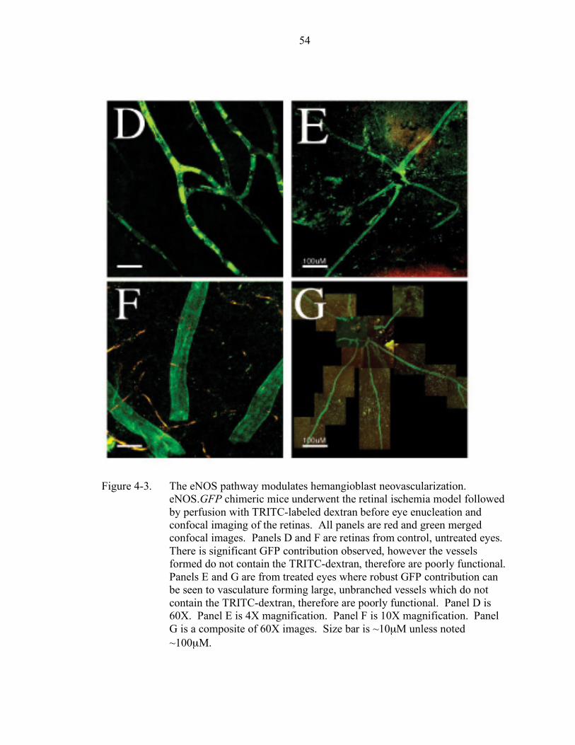
54
Figure 4-3. The eNOS pathway modulates hemangioblast neovascularization. eNOS.GFP chimeric mice underwent the retinal ischemia model followed by perfusion with TRITC-labeled dextran before eye enucleation and confocal imaging of the retinas. All panels are red and green merged confocal images. Panels D and F are retinas from control, untreated eyes. There is significant GFP contribution observed, however the vessels formed do not contain the TRITC-dextran, therefore are poorly functional. Panels E and G are from treated eyes where robust GFP contribution can be seen to vasculature forming large, unbranched vessels which do not contain the TRITC-dextran, therefore are poorly functional. Panel D is 60X. Panel E is 4X magnification. Panel F is 10X magnification. Panel G is a composite of 60X images. Size bar is ~10µM unless noted ~100µM.

55
The NOS pathway affects blood vessel branching characteristics.
To further examine the role of NOS in neovascularization, retinas from the non-
treated contralateral eyes were compared to the injured retinas of WT, iNOS-/-, and
eNOS-/- recipients. This was done in order to elucidate whether NOS could drive HSC
formation of vasculature without ischemic injury and growth factor administration.
iNOS-/- animals responded in a similar fashion to WT animals with production of GFP+
HSC derived vessels in the injured retina (Fig. 4-2 E & G), but little or no contribution
could be found in the retinas from the contralateral untreated eye (Fig. 4-2 D & F).
Unexpectedly, retinas from eNOS-/- recipients, which as described in other studies have
systemic vascular dysfunction, demonstrated robust GFP+ HSC derived contribution to
the preexisting vascular endothelium of both test (Fig. 4-3 E & G) and control eyes (Fig.
4-3 D & F).
After induction of retinal ischemia by laser ablation injury, C57BL6.GFP chimeras
produced a variety of GFP+ blood vessels at the sites of injury ranging from small
capillaries to larger vessels. In C57BL6.GFP chimeras, size was most likely dictated by
the degree of the laser injury (Fig. 3-5 D-F) and no GFP+ contribution to vasculature was
observed in control eyes (Fig. 3-5 B). iNOS.GFP chimeras produced primarily small,
highly branched blood vessels that perfused readily in treated eyes (Fig. 4-2 D & F).
These animals had limited donor EPC contribution in contralateral untreated eyes (Fig. 4-
2 E & G). Strikingly, eNOS.GFP chimeras only produced relatively large and
unbranched vessels regardless of ischemic insult (Fig. 4-3 D through G). The branching
characteristics of the three strains were markedly different suggesting that the NO
pathway functions in vessel organization. Total branch points of GFP vessels per 60X
field of view were counted for each genotype (Figure 4-4). C57BL6 model control
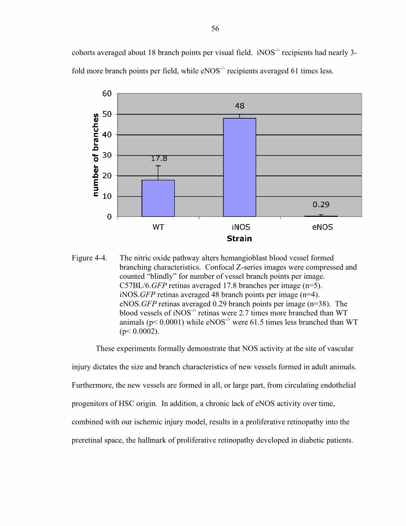
56
cohorts averaged about 18 branch points per visual field. iNOS-/- recipients had nearly 3-
fold more branch points per field, while eNOS-/- recipients averaged 61 times less.
Figure 4-4. The nitric oxide pathway alters hemangioblast blood vessel formed branching characteristics. Confocal Z-series images were compressed and counted “blindly” for number of vessel branch points per image. C57BL/6.GFP retinas averaged 17.8 branches per image (n=5). iNOS.GFP retinas averaged 48 branch points per image (n=4). eNOS.GFP retinas averaged 0.29 branch points per image (n=38). The blood vessels of iNOS-/- retinas were 2.7 times more branched than WT animals (p< 0.0001) while eNOS-/- were 61.5 times less branched than WT (p< 0.0002).
These experiments formally demonstrate that NOS activity at the site of vascular
injury dictates the size and branch characteristics of new vessels formed in adult animals.
Furthermore, the new vessels are formed in all, or large part, from circulating endothelial
progenitors of HSC origin. In addition, a chronic lack of eNOS activity over time,
combined with our ischemic injury model, results in a proliferative retinopathy into the
preretinal space, the hallmark of proliferative retinopathy developed in diabetic patients.

57
NO production affect on vasculature in non-ocular tissue
The finding that HSC have the ability to contribute to vascular tissue in non-treated
eyes during a disease state lends to the examination of tissues far removed and unrelated
to the eye. To determine the extent of donor GFP+ HSC contribution to the overall
vascular system multiple tissues (spleen, thymus, brain, kidney, liver, muscle, skin, and
gut) from the C57BL6.GFP, iNOS.GFP and eNOS.GFP chimeras (n=10 per cohort) were
harvested. Each of these animals had demonstrated long-term, multilineage
hematopoietic engraftment and had undergone the retinal ischemia model. At one month
after the induction of retinal ischemia the animals were euthanized and perfused with
tetramethyl rhodamine isothiocyanate-conjugated dextran (TRITC, a red fluorescent dye)
through the left ventricle. Tissues were harvested and immediately placed in optimum
cutting temperature medium and frozen to preserve GFP. 10 micron thick sections were
then cut and mounted with DAPI to stain the nuclei. Sections were examined by
fluorescent microscopy for GFP+ contributions to the vasculature. Results for the spleen,
thymus and brain are shown (Fig. 4-5). In all cases the C57BL6.GFP and iNOS.GFP
yielded similar results: limited evidence for GFP+ cells being incorporated into blood
vessels in any tissue outside of the treated retina (Fig. 4-5 A-F, and data not shown).
This indicates that whole body irradiation alone is not sufficient for induction of HSC
contribution to vasculature in tissues which are not treated further. In contrast,
eNOS.GFP chimeras exhibited robust GFP+ contributions to the vasculature (as
determined by co-localization with the perfused red fluorescent dye) in all tissues
examined (Fig. 4-5 G-L, and data not shown). Lack of eNOS creates a pathologic
vascular condition where HSC are induced to contribute to vascular repair throughout an
organism.

58
Figure 4-5. Chronic vascular injury in eNOS.GFP chimeras induces widespread hemangioblast activity from adult HSC. NOS knockout animals which underwent the neovascularization model, and spleen (A, G, B &H), thymus (C, I, D & J), and brain (E, K, F & L) were harvested from TRITC perfused animals. 10µM cryosections were prepared and mounted with Vectashield plus DAPI. iNOS.GFP (A-F) and eNOS.GFP (G-L) chimeras were examined by fluorescence microscopy. The donor GFP HSC derived cells are green, and the TRITC-labeled dextran perfusant is red. Panels A & G are magnification X40. All remaining panels are all magnification X64.

59
To ascertain the endothelial cell nature of the GFP+ cells surrounding the vessel lumens
tissue sections were stained for the pan endothelial cell marker MECA-32. Ten micron
frozen sections were stained with a primary antibody to MECA-32, followed by a Texas
Red conjugated secondary antibody and DAPI. Endothelial cells were then scored for the
presence of both MECA-32+ and GFP+ cells via fluorescent microscopy. Splenic
sections demonstrate the characteristic results observed in all tissues studied (Fig. 4-6).
Donor derived hematopoietic cells in the spleen serve as internal negative staining
controls for MECA-32 in each section. WT animals showed occasional GFP+, MECA-
32+ endothelial cells in the brain (closest organ to the site of VEGF administration) with
the majority of tissues such as the spleen (Fig. 4-6 A-D), kidney, liver, and muscle
showing no donor derived endothelial cells. iNOS-/- animals, which exhibit minor
systemic vascular defects, had occasional GFP+, MECA-32+ endothelial cells in the
spleen (Fig. 4-6 E-H) and other tissues.
Overall GFP+ HSC derived contribution to the vasculature of iNOS-/- animals,
outside the area of retinal ischemia, was at most 1% in >150 sections examined for
MECA-32+ vessels. Robust GFP+ donor derived endothelial cell production was
observed in eNOS-/- recipients which have been demonstrated to have chronic and severe
vascular pathology. Most vessels were quite large, and most showed extensive GFP+
HSC derived MECA-32+ endothelial cell contributions in the spleen (Fig. 4-6 I-P) and
other tissues examined. The HSC contribution to vasculature detected in untreated tissue
was analogous to that observed in the treated retinas demonstrating that chronic vascular
injury appears to be sufficient to induce the hemangioblast activity of adult HSC.
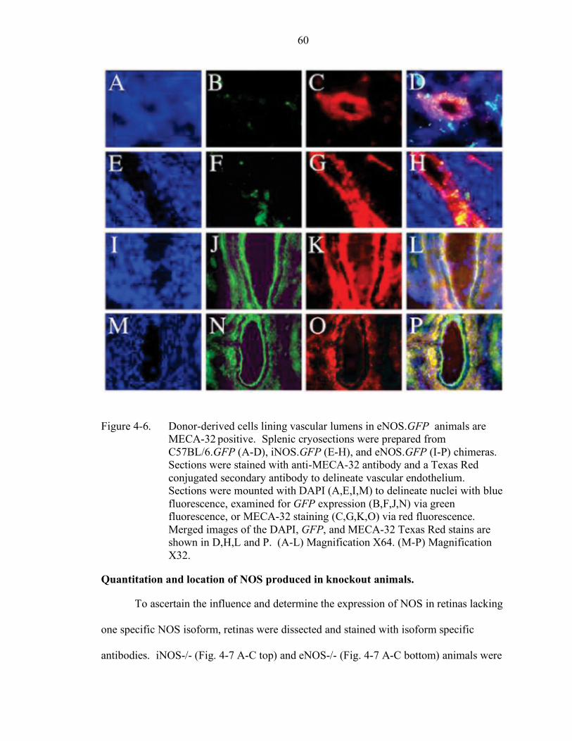
60
Figure 4-6. Donor-derived cells lining vascular lumens in eNOS.GFP animals are MECA-32 positive. Splenic cryosections were prepared from C57BL/6.GFP (A-D), iNOS.GFP (E-H), and eNOS.GFP (I-P) chimeras. Sections were stained with anti-MECA-32 antibody and a Texas Red conjugated secondary antibody to delineate vascular endothelium. Sections were mounted with DAPI (A,E,I,M) to delineate nuclei with blue fluorescence, examined for GFP expression (B,F,J,N) via green fluorescence, or MECA-32 staining (C,G,K,O) via red fluorescence. Merged images of the DAPI, GFP, and MECA-32 Texas Red stains are shown in D,H,L and P. (A-L) Magnification X64. (M-P) Magnification X32.
Quantitation and location of NOS produced in knockout animals.
To ascertain the influence and determine the expression of NOS in retinas lacking
one specific NOS isoform, retinas were dissected and stained with isoform specific
antibodies. iNOS-/- (Fig. 4-7 A-C top) and eNOS-/- (Fig. 4-7 A-C bottom) animals were

61
quantitated for NOS expression in parallel. Animals were sacrificed and the eyes
enucleated as described. The dissected retinas were then imaged through confocal
microscopy. Figure 4-7 (A top and bottom) demonstrates that in each knockout strain the
isoform which is deleted is not expressed in vivo at detectable levels. The iNOS -/- has
relative amounts of NOS expressed (B and C top), while the eNOS -/- retinas
demonstrate an increase in iNOS expression as seen throughout the large, and particularly
the smaller vessels (B and C bottom). This confirms that there is an upregulation of
iNOS expression in eNOS knockout retinas indicating a dysregulation in amount of NO
produced resulting in the pathologic blood vessel formation observed in these animals.
Discussion
Through growth factor administration and ischemic injury to the retina, HSC can
be induced to transdifferentiate into vascular endothelium. Furthermore,
transdifferentiation can also be observed during a pathologic disease state of chronic
vascular injury. The substantial role NO plays in vascular tone, and the presence of a
NOS isoform specifically found in endothelial cells hinted at a role of NO in blood vessel
formation and remodeling. NOS activity can also dictate the general size and branch
characteristics of new blood vessels formed in response to ischemic injury and growth
factor administration. Using the neovascular model of inducing HSC hemangioblast
activity to promote blood vessel formation in the adult retina, donor WT HSC
transplanted into iNOS-/- recipients produced highly branched vessels that are generally
smaller in size. These HSC are functioning in an environment where local NO
production is similar to what is seen in wild type, non-infection conditions due to the
eNOS isoform which is constitutively active in endothelial cells. A wide variety of
vessel sizes are formed which are functional as measured by perfusion of marker dye.

62
Figure 4-7. Nitric oxide production is dysregulated in eNOS knockout animals. iNOS-/- and eNOS-/- retinas were stained with NOS isoform specific antibodies. Vessels were illuminated by agglutin staining (red) and regions which were positive for the NOS antibody are green. In the top row, panel A depicts an iNOS-/- retina stained with iNOS specific antibody. Panels B and C are iNOS-/- stained with eNOS isoform specific antibody. In the bottom row, Panel A depicts an eNOS-/- retinas stained with eNOS isoform specific antibody. Panel B and C are eNOS-/- stained with iNOS isoform specific antibody.
The branch patterning is similar to normal mouse vasculature, although slightly
increased. Contrastingly, eNOS-/- recipients produced primarily unbranched vessels of
large size. This indicates that the local NO production due to the eNOS isoform activity
is necessary for normal hemangioblast derived blood vessel formation. Modification of
NO production via the eNOS isoform, which can now be specifically targeted with
pharmacological agents, could provide a means to influence neovascularization and

63
angiogenesis in pathologic diseases such as Diabetic Retinopathy, and Retinopathy of
Prematurity. This altered HSC response was in addition to the widespread vascular
remodeling by donor HSC seen throughout the eNOS-/- recipients even in non-injured
organs and tissues. The vessels of eNOS-/- recipients were difficult to perfuse indicating
their general vascular dysfunction. This further emphasizes the crucial nature of the
eNOS isoform’s NO production to ensure proper vessel formation and functionality
foreshadowing studies occurring outside the realm of the eye on which our model
focuses.
This chapter reiterates work demonstrating that an injury state, whether it be acute
as seen in the photocoagulation of blood vessels, or chronic as seen in the vascular
pathology observed in the eNOS knockout animals, is required for HSC plasticity.85
Furthermore, local mediators, including VEGF and NO, can greatly influence not only
the size and amount of new blood vessels formed but also their functionality. The
regulation of NOS activity as a means to influence the remodeling of vascular beds may
provide specific treatment regimes. It may prove beneficial to selectively inhibit a
particular NOS isoform to correct an imbalance thus altering the development of new
blood vessels. Whether a deregulation of NOS or NO activity is a causal effect in human
proliferative retinopathy remains to be determined. If this is the case, pharmaceuticals
that affect these activities, some already in use for non-related disease treatment, may
provide an effective therapy or preventative for human diseases in relation to proliferative
or pathological blood vessel formation due to hemangioblast activity.

64
CHAPTER 5 LIMITATIONS OF STEM CELL RESEARCH AND ETHICAL CONSIDERATIONS
While the promise of stem cells as therapy is considerable, there are limitations to
their usage including their biological activity and ethical implications. The biological
limitations of stem cell plasticity are related to their ability of to transdifferentiate and
their potential to form tissues. The ethical implications are many, and I will address
some of them in this chapter.
Biological limitations
In the current research environment, there is currently heated controversy over the
reported plasticity of stem cells, particularly the HSC in relation to cardiac muscle, liver,
and the nervous system.121 The limitations of adult stem cells include :their diminished
capacity to proliferate when compared to embryonic stem cells. Autologous
transplantation of cells back into a patient will still retain genetic abnormalities.
Correspondingly, ex vivo epanded or genetically modified cells used for therapy may
produce unforeseen consequences. Some tissues arise through a complex developmental
fate, such as the pancreas which is derived from the infolding of several tissue layers
which will not be easily mimicked in vitro. The resident stem cells within a tissue type
are extremely rare. This paucity of cells makes them extremely difficult to identify,
isolate, and purify. In addition, it had proven difficult to culture adult stem cells
compared to embryonic ste cells in vitro limiting their use as potential therapy. Finally
fusion It is believed that cell fusion may be a potential method for introducing new
genetic material to correct mutated or malfunctioning genes that cause disease. Cell

65
fusion occurs when two or more cells combine to form one cell which then contains more
genetic material than normal. Fusion has been shown to occur in embryonic along with
adult stem cells.122, 123 In adult mice, fused liver cells may contain 80 chromosomes,
double the amount found in a normal mouse liver cells. Other cell types, including
megakaryocytes and muscle cells function with an increased ploidy as well. In most cell
types, however, aneuploidy would be detrimental often inducing apoptosis or cell
transformation. The resulting imbalance in gene dosage could lead to nonfunctional
tissue or cancer. It is not clearly understood, nor has it been definitively proven whether
fusion is a pathway for stem cell plasticity with research indicating that fusion can result
in, but is not necessary for plasticity.124-126 If the former is the case, then investigation
into the effect of producing cells with an increased ploidy at any time must be done. A
fusion event during contribution to the regenerated tissue could preclude stem cell based
therapy.
The first work characterizing fusion done by Terada et al. found that fusion events
in vitro were extremely rare.122 Consequently, the robust amount of donor derived
contribution is unlikely to arise from such rare events. Recent work from our lab
demonstrated that the cells derived are diploid, and any unresolved fusion of the HSC
would have resulted in an increase in ploidy shown in figure 5-1.127 The circulating
endothelial precursor cells derived from the donor marrow exhibit normal 2N ploidy
when stained with the DNA dye propidium iodide. This does not rule out the possibility
of a fusion event which was resolved resulting in normal ploidy, however, and special
specific experiments much be done to ascertain if this fusion resolution is occurring.

66
Figure 5-1. Propidium iodide staining of circulating EPC does not indicate abnormal ploidy. Long-term GFP engrafted C57BL/6 recipients underwent the neovascularization model. Animals were bled, and FICOLL enriched peripheral blood was stained and FACS enriched for VEGFR2 expression. Cells were stained with propidium iodide and analyzed by FACS. Left panel depicts EPC from a nontransplanted C57BL/6 animal bled in parallel. Right depicts a test animal. Both exhibit classical diploid staining profiles.
Ethics
Any comprehensive analysis of the stem cell field would be remiss to not include
the ethical implications for their use in therapy. The field has proven to be a polarizing
issue which influences many religious and political referenda born simultaneously with
Dolly, the first cloned mammal. The debate arises from the use of embryonic stem cells
which at this time can only be isolated from an embryo resulting in the embryo’s
destruction. These cells are attractive, however, because of their ability to produce all of
the cell types in an adult animal or provide an environment in which DNA can be
transferred in nuclear transfer. To date no single adult cell has been shown to have the
pluripotency of embryonic cells along with providing the intracellular environmental cues
necessary to reprogram transferred DNA. Adult stem cells do not have the same capacity

67
to produce any tissue or cell types. This inherent extraordinary therapeutic potential
resulting from the destruction of an embryo leads to opposition based on the idea of when
life begins.
In the United States, many Fundamentalist Christian groups are strongly opposed to
embryonic stem (ES) cell research as the destruction of the embryo is considered
abortion, or murder. They believe that any and all research using human stem cells is
morally unacceptable. Other religions, however, are supportive of embryonic research.
Many Jewish groups of differing denominations do not view an early stage embryo as a
human being, therefore usage of embryonic tissue is not destruction of a human. Many
Humanists, Unitarian Univeraslists, and Muslim clerics have also come out in favor of
stem cell research. In addition, proponents point out that stem cell research uses
discarded embryos from in vitro fertilization, and that fertility clinics routinely destroy
thousands of embryos. These unused embryos would normally be discarded or kept
frozen indefinitely if not used in research. There is no general consensus among religious
groups which gives rise to many concerns over the use of ES cells.
Concerns Over Stem Cell Use
Certainly, stem cells are not the first human discovery to revolutionize scientific
knowledge and create waves of ethical debate. Since ancient times society has
admonished man for approaching these boundaries as exemplified in the Greek myth of
Icarus who did not heed his father’s command; he reveled in the “unnatural” sensation of
flight and then plummeted to his death after the sun melted his wings. This Greek myth
embodies our apprehensions about interfering with nature. Galileo Galilei expanded the
frontiers of astronomy and posited that the Earth rotates on its axis and revolves around
the Sun. This led to his eventually condemnation for heresy. In Victorian times, society

68
grappled with the balance between the knowledge gained from performing autopsies for
crucial understanding of human anatomy versus the desecration of those who were dead.
Even recently, complete consent to produce recombinant DNA for lifesaving medications
such as insulin has been granted but only after vehement protestations over genetic
engineering. There are shared concerns among all instances of testing medical
boundaries, and the concerns of stem cell technology include issues of safety, efficacy,
and resource allocation. For decades, patients have undergone adult HSC transplantation
in the treatment of immune deficiencies and cancer. Although graft-versus-host disease
and posttransplantation infections are major risks of allogeneic bone marrow transplants,
investigators have worked to minimize these consequences and many patients accept
these risks in the hope of the lifesaving benefit of disease eradication. However, the field
of stem cell therapy is still in its infancy and researchers are incrementally improving
safety, efficacy, and applicability to a wider spectrum of disease. In all instances of
expanding the horizons of our knowledge a societal consciousness was at play, often
times encumbering progress and questioning techniques of intervention.
Stem cell therapy differs from previous technologies in how these potential sources
of regenerating tissue are tapped. Adult stem cells are typically acquired by harvesting
adult tissues. Patients give informed consent and usually undergo little risk at donation.
In contrast, human embryonic stem cells (hES) are obtained by culturing cells from the
inner cell mass of a blastocyst. This blastocyst is usually acquired from an unused human
embryo produced by in vitro fertilization or from an aborted fetus. The harvesting
process requires dissolving the blastocyst bringing into question the moral and legal
status of the human embryo.

69
Many religious perspectives consider the human fetus an individual human entity.
However, there is substantial debate regarding at which specific stage dignity is conferred
in development ranging from conception, to primitive streak development, implantation,
or birth. Taking into account the many perspectives on the moral status of the human
embryo weighed against the scientific promises of a healthier tomorrow through stem cell
technology, our society has attempted to define the legal status of the human embryo. In
the United States, the first mandate was outlined in 1973 when the US Supreme Court
ruled that a fetus is not a person in terms of constitutional protection (Roe v Wade, 410
US 113 [1973]). For a better examination of the decision’s effect on research, the
National Institutes of Health (NIH) imposed a moratorium on fetal research, and
Congress founded the National Commission charging it to put together policy and
guidelines on fetal research. The commission published a report encouraging fetal
research due to its potential, provided that the research risks to the fetus were minimal
and were only those that would be accepted for a term fetus. Thus, despite Roe v Wade,
the commission extended protection to a fetus equal to adult patients in research. This
included fetuses planned for elective abortion.
The NIH moratorium was lifted in 1975, however during President Ronald W.
Reagan’s second term Congress enacted legislation that further protected the fetus by
ending federal support of fetal research involving any level of risk. In 1996, Congress
extended this restriction by banning federal funding for “the creation of a human embryo
or embryos for research purposes.” This led the NIH to distinguish between deriving and
using existing human embryos to support embryonic stem cell research. Under these
guidelines researchers using already established hES cell lines derived from private

70
sector support can receive public sector monies provided that the fertilized embryos
would otherwise have been discarded after IVF or were from already aborted fetuses,
donors are aware of the research use, and no payment was made to the donors.
President William J. Clinton created The National Bioethics Advisory Commission
(NBAC) to thoroughly review moral and legal issues of stem cell re search. This
commission largely framed its moral position based on a utilitarianism argument—the
good of many outweigh the status of one. In addition, it drew on medicine’s aims to heal
and prevent disease urging consideration of a long-term benefit-to-harm balance. The
NBAC conclusion recommended allowing federal funding for hES research on excess
IVF embryos. Reasons supporting this position include the potential of ES cells in
regenerative and reproductive medicine and the need for federal support to avoid private
sector conflicts of interest which sometimes invokes secrecy, limiting the spread of
knowledge, and places shareholders interests ahead of public good. Taking all these
guidelines and perspectives into account, President George W. Bush made an executive
order on August 9, 2001 to limit federal funding of hES research to cell lines already
derived from 64 embryos.
The cost to society of foregoing use of this technology, either by failure to correct
genetic abnormalities or by improving the success of lifesaving organ transplantations,
may be equal to or greater than the perceived costs to the dignity of life due to destruction
of a human embryo. There must be a balance between the perceived costs to the dignity
of life held by those with the most sacrosanct concept of the moral status of embryos and
those who would directly benefit from stem cell based therapy. The climate in which
stem cells are explored can be nurturing or profoundly limiting. As medical scientists,

71
we must not make judgments or ethical decisions on our own; rather, we must ensure full
informed consent of the population as a whole. This approach may limit quick progress
and may disqualify avenues of research and therapy, but as responsible researchers we
must use the resources of society in a worthy manner to explore fully the tremendous
potential of embryonic stem cells.
The use of adult stem cells eliminates any ethical concerns as the cells used for
therapy can be obtained with only slight discomfort, and potentially from the individual
patient themselves. This is attractive due to the fact that there would be no human
leukocyte antigen mismatching, and therefore no need for immune suppressing drugs or
the fear of tissue rejection and graft versus host disease. If adult stem cells prove to have
the same potentiality as ES cells, either individually or as a collection, their use would
end the need for embryonic tissue and their subsequent destruction eliminating any
ethical concerns over the ends justifying the means.

72
CHAPTER 6 GENERAL CONCLUSIONS
The goal of this work was to characterize the hematopoietic stem cell’s plastic
ability and describe biological pathways which can modulate this ability. In a historic
context, this work spans several stages of the he stem cell field as it matured from the
beginning flurry of activity into its current stage of careful evaluation. The pioneering
work demonstrated the exciting potential of the field as an alternative and powerful tool
for use as a cell-based therapy for many diseases ranging from cancer to diabetes, heart
attack, and Parkinson’s disease. Those founders demonstrated that these cells,
specifically the hematopoietic stem cell, was capable of producing many different tissue
types. It was found that the HSC is capable of producing not only all of the blood
lineages, but also muscle, pancreatic cells, heart, intestinal epithelium, brain, and blood
vessel endothelium. These exciting results sparked a revolution in the scientific field
with new findings constantly being published in peer reviewed scientific journals along
with the front pages of popular newspapers and magazines. This initial impetus soon
played out, however, as the heralded plasticity of stem cells came under thorough and
critical scrutiny, and justifiably so. It became apparent that the cell types produced were
not necessarily functional, nor could there be certainty of the source of the donor cells—
several different cell types within the transplant could be individually contributing to the
observed tissue, therefore no one cell would be capable of forming several tissue types.
This critique was addressed in several studies which began the second stage of stem cell
research. Krause et al. did a single cell transplant homing assay to prove several tissue

73
types could arise from one cell.82 Grant et al. also did single cell transplants and
demonstrated the functional ability of endothelial cells which clonally arose from the
HSC to carry red fluorescent dye perfused into the circulator system.30 In addition,
Lagasse et al. rescued liver function in a metabolic liver disease with HSC.84 Clearly,
stem cells, particularly the HSC, are capable of providing functional rescue of disease
conditions in a clonal manner. With this understanding, we now embark on the third
stage of stem cell plasticity research—defining the genes and biological pathways
involved in regulating or controlling stem cell activity.
Many genes have been shown to maintain the “stemness” of stem cells by
controlling their self-renewing and proliferating capacity. In addition, we are starting to
understand factors, such as the nitric oxide pathway, which play a role in stem cell
homing and functional behavior.128 This work, along with other exciting work done in
our lab on chemokines such as stromal-derived factor-1’s involvement in homing,
highlights the therapeutic benefit of not only stem cell research, but also fortifies the
foundation for cell-based therapy.129 This third age of directed stem cell repair of
damaged or non functional tissue has the potential for direct translation into disease
therapy along with opening exciting new avenues of original research. This body of
work chronicles the relatively new field of stem cell plasticity research from the initial
characterizing of HSC plasticity up to describing biological pathways which can
orchestrate HSC activity. The founding work and initial application to HSC plasticity
described here can lead to many novel stem-cell therapy strategies for debilitating
conditions and diseases. Additional effort will focus on direct translation of this
knowledge into human disease therapy.

74
LIST OF REFERENCES
1. Palis, J., Robertson, S., Kennedy, M., Wall, C. & Keller, G. Development of erythroid and myeloid progenitors in the yolk sac and embryo proper of the mouse. Development 122, 1363-1371 (1999).
2. Wong, P., Chung, S., Chui, D. & Eaves, C. Properties of the earliest clonogenic
hemopoietic precursor to appear in the developing murine yolk sac. Proc Natl Acad Sci 83, 3851-3854 (1986).
3. Drake, C. & Fleming, P. Vasculogenesis in the day 6.5 to 9.5 mouse embryo.
Blood 95, 1671-1679 (2000). 4. Haar, J. & Ackerman, G. A phase and electron microscopic study of
vasculogenesis and erythropoiesis in the yolk sac of the mouse. Anat Rec 170, 199-223 (1971).
5. Risau, W. & Flamme, I. Vasculogenesis. Annual Review of Cell &
Developmental Biology 11, 73-91 (1995). 6. Flamme, I. & Risau, W. Induction of vasculogenesis and hematopoiesis in vitro.
Development 116, 435-9 (1992). 7. Godin, I., Garcia-Porero, J., Coutinho, A., Dieterlen-Lievre, F. & Marcos, M.
Para-aortic splanchnopleura from early mouse embryos contains B1a cell progenitors. Nature 364, 67-70 (1993).
8. Godin, I., Garcia-Porrero, J., Dieterlen-Lievre, F. & Cumano, A. Stem cell
emergence and hematopoietic activity are incompatible in mouse intraembryonic sites. J Exp Med 190, 43-52 (1999).
9. Godin, I., Dieterlen-Lievre, F. & Cumano, A. Emergence of multipotent
hemopoietic cells in the yolk sac and paraaortic splanchnopleura in mouse embryos, beginning at 8.5 days postcoitus. Proceedings of the National Academy of Sciences of the United States of America 92, 773-7 (1995).
10. Medvinsky, A., Samoylina, N., Muller, A. & Dzierzak, E. An early pre-liver intra-
embryonic source of CFU-S in the developing mouse. Nature 364, 64-70 (1993). 11. Takahashi, T. Kalka C, Masuda H, Chen D, Silver M, Kearney M, Magner M,
Isner J.M., & Asahara T. Ischemia- and cytokine-induced mobilization of bone marrow-derived endothelial progenitor cells for neovascularization. Nat Med 5, 434-8 (1999).
75
12. Nishikawa, S. I., Nishikawa S, Kawamoto H, Yoshida H, Kizumoto M, Kataoka H, & Katsura Y. In vitro generation of lymphohematopoietic cells from endothelial cells purified from murine embryos. Immunity 8, 761-9 (1998).
13. Muller, A. M., Medvinsky, A., Strouboulis, J., Grosveld, F. & Dzierzak, E.
Development of hematopoietic stem cell activity in the mouse embryo. Immunity 1, 291-301 (1994).
14. Yoder, M., Hiatt K, Dutt P, Mukherjee P, Bodine D.M., & Orlic D.
Characterization of definitive lymphohematopoietic stem cells in the day 9 murine yolk sac. Immunity 7, 335-344 (1997).
15. Folkman, J. & Shing, Y. Angiogenesis. J Biol Chem 267, 10931-4. (1992). 16. George, F., Brisson C, Poncelet P, Laurent J.C., Massot O, Arnoux D, Ambrosi P,
Klein-Soyer C, Cazenave J.P., & Sampol J. Rapid isolation of human endothelial cells from whole blood using S- Endo1 monoclonal antibody coupled to immuno-magnetic beads: demonstration of endothelial injury after angioplasty. Thromb Haemost 67, 147-53. (1992).
17. Asahara, T., Masuda H, Takahashi T, Kalka C, Pastore C, Silver M, Kearne M,
Magner M, & Isner J.M. Bone marrow origin of endothelial progenitor cells responsible for postnatal vasculogenesis in physiological and pathological neovascularization. Circ Res 85, 221-8. (1999).
18. Asahara, T., Murohara T, Sullivan A, Silver M, van der Zee R, Li T,
Witzenbichler B, Schatteman G, & Isner J.M. Isolation of putative progenitor endothelial cells for angiogenesis. Science 275, 964-7 (1997).
19. Shi, Q., Rafii S, Wu M.H., Wijelath E.S., Yu C, Ishida A, Fujita Y, Kothari S,
Mohle R, Sauvage L.R., Moore M.A., Storb R.F., & Hammond W.P. Evidence for circulating bone marrow-derived endothelial cells. Blood 92, 362-7 (1998).
20. Reyes, M., Dudek A, Jahagirdar B, Koodie L, Marker P.H., & Verfaillie C.M.
Origin of endothelial progenitors in human postnatal bone marrow. J Clin Invest 109, 337-46. (2002).
21. Lin, Y., Weisdorf, D. J., Solovey, A. & Hebbel, R. P. Origins of circulating
endothelial cells and endothelial outgrowth from blood. J Clin Invest 105, 71-7. (2000).
22. Isner, J. M. & Asahara, T. Angiogenesis and vasculogenesis as therapeutic
strategies for postnatal neovascularization. J Clin Invest 103, 1231-6 (1999).

76
23. Shaw, J. P., Basch, R. & Shamamian, P. Hematopoietic stem cells and endothelial cell precursors express Tie-2, CD31 and CD45. Blood Cells Mol Dis 32, 168-75 (2004).
24. Hristov, M., Erl, W. & Weber, P. C. Endothelial progenitor cells: mobilization,
differentiation, and homing. Arterioscler Thromb Vasc Biol 23, 1185-9 (2003). 25. Kawamoto, A., Gwon H.C., Iwaguro H, Yamaguchi J.I., Uchida S, Masuda H,
Silver M, Ma H, Kearney M, Isner J.M., & Asahara T. Therapeutic potential of ex vivo expanded endothelial progenitor cells for myocardial ischemia. Circulation 103, 634-7. (2001).
26. Yin, A. H., Miraglia S, Zanjani E.D., Almeida-Porada G, Ogawa M, Leary A.G.,
Olweus J, Kearney J, & Buck D.W. AC133, a novel marker for human hematopoietic stem and progenitor cells. Blood 90, 5002-12. (1997).
27. Peichev, M., Naiyer A.J., Pereira D, Zhu Z, Lane W.J., Williams M, Oz M.C.,
Hicklin D.J., Witte L, Moore M.A., & Rafii S. Expression of VEGFR-2 and AC133 by circulating human CD34(+) cells identifies a population of functional endothelial precursors. Blood 95, 952-8. (2000).
28. Quirici, N., Soligo D, Caneva L, Servida F, Bossolasco P, & Deliliers G.L.
Differentiation and expansion of endothelial cells from human bone marrow CD133(+) cells. Br J Haematol 115, 186-94. (2001).
29. Crosby, J. R., Kaminski W.E., Schatteman G, Martin P.J., Raines E.W., Seifert
R.A., & Bowen-Pope D.F. Endothelial cells of hematopoietic origin make a significant contribution to adult blood vessel formation. Circ Res 87, 728-30. (2000).
30. Grant, M. B., May W.S., Caballero S, Brown G.A., Guthrie S.M., Mames R.N.,
Byrne B.J., Vaught T, Spoerri P.E., Peck A.B., & Scott E.W. Adult hematopoietic stem cells provide functional hemangioblast activity during retinal neovascularization. Nat Med 8, 607-12 (2002).
31. Gloor, B. P., Rokos, L. & Kaldarar-Pedotti, S. Cell cycle time and life-span of
cells in the mouse eye. Measurements during the postfetal period using repeated 3H-thymidine injections. Dev Ophthalmol 12, 70-129 (1985).
32. Dignat-George, F. & Sampol, J. Circulating endothelial cells in vascular
disorders: new insights into an old concept. Eur J Haematol 65, 215-20 (2000). 33. Hanahan, D. & Folkman, J. Patterns and emerging mechanisms of the angiogenic
switch during tumorigenesis. Cell 86, 353-64 (1996).

77
34. Plate, K. H., Breier, G., Weich, H. A. & Risau, W. Vascular endothelial growth factor is a potential tumour angiogenesis factor in human gliomas in vivo. Nature 359, 845-8 (1992).
35. Bouck, N. Tumor angiogenesis: the role of oncogenes and tumor suppressor
genes. Cancer Cells 2, 179-85 (1990). 36. Klagsbrun, M. & D'Amore, P. A. Regulators of angiogenesis. Annu Rev Physiol
53, 217-39 (1991). 37. Pepper, M. S., Sappino, A. P., Montesano, R., Orci, L. & Vassalli, J. D.
Plasminogen activator inhibitor-1 is induced in migrating endothelial cells. J Cell Physiol 153, 129-39 (1992).
38. Madri, J. A., Pratt, B. M. & Tucker, A. M. Phenotypic modulation of endothelial
cells by transforming growth factor-beta depends upon the composition and organization of the extracellular matrix. J Cell Biol 106, 1375-84 (1988).
39. Nicosia, R. F., Nicosia, S. V. & Smith, M. Vascular endothelial growth factor,
platelet-derived growth factor, and insulin-like growth factor-1 promote rat aortic angiogenesis in vitro. Am J Pathol 145, 1023-9 (1994).
40. Kumar, R., Yoneda, J., Bucana, C. D. & Fidler, I. J. Regulation of distinct steps of
angiogenesis by different angiogenic molecules. Int J Oncol 12, 749-57 (1998). 41. Rmali, K. A., Al-Rawi, M. A., Parr, C., Puntis, M. C. & Jiang, W. G.
Upregulation of tumour endothelial marker-8 by interleukin-1beta and its impact in IL-1beta induced angiogenesis. Int J Mol Med 14, 75-80 (2004).
42. Jones, A. V. & Cross, N. C. Oncogenic derivatives of platelet-derived growth
factor receptors. Cell Mol Life Sci 61, 2912-23 (2004). 43. Loukinova, E., Dong G, Enamorado-Ayalya I, Thomas G.R., Chen Z, Schreiber
H, & Van Waes C. Growth regulated oncogene-alpha expression by murine squamous cell carcinoma promotes tumor growth, metastasis, leukocyte infiltration and angiogenesis by a host CXC receptor-2 dependent mechanism. Oncogene 19, 3477-86 (2000).
44. Lewis, C. E., Leek, R., Harris, A. & McGee, J. O. Cytokine regulation of
angiogenesis in breast cancer: the role of tumor-associated macrophages. J Leukoc Biol 57, 747-51 (1995).
45. Noel, A., Maillard C, Rocks N, Jost M, Chabottaux V, Sounni N.E., Maquoi E,
Cataldo D, & Foidart J.M. Membrane associated proteases and their inhibitors in tumour angiogenesis. J Clin Pathol 57, 577-84 (2004).

78
46. Ausprunk, D. H. & Folkman, J. Migration and proliferation of endothelial cells in preformed and newly formed blood vessels during tumor angiogenesis. Microvasc Res 14, 53-65 (1977).
47. Isner, J. M., Kalka, C., Kawamoto, A. & Asahara, T. Bone marrow as a source of
endothelial cells for natural and iatrogenic vascular repair. Ann N Y Acad Sci 953, 75-84 (2001).
48. Edelberg, J. M., Tang, L., Hattori, K., Lyden, D. & Rafii, S. Young adult bone
marrow-derived endothelial precursor cells restore aging-impaired cardiac angiogenic function. Circ Res 90, E89-93 (2002).
49. Pesce, M., Orlandi A, Iachininoto M.G., Straino S, Torella A.R., Rizzuti V,
Pompilio G, Bonanno G, Scambia G, & Capogrossi M.C. Myoendothelial differentiation of human umbilical cord blood-derived stem cells in ischemic limb tissues. Circ Res 93, e51-62 (2003).
50. Orlic, D., Kajstura J, Chimenti S, Jakoniuk I, Anderson S.M., Li B, Pickel J,
McKay R, Nadal-Ginard B, Bodine D.M., Leri A, & Anversa P. Bone marrow cells regenerate infarcted myocardium. Nature 410, 701-5 (2001).
51. Orlic, D., Kajstura J, Chimenti S, Limana F, Jakoniuk I, Quaini F, Nadal-Ginard
B, Bodine D.M., Leri A, & Anversa P. Mobilized bone marrow cells repair the infarcted heart, improving function and survival. Proc Natl Acad Sci U S A 98, 10344-9 (2001).
52. Fujita, M., Ikemoto M, Kishishita M, Otani H, Nohara R, Tanaka T, Tamaki S,
Yamazato A, & Sasayama S. Elevated basic fibroblast growth factor in pericardial fluid of patients with unstable angina. Circulation 94, 610-3 (1996).
53. Schaper, W. & Ito, W. D. Molecular mechanisms of coronary collateral vessel
growth. Circ Res 79, 911-9 (1996). 54. Banai, S., Shweiki D, Pinson A, Chandra M, Lazarovici G, & Keshet E.
Upregulation of vascular endothelial growth factor expression induced by myocardial ischaemia: implications for coronary angiogenesis. Cardiovasc Res 28, 1176-9 (1994).
55. Hashimoto, E., Ogita T, Nakaoka T, Matsuoka R, Takao A, & Kira Y. Rapid
induction of vascular endothelial growth factor expression by transient ischemia in rat heart. Am J Physiol 267, H1948-54 (1994).
56. Li, J., Brown L.F., Hibberd M.G., Grossman J.D., Morgan J.P., & Simons M.
VEGF, flk-1, and flt-1 expression in a rat myocardial infarction model of angiogenesis. Am J Physiol 270, H1803-11 (1996).

79
57. Ladoux, A. & Frelin, C. Hypoxia is a strong inducer of vascular endothelial growth factor mRNA expression in the heart. Biochem Biophys Res Commun 195, 1005-10 (1993).
58. Cohen, M. V., Vernon, J., Yaghdjian, V. & Hatcher, V. B. Longitudinal changes
in myocardial basic fibroblast growth factor (FGF-2) activity following coronary artery ligation in the dog. J Mol Cell Cardiol 26, 683-90 (1994).
59. Rafii, S. & Lyden, D. Therapeutic stem and progenitor cell transplantation for
organ vascularization and regeneration. Nat Med 9, 702-12 (2003). 60. Clark, E. R. & Clark, E. L. Growth and behavior of epidermis as observed
microscopically in observation chambers inserted in the ears of rabbits. Am J Anat 93, 171-219 (1953).
61. Cliff, W. J. Kinetics Of Wound Healing In Rabbit Ear Chambers, A Time Lapse
Cinemicroscopic Study. Q J Exp Physiol Cogn Med Sci 50, 79-89 (1965). 62. Schoefl, G. I. Studies On Inflammation. Iii. Growing Capillaries: Their Structure
And Permeability. Virchows Arch Pathol Anat Physiol Klin Med 337, 97-141 (1963).
63. Ignarro, L. J. Nitric oxide. A novel signal transduction mechanism for
transcellular communication. Hypertension 16, 477-83. (1990). 64. Moncada, S. & Higgs, A. The L-arginine-nitric oxide pathway. N-Engl-J-Med
329, 2002-12 (1993). 65. Phillips, P. G., Birnby, L. M., Narendran, A. & Milonovich, W. L. Nitric oxide
modulates capillary formation at the endothelial cell- tumor cell interface. Am J Physiol Lung Cell Mol Physiol 281, L278-90. (2001).
66. Sieber, C. C., Sumanovski, L. T., Stumm, M., van der Kooij, M. & Battegay, E. In
vivo angiogenesis in normal and portal hypertensive rats: role of basic fibroblast growth factor and nitric oxide. J Hepatol 34, 644-50. (2001).
67. Alderton, W. K., Cooper, C. E. & Knowles, R. G. Nitric oxide synthases:
structure, function and inhibition. Biochem J 357, 593-615 (2001). 68. Hobbs, A. J., Higgs, A. & Moncada, S. Inhibition of nitric oxide synthase as a
potential therapeutic target. Annu Rev Pharmacol Toxicol 39, 191-220 (1999). 69. Berka, V., Chen, P. F. & Tsai, A. L. Spatial relationship between L-arginine and
heme binding sites of endothelial nitric-oxide synthase. J Biol Chem 271, 33293-300 (1996).

80
70. Stone, J. R. & Marletta, M. A. Spectral and kinetic studies on the activation of soluble guanylate cyclase by nitric oxide. Biochemistry 35, 1093-9 (1996).
71. Epe, B., Ballmaier, D., Roussyn, I., Briviba, K. & Sies, H. DNA damage by
peroxynitrite characterized with DNA repair enzymes. Nucleic Acids Res 24, 4105-10 (1996).
72. Risau, W. Mechanisms of angiogenesis. Nature 386, 671-4. (1997). 73. Papapetropoulos, A., Desai K.M., Rudic R.D., Mayer B, Zhang R, Ruiz-Torres
M.P., Garcia-Cardena G, Madri J.A., & Sessa W.C. Nitric oxide synthase inhibitors attenuate transforming-growth-factor-beta 1-stimulated capillary organization in vitro. Am-J-Pathol 150, 1835-44 (1997).
74. Ziche, M., Morbidelli L, Masini E, Amerini S, Granger H.J., Maggi C.A.,
Geppetti P, Ledda F. Nitric oxide mediates angiogenesis in vivo and endothelial cell growth and migration in vitro promoted by substance P. J-Clin-Invest 94, 2036-44 (1994).
75. Shesely, E. G., Maeda N, Kim H.S., Desai K.M., Krege J.H., Laubach V.E.,
Sherman P.A., Sessa W.C., & Smithies O. Elevated blood pressures in mice lacking endothelial nitric oxide synthase. Proc Natl Acad Sci U S A 93, 13176-81 (1996).
76. Brooks, S. E., Gu X, Samuel S, Marcus D.M., Bartoli M, Huang P.L., & Caldwell
R.B. Reduced severity of oxygen-induced retinopathy in eNOS-deficient mice. Invest Ophthalmol Vis Sci 42, 222-8. (2001).
77. Bussolati, B., Dunk C, Grohman M, Kontos C.D., Mason J, & Ahmed A.
Vascular Endothelial Growth Factor Receptor-1 Modulates Vascular Endothelial Growth Factor-Mediated Angiogenesis via Nitric Oxide. Am J Pathol 159, 993-1008. (2001).
78. Fecker, L. F., Eberle, J., Orfanos, C. E. & Geilen, C. C. Inducible nitric oxide
synthase is expressed in normal human melanocytes but not in melanoma cells in response to tumor necrosis factor-alpha, interferon-gamma, and lipopolysaccharide. J Invest Dermatol 118, 1019-25 (2002).
79. Sennlaub, F., Courtois, Y. & Goureau, O. Nitric oxide synthase-II is expressed in
severe corneal alkali burns and inhibits neovascularization. Invest Ophthalmol Vis Sci 40, 2773-9 (1999).
80. Sennlaub, F., Courtois, Y. & Goureau, O. Inducible nitric oxide synthase mediates
the change from retinal to vitreal neovascularization in ischemic retinopathy. J Clin Invest 107, 717-25. (2001).

81
81. Hadjantonakis, A. K., Gertsenstein, M., Ikawa, M., Okabe, M. & Nagy, A. Generating green fluorescent mice by germline transmission of green fluorescent ES cells. Mech Dev 76, 79-90 (1998).
82. Krause, D. S., Theise N.D., Collector M.I., Henegariu O, Hwang S, Gardner R,
Neutzel S, & Sharkis S.J. Multi-organ, multi-lineage engraftment by a single bone marrow-derived stem cell. Cell 105, 369-77. (2001).
83. Petersen, B. E., Bowen W.C., Patrene K.D., Mars W.M., Sullivan A.K., Murase
N, Boggs S.S., Greenberger J.S., & Goff J.P. Bone marrow as a potential source of hepatic oval cells. Science 284, 1168-70. (1999).
84. Lagasse, E., Connors H, Al-Dhalimy M, Reitsma M, Dohse M, Osborne L, Wang
X, Finegold M, Weissman I.L., & Grompe M. Purified hematopoietic stem cells can differentiate into hepatocytes in vivo. Nat Med 6, 1229-34. (2000).
85. Wagers, A. J., Sherwood, R. I., Christensen, J. L. & Weissman, I. L. Little
evidence for developmental plasticity of adult hematopoietic stem cells. Science 297, 2256-9 (2002).
86. Gehling, U. M., Ergun S, Schumacher U, Wagener C, Pantel K, Otte M, Schuch
G, Schafhausen P, Mende T, Kilic N, Kluge K, Schafer B, Hossfeld D.K., & Fiedler W. In vitro differentiation of endothelial cells from AC133-positive progenitor cells. Blood 95, 3106-12. (2000).
87. Bhattacharya, V., McSweeney P.A., Shi Q, Bruno B, Ishida A, Nash R, Storb
R.F., Sauvage L.R., Hammond W.P., & Wu M.H. Enhanced endothelialization and microvessel formation in polyester grafts seeded with CD34(+) bone marrow cells. Blood 95, 581-5. (2000).
88. Asahara, T., Takahashi T, Masuda H, Kalka C, Chen D, Iwaguro H, Inai Y, Silver
M, & Isner J.M. VEGF contributes to postnatal neovascularization by mobilizing bone marrow-derived endothelial progenitor cells. Embo J 18, 3964-72. (1999).
89. Kalka, C., Masuda H, Takahashi T, Gordon R, Tepper O, Gravereaux E, Pieczek
A, Iwaguro H, Hayashi S.I., Isner J.M., & Asahara T. Vascular endothelial growth factor(165) gene transfer augments circulating endothelial progenitor cells in human subjects. Circ Res 86, 1198-202. (2000).
90. Dimmeler, S., Aicher A, Vasa M, Mildner-Rihm C, Adler K, Tiemann M, Rutten
H, Fichtlscherer S, Martin H, & Zeiher A.M. HMG-CoA reductase inhibitors (statins) increase endothelial progenitor cells via the PI 3-kinase/Akt pathway. J Clin Invest 108, 391-7. (2001).

82
91. Murohara, T., Ikeda H, Duan J, Shintani S, Sasaki K, Eguchi H, Onitsuka I, Matsui K, & Imaizumi T. Transplanted cord blood-derived endothelial precursor cells augment postnatal neovascularization. J Clin Invest 105, 1527-36. (2000).
92. Kocher, A. A., Schuster M.D., Szabolcs M.J., Takuma S, Burkhoff D, Wang J,
Homma S, Edwards N.M., & Itescu S. Neovascularization of ischemic myocardium by human bone-marrow-derived angioblasts prevents cardiomyocyte apoptosis, reduces remodeling and improves cardiac function. Nat Med 7, 430-6. (2001).
93. Choi, K. Hemangioblast development and regulation. Biochem Cell Biol 76, 947-
56 (1998). 94. Noden, D. M. Embryonic origins and assembly of blood vessels. Am Rev Respir
Dis 140, 1097-103. (1989). 95. Beck, L., Jr. & D'Amore, P. A. Vascular development: cellular and molecular
regulation. Faseb J 11, 365-73. (1997). 96. Takakura, N., Watanabe T, Suenobu S, Yamada Y, Noda T, Ito Y, Satake M, &
Suda T. A role for hematopoietic stem cells in promoting angiogenesis. Cell 102, 199-209. (2000).
97. Koc, O. N., Gerson S.L., Cooper B.W., Dyhouse S.M., Haynesworth S.E., Caplan
A.I., & Lazarus H.M. Rapid hematopoietic recovery after coinfusion of autologous-blood stem cells and culture-expanded marrow mesenchymal stem cells in advanced breast cancer patients receiving high-dose chemotherapy. J Clin Oncol 18, 307-16. (2000).
98. Brazelton, T. R., Rossi, F. M., Keshet, G. I. & Blau, H. M. From marrow to brain:
expression of neuronal phenotypes in adult mice. Science 290, 1775-9 (2000). 99. Castro, R. F., Jackson K.A., Goodell M.A., Robertson C.S., Liu H, & Shine H.D.
Failure of bone marrow cells to transdifferentiate into neural cells in vivo. Science 297, 1299 (2002).
100. Ogata, H., Bradley W.G., Inaba M, Ogata N, Ikehara S, & Good R.A. Long-term
repopulation of hematolymphoid cells with only a few hemopoietic stem cells in mice. Proc Natl Acad Sci U S A 92, 9432 (1995).
101. Goodell, M. A., Jackson K.A., Majka S.M., Mi T, Wang H, Pocius J, Hartley C.J.,
Majesky M.W., Entman M.L., Michael L.H., & Hirschi K.K. Stem cell plasticity in muscle and bone marrow. Ann N Y Acad Sci 938, 208-18; discussion 218-20. (2001).

83
102. Otani, A., Kinder K, Ewalt K, Otero F.J., Schimmel P, & Friedlander M. Bone marrow-derived stem cells target retinal astrocytes and can promote or inhibit retinal angiogenesis. Nat Med 8, 1004-10 (2002).
103. Vodovotz, Y., Kwon N.S., Pospischil M, Manning J, Paik J, & Nathan C.
Inactivation of nitric oxide synthase after prolonged incubation of mouse macrophages with IFN-gamma and bacterial lipopolysaccharide. J Immunol 152, 4110-8 (1994).
104. Ando, A., Yang A, Mori K, Yamada H, Yamada E, Takahashi K, Saikia J, Kim
M, Melia M, Fishman M, Huang P, & Campochiaro P.A. Nitric oxide is proangiogenic in the retina and choroid. J Cell Physiol 191, 116-24 (2002).
105. Murohara, T., Asahara T, Silver M, Bauters C, Masuda H, Kalka C, Kearney M,
Chen D, Symes J.F., Fishman M.C., Huang P.L., & Isner J.M. Nitric oxide synthase modulates angiogenesis in response to tissue ischemia. J Clin Invest 101, 2567-78 (1998).
106. Fukumura, D., Gohongi T, Kadambi A, Izumi Y, Ang J, Yun C.O., Buerk D.G.,
Huang P.L., & Jain R.K. Predominant role of endothelial nitric oxide synthase in vascular endothelial growth factor-induced angiogenesis and vascular permeability. Proc Natl Acad Sci U S A 98, 2604-9 (2001).
107. Ziche, M., Morbidelli L, Choudhuri R, Zhang H.T., Donnini S, Granger H.J., &
Bicknell R. Nitric oxide synthase lies downstream from vascular endothelial growth factor-induced but not basic fibroblast growth factor-induced angiogenesis. J Clin Invest 99, 2625-34 (1997).
108. Morbidelli, L., Chang C.H., Douglas J.G., Granger H.J., Ledda F, & Ziche M.
Nitric oxide mediates mitogenic effect of VEGF on coronary venular endothelium. Am-J-Physiol 270, H411-5 (1996).
109. Noiri, E., Lee E, Testa J, Quigley J, Colflesh D, Keese C.R., Giaever I, &
Goligorsky M. Podokinesis in endothelial cell migration: role of nitric oxide. Am J Physiol 274, C236-44 (1998).
110. Parenti, A., Morbidelli L, Cui X.L., Douglas J.G., Hood J.D., Granger H.J., Ledda
F, & Ziche M. Nitric oxide is an upstream signal of vascular endothelial growth factor-induced extracellular signal-regulated kinase1/2 activation in postcapillary endothelium. J Biol Chem 273, 4220-6 (1998).
111. Tsurumi, Y., Murohara T, Krasinski K, Chen D, Witzenbichler B, Kearney M,
Couffinhal T, & Isner J.M. Reciprocal relation between VEGF and NO in the regulation of endothelial integrity. Nat Med 3, 879-86 (1997).
84
112. Tuder, R. M., Flook, B. E. & Voelkel, N. F. Increased gene expression for VEGF and the VEGF receptors KDR/Flk and Flt in lungs exposed to acute or to chronic hypoxia. Modulation of gene expression by nitric oxide. J Clin Invest 95, 1798-807 (1995).
113. Gregg, A. R., Schauer A, Shi O, Liu Z, Lee C.G., & O'Brien W.E. Limb reduction
defects in endothelial nitric oxide synthase-deficient mice. Am J Physiol 275, H2319-24 (1998).
114. Godecke, A., Decking U.K., Ding Z, Hirchenhain J, Bidmon H.J., Godecke S, &
Schrader J. Coronary hemodynamics in endothelial NO synthase knockout mice. Circ Res 82, 186-94 (1998).
115. Lee, P. C., Salyapongse A.N., Bragdon G.A., Shears L.L. 2nd, Watkins S.C.,
Edington H.D., & Billiar T.R. Impaired wound healing and angiogenesis in eNOS-deficient mice. Am J Physiol 277, H1600-8 (1999).
116. Babaei, S., Teichert-Kuliszewska K, Monge J.C., Mohamed F, Bendeck M.P., &
Stewart D.J. Role of nitric oxide in the angiogenic response in vitro to basic fibroblast growth factor. Circ Res 82, 1007-1015 (1998).
117. Rudic, R. D., Shesely E.G., Maeda N, Smithies O, Segal S.S., & Sessa W.C.
Direct evidence for the importance of endothelium-derived nitric oxide in vascular remodeling. J Clin Invest 101, 731-6 (1998).
118. Ferrara, N. & Davis Smith, T. The biology of vascular endothelial growth factor.
Endocr Rev 18, 4-25. (1997). 119. Smith, R. S., Jr., Lin, K. F., Agata, J., Chao, L. & Chao, J. Human endothelial
nitric oxide synthase gene delivery promotes angiogenesis in a rat model of hindlimb ischemia. Arterioscler Thromb Vasc Biol 22, 1279-85 (2002).
120. Wink, D. A. & Mitchell, J. B. Chemical biology of nitric oxide: Insights into
regulatory, cytotoxic, and cytoprotective mechanisms of nitric oxide. Free Radic Biol Med 25, 434-56 (1998).
121. Alvarez-Dolado, M., Pardal R, Garcia-Verdugo J.M., Fike J.R., Lee H.O., Pfeffer
K, Lois C, Morrison S.J., & Alvarez-Buylla A. Fusion of bone-marrow-derived cells with Purkinje neurons, cardiomyocytes and hepatocytes. Nature 425, 968-73 (2003).
122. Terada, N., Hamazaki T, Oka M, Hoki M, Mastalerz D.M, Nakano Y, Meyer
E.M., Morel L, Petersen BE, Scott EW. Bone marrow cells adopt the phenotype of other cells by spontaneous cell fusion. Nature 416, 542-5 (2002).

85
123. Ying, Q. L., Nichols, J., Evans, E. P. & Smith, A. G. Changing potency by spontaneous fusion. Nature 416, 545-8 (2002).
124. Vassilopoulos, G., Wang, P. R. & Russell, D. W. Transplanted bone marrow
regenerates liver by cell fusion. Nature 422, 901-4 (2003). 125. Wang, X., Willenbring H, Akkari Y, Torimaru Y, Foster M, Al-Dhalimy M,
Lagasse E, Finegold M, Olson S, & Grompe M. Cell fusion is the principal source of bone-marrow-derived hepatocytes. Nature 422, 897-901 (2003).
126. Jang, Y. Y., Collector, M. I., Baylin, S. B., Diehl, A. M. & Sharkis, S. J.
Hematopoietic stem cells convert into liver cells within days without fusion. Nat Cell Biol 6, 532-9 (2004).
127. Cogle, C. R., Wainman D.A., Jorgensen M.L., Guthrie S.M., Mames R.N., &
Scott E.W. Adult human hematopoietic cells provide functional hemangioblast activity. Blood (2003).
128. Guthrie, S. M., Curtis L.M., Mames R.N., Simon G.G., Grant M.B., & Scott E.W.
The nitric oxide pathway modulates hemangioblast activity of adult hematopoietic stem cells. Blood 105, 1916-22 (2005).
129. Butler, J. M., Guthrie S.M., Koc M, Afzal A, Caballero S, Brooks H.L., Mames
R.N., Segal M.S., Grant M.B., Scott E.W. SDF-1 is both necessary and sufficient to promote proliferative retinopathy. J Clin Invest 115, 86-93 (2005).
130. Sir Arthur Conan Doyle. Memoirs of Sherlock Holmes. Oxford World's Classics,
Oxford University Press. Corby, United Kingdom. Aug. 1893.

86
BIOGRAPHICAL SKETCH
Steve Guthrie was born and raised in Lancaster, Pennsylvania. He attended
Albright College in Reading, Pennsylvania where he graduated in 1998 with two majors
(biology and philosophy) receiving the Gary Kennis Philosophy Award and Ernest J.
Pastorello Biology Prize. He then moved to Gainesville, Florida, where he worked as a
lab technician for Dr. Alfred Lewin, and then as a biological scientist for Dr. Edward
Scott for 2 years. He joined the Interdisciplinary Program in Biomedical Sciences at the
University of Florida College of Medicine in 2000 where he began his doctoral study
under the guidance of Dr. Edward Scott in the Department of Molecular Cell Biology.
He was awarded a Grinter Fellowship, and received first place in his department, and
fifth place overall at the 2003 Medical Guild Research Day sponsored by College of
Medicine. Steve will be doing post-doctoral research at the University of Alabama at
Birmingham beginning Summer of 2005.